What is acrocyanosis
Acrocyanosis is a painless blue discoloration in the hands, feet and sometimes the face due to reduced oxygen in the blood (diminished oxyhemoglobin) 1. Other peripheral part like ear, nose, lips and nipple can also be affected 2. Acrocyanosis may be due to central or local tissue oxygenation defects. The changes may be transient after cold exposure but frequently persist during winter and even in summer. Acrocyanosis is often present in infant in a cool environment. In response to cold temperatures, the body adapts by restricting blood flow to the skin. This is done as a thermoregulating mechanism to prevent further loss of body heat and to sustain the core body temperature.
Acrocyanosis can be both primary and secondary to psychiatric, neurologic, autoimmune, infective, metabolic and other causes 1. The existing hypothesis suggests the prevailing role of vasospastic reaction over possible blood rheology impairment 3. As per the current line of thinking, it is due to chronic vasospasm of small cutaneous arteries, and arterioles along with compensatory dilatation in the capillary and post capillary venules causes cyanosis and sweating.
There is no standard and curative medical or surgical treatment of acrocyanosis 1. In mild cases, it is unnecessary to give any drug treatment. Life style modification, dietary and hygiene counseling, avoidance of cold and reassurance that the bluish skin discoloration does not indicate any serious illness is all that is necessary.
Acrocyanosis vs Raynaud’s
In Raynaud phenomenon, blood-flow restriction occurs during cold temperatures and emotional stress causing reversible discomfort and color changes (pallor, cyanosis, erythema, or a combination) in one or more digits. Occasionally, other acral parts (eg, nose, tongue) are affected. Specifically, in Raynaud phenomenon, there is vasoconstriction of the digital arteries and cutaneous arterioles 4. Overall, Raynaud phenomenon is a transient and peripheral vasoconstrictive response to cold temperatures or emotional stress. Raynaud phenomenon can be categorized as either primary or secondary 5.
Primary Raynaud syndrome is much more common (> 80% of cases) than secondary; it occurs without symptoms or signs of other disorders. In the remaining 20% of patients with Raynaud symptoms, a causative underlying disease (eg, systemic sclerosis) will be evident at initial presentation or diagnosed subsequently.
Secondary Raynaud phenomenon is associated with different causes. It is most commonly associated with connective tissue disorders such as scleroderma, systemic lupus erythematosus, Sjogren syndrome, and antiphospholipid syndrome 6.
Drugs, such as antimigraine medications, interferon alpha and beta, cyclosporine, and nonselective beta blockers, can cause secondary Raynaud phenomenon.
Occupations that result in overt vibrational exposure from vibrating machinery mostly affect males. This is known as hand-arm vibration syndrome. Exposure to polyvinyl chloride, cold injury from work, or ammunition work are other occupational-associated causes of secondary Raynaud phenomenon.
In the population of patients older than 60 years, the obstructive vascular disease is a frequent cause of Raynaud phenomenon. Obstructive vascular disease causes include thromboangiitis obliterans, microemboli, diabetic angiopathy, or atherosclerosis.
Infections associated with secondary Raynaud phenomenon include parvovirus B19, cytomegalovirus, hepatitis B, and hepatitis C.
Other causes of secondary Raynaud phenomenon include fibromyalgia, polycythemia, arteriovenous fistula, myalgic encephalitis, or malignancy.
Acrocyanosis causes
Acrocyanosis is caused by vasospasm of the small vessels of the skin in response to cold. It can be primary or secondary acrocyanosis 7.
Primary acrocyanosis also called Idiopathic or Essential acrocyanosis, is genetically determined or of unknown origin. Primary acrocyanosis is not associated with occlusive arterial disease. Chronic vasospasm of small cutaneous arterioles or venules results in secondary dilatation of capillaries and the sub-papillary venous plexus.
Primary acrocyanosis most often presents in adolescents and young adults (onset is often in the 20s–30s) 8
- It is more common in females than in males.
- Acrocyanosis is rarely seen in children or post-menopausal women.
- It may co-exist with chilblains, erythromelalgia or Raynaud phenomenon.
Secondary acrocyanosis occurs in association with a specific disease or after drug administration. Secondary acrocyanosis can present at any age, depending on the underlying cause.
Contributing factors leading to secondary acrocyanosis can include:
- Hypoxaemia, often due to lung disease or smoking
- Connective tissue diseases (eg, granulomatosis with polyangiitis, rheumatoid arthritis, lupus erythematosus, overlap syndrome)
- Neoplasms (eg, Hodgkin lymphoma, ovarian cancer)
- Occlusive arterial diseases (peripheral vascular disease)
- Eating disorders and malnutrition
- Hematological disorders
- Drugs and toxins
- Infections
- Atopic dermatitis
- Heritable diseases (e.g., mitochondrial disease, Down syndrome, Ehlers-Danlos syndrome)
- Infancy
- Buerger disease
- Spinal cord injury
- Atrophic rhinitis
- Psychiatric/psychological reasons
What are the clinical features of acrocyanosis?
Primary acrocyanosis
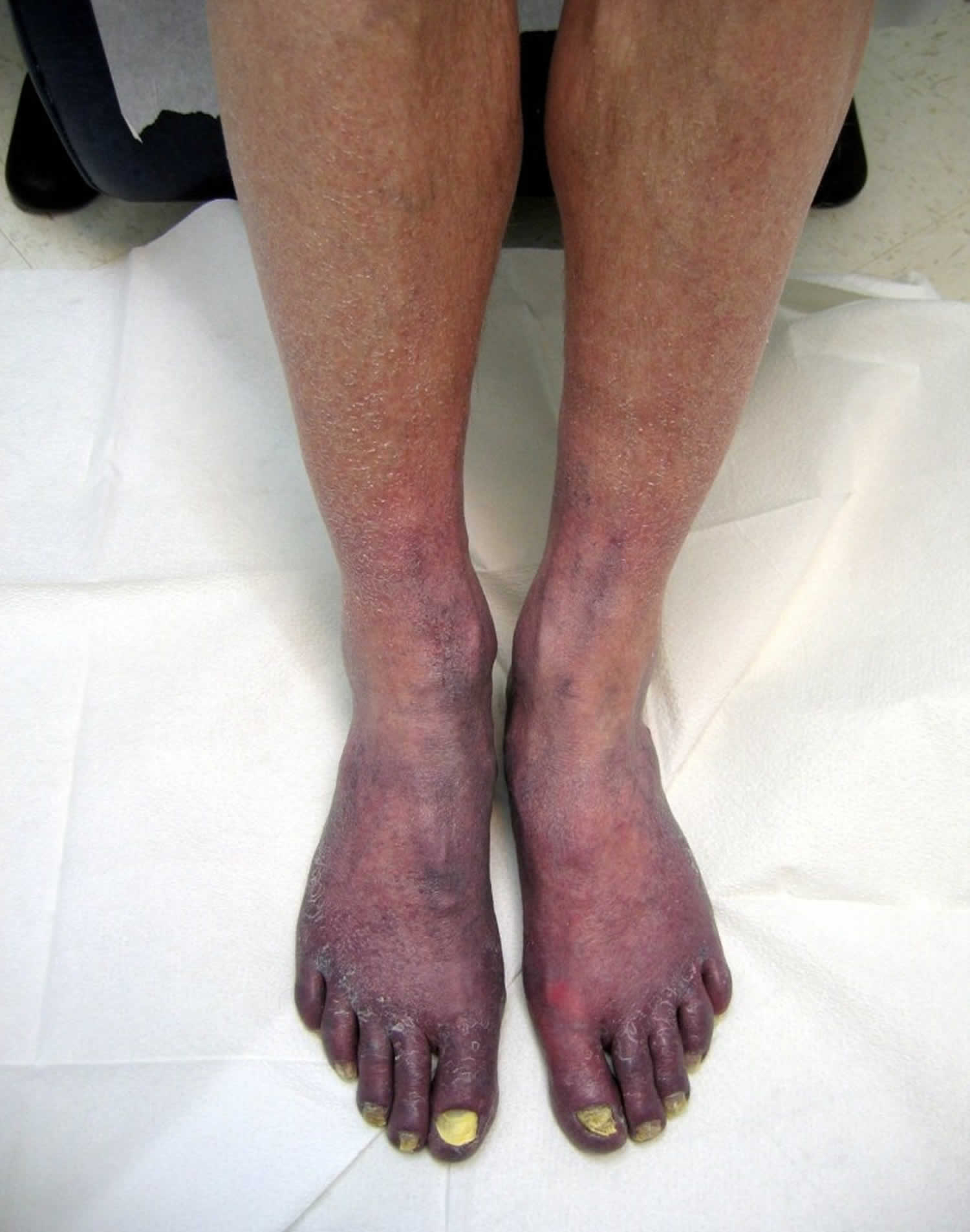
Primary acrocyanosis presents with bilateral, symmetrical, painless and persistent blue discolouration of fingers and toes. Acrocyanosis may extend to include hands, feet and face. Pressure on an area of blanching leads to a slow and irregular return of blood from the periphery towards the centre (Crocq sign). Arterial pulses are normal, and there is no proximal pallor, ulceration or gangrene.
Clinical features of acrocyanosis may also include:
- Hyperhidrosis of hands and feet
- Cold hands and feet
- Swelling of digits
Secondary acrocyanosis
The presentation of secondary acrocyanosis varies depending on the underlying cause. Secondary acrocyanosis is often asymmetrical and is associated with pain and tissue damage (blue toe syndrome).
Signs of a primary disorder may be present.
Acrocyanosis diagnosis
Clinical diagnosis of acrocyanosis is based on the patient’s general appearance and the distribution and persistence of cyanosis.
In young adults, where the diagnosis is most likely to be primary acrocyanosis, only limited investigations are required.
In older adults, or if there are atypical features such as pain and asymmetry of acrocyanosis, a targeted history, examination and investigations will be required to determine the cause. Investigations may include:
- Pulse oximetry
- Urinalysis
- Full blood count, C-reactive protein, ESR
- Standard biochemistry including liver and kidney function
- Streptococcal titers
- Autoantibodies
- Immunoglobulins and plasma electrophoresis
- Complement studies
- Chest X-ray
- Arterial and venous blood gas measurements
- Skin biopsy
- Nailfold capillaroscopy (to distinguish primary cyanosis from early stage connective tissue disorders).
- Screening for antinuclear factor, rheumatoid factor, cardiolipin antibody and lupus antibody may be required for connective tissue disease and antiphospholipid antibody (APLA) syndrome.
Acrocyanosis treatment
There is no standard and curative medical or surgical treatment of acrocyanosis. In mild cases, it is unnecessary to give any drug treatment.
Primary acrocyanosis
Treatment is not required for the majority of patients with acrocyanosis and pharmacological intervention is rarely necessary 9. The patient should be reassured that the condition is harmless. Options for active management may include:
- Behavioral measures, such as avoidance of cold exposure and trauma.
- Thick insulated clothing like insulated boots, thin polypropylene liner socks (to wick the moisture away from the skin and to maintain the normal skin temperature) is necessary for persons who have the illness and works at outdoor in cold climate.
- Psychophysiological measures, such as biofeedback training, reflex conditioning and hypnosis.
Note that vasodilators, e.g., calcium channel blockers, are not helpful (unlike in Raynaud phenomenon).
- There is limited evidence for benefit from bioflavonoids, nicotinic acid derivatives, adrenergic blockers, topical minoxidil, cyclandelate, rutin compounds and bromocriptine.
- Sympathectomy may be considered in very severe cases.
In clinical practice calcium channel blockers like nifedipine and diltiazem are not very effective 10, but reasonable improvements have been seen in some patients. Topically applied nicotinic acid derivatives and minoxidil can be beneficial 11. Sympathectomy or selectively disrupting only the fibers supplying the involved area will usually alleviate the cyanosis but such an extreme procedure would rarely be appropriate 12.
Secondary acrocyanosis
Treatment of secondary acrocyanosis depends on the underlying cause.
Acrocyanosis prognosis
Primary acrocyanosis is a benign condition that usually resolves in middle age with no long-term sequelae.
Secondary acrocyanosis may resolve with treatment of primary cause. As the pathological mechanisms that cause secondary acrocyanosis vary, so does prognosis.
References- Das S, Maiti A. Acrocyanosis: an overview. Indian J Dermatol. 2013;58(6):417–420. doi:10.4103/0019-5154.119946 https://www.ncbi.nlm.nih.gov/pmc/articles/PMC3827510
- Chronic idiopathic acrocyanosis. Nousari HC, Kimyai-Asadi A, Anhalt GJ. J Am Acad Dermatol. 2001 Dec; 45(6 Suppl):S207-8.
- James WD, Berger TG, Elston DM. 10th ed. Chap 3. Canada: Sunders Elsevier; 2006. In: Dermatoses Resulting From Physical Factors; Andrews’ diseases of the skin Clinical Dermatology; pp. 21–49.
- Musa R, Qurie A. Raynaud Disease (Raynaud Phenomenon, Raynaud Syndrome) [Updated 2019 Feb 14]. In: StatPearls [Internet]. Treasure Island (FL): StatPearls Publishing; 2019 Jan-. Available from: https://www.ncbi.nlm.nih.gov/books/NBK499833
- Ventura I, Reid P, Jan R. Approach to Patients with Suspected Rheumatic Disease. Prim. Care. 2018 Jun;45(2):169-180
- Wollina U, Koch A, Langner D, Hansel G, Heinig B, Lotti T, Tchernev G. Acrocyanosis – A Symptom with Many Facettes. Open Access Maced J Med Sci. 2018 Jan 25;6(1):208-212.
- Kurklinsky AK, Miller VM, Rooke TW. Acrocyanosis: The Flying Dutchman. Vasc Med. 2011 Aug 1;16(4):288–301.
- Dyer JA. Chapter 94: Cold Injuries. In: Fitzpatrick’s Dermatology in General Medicine. 8th ed. McGraw-Hill Education; 2012.
- Chadachan V, Eberhardt RT. Chapter 49: Acrocyanosis. In: Loscalzo MACAB, editor. Vascular Medicine: A Companion to Braunwald’s Heart Disease (Second Edition). Philadelphia: W.B. Saunders; 2013.
- Nousari HC, Kimyai-Asadi A, Anhalt GJ. Chronic idiopathic acrocyanosis. J Am Acad Dermatol. 2001;45(Suppl 6):S207–8
- Dowd PM. Reactions to cold. In: Burns T, Stephen B, Cox N, Christopher G, editors. Rook’s Text book of Dermatology. 7th ed. Vol. 2. Malden MA: Blackwell Science; 2008. pp. 23.1–23.17
- Creager MA, Dzau VJ. Vascular diseases of the extremities. In: Kasper DL, Fauci AS, Longo DL, Braunwald E, Hauser SL, Jameson JL, editors. Harrison’s Prins of Internal Medicine. 16th ed. New York: McGraw-Hill; 2005. p. 1490


