Angiokeratoma
Angiokeratoma literally means a collection of blood vessels, horn and tumor respectively refers to the presence of one or more dilated vessel(s) in the papillary dermis accompanied by an overlying acanthotic and/or hyperkeratotic epidermis 1. Angiokeratomas are characterized by red to blue papules and consist of dilated dermal vessels with an overlying acanthotic epidermis 2. It may have a rough scaly surface. It is composed of surface blood vessels (dilated capillaries). Often unnoticed, an angiokeratoma may become crusty and bleed if accidentally scratched or damaged, or a harmless clot may form in the lesion (thrombosis), changing the color to dark purple or black overnight.
There are five clinical types of angiokeratomas but other atypical forms have also been described 3:
- Sporadic angiokeratoma with an onset in adult life
- Angiokeratoma of Mibelli in which cold temperature may precipitate acral lesions
- Angiokeratoma of Fordyce, the predominantly genital variety
- Angiokeratoma circumscriptum neviforme which is nevoid with an early age of onset
- Fabry disease (angiokeratoma corporis diffusum), a systemic variety associated with widespread cutaneous lesions due to deposition of glycosphingolipids chiefly in the vascular endothelium and smooth muscle cells
The worldwide prevalence is estimated to be about 0.16% with a female preponderance (3:1). Apart from Fabry disease (angiokeratoma corporis diffusum), which is caused by mutations in the GLA gene, the cause of other angiokeratomas is unknown.
Angiokeratomas are harmless surface vascular lesions that can usually be left alone. As the black spots sometimes resemble melanoma, a skin biopsy may be performed to rule out malignancy and allay any fears.
If the bleeding becomes a concern or treatment is requested for cosmetic purposes, they can be removed. Surgical options include excision, laser therapy, cryotherapy or electrocautery.
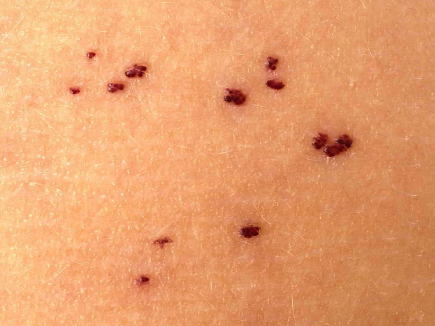
Sporadic angiokeratoma
- Solitary lesions
- Common in those over 40 years
Figure 1. Sporadic angiokeratoma
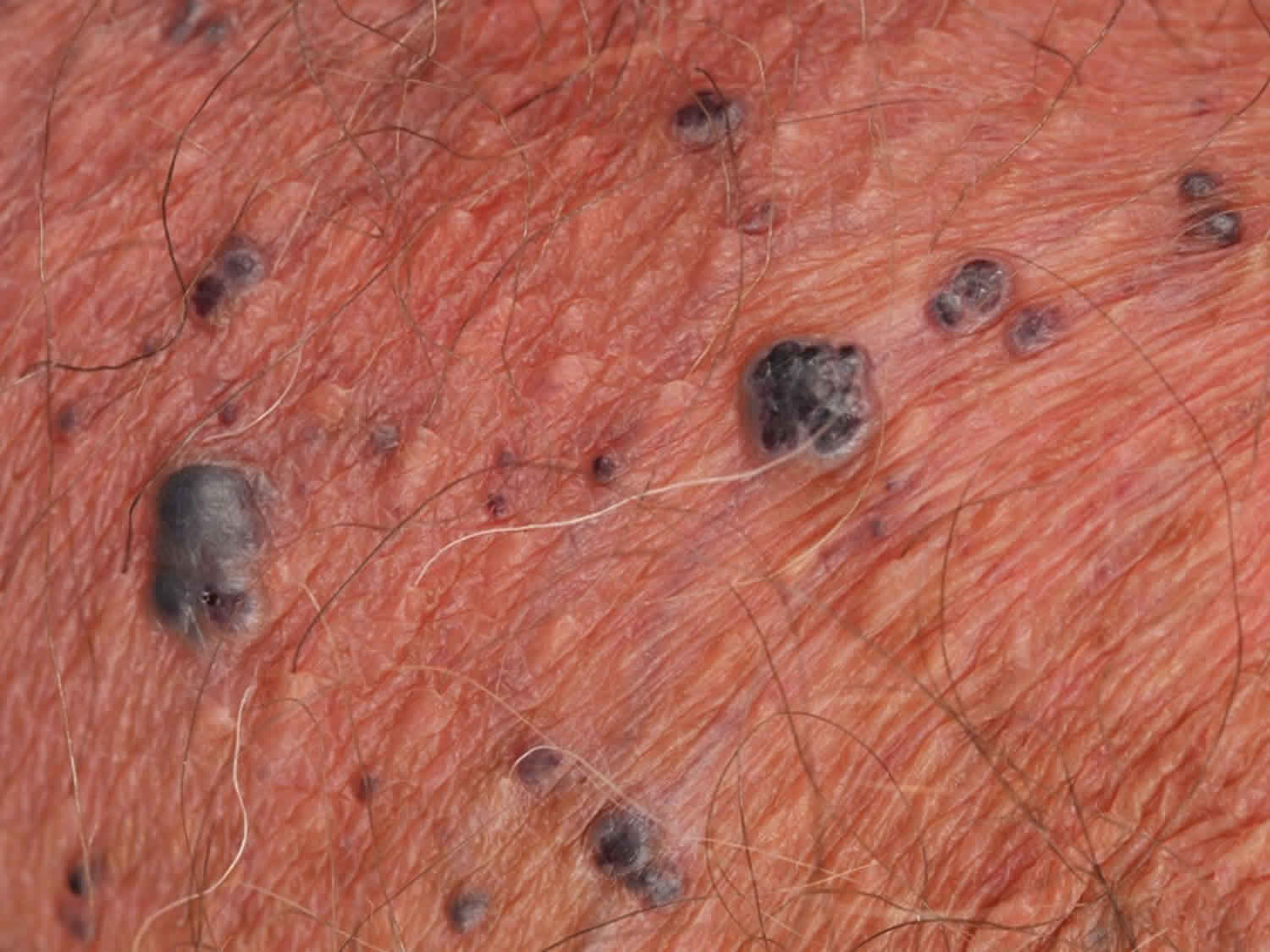
Angiokeratoma of Mibelli
- Multiple red to blue, dry to warty papules on fingers and toes, hands and feet, or less often knees, elbows, breasts
- Lesions appear in childhood or adolescent
- May be familial
- More common in females than in males
- Lesions may bleed with minor trauma
- Associated with chilblains
Figure 2. Angiokeratoma of Mibelli
Angiokeratoma circumscriptum
Angiokeratoma circumscriptum is the rarest variant. Maybe present at birth but can occur later in childhood or adulthood. Clinically, it is manifested as dark red to blue-black nodules or plaques presenting unilaterally over the lower extremities. Other sites of presentation include thighs, buttocks, and so on. Over time lesions may darken in color and change shape and size. The lesions often become keratotic and show no tendency for spontaneous remission, posing a concern for cosmesis to the patient 4. Females are more affected than men 3:1.
Figure 3. Angiokeratoma circumscriptum neviforme
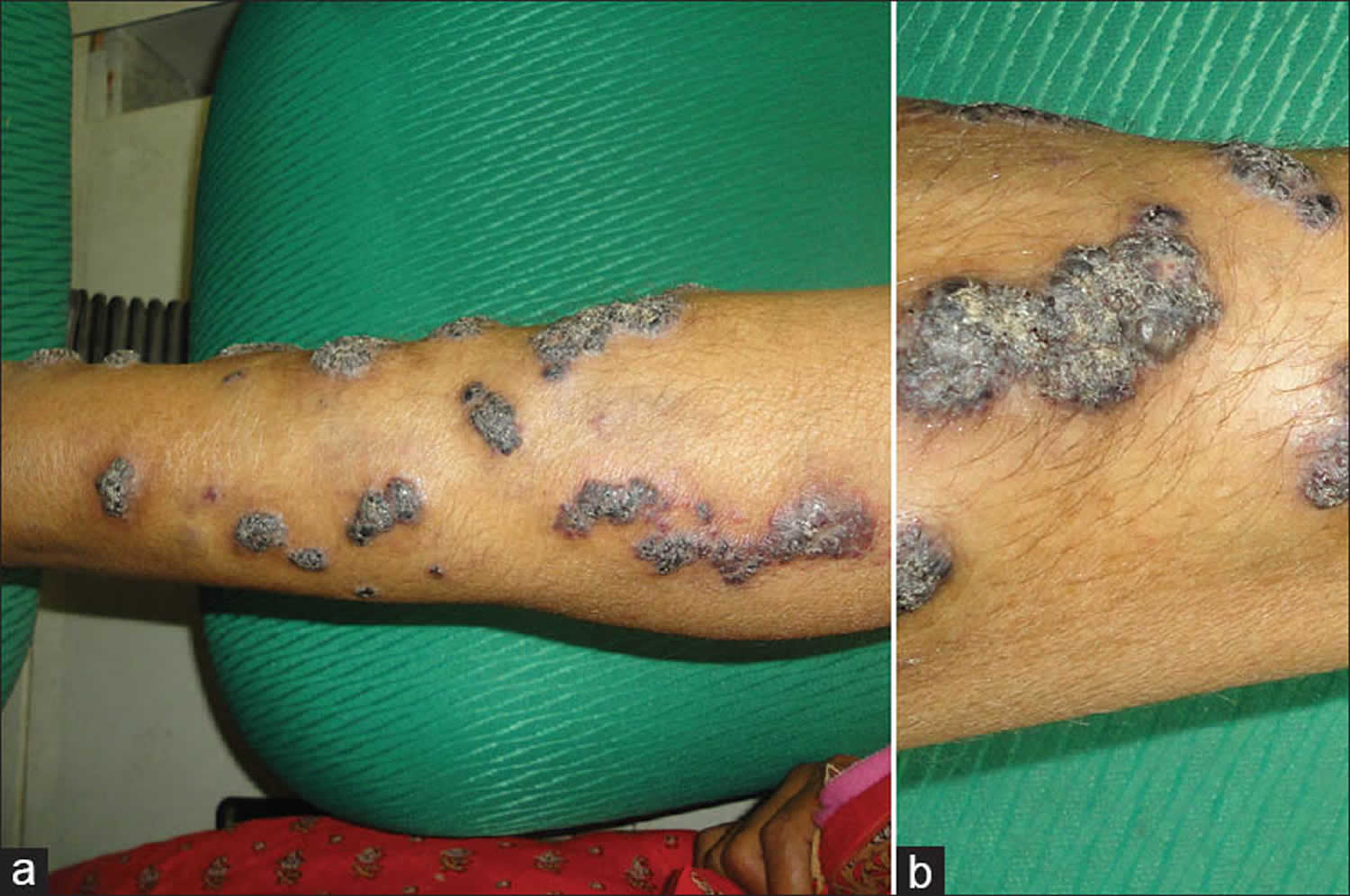
Footnote: Linear, verrucous, well-circumscribed plaques on the left lower leg (a) and close-up view of one of the lesions over the leg showing verrucous and hyperkeratotic surface (b)
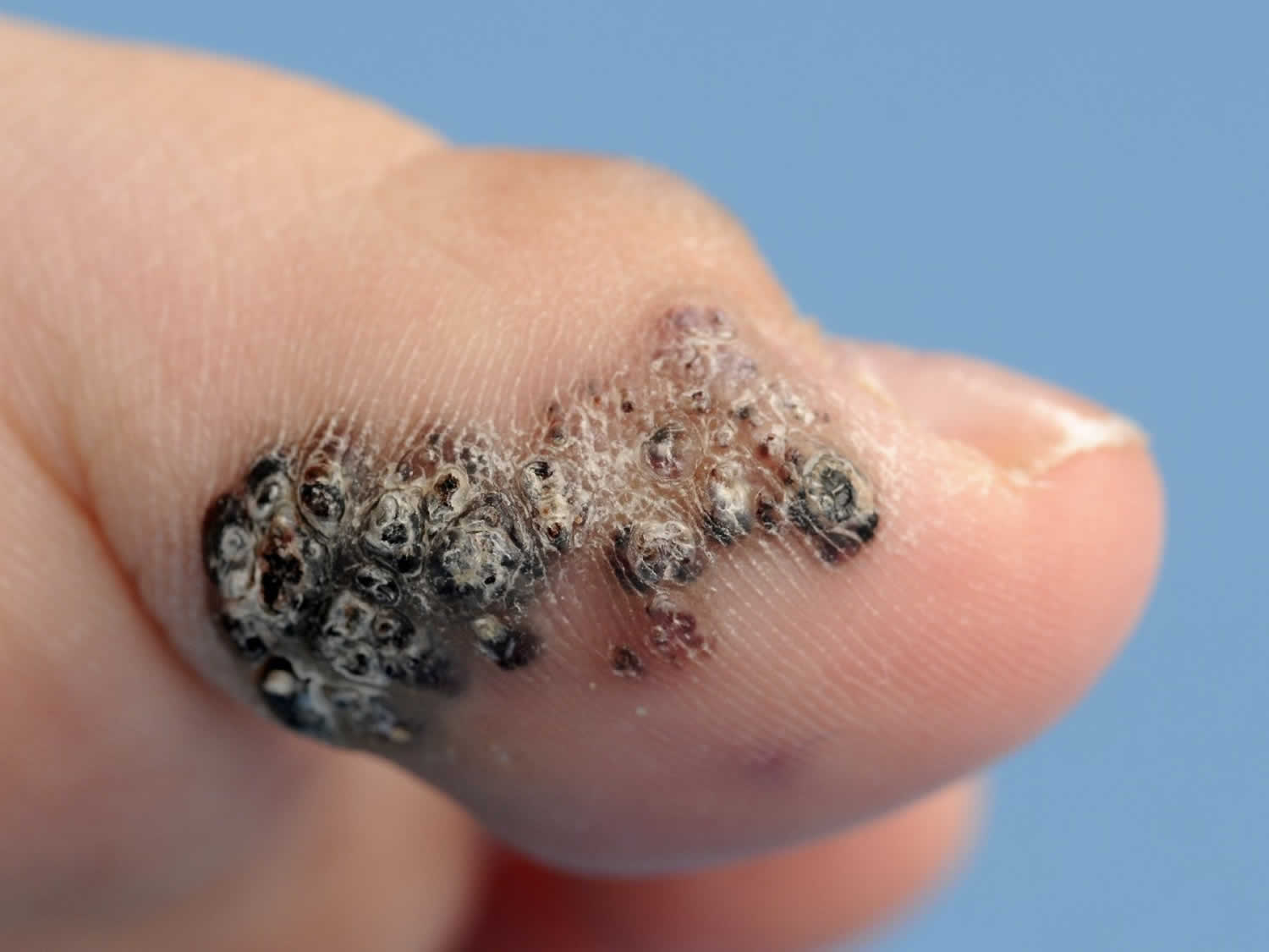
[Source 5 ]Angiokeratoma of Fordyce
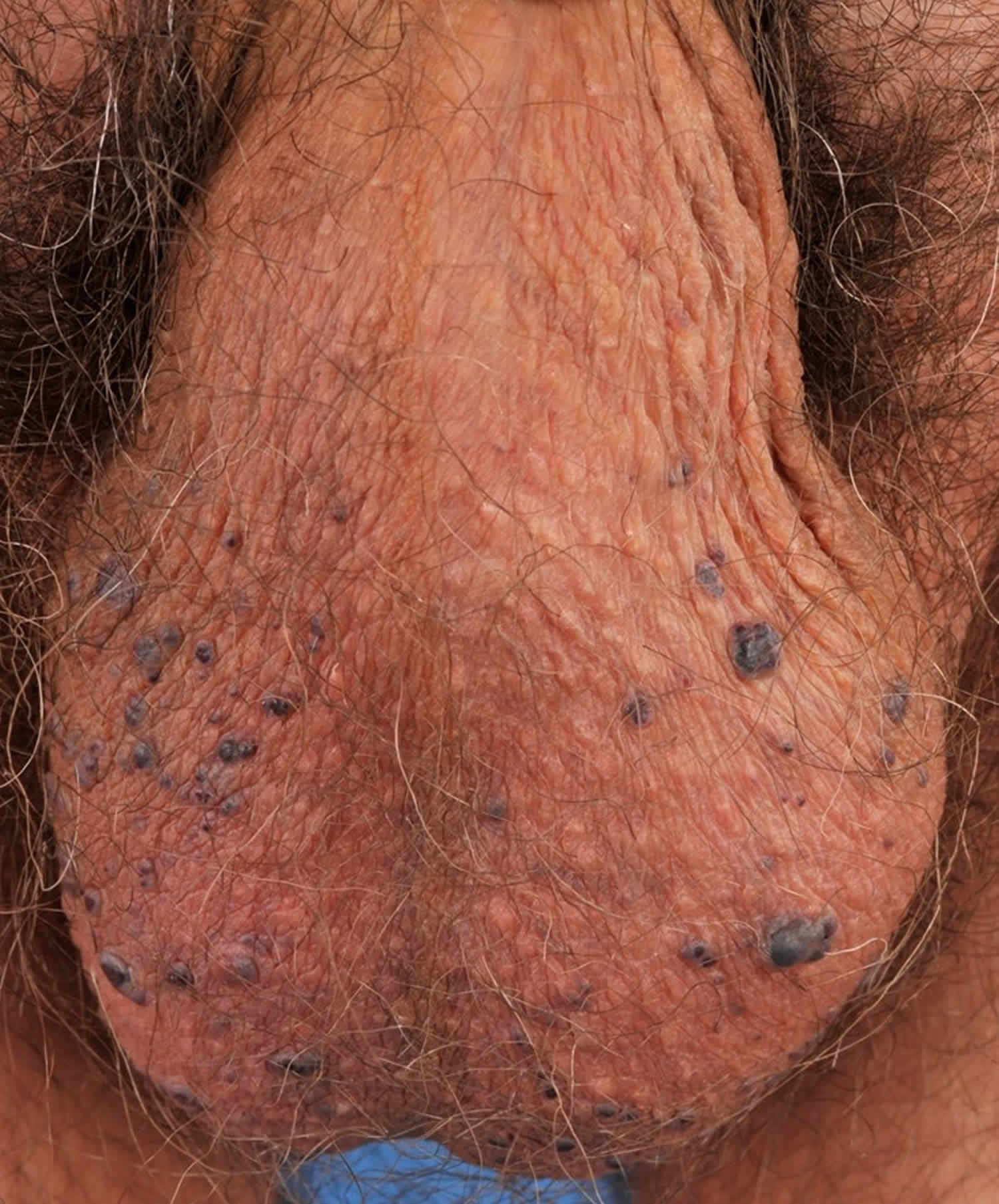
- Most commonly found on the scrotum. Also found on the shaft of the penis, labia majora of the vulva, inner thigh and lower abdomen
- Most prevalent in those over 40 years
- Men are more often affected than women
- Can present as a single lesion or multiple lesions (> 100)
- Lesions are small, red and less scaly in younger patients whilst in older patients, they tend to be larger, blue/black and with overlying scales
- Usually symptomless and may only be noticed when they bleed after scratching or intercourse
Figure 4. Angiokeratoma of Fordyce
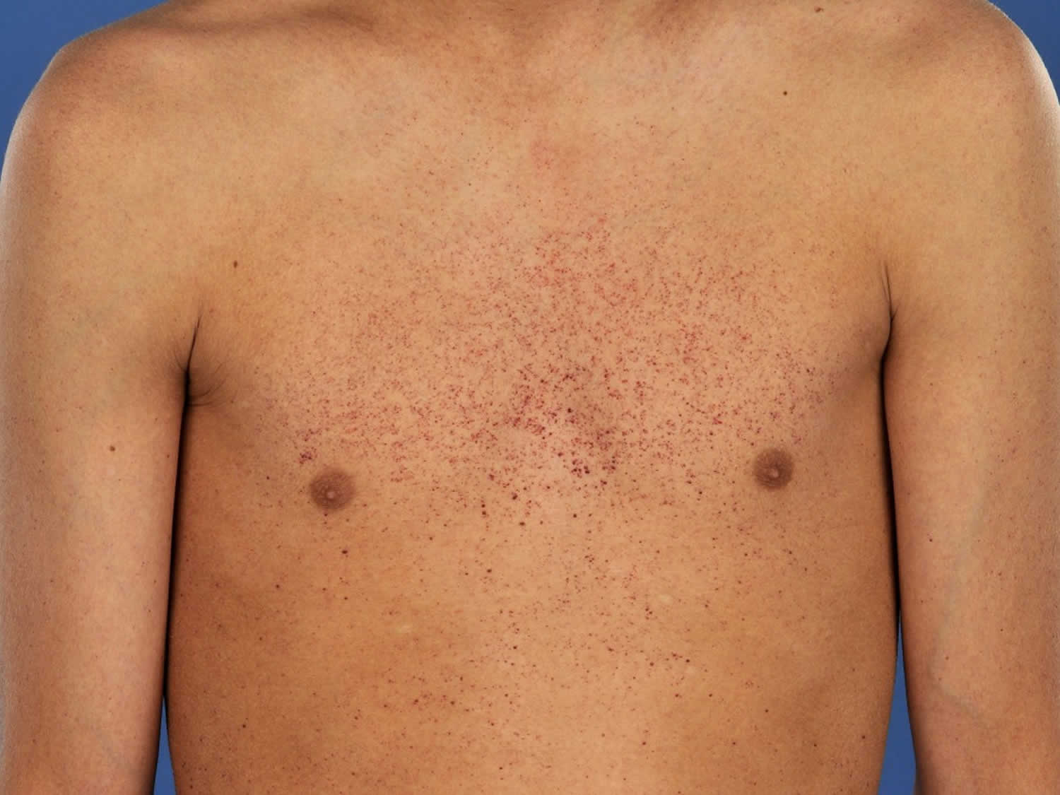
Fabry disease angiokeratoma
Fabry disease (angiokeratoma corporis diffusum) is an inherited disorder caused by a deficiency of an alpha-galactosidase enzyme, ceramide trihexosidase, that results from the buildup of a particular type of fat, called globotriaosylceramide, in the body’s cells. Beginning in childhood, this buildup causes signs and symptoms that affect many parts of the body. Characteristic features of Fabry disease include episodes of pain, particularly in the hands and feet (acroparesthesias); clusters of small, dark red spots on the skin called angiokeratomas; a decreased ability to sweat (hypohidrosis); cloudiness or streaks in the front part of the eye (corneal opacity or corneal verticillata); problems with the gastrointestinal system; ringing in the ears (tinnitus); and hearing loss. Angiokeratomas are widespread, most numerous on lower trunk and groin area. Fabry disease also involves potentially life-threatening complications such as progressive kidney damage, heart attack, and stroke. Some affected individuals have milder forms of the disorder that appear later in life and affect only the heart or kidneys.
Fabry disease affects an estimated 1 in 40,000 to 60,000 males 6. Fabry disease angiokeratoma also occurs in females, although the prevalence is unknown. Milder, late-onset forms of Fabry disease are probably more common than the classic, severe form.
Fabry disease (angiokeratoma corporis diffusum) is caused by mutations in the GLA gene. This gene provides instructions for making an enzyme called alpha-galactosidase A. This enzyme is active in lysosomes, which are structures that serve as recycling centers within cells. Alpha-galactosidase A normally breaks down a fatty substance called globotriaosylceramide. Mutations in the GLA gene alter the structure and function of the enzyme, preventing it from breaking down this substance effectively. As a result, globotriaosylceramide builds up in cells throughout the body, particularly cells lining blood vessels in the skin and cells in the kidneys, heart, and nervous system. The progressive accumulation of this substance damages cells, leading to the varied signs and symptoms of Fabry disease.
GLA gene mutations that result in an absence of alpha-galactosidase A activity lead to the classic, severe form of Fabry disease. Mutations that decrease but do not eliminate the enzyme’s activity usually cause the milder, late-onset forms of Fabry disease that typically affect only the heart or kidneys.
Figure 5. Fabry disease angiokeratoma (angiokeratoma corporis diffusum)
Angiokeratoma causes
Apart from Fabry disease (angiokeratoma corporis diffusum), which is caused by mutations in the GLA gene, the cause of other angiokeratomas is unknown.
Angiokeratoma treatment
Angiokeratomas are harmless surface vascular lesions that can usually be left alone. As the black spots sometimes resemble melanoma, a skin biopsy may be performed to rule out malignancy and allay any fears.
If the bleeding becomes a concern or treatment is requested for cosmetic purposes, they can be removed. Surgical options include excision, laser therapy, cryotherapy or electrocautery.
References- Angiokeratoma circumscriptum neviforme: Revisiting a rare entity. Indian J Paediatr Dermatol 2015;16:246-8 http://www.ijpd.in/temp/IndianJPaediatrDermatol164246-3311203_091152.pdf
- Baker C, Kelly R. Other vascular disorders. In: Bolognia JL, Jorizzo JL, Rapini R, editors. Dermatology. 2 nd ed. London: Mosby; 2008. p. 1615-25.
- Zaballos P, Daufν C, Puig S, Argenziano G, Moreno-Ramνrez D, Cabo H, et al . Dermoscopy of solitary angiokeratomas: A morphological study. Arch Dermatol 2007;143:318-25.
- Ghosh SK, Bandyopadhyay D, Ghoshal L, Haldar S. Angiokeratoma circumscriptum naeviforme: A case report of a rare disease. Dermatol Online J 2011;17:11.
- Das A, Mondal AK, Saha A, Chowdhury SN, Gharami RC. Angiokeratoma circumscriptum neviforme: An entity, few and far between . Indian Dermatol Online J 2014;5:472-4
- Fabry disease. https://ghr.nlm.nih.gov/condition/fabry-disease


