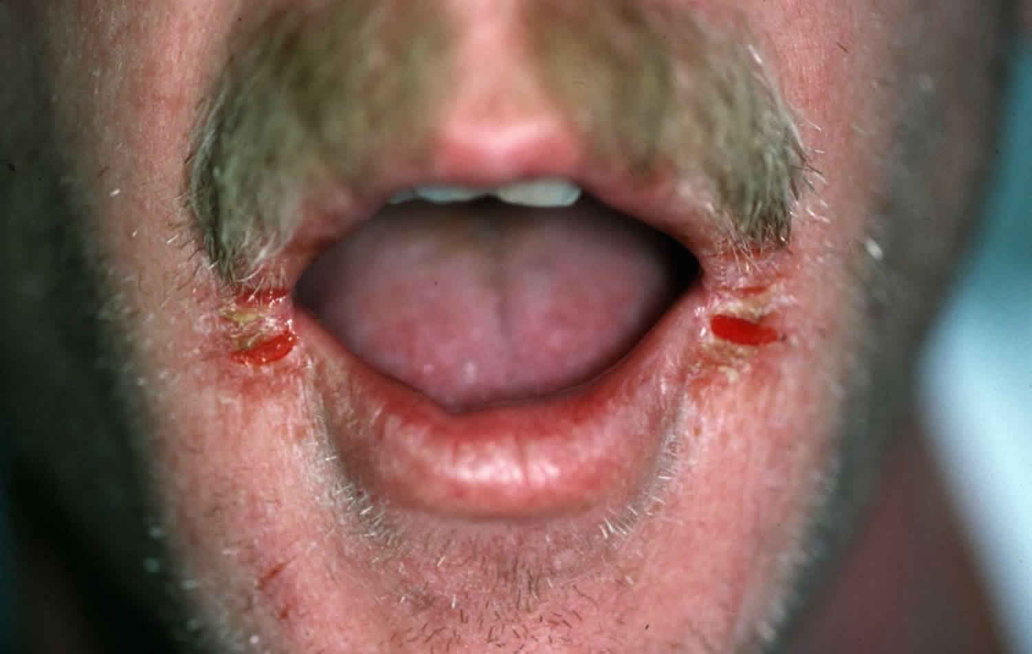
Angular cheilitis
Angular cheilitis also called angular stomatitis, angular cheilosis, commissural stomatitis, rhagades or perleche (from the French for “through licking”), is a common inflammatory condition affecting the corners of the mouth or oral commissures. Rhagades is a general term for fissuring of the skin in areas of motion, especially the labial commissures and nose. The areas are generally slightly painful. It is made worse by licking the lips. Depending on underlying causes, it may last a few days or persist indefinitely. Angular cheilitis is usually associated with a fungal (Candidal) or bacterial (Staphylococcal) infection, those affected may also have thrush (oral candidiasis). The condition can last from days to months, depending upon whether or not the affected person seeks treatment.
Angular cheilitis may affect people of all ages. Chronic pooling of saliva encourages fungal and bacterial growth, and patients who are immunocompromised, have undergone head and neck radiation, or have diabetes mellitus are also prone to this condition.
The angles of the mouth are points of interface for squamous epithelium of the face and oral mucosa. They are also a mechanically dynamic hinge for the oral aperture that endures more motion and tensile forces than the rest of the lips. Thus, the commissures are especially susceptible to certain stresses.
The following are alterations in mouth structure leading to changes in lip approximation and increased salivary pooling and maceration at the labial commissures (corners of the mouth):
- The normal loss of skin turgor due to aging, smoking, or rapid weight loss
- Loss of vertical dimension of the face due to severe tooth wear, edentulous states and mal-fitted dentures increasing overhang of the upper lips onto the lower (overclosure)
- Retrognathic malocclusion
- Deepened furrowing of the skin dependent to the commissures (marionette lines)
- Conditions associated with enlarged lips such as oro-facial granulomatosis. Up to 20% of oro-facial granulomatosis patients suffer from angular cheilitis, but Candida is usually not isolated from lesions.
- Down syndrome: 25% of patients have angular cheilitis due to macroglossia leading to tongue protrusion and drooling 1.
Patients who are predisposed to angular cheilitis also may have problems with:
- Iron deficiency
- Vitamin B12 deficiency
- Folate deficiency (vitamin B9)
- Drooping of the corners of the mouth caused by dentures that do not adequately support the facial musculature.
Dentures may act as reservoirs of infection. To prevent angular cheilitis, try soaking dentures overnight in a solution made up of 10 parts water to 1 part household bleach. For metal dentures that may become discolored by bleach, the use of a sodium benzoate or chlorhexidine mouth rinse is a good option.
Angular cheilitis key points
- Every case of angular cheilitis should prompt an investigation for oral candidiasis. A finding of oral candidiasis should prompt an investigation for diabetes or immunosuppression including, but not limited to, steroid use.
- Do not use topical steroid monotherapy without excluding infectious angular cheilitis.
- Unilateral angular cheilitis may be due to local trauma, herpetic lesions, or syphilitic papule. Cases that do not respond to treatment may also be a manifestation of malignancy.
If persistent lip irritation, painful cracking, or fissuring at the corners of the mouth is present, seek evaluation from your primary care provider or dermatologist.
Angular cheilitis staging
Angular cheilitis stages 2:
- Type I: Small rhagades limited to the corner of the mouth, adjacent skin slightly involved
- Type II: Lesion with ragged border more extensive in length and depth than Type I lesion
- Type III: Lesion consisting of several rhagades radiating from the corner of the mouth into the adjacent skin
- Type IV: Lesion presenting no rhagades, but erythema of skin contagious to the vermilion border
Angular cheilitis causes
Most cases of angular cheilitis are ultimately due to physical maceration at the angular commissures due to overexposure to saliva. The digestive enzymes in saliva can act even on body tissues in left if allowed prolonged contact. Continued saliva exposure induces a contact dermatitis and eczematous reaction at the commissures. The compromised integrity of the stratum corneum epithelium allows local commensal organisms to infect the area. Frequently, colonizing Candida albicans establishes and invades the susceptible tissue. This may then allow bacterial superinfection with staph and strep species. Thus, risk factors are those that increase saliva retention at the commissures, increase exposure to culprit microbes, cause direct tissue inflammation, or inhibit wound healing and immunity.
Angular cheilitis is due to one or more of the following factors:
- A dribble of saliva causing eczematous cheilitis, a form of contact irritant dermatitis
- An overhang of upper lip resulting in deep furrows (marionette lines)
- Dry chapped lips
- Proliferation of bacteria (impetigo), yeasts (thrush) or virus (cold sores).
Angular cheilitis is common and affects children and adults, especially when they are in poor health. Predisposing factors include:
- Oral thrush: infancy, old age, diabetes, systemic corticosteroid or antibiotic use
- Dentures, especially if they are poor fitting, and there is associated gum recession
- Poor nutrition: coeliac disease, iron deficiency, riboflavin deficiency
- Systemic illness, particularly inflammatory bowel disease (ulcerative colitis and Crohn disease)
- Sensitive skin, especially atopic dermatitis
- Genetic predisposition, for example in Down syndrome
- Oral retinoid medication: isotretinoin for acne, acitretin for psoriasis.
Atopic dermatitis
Allergic or irritant contact dermatitis causes up to 22% of cases of angular cheilitis and 25% to 34% of generalized cheilitis. Common causes include nickel (in individuals with orthodontic braces) 3, foods (due to flavorings and preservatives), toothpaste, mouthwash, the sunscreen component of expired lip balm, lip cosmetics (due to preservatives, sodium laurel sulfate, emollients, colophony, Cocamidopropyl betaine), acne products, and chewing gum. It may be impossible to distinguish irritant and allergic contact dermatitis without a patch test 4.
Immune deficiency
Immune deficiency causes angular cheilitis, often via the development of oral candidiasis (thrush) with extension to the labial commissures. Chronic steroid use (inhaled or oral), HIV/AIDS, thymic aplasia, severe combined immunodeficiency syndrome (SCID), DiGeorge syndrome, hereditary myeloperoxidase deficiency, and Chediak-Higashi syndrome. Blood dyscrasias and malignancies probably also imbue some immune suppression as seen in acute leukemia and agranulocytosis.
Nutritional deficiencies
Nutritional deficiencies are less common in developed countries but exist in susceptible populations such as the elderly, celiac disease patients, the impoverished, the mentally ill, vegans and their solely breastfed infants not receiving vitamin supplementation, bariatric surgery and ileal resection patients, chronic gastritis and chronic pancreatitis sufferers, Crohn disease patients, and those with pernicious anemia. Up to 25% of angular cheilitis has iron or vitamin B deficiency.
The following are associated with angular cheilitis:
- Vitamin B deficiencies (especially cyanocobalamin, folate, riboflavin) 5
- Trace mineral deficiencies (zinc or iron)
- General protein malnutrition
Ariboflavinosis (vitamin B2 deficiency): Cheilosis, angular cheilitis, photosensitivity, magenta glossitis, stomatitis, pharyngitis, and pseudo-syphilis (seborrhea-like dermatitis of the scrotum, vulva, philtrum, or nasolabial folds). As iron absorptions may be impaired, this deficiency can also lead to normocytic anemia.
Pellagra (hypovitaminosis B3): Classic signs of are dermatitis, diarrhea, dementia, and glossitis. angular cheilitis can occur.
Vitamin B5 deficiency: Rare; angular cheilitis, glossitis, seborrheic dermatitis-like rash around the eyes, nose, and mouth
Vitamin B6 deficiency: Sideroblastic anemia, cognitive or psychiatric depression, hypertension, and hyperhomocysteinemia, neuropathy, conjunctivitis, oral (aphthous-like) ulcers called stomatitis, atrophic glossitis, angular cheilitis, and intertrigo.
Folate (vitamin B9) and vitamin B12 deficiency: Megaloblastic anemia (pernicious anemia) and neurological symptoms. The latter include peripheral neuropathy (paresthesia, ataxia, decreased sensation), cognitive impairment/dementia, motor deficits (absent reflexes.) Lack of vitamin B12 can also cause glossitis and angular cheilitis. Patients susceptible to hypovitaminosis B12 also usually are folate deficient.
Iron deficiency: Microcytic anemia (fatigue, exertional dyspnea, palpitations, headache, pica), glossitis, angular cheilitis, koilonychia (spoon nails), and alopecia areata (non-scarring hair loss). If the patient is also experiencing dysphagia, a barium swallow or upper endoscopy may reveal esophageal webs, making the diagnosis of Plummer-Vinson syndrome (sideropenic dysphagia or Paterson-Brown-Kelly syndrome).
Zinc deficiency: Constellation of angular cheilitis along with alopecia, diarrhea, dermatitis, and oral ulcers (especially on the tongue and buccal surfaces). Causes include acrodermatitis enteropathica, an autosomal recessive condition that impairs zinc absorption.
Manifestations of systemic disease
Sjogren syndrome
Angular cheilitis is the most common oral lesion found in Sjogren syndrome, followed by atrophic glossitis and oral candidiasis. This is according to a systematic review by Serrano, et al. which incorporate the data of 2426 patients with Sjogren syndrome. The prevalence of angular cheilitis ranged from 2% to 81%, with the largest population reporting frequency of 20-40%. Sjogren syndrome is a rheumatologic disease characterized by xerostomia (“dry mouth”) and hyposialia (“decreased salivation”). This results from lymphocytic infiltration and destruction of salivary glands. Surprisingly, in this patient population, there is an inverse relationship between salivary flow and the presence of candidiasis, which is opposite of what is the case for non-Sjogren’s angular cheilitis. This may be explained by proper levels of saliva allowing for mucosal lubrication, tissue healing, and local immunity. Salivary IgA inhibits binding of candida species to mucosal surfaces and flushes Candida from the oral cavity. Denture-wearing still predisposes Sjogren syndrome patients to angular cheilitis, since dental orthotics act as a reservoir for Candida and was a risk factor for oral candidiasis 6.
Inflammatory bowel disease
Inflammatory bowel disease (Crohn disease and ulcerative colitis) can contribute to angular cheilitis in several ways, one of which is general malnutrition impeding wound healing.
Infection
The most common cause of angular cheilitis, with the organisms below isolated in over 50-80% of lesions 7.
- Risk factors include:
- Increased exposure to infecting microbes or factors that increase the microbial burden of skin flora such as poor hygiene, oral thrush, gingival disease/poor dentition
- Diabetes causing overgrowth of Candida due to increased salivary glucose levels and increased adherence to mucous membranes 8
- Decrease local immunity due to an immunocompromised state from chronic steroid use, chemotherapy, or HIV/AIDS
- Depletion of normal oral flora from prolonged antibiotic use enabling proliferation of Candida species
- Specific organisms:
- Candida (especially Candida albicans): the most common cause of angular cheilitis. Candida is a normal commensal flora of the mouth in the yeast form, with 40% to 60% of healthy individuals acting as a reservoir. This explains why Candida is found in 93% of cases of angular cheilitis but is described as the sole pathogen in only about 20% to 50% of the cases. Poor oral hygiene plays a role by increasing colony burden, especially when dentures are present. Since the hyphal form of this dimorphic yeast is the pathogenic variant, potassium iodide staining can help distinguish whether Candida is an innocent bystander yeast or a culprit pathogen. Uncontrolled diabetes mellitus is a major risk factor because of relative immunodeficiency and the increased availability of glucose as a substrate for fungi. Candida is typically the primary pathogen invading macerated labial commissures, setting the stage for subsequent bacterial infection. Infantile angular cheilitis is almost always associated with concomitant oral candidiasis (thrush) and needs treatment to prevent recurrence.
- Staphylococcus aureus is the sole pathogen in approximately 20% of cases. The anterior nares harbor Staphylococcus and decolonization therapy with anti-staphylococcal ointments should be applied to the nares if Staphylococcus aureus is identified as a causative agent.
- Beta-hemolytic streptococci, isolated in 8% to 15% of cases, but less commonly as a sole pathogen. The anterior nares can harbor Streptococcus and Staphylococcus.
- Polymicrobial infections cause most angular cheilitis, with 60% to 75% causes by a combination of Candida albicans and Staphylococcus aureus.
Recurrent mechanical, chemical, or thermal injury
Recurrent mechanical, chemical, or thermal injury to labial commissures or conditions that make the angles of the mouth susceptible to insults:
- Xerostomia contributes to 5% of cases of angular cheilitis
- Radiation treatment
- Sjogren syndrome
- Medications causing xerostomia and xeroderma such as isotretinoin, acitretin, indinavir, sorafenib, anticholinergic medications, and anticancer drugs
- Hypervitaminosis A
- Environmental exposures (dry heat, cold)
- Repetitive behaviors leading to salivary overexposure and commissural maceration. Habit-induced cheilitis is sometimes considered a distinct entity from angular cheilitis and termed either “factitious cheilitis” or “perleche.”
- Nervous tics such as over-licking of lips
- Thumb-sucking, lollipops
- Sialorrhea, drooling, or hypersialia
- Aggressive dental flossing 9
- Smoking
- Acute mechanical stress at the labial commissures such as post-tonsillectomy
Idiopathic angular cheilitis with no identifiable cause
Since infection is the most common cause and maceration from saliva exposure the most common risk factor, empiric treatment with antifungal and/or antibiotic creams are reasonable but, long-term emollient therapy may be necessary in unresponsive or recurrent cases. Any case of idiopathic angular cheilitis, after it has undergone adequate investigation, should raise a red flag for nutritional deficiencies or malignancy (the latter, especially in unilateral cases that fail to respond to any therapy.) A rare cause of angular cheilitis is glucagonoma – a pancreatic endocrine tumor that causes a syndrome of dermatitis, diabetes, weight loss, anemia, and angular cheilitis.
Angular cheilitis symptoms
Angular cheilitis can be found in the corners of the mouth. Angular cheilitis may result in the following symptoms and signs at the corners of the mouth:
- Painful cracks and fissures of the corners of the mouth, with redness
- Ulceration
- Blisters, erosions, oozing, crusting
- Redness
- Drainage of pus
- Tissue softness and tenderness
- Bleeding.
Angular cheilitis may progress to more widespread impetigo or candidiasis on the adjacent skin and elsewhere.
Angular cheilitis complications
Longstanding angular cheilitis can cause tissue atrophy and permanent scarring or discoloration.
Angular cheilitis diagnosis
The diagnosis of angular cheilitis is often purely clinical. Therefore, laboratory investigation is usually only performed after treatment failure. However, because an infection is the most common cause, testing for Candida or bacterial culture can be performed at diagnosis.
The culture of swabs taken from the corners of the mouth may reveal:
- Candida albicans
- Staphylococcus aureus
- Herpes simplex
Investigation of underlying medical conditions that may contribute (nutritional deficiencies, immunocompromised states, systemic diseases) is at the discretion of the medical provider. If first-line antifungal/antibiotic combination yields no clinical improvement in 2 to 3 weeks, testing should include Hgb level with MCV, iron profile with ferritin, folate, and vitamin B2/B6/B12 levels, and fasting blood glucose.
Candidal angular cheilitis suspected:
- Light microscopy: For confirmation, lesion scrapings can be taken and stained with periodic acid Schiff (PAS) technique. Positive results show red/purple hyphae (indicative of infection) or yeast (which can be simple colonization). Gram stain shows these bodies are dark blue. Hyphae and yeast are also visible on KOH slides.
- Germ tube test (Reynolds-Braude phenomenon) in sheep/human serum at 37 °C for 2 to 4 hours
- Chlamydospore formation in CMA or rice starch agar incubated at 25 °C for 2 to 3 days
- Sugar assimilation assay: Test for fermentation of glucose and maltose (but not sucrose or lactose), yielding pale pink coloration in the presence of tetrazolium reduction medium
- Fungal culture: Sabouraud dextrose agar and antibiotics, cornmeal agar, or CHROM agar
- Candida strain typing: Serotyping, isoenzyme profiles, morphotyping, resistance pattern
- Immunodiagnosis: ELISA, RIA, CIE, PHA, and LPA
Bacterial angular cheilitis suspected: Bacterial culture with sensitivities
Oral candidiasis confirmed:
- HIV testing
- Diabetes testing (random blood glucose, fasting blood glucose, glucose tolerance test, or HgbA1c testing)
Nutritional deficiency angular cheilitis suspected:
- Folic acid: Serum level
- Vitamin B12: Serum level; serum homocysteine and serum or urinary methylmalonic acid levels are more reliable indicators of deficiency than vitamin B12 serum levels
- Vitamin B2: Urinary riboflavin excretion or erythrocyte glutathione reductase activity assay
- Iron: Serum iron profile (iron level, iron saturation, ferritin level, TIBC
- Zinc: Serum zinc level
Allergic or irritant contact angular cheilitis suspected: Patch testing to distinguish allergic contact dermatitis
Malignancy suspected: Biopsy
Angular cheilitis treatment
Treatment depends on non-infectious or infectious cause. In many cases, no treatment is needed and angular cheilitis resolves by itself. Depending on the specific cause, the following treatments may be useful:
- Lip balm or thick emollient ointment, applied frequently
- Topical antiseptics
- Topical or oral antistaphylococcal antibiotic
- Topical antifungal cream
- Oral antifungal medication
- Topical steroid ointment
- Nutritional supplements
- Filler injections or implants to build up the oral commissures
- Botulinum toxin to smooth out the lines.
- Xylitol or chlorhexidine acetate/xylitol gum decreased angular cheilitis in patients 60 years of age and older in a clinical trial by Simons et al. 10
- Allergen avoidance is necessary for allergic contact dermatitis-induced angular cheilitis
- Rinsing of the mouth after inhaled corticoid steroid administration in COPD/asthma patients decreases the risk of oral candidiasis
- Eradicate of staph and strep with mupirocin or bacitracin application to the anterior nares in colonized individuals who suffer from bacterial angular cheilitis
- Identify reservoirs of infection such as dentures and ensure proper cleaning and possibly overnight storage in hypochlorite or chlorhexidine
- Early referral to prosthodontist is imperative if dentures are a contributing risk factor
- In edentulous and immunosuppression patients, preventative therapy of barrier ointments (petrolatum, zinc oxide) or daily imidazole cream
Treatment of angular cheilitis is usually undertaken with topical antifungals such as nystatin, clotrimazole, or econazole. Combinations of a topical antifungal and a topical steroid – such as Mycostatin and triamcinolone or iodoquinol and hydrocortisone – may also be prescribed. In persistent cases, oral antifungals may be used to treat the condition.
Fungicidal medications
Fungal infections require topical fungicidal medications applied to the labial commissures, usually 3 times daily for 2 weeks.
- Nystatin 100,000 units/mL ointment topically twice per day (BID)
- Gentian violet solution topically twice per day to 3 times per day (TID) is effective in children if a purple discoloration is acceptable
- Ketoconazole 2% cream topically
- Clotrimazole 1% cream topically
- Miconazole 2% cream topically (with or without hydrocortisone 1%): Mixed staphylococcal and candidal infections respond best to this treatment because of its inherent gram-positive bacteriostatic activity, thus being used as first-line treatment by some providers
- Iodoquinol 1% cream topically twice per day to 3 times per day, usually combined with hydrocortisone 1% cream.
Oral (systemic) antifungals
Nystatin is used in mild cases or thrush and those isolated to the oral cavity. Triazoles treat moderate and severe cases of oral candidiasis and any cases extending into the esophagus. When triazoles are used, they obviate the need for topical antifungals. However, they are inhibitors of hepatic cytochrome P450 system and may interact with other drugs. Fluconazole has the highest level of evidence 11.
- Nystatin 5 mL of 100,000 units/mL suspension, swish and swallow 4 times per day for 7 to 14 day for oral candidiasis (no oral bioavailability)
- Clotrimazole 1 troche sucked on 5 times per day for 7 to 14 days for mild oropharyngeal candidiasis refractory to nystatin
- Fluconazole 200 mg orally (PO) for 1 day, then 100 mg orally daily for 7 to 14 days (can be increased to 200 mg daily for severe cases or immunosuppression). This is more effective than nystatin in immunocompromised patients.
- Itraconazole 200 mg orally daily for 2 to 4 weeks or 200 mg swish and swallow 4 times per day without food for 7 to 14 days
- Posaconazole 100 mg orally twice per day for 1 day, then 100 mg orally daily for 7 to 14 days but the dose can be increased to 400 mg orally BID for fluconazole/itraconazole-refractory cases
- Voriconazole: Only recommended when treatment with fluconazole and either itraconazole or posaconazole have failed
- Caspofungin 70 mg orally once, then 50 mg orally daily until 2 days after symptoms/lesions resolved
- Amphotericin 30 to 40 g per d until 2 days after symptoms/lesions resolved (40 to 50 g per day for neutropenic patients)
- Further discussion for the systemic treatment of oral candidiasis are beyond the scope of this review
Topical antiseptics or antibiotics
Bacterial infections require topical antiseptics or antibiotics. Application of the same preparation to the anterior nares (usually 4 to 5 times daily) can prevent recurrent infection when colonization is present. Treatment course is for 1 to 2 weeks.
- Mupirocin 2% ointment 3 times per day to 4 times per day (QID)
- Fusidic acid 2% cream (with or without hydrocortisone 1%) applied 4 times per day topically as an antistaphylococcal regimen
Oral (Systemic) Antibiotics
These rarely warranted unless lesions are extensive or treatment failure to topical antibiotics; should warrant culture, sensitivities, and consideration of an alternative diagnosis
Topical glucocorticoids
Topical glucocorticoids are monotherapy in strictly inflammatory processes or add-on therapy to anti-candidal or antibacterial regimens to decrease inflammation, enhance healing of erosions, and prevent relapses.
- Desonide 0.05% ointment topically
- Clotrimazole cream topically twice per day for 2 weeks
- Hydrocortisone 1% ointment topical twice per day to three times per day for 2 weeks (added to iodoquinol 1% cream or fusidic acid 2% cream)
Nutritional replacement or supplementation
Nutritional replacement or supplementation is necessary in cases of avitaminosis, mineral deficiencies, or general malnutrition.
Dental
A dentist should refit ill-fitting dentures or other dental apparati to restore facial contour. As the functional reservoir of Candida, treat dentures with an antifungal and cleaned frequently. In chronically debilitated patients, a cannula incorporated into the dentures can channel salivary flow into the oropharynx.
Sometimes, malocclusion persists despite dental realignment or is not a viable option for a patient. Other times, depressions at the commissures exist and are amenable to dermal filler therapy. Injectable fillers (collagen, hyaluronic acid) or surgical implants can change mouth shape and restore commissural anatomy. This makes saliva less likely to accumulate at the fissures. A practitioner who is well-versed in the administration of fillers should apply these fillers since the purpose is beyond the normal cosmetic application.
Improved control of chronic medical conditions that contribute to angular cheilitis
- Blood sugar control in diabetes as HgbA1c directly correlates with the incidence of angular cheilitis
- Anti-retroviral therapy in HIV/AIDS as immune status indirectly correlated with the incidence of angular cheilitis
Elimination of behavioral practices that contribute to angular cheilitis
- Tobacco smoking
- Lip licking
Treatment failures
- Failure to identify/eradicate oral candidiasis
- Resistant species of Candida
- Resistant strains of Staphylococcus or Streptococcus
- Failure to remediate underlying modifiable risk factors such as hygienic issues, ill-fitting dentures, behaviors.
- Persistent, unmodifiable risk factors
- Undiagnosed nutritional deficiency
- Undiagnosed systemic inflammatory conditions (Sjogren syndrome or inflammatory bowel disease)
- Unidentified immunosuppression
- Undiagnosed malignancy
Follow up in 2 weeks recommended.
Angular cheilitis prognosis
Angular cheilitis is a highly manageable condition. Angular cheilitis is mostly curable and poses no inherent risk to life and rarely results in permanent disfigurement 12. Angular cheilitis improves within the first several days of successful treatment and typically resolves by two weeks, thus schedule a follow up then. Chronic cases can provoke atrophy or granulation formation at the angles of the mouth. In one study done over 5 years, angular cheilitis had a recurrence rate of 80%. Identification and management of underlying risk factors is a necessity to prevent recurrence. When non-modifiable risk factors exist, when modifiable risk factors go unaddressed, or when the treatment course is incomplete, repeat bouts of angular cheilitis are commonplace. Common reasons for recurrence are failure to identify and treat oral candidiasis, continued poor oral and denture hygiene. If relapses are frequent, prolong treatment past 2 weeks and use preventive measures with topical emollients or antifungals.
References- Scully C, van Bruggen W, Diz Dios P, Casal B, Porter S, Davison MF. Down syndrome: lip lesions (angular stomatitis and fissures) and Candida albicans. Br. J. Dermatol. 2002 Jul;147(1):37-40.
- Oza N, Doshi JJ. Angular cheilitis: A clinical and microbial study. Indian J Dent Res. 2017 Nov-Dec;28(6):661-665.
- Cross D, Eide ML, Kotinas A. The clinical features of angular cheilitis occurring during orthodontic treatment: a multi-centre observational study. J Orthod. 2010 Jun;37(2):80-6.
- Yesudian PD, Memon A. Nickel-induced angular cheilitis due to orthodontic braces. Contact Derm. 2003 May;48(5):287-8.
- Rose JA. Folic-acid deficiency as a cause of angular cheilosis. Lancet. 1971 Aug 28;2(7722):453-4.
- Serrano J, Lopez-Pintor RM, Gonzalez-Serrano J, Fernandez-Castro M, Casanas E, Hernandez G. Oral lesions in Sjogren’s syndrome: A systematic review. Med Oral Patol Oral Cir Bucal. 2018 Jul 01;23(4):e391-e400.
- MacFarlane TW, Helnarska SJ. The microbiology of angular cheilitis. Br Dent J. 1976 Jun 15;140(12):403-6.
- Dorocka-Bobkowska B, Zozulinska-Ziolkiewicz D, Wierusz-Wysocka B, Hedzelek W, Szumala-Kakol A, Budtz-Jörgensen E. Candida-associated denture stomatitis in type 2 diabetes mellitus. Diabetes Res. Clin. Pract. 2010 Oct;90(1):81-6.
- Kahana M, Yahalom R, Schewach-Millet M. Recurrent angular cheilitis caused by dental flossing. J. Am. Acad. Dermatol. 1986 Jul;15(1):113-4.
- Simons D, Brailsford SR, Kidd EA, Beighton D. The effect of medicated chewing gums on oral health in frail older people: a 1-year clinical trial. J Am Geriatr Soc. 2002 Aug;50(8):1348-53.
- Devani A, Barankin B. Dermacase. Angular cheilitis. Can Fam Physician. 2007 Jun;53(6):1011, 1022-3.
- Federico JR, Basehore BM, Zito PM. Angular Chelitis. [Updated 2019 Sep 11]. In: StatPearls [Internet]. Treasure Island (FL): StatPearls Publishing; 2020 Jan-. Available from: https://www.ncbi.nlm.nih.gov/books/NBK536929


