Cafe au lait spots
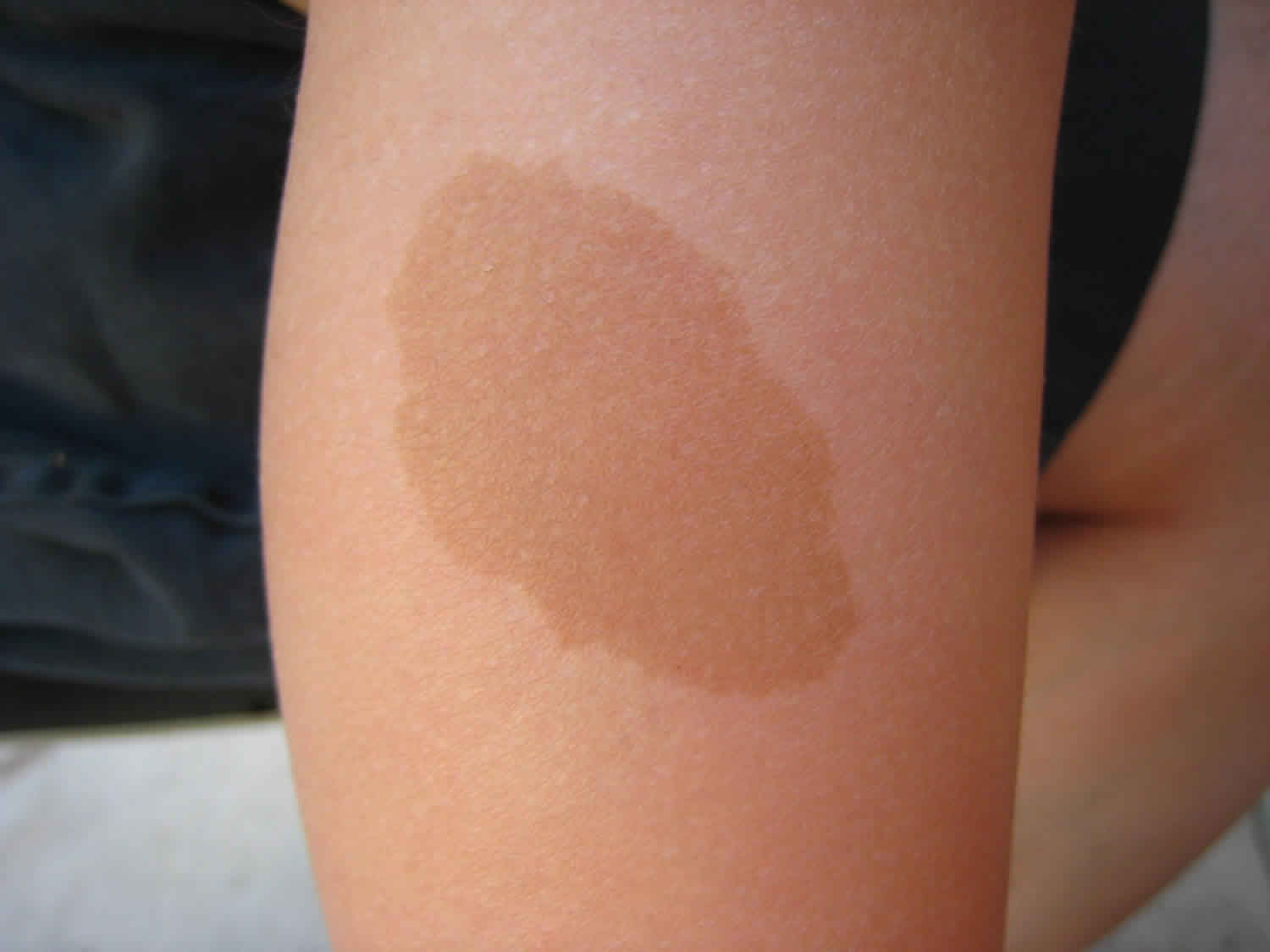
Cafe au lait spots also called cafe au lait macules, are common birthmark presenting as hyperpigmented lesions that may vary in color from light brown to dark brown; this is reflected by the name of the condition, which means “coffee with milk” 1. The borders may be smooth or irregular.
The size and number of café au lait skin lesions widely vary and are usually the earliest manifestations of neurofibromatosis 2. Café-au-lait spots are usually present at birth (congenital) or appear in early infancy. Café-au-lait spots sometimes become apparent later in infancy, especially after exposure to the sun, which darkens the color.
Cafe au lait spots may be isolated or associated with systemic diseases such as neurofibromatosis, McCune Albright syndrome, Legius syndrome, and Noonan syndrome with multiple lentigines syndrome.
The overall prevalence of cafe au lait macules varies with race.
- 0.3% of Caucasians
- 0.4% of Chinese
- 3% of Hispanics
- 18% of African Americans
Isolated cafe au lait macules are invariably solitary. More than 3 in a Caucasian or more than 5 in an African American are uncommon and should lead to systemic evaluation, referral and close follow-up.
Cafe au lait spots causes
The brown color of a cafe au lait macule is due to a pigment called melanin, which is produced in the skin by cells called melanocytes.
- The epidermal melanocytes of an isolated cafe au lait macule have excessive numbers of melanosomes (intracellular pigment granules). This is known as epidermal melanotic hypermelanosis.
- The cafe au lait macules associated with neurofibromatosis type 1 and Leopard syndrome have increased proliferation of epidermal melanocytes (epidermal melanocytic hyperplasia).
- A cafe au lait macules is not classified as a congenital melanocytic naevus.
Multiple cafe au lait macules are related to several genetic syndromes.
Neurofibromatosis type 1
About half of those with neurofibromatosis type 1 (NF1) have an inherited mutation of the NF1 gene on chromosome 17. NF1 codes for neurofibromin, a tumor suppressor gene. Others have a sporadic mutation of the same gene.
Neurofibromatosis type 2
Like NF1, autosomal dominant and sporadic mutations of the NF2 gene are equally common. NF2 gene codes for Merlin protein, whose physiologic function is still under investigation.
Legius syndrome
Legius syndrome is caused by SPRED gene mutation, which generally controls the RAS pathway and interacts with neurofibromin.
McCune Albright syndrome
McCune Albright syndrome is caused by mutation of Gs protein, activating adenylate cyclase.
Noonan syndrome with multiple lentigines
Noonan syndrome with multiple lentigines is due to the autosomal dominant inheritance of mutated PTPN11 gene on chromosome 12. The gene codes protein tyrosine phosphatase SHP-2.
The next three syndromes are much rarer than those described above.
Watson syndrome
Watson syndrome is linked to mutation of NF1 gene, or is at least allelic to NF1, or is caused by mutation of contiguous genes to NF1.
Bloom syndrome
Bloom syndrome is due to the autosomal recessive mutation in BLM gene on chromosome 15. The gene product is DNA helicase, an enzyme essential to DNA repair to prevent chromosomal breakage.
Silver-Russell syndrome
There are several genetic abnormalities associated with Silver-Russell syndrome. The 2 most common are:
- The absence of methylation in the genetic imprinting process of H19 and IGF2 genes on chromosome 11. They are responsible for normal cell growth. This abnormality is found in 30% of cases of Silver-Russell syndrome.
- Inheritance of both chromosome 7s from mother (maternal uniparental disomy).
Cafe au lait spots symptoms
Cafe au lait spots or macules:
- Are light brown in color.
- The pigment is evenly distributed.
- They are well demarcated with a smooth or irregular border
- Their shape is either round or oval.
The distribution and configuration of cafe au lait macules can be a clue to an underlying syndrome.
Neurofibromatosis type 1
Neurofibromatosis type 1 (NF1) is highly variable in appearance. The main 2 National Institute of Health Consensus criteria for the diagnosis of NF1 are:
- 6 or more cafe au lait macules with diameter > 5 mm in children and > 15 mm in adults. They may be on the trunk or extremities.
- Axillary or inguinal freckling. These are small cafe au lait macules and have the same microscopic appearance.
There are 5 other National Institute of Health criteria:
- Cutaneous neurofibromas (> 2) or a plexiform neurofibroma (> 1).
- Cutaneous neurofibromas are present in > 90% of adults with neurofibromatosis type 1. They are soft tumours that move with the skin, are not painful and do not have malignant potential.
- Plexiform neurofibromas are found in 25% of neurofibromatosis type 1 patients. They are soft, painful, hyperpigmented plaques and sometimes have excessive hair (hypertrichosis). If large enough, they can cause distortion of surrounding structures. Plexiform neurofibromas have the potential for malignant transformation.
- Lisch nodules on iris (> 2)
- Optic pathway glioma
- Osseous dysplasia: sphenoid dysplasia; thinning and bowing of long bone; pseudoarthrosis
- First degree relatives diagnosed with neurofibromatosis type 1 using these criteria
At least two criteria are required to make a working diagnosis of neurofibromatosis. The definitive diagnosis is made by confirming the presence of a genetic mutation.
Neurofibromatosis type 2
Neurofibromatosis type 2 presents with unilateral or bilateral acoustic schwannoma (vestibular schwannoma). Patients develop hearing problems, ringing in the ears (tinnitus), and dizziness. By the age of 30, nearly all patients with neurofibromatosis type 2 have bilateral vestibular schwannoma.
- Other nervous system tumors in neurofibromatosis type 2 include cranial and peripheral nerve schwannomas, meningiomas, ependymomas, and astrocytomas.
- 60–80% of patients with neurofibromatosis type 2 suffer from presenile posterior subcapsular lenticular opacities (cataracts).
- The main skin lesion arising in neurofibromatosis type 2 is an elastic, firm, well-demarcated subcutaneous neurilemmoma. cafe au lait macules are less common.
Legius syndrome
Patients with Legius syndrome have multiple cafe au lait macules (> 5mm in children and > 15 mm in adults). Axillary freckling less common.
They may rarely have macrocephaly, cognitive disabilities, and several congenital malformations such as Noonan-like facies, pectus excavatum/carinatum, and lipomas.
McCune Albright syndrome
Cafe au lait macules in McCune Albright syndrome are fewer than in neurofibromatosis type 1, with more irregular borders. They are classically found on the midline.
The clinical diagnosis of McCune Albright syndrome is established by a triad of abnormalities:
- Polyostotic or monostotic fibrous dysplasia
- Cafe au lait macules
- Hyperfunctioning hormonal disorders such as precocious puberty, hyperthyroidism, hypercortisolism, hyperomatotropism, and hypophosphatemic rickets.
Noonan syndrome with multiple lentigines
Noonan syndrome with multiple lentigines is also known as LEOPARD syndrome; the L of LEOPARD syndrome refers to prominent lentigines. These are multiple < 5 mm, well-demarcated, brown macules presenting on the whole skin without mucous membrane involvement. They appear at birth and continue increasing in number until puberty.
LEOPARD is an acronym referring to the clinical findings required to make the diagnosis:
- Lentigines
- Electrocardiogram conduction defects
- Ocular hypertelorism
- Pulmonary stenosis
- Abnormalities of genitalia
- Retardation of growth
- Sensorineural deafness
Patients with Noonan syndrome with multiple lentigines may also develop cafe au lait macules, nail malformation, and hyperelastic skin.
Watson syndrome
Watson syndrome is extremely rare with only 4 families described between the 1960s to early 1990s. Their cafe au lait macules in Watson syndrome had similar characteristics to neurofibromatosis type 1. Other features of Watson syndrome are:
- Stenosis of the pulmonary valve
- Mental retardation
- Short stature
- Relative macrocephaly
- Lisch nodules on the iris
- Neurofibromas
Bloom syndrome
Cafe au lait macules are not the main clinical finding in Bloom syndrome. Key characteristics of Bloom syndrome are:
- Growth deficiency
- Immunodeficiency
- Malignancies
- Predilection to diabetes
- Distinctive narrow facies
- High-pitched voice
- Hypogonadism and/or infertility
Silver-Russell syndrome
Even though cafe au lait macules are not essential for diagnosis, they are common in children with Silver-Russell syndrome. They are mainly located on chest, stomach, and extremities.
The main features of Silver-Russell syndrome are:
- Severe retardation of intrauterine and postnatal growth
- Relative macrocephaly
- Small, triangular facies and prominent forehead
- Other congenital malformations, including clinodactyly V, hemihypoplasia, micrognathia, and ear anomalies
Cafe au lait spots diagnosis
Cafe au lait spots or macules are diagnosed clinically. If significant in number and size, a complete clinical examination should be undertaken to determine whether an associated syndrome may be present.
Syndromes may be diagnosed from their clinical manifestations or by genetic testing.
Cafe au lait spots treatment
No medical care is required to treat cafe au lait spots or macules. Lasers reported to have successfully faded cafe au lait macules include:
- Pulsed-dye laser
- Er:YAG laser
- Q-switched Nd:YAG laser
- Q-switched ruby or alexandrite laser
Results are inconsistent. One group has found lesions with an irregular margin respond better than those with a smooth, well-defined border. Risks for laser surgery include transient/permanent hyperpigmentation, hypopigmentation, and scarring.
Treatment of underlying syndromes may be complex and require multidisciplinary care.
Cafe au lait spots prognosis
Without treatment, cafe au lait macules persist lifelong. No reports indicate that cafe au lait macules undergo malignant change. Café au lait macules are benign and produce no mortality or morbidity, although the associated syndromes may have significant manifestations. Results from lasers are not consistent. However, for those who responded to initial treatment, recurrence rates are reported to be low.
References


