close

Small and Large Intestine

BladderConditions & DiseasesSmall and Large IntestineUrinary System

Cloacal exstrophy

Cloacal exstrophy

Cloacal exstrophy

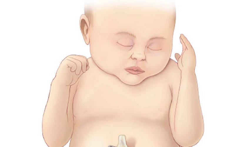
Cloacal exstrophy, also known as OEIS Syndrome, occurs when a portion of the large intestine lies outside of the body and on either side of it and connected to it are the two halves of the bladder. The intestine may be short and the anus may not open. The bony pelvis is also split open like a book. In males, the penis is usually flat and short, with the exposed inner surface of the urethra on top. The penis is sometimes split into a right and left half. In girls, the clitoris is split into a right half and left half and there may be one or two vaginal openings. Cloacal exstrophy (OEIS syndrome) is a very rare birth defect, affecting 1 in every 250,000 births. Although cloacal exstrophy is a serious condition and requires a series of operations, the long-term outcome is good for many children. Patients and families need to be counseled about the complexity of the anomaly, the need for multiple procedures, and long-term expectations for continence, sexual function, and fertility.

Cloacal exstrophy is known as OEIS Syndrome because of the four features that are typically found together 1:

  • Omphalocele: Some of the abdominal organs protrude through an opening in the abdominal muscles in the area of the umbilical cord. The omphalocele may be small, with only a portion of the intestine protruding outside the abdominal cavity, or large, with many of the abdominal organs (including intestine, liver and spleen) protruding outside the abdominal cavity.
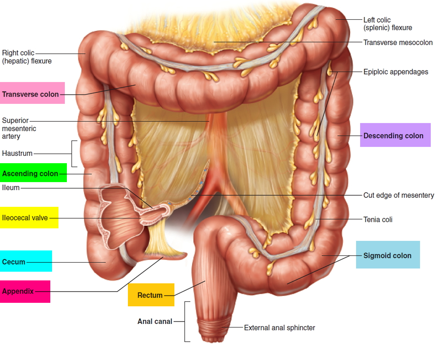
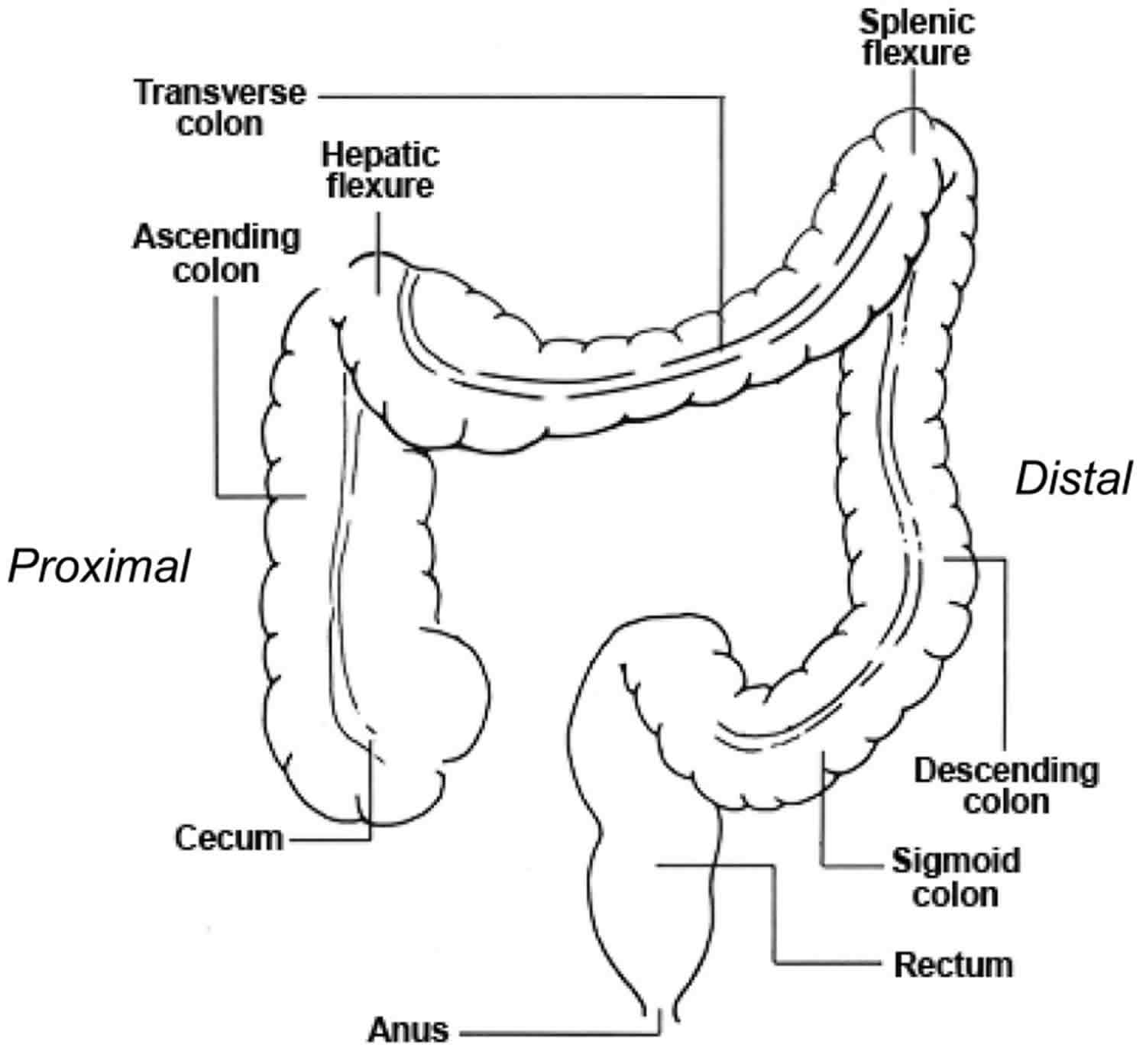
  • Exstrophy of the bladder and rectum: The bladder is open and separated into two halves. The rectum and colon are similarly open and the segment of the rectum is placed between the bladder halves on the surface of the abdomen.
  • Imperforate anus: The anus has not been formed or perforated and the colon connects to the bladder.
  • Spinal defects: These defects may either be major or minor. Often children born with cloacal exstrophy are also born with some degree of spina bifida.

With cloacal exstrophy there are often other birth defects, like spina bifida. This occurs in up to 75 percent of cases. Kidney abnormalities and omphalocele are also common. An omphalocele is when an infant’s intestine or other abdominal organs are open to the outside the body. This is from a hole in the belly button (navel) area. The intestines are covered only by a thin layer of tissue and can be easily seen.

Cloacal exstrophy (OEIS Syndrome) is a complex anomaly that often requires several surgical procedures and requires lifelong medical follow-up care.

As soon as possible, surgical reconstruction is done. Surgery is major, and often done in parts. The schedule of surgery depends on the child’s condition and overall health. Surgery can return the bladder and bowel organs back into the body, to a healthy position. It can provide ways for bowel and urinary control, better kidney function, and improve the way the sex organs or genitals look.

Reconstruction surgery often starts within the first few days of life. It is sometimes delayed to allow the baby to grow and develop. Surgical repair is generally divided into steps and include:

  • Repair of spinal abnormalities, and if needed, the repair of a large omphalocele.
  • Once the child has recovered from spinal surgery, the gastrointestinal tract is treated. Many babies require a stoma because the colon is not normal, and the anus is not formed. The stoma will allow for waste to be released from the intestines to a pouch on the outside of the body.
  • Closure of the exposed bladder and bowel and reconstruction of the genitals are next. This may be done in steps if the pelvic bones are widely separated. For a successful closure, a pelvic osteotomy (cutting the bones to allow the pelvis to close more easily) is critical. In some cases, the abdominal wall, the bladder and genitals (genitourinary system) and the bowel may be repaired at the same time. Bladder reconstruction often includes the use of a catheter for some time.

Figure 1. Cloacal exstrophy

cloacal exstrophy

Cloacal exstrophy

[Source 2 ]

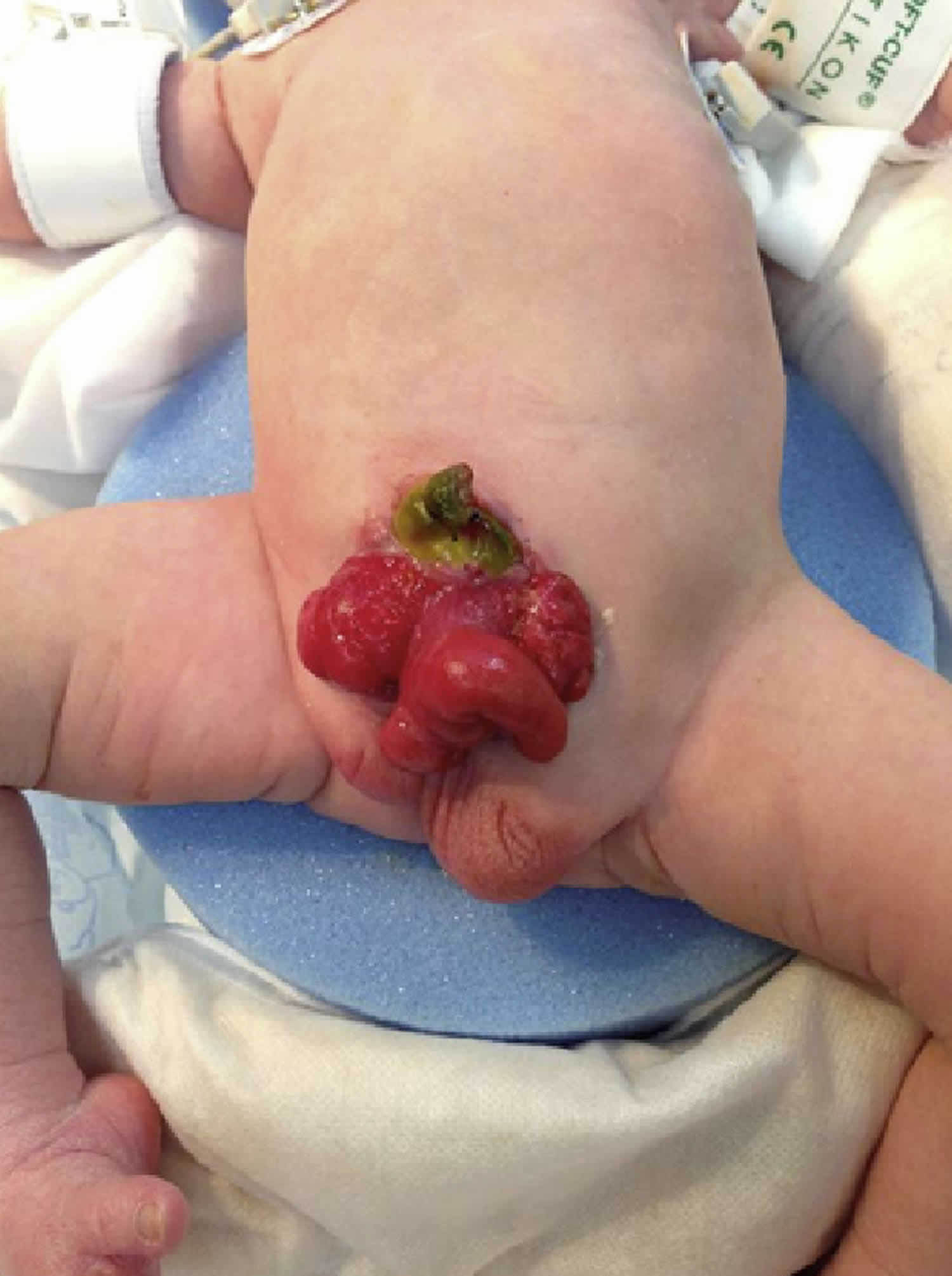
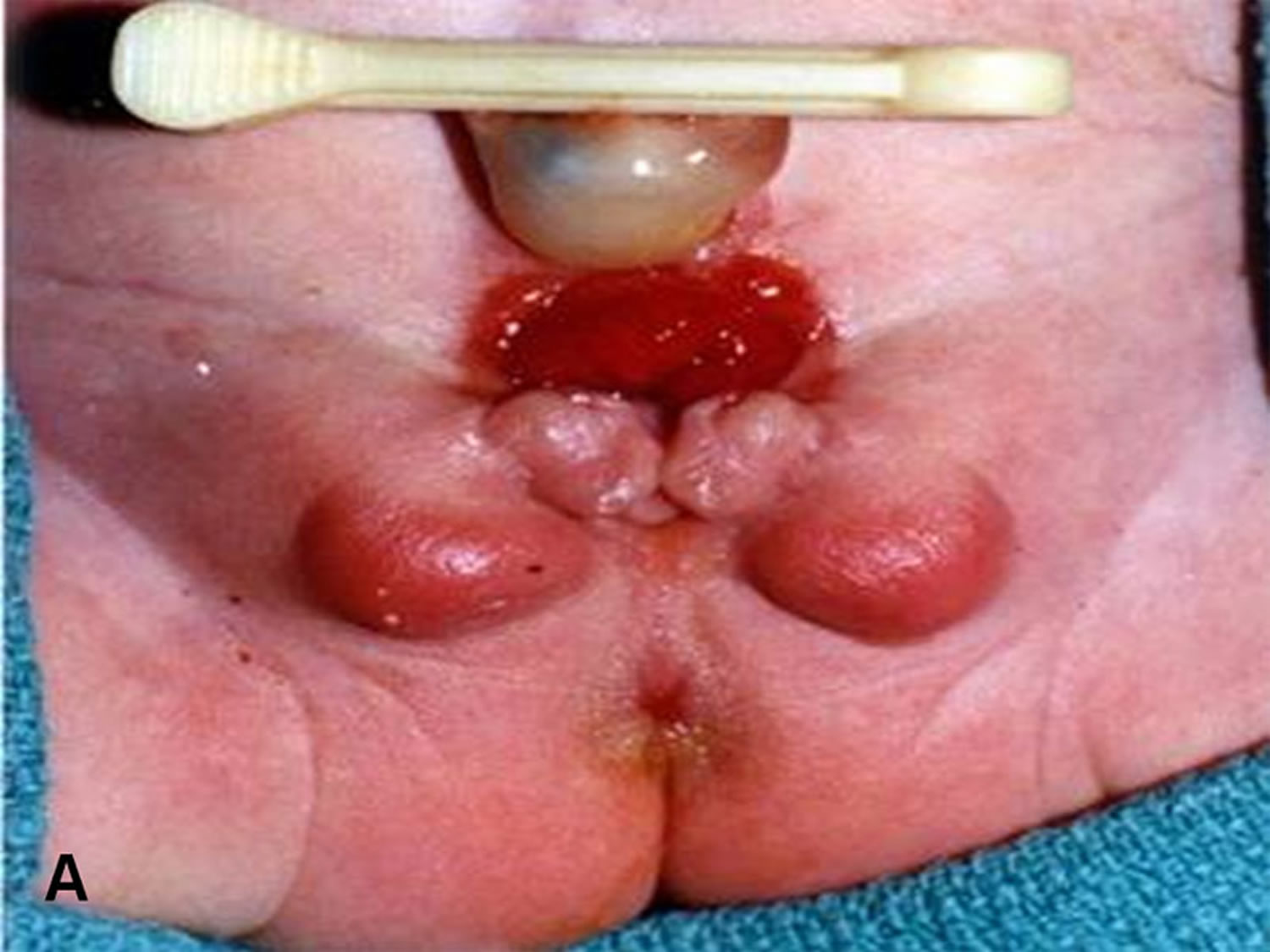
Figure 2. Cloacal exstrophy ultrasound

Cloacal exstrophy ultrasound

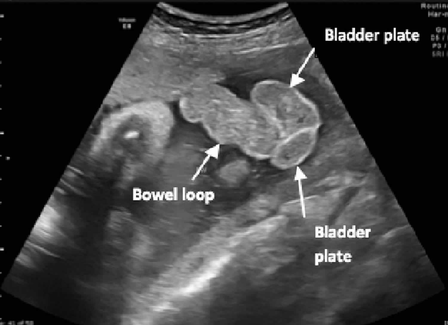
Footnote: Fetal ultrasound at 36 weeks’ gestation demonstrating bowel loops herniating between 2 bladder plates (“elephant trunk”).

[Source 3 ]

Figure 3. Cloacal bladder exstrophy female infant

cloacal bladder exstrophy female infant

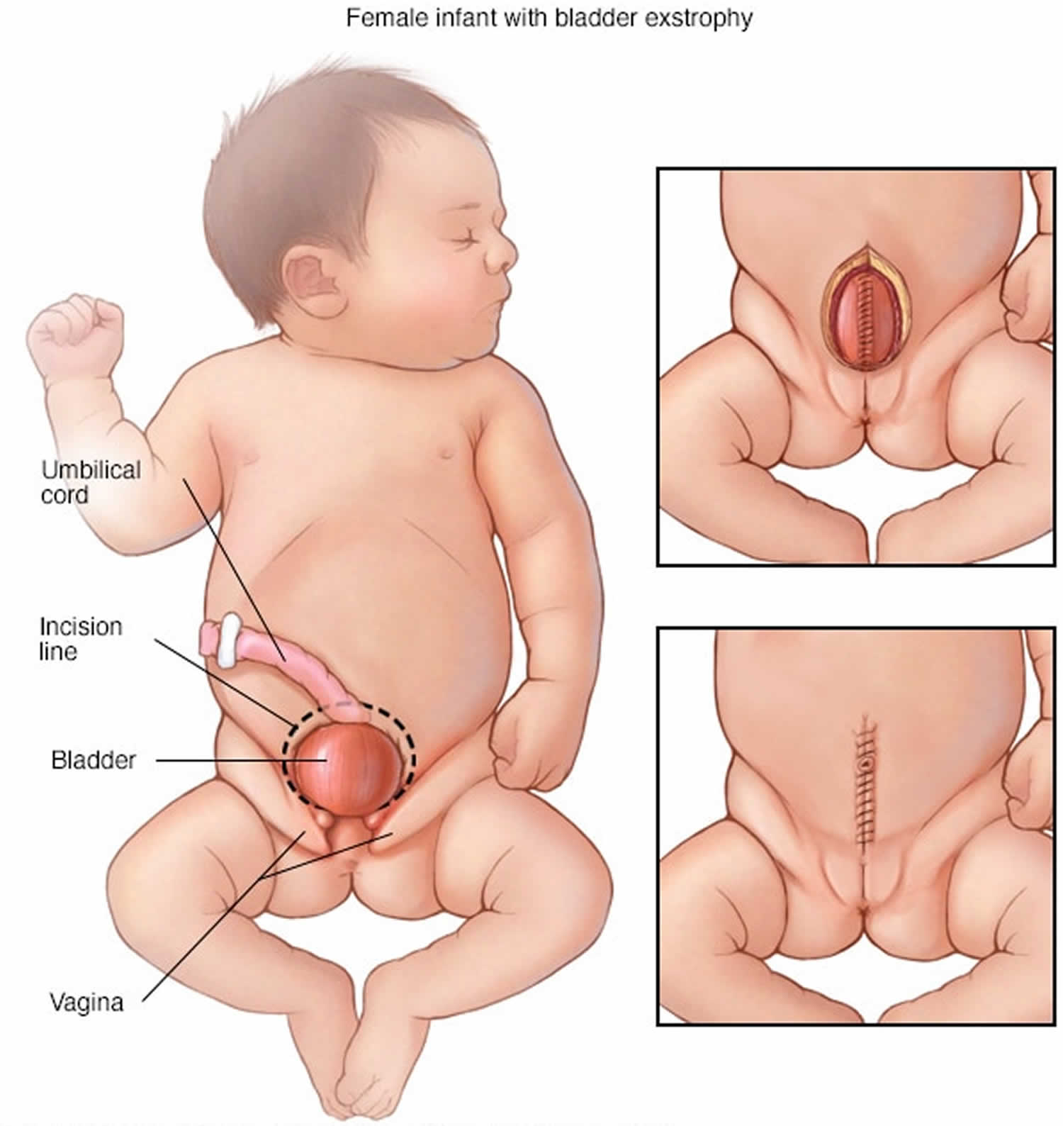
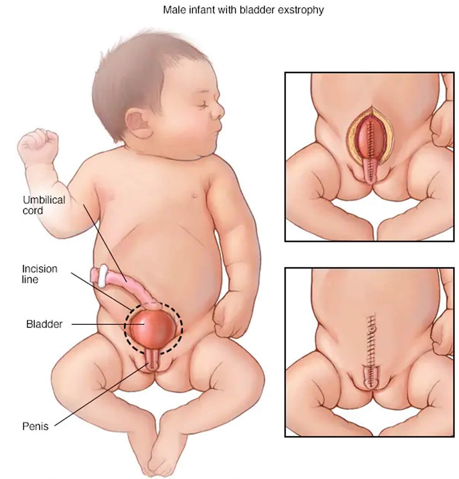
Figure 4. Cloacal bladder exstrophy male infant

Cloacal bladder exstrophy male infant

Will my child be able to have children when they reach adulthood?

In many cases, the answer to this question is: yes. But almost always, assisted fertility is necessary for adults.

With regard to sexuality, males are generally potent, but some report inadequate phallus or residual curvature. Females report normal sexual function 4.

With respect to fertility and childbearing, retrograde ejaculation or iatrogenic obstruction of the ejaculatory ducts or vas deferens after surgical reconstruction may result in abnormal semen analysis. Antegrade ejaculation is preserved after single-stage repair, but abnormal semen parameters are common. However, fertilization, with viable pregnancy, has been achieved by male patients with cloacal exstrophy 5.

Females have had successful pregnancies 6. Cesarean delivery is recommended to avoid injury to continence mechanism. Postpartum uterine prolapse is common because of aggravation of preexisting abnormal pelvic support.

Cloacal exstrophy causes

The cause of cloacal exstrophy is currently unknown, so there is also no known way to prevent it. On the basis of the known embryologic principles of cloacal development, any inciting event would have to occur early in pregnancy.

There is a higher incidence of cloacal exstrophy in families in which one member is affected as compared with the general population. Offspring of patients with exstrophy-epispadias complex have a 1 in 70 risk (500 times that of the general population) of being affected. Nevertheless, familial occurrence is uncommon in large series 7. The heritability of cloacal exstrophy has not been established, because no offspring have been reported. Moreover, there’s no evidence to suggest that anything done by expectant parents leads to the condition.

At present, 22q11.2 duplication is the genetic variant most commonly associated with bladder exstrophy-epispadias complex 8.

Cloacal exstrophy has been reported in twins. Concordance rates show strong evidence of genetic effects 9, but less than 100% concordance among identical twins suggests some role for environmental effect on development of exstrophy-epispadias.

A higher incidence of bladder exstrophy is observed in infants of younger mothers and in those with relatively high parity.

Maternal tobacco exposure is associated with more severe defects (cloacal vs classic exstrophy).

Growing evidence suggests an increased incidence of cloacal exstrophy and bladder exstrophy-epispadias with in-vitro fertilization (IVF) pregnancies 10.

Cloacal exstrophy symptoms

In some cases, cloacal exstrophy is detected from a routine prenatal ultrasound. In other cases, it isn’t diagnosed until birth, when physicians can clearly see the exposed organs.

Antenatal ultrasound findings suggestive of exstrophy-epispadias complex include the following:

  • Repeated failure to visualize the bladder on ultrasound
  • Lower-abdominal-wall mass
  • Low-set umbilical cord
  • Abnormal genitalia
  • Increased pelvic diameter

Additional antenatal ultrasound findings suggestive of cloacal exstrophy include the following:

  • Omphalocele
  • Limb abnormalities
  • Myelomeningocele
  • Trunk sign from prolapsed intestine

Increased use of fetal magnetic resonance imaging (MRI) may further improve the accuracy of antenatal diagnosis, but this test is not necessary if suspicion is high on the basis of ultrasound findings.

Classic bladder exstrophy and cloacal exstrophy are obvious to all in the delivery room. Variants of the exstrophy-epispadias complex exist, including skin-covered bladder exstrophy, duplicate bladders, superior vesical fistula, and epispadias with major bladder prolapse 11. Most exstrophy variants and epispadias are also identifiable at birth. Unrecognized female epispadias may present as persistent childhood incontinence. Unrecognized split-symphysis variants of exstrophy may be identified in childhood only because of persistent incontinence or a waddling gait.

Physical examination

Patients with classic bladder exstrophy or epispadias typically appear as term infants. Patients with cloacal exstrophy, however, are often preterm. They may have respiratory embarrassment requiring mechanical ventilation.
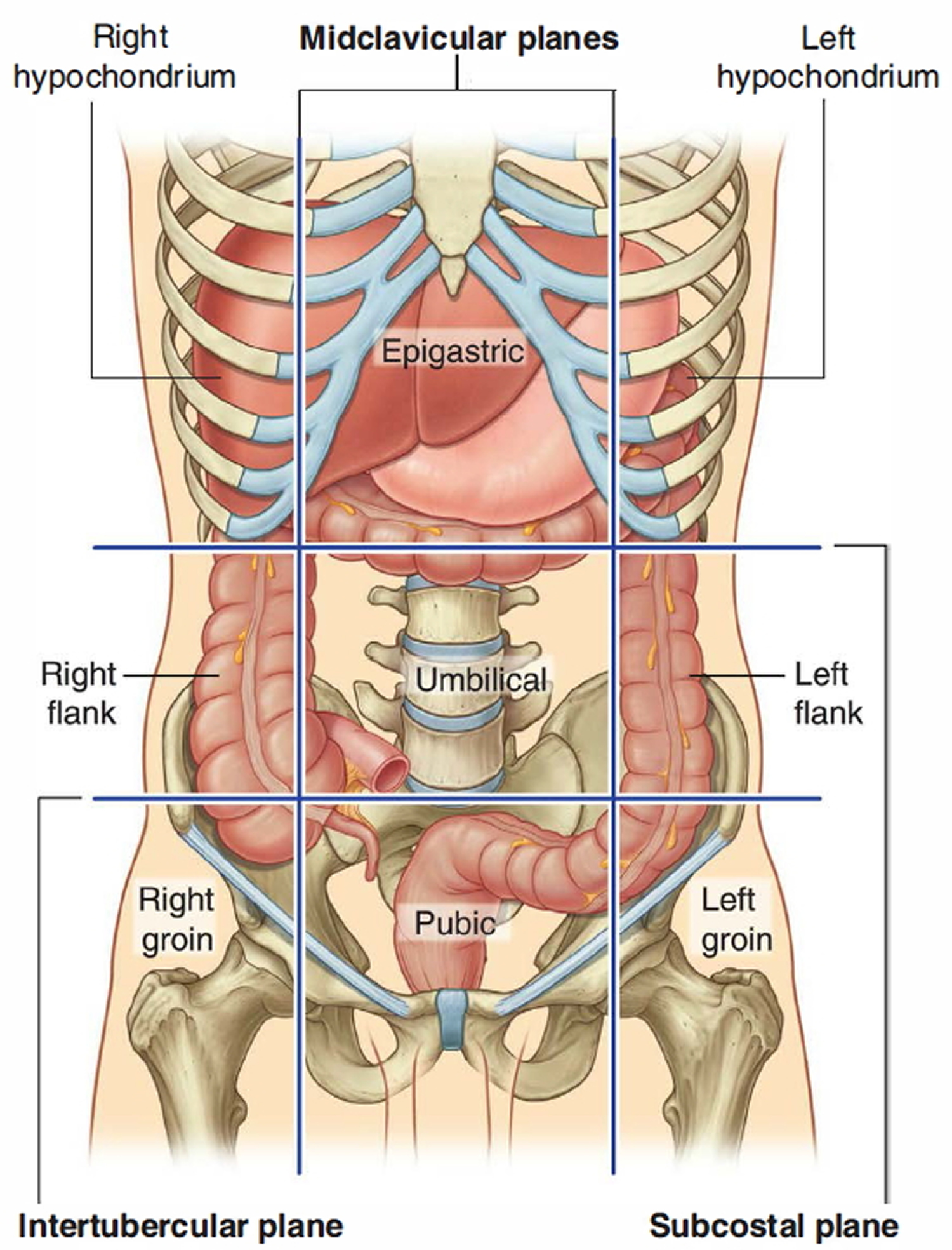
Abdominal findings

In classic cloacal bladder exstrophy (see the images above), the bladder is open on the lower abdomen, with mucosa fully exposed through a triangular fascial defect. The abdominal wall appears long because of a low-set umbilicus on the upper edge of the bladder plate. The distance between the umbilicus and anus is foreshortened. The recti diverge distally, attaching to the widely separated pubic bones. Indirect inguinal hernias are frequent (>80% of males, >10% of females) because of wide inguinal rings and the lack of an oblique inguinal canal.

Nearly all patients with cloacal exstrophy have an associated omphalocele. The bladder is open and separated into two halves, flanking the exposed interior of the cecum. Openings to the remainder of the hindgut and to one or two appendices are evident within the cecal plate. Terminal ileum may prolapse as a “trunk” of bowel onto the cecal plate.

In cloacal exstrophy variants, the pubic symphysis is widely separated, and the recti diverge distally. The umbilicus is low or elongated. A small superior bladder opening or a patch of isolated bladder mucosa may be present. The intact bladder may be externally covered by only a thin membrane. Isolated ectopic bowel segments have been reported.

Genital findings

In describing the anatomy of the penis, the terms dorsal and ventral refer to a normal phallus in the erect state. The dorsal surface is in continuity with the abdominal wall, and the ventral surface is in continuity with the scrotum.

In cloacal exstrophy, the penis is generally quite small and bifid, with a hemiglans located just caudal to each hemibladder. Infrequently, the phallus may be intact in the midline. In females, the clitoris is bifid, and two vaginas are present. The anus is absent.

In exstrophy variants, the genitalia generally are intact (see the image below), though epispadias can occur.

In classic bladder exstrophy in males, the phallus is short and broad with upward curvature (dorsal chordee). The glans lies open and flat like a spade, and the dorsal component of the foreskin is absent. The urethral plate extends the length of the phallus without a roof. The bladder plate and urethral plate are in continuity, with the verumontanum and ejaculatory ducts visible within the prostatic urethral plate. The anus is anteriorly displaced with a normal sphincter mechanism.

In classic bladder exstrophy in females, the clitoris is uniformly bifid with divergent labia superiorly. The open urethral plate is in continuity with the bladder plate. The vagina is anteriorly displaced. The anus is anteriorly displaced with a normal sphincter mechanism.

In male epispadias, the phallus is short and broad with upward curvature (dorsal chordee). The glans lies open and flat like a spade, and the dorsal component of the foreskin is absent. The urethral meatus is located on the dorsal penile shaft, anywhere between the penopubic angle and the proximal margin of the glans.

In female epispadias, the clitoris is most often bifid with divergent labia superiorly. The dorsal aspect of the urethra is open distally. The urethra and bladder neck are patulous and may allow visualization of bladder. Bladder mucosa may prolapse through the bladder neck.

Musculoskeletal findings

In classic bladder exstrophy, the pubic symphysis is widely separated. Divergent rectus muscles remain attached to the pubis. External rotation of the innominate bones results in a waddling gait in ambulatory patients but does not appear to result in orthopedic problems later in life.

In cloacal exstrophy, the examination is the same as for bladder exstrophy. As many as 65% of patients have a clubfoot or major deformity of a lower extremity. As many as 80% of patients have vertebral anomalies.

In split-symphysis variants of exstrophy, the pubic symphysis is widely separated (see the image below), and the rectus muscles are divergent.

Neurologic findings

In cloacal exstrophy, as many as 95% of patients have myelodysplasia, which may include myelomeningocele, lipomeningocele, meningocele, or other forms of occult dysraphism. These patients are at risk for neurologic deterioration, and they should be observed closely. Early neurosurgical consultation is appropriate if a radiographic abnormality of the spinal cord or canal is observed.

Cloacal exstrophy diagnosis

Cloacal exstrophy can usually be diagnosed by fetal ultrasound before an infant is born. Upon birth, a physical exam will confirm the diagnosis.

Laboratory studies

Before complex reconstruction of the urinary tract, it is important to obtain information about the patient’s baseline renal function. In patients with cloacal exstrophy, losses from the terminal ileum short-gut physiology can result in significant electrolyte abnormalities.

Imaging studies

Baseline examination of the kidneys with ultrasonography is recommended for all patients with exstrophy because increased bladder pressure after bladder closure can lead to hydronephrosis and upper urinary tract deterioration. Congenital upper urinary tract anomalies are uncommon with classic exstrophy and epispadias but are present in approximately one third of patients with cloacal exstrophy (eg, ectopic pelvic kidney, renal agenesis, or hydronephrosis).

Spinal ultrasound or radiography may be helpful. Myelodysplasia should be excluded in newborns with cloacal exstrophy. This can be accomplished by means of ultrasound early in life. In cloacal exstrophy, magnetic resonance imaging (MRI) is recommended to help identify occult abnormalities that may predispose to symptomatic spinal cord tethering.

Bilateral vesicoureteral reflux (VUR) is present in nearly all patients with classic bladder exstrophy. Voiding cystourethrography (VCUG) is performed in early childhood to assess bladder capacity in preparation for reconstructive continence surgery. Evaluation of the bladder neck and proximal urethra is recommended in patients with epispadias in order to plan surgical management.

Managing pregnancy after a cloacal exstrophy diagnosis

Because cloacal exstrophy is a high-risk condition, you will need to be monitored throughout your pregnancy. In some cases, pregnancy may be complicated by polyhydramnios (excess amniotic fluid) during the third trimester, which may trigger preterm labor and delivery. Your delivery should also be planned at our state-of-the-art facility. This way, our delivery team can address any complications should they arise and the baby will have immediate access to treatment and the best surgical professionals.

Cloacal exstrophy treatment

Cloacal exstrophy is treated through surgical repair after birth, usually in stages to address each defect. This requires an in-depth treatment plan to be created for your child’s specific needs. The extent of cloacal exstrophy surgery required for your baby depends on the type and severity of his or her abnormalities.

Your child will undergo a series of surgeries over a number of years — referred to as staged reconstruction. The exact timing, nature and outcome of each cloacal exstrophy surgery will depend on your child’s particular situation. Your child’s surgeons will create a treatment plan based on the type and the extent of your child’s condition and discuss the plan with you. Usually surgery begins in the first days of life with the highest-priority procedure. Surgeons usually repair the bladder, create a colostomy (an opening in the colon with an attached “bag” that allows stool to pass) and repair the abdominal wall defect.

Babies with spinal defects usually have them repaired sometime in the first few days of life. Later surgeries include urinary and genital reconstruction, as well as an operation to create a rectum and close the colostomy opening. There are no fetal interventions (surgical procedures while inside the uterus) for cloacal exstrophy.

Treatment may include:

  • Abdominal repair: Typically, soon after your child is born, the surgeons will repair the omphalocele by closing the bladder and creating a colostomy so your child can eliminate stool. With a colostomy, the large intestine is separated from the bladder halves and reclosed. The two halves of the bladder are brought together and placed into the abdomen. The end of the large intestine is brought to the surface of the skin through an opening in the abdomen. A plastic bag, called a colostomy pouch, is placed over the opening to collect the stool.
  • Other surgery, such as surgery to repair the spine, may be planned around the initial stage of the abdominal repair.

After the initial surgery, your child will remain in the hospital where we will monitor the intestine as it begins to function. Our team will work with you and your family to ensure that the plan for your child is clear and that you have access to the supports you need.

  • Osteotomies: Once your child has healed from his first procedure and had some time to grow, we will schedule the second stage of the repair. This primarily involves working on the bladder. The orthopedic surgeon on our team will perform osteotomies to help ensure that your child’s pelvis can best support the bladder over time. During the osteotomy the hip bones are cut and adjusted. Your child will need to be in traction or in a spica cast for several weeks following this surgery.
  • Pull-through procedure: If your child was born with a significant amount of colon and is capable of forming solid stool, a surgical procedure, known as a “pull through” may eventually be performed. The purpose of this procedure is to connect the colon to the rectum.

Subsequent surgeries may also involve major urinary reconstructive surgery and further genital reconstruction. These issues will be discussed with you and your family as your child grows up.

Cloacal exstrophy repair

Reconstruction of exstrophy-epispadias complex remains one of the greatest challenges facing the pediatric urologist. Many modifications in surgical procedures have improved outcomes, but the optimal approach remains uncertain. Longitudinal prospective assessment of the two main current surgical approaches (staged procedure and total reconstruction) is critical for optimizing functional and cosmetic outcomes.

Complete primary reconstruction is now more than 20 years old; however, each approach is in a constant state of minor modification. Data on this approach continue to mature and are updated almost yearly 12. Analysis of each experience focuses on daytime continence with volitional voiding, need for further surgical procedures, and complication rates. In experienced hands, the safety and efficacy of the different approaches are comparable.

Goals of therapy include provision of urinary continence with preservation of renal function and reconstruction of functional and cosmetically acceptable genitalia. Creation of a neoumbilicus is also important to many of these patients.

Surgical techniques used in the treatment of exstrophy-epispadias complex include the following:

  • Staged functional closure for classic bladder exstrophy (ie, modern staged repair of exstrophy) 13
  • Complete primary repair for classic bladder exstrophy
  • Urinary diversion for classic bladder exstrophy
  • Closure for cloacal exstrophy
  • Gender reassignment

Staged functional closure for classic bladder exstrophy

Modern staged repair of exstrophy, which represents the traditional surgical approach, comprises a series of operations. Initial bladder closure is completed within 72 hours of birth. If this is delayed, pelvic osteotomies are required to facilitate successful closure of the abdominal wall and to allow the bladder to lie within a closed and supportive pelvic ring.

Epispadias repair with urethroplasty is performed at age 12-18 months. This allows enough increase in bladder outlet resistance to improve the bladder capacity.

Bladder neck reconstruction (typically a modified Young-Dees-Leadbetter repair) is performed at age 4 years. This allows continence and correction of vesicoureteral reflux (VUR). Multiple modifications have been proposed. The procedure is delayed until bladder capacity is adequate; better results are reported with a capacity greater than 85 mL.

Chua et al retrospectively studied a modification of staged exstrophy repair aimed at incorporating the advantages of complete primary repair for classic bladder exstrophy by avoiding concurrent epispadias repair and adding bilateral ureteral reimplantation and bladder neck tailoring (staged repair of bladder exstrophy with bilateral ureteral reimplantation) at the initial repair 14. They found staged repair of bladder exstrophy with bilateral ureteral reimplantation to be a safe alternative for exstrophy-epispadias repair, preventing penile tissue loss and yielding long-term outcomes comparable to those of complete primary repair for classic bladder exstrophy.

The radical soft-tissue mobilization (radical soft-tissue mobilization) procedure, also referred to as the Kelly repair, has been suggested as an alternative approach to staged reconstruction of bladder exstrophy 15. Radical soft-tissue mobilization has been performed not only as the second part of a two-step strategy (after bladder closure) but also as part of a combined procedure that includes delayed bladder closure and radical soft-tissue mobilization in a single stage without pelvic osteotomy 16.

Complete primary repair for classic bladder exstrophy

Compared with modern staged repair of exstrophy, complete primary repair for classic bladder exstrophy is a newer approach to exstrophy closure. Primary bladder closure, urethroplasty, and genital reconstruction are performed in a single stage in newborns. This procedure involves complete penile disassembly in males and mobilization of the urogenital complex in females. Hypospadias is a common outcome in males and requires subsequent reconstruction.

The goal is early bladder cycling. A subset of patients have achieved continence without bladder neck reconstruction.

In a study of 34 boys treated with a modified penile disassembly technique (15 with bladder exstrophy who underwent complete primary repair for classic bladder exstrophy, 11 with penopupic epispadias after previous closure of bladder exstrophy, and eight with isolated complete epispadias), Anwar et al found the modified technique to yield excellent cosmetic results 17. Preservation of the distal urethral plate along with both hemiglans avoided shortening and prevented occurrence of hypospadias.

Urinary diversion for classic bladder exstrophy

Urinary diversion was the original surgical treatment of choice. Diversion may be performed in a patient with an extremely small bladder plate not suitable for functional closure 18. In Europe, early diversion has been widely used, with success for most exstrophy patients.

Closure for cloacal exstrophy

Treatment of myelodysplasia and gastrointestinal anomalies has priority over management of urinary and genital anomalies.

Closure can be either staged or performed in a single stage, depending on the overall condition of the child and the severity of the abdominal wall defect. If a large omphalocele is present, successful closure of the abdomen and the bladder in one stage may be difficult to accomplish.

The first stage involves separation of the gastrointestinal and genitourinary (genitourinary) tracts, closure of the colon, creation of a colostomy, and closure of the omphalocele. The bladder plates are brought together in the midline.

Because virtually all of these patients have some element of short-gut syndrome, the hindgut should be incorporated into the gastrointestinal tract to maximize absorptive surface area. Ileostomy should be avoided because of the high incidence of recurrent hospitalizations for dehydration and severe electrolyte abnormalities. The decision between rectal pull-through and permanent colostomy is based on the surgeon’s preference and the projected potential for social fecal continence 19.

Subsequent bladder closure is carried out as in surgical management of classic bladder exstrophy. The principles of complete primary repair have been applied at this point as well. Consideration may be given to continent diversion as the second stage, on the basis of poor potential for volitional voiding and continence.

Because of more severe pubic diastasis, pelvic osteotomies are required. Staged pelvic osteotomy (staged pelvic osteotomy) with gradual closure of the pelvis may be needed in severe cases 20. In a study comparing staged pelvic osteotomy before bladder closure with combined pelvic osteotomy (combined pelvic osteotomy) at the time of closure in cloacal exstrophy patients, Inouye et al found that staged pelvic osteotomy reduced preoperative diastasis more than combined pelvic osteotomy did, without appearing to incur increased rates of complication, closure failure, or incontinence 21.

Gender reassignment

Historically, all males with cloacal exstrophy underwent early gender conversion because of inadequate male genitalia. Testicular histology is normal despite frequent cryptorchidism.

Evidence suggesting that testosterone in utero has a significant impact on the developing brain has led to a change in surgical philosophy, as has anecdotal evidence suggesting that raising a 46,XY cloacal exstrophy patient as female can result in significant gender dysphoria. Cloacal exstrophy is now included as a subset of disorders of sex development 22. Multidisciplinary evaluation and both early and long-term counseling should be offered.

Intraoperative concerns

Multiple or lengthy surgical procedures with exposure to latex antigens increase the risk of latex sensitization or allergy 23. Approximately 30% of patients with bladder exstrophy have demonstrated symptoms of latex allergy, and 70% reveal sensitization (elevation of specific immunoglobulin E [IgE] antibody) to latex antigens. For practical purposes, all patients with exstrophy-epispadias complex should be considered to be latex-sensitive.

Full latex precautions are recommended in the operating room, beginning with preparation for the first operative procedure. Potential latex-containing materials in the operating room include gloves, catheters, drains, masks, anesthesia materials, bandages, and thromboembolic stockings. Polyvinyl chloride and silicone are acceptable alternatives. Latex allergy should be considered seriously in the event of intraoperative anaphylaxis. The offending agent should be removed and the surgical procedure aborted if necessary.

Treatment includes cardiopulmonary resuscitation with fluids, epinephrine, steroids, and histamine blockade. In those with a known latex allergy, premedication with steroids and histamine H1 and H2 blockers should be considered.

After cloacal exstrophy repair

The goal of surgeons and doctors is to help improve the child’s quality of life. Better tools for anesthesia and infant nutrition have helped to increase the survival rate for newborns with this condition.

Postoperatively, patients with exstrophy remain in the hospital in modified Bryant traction (legs adducted and pelvis slightly elevated) for 3 weeks after bladder closure. Alternative techniques of immobilization may be used, based on osteotomies or institutional protocol.

Bladder and kidneys are drained fully with multiple catheters during the first few weeks after closure.

Nutritional support is mandatory for patients with cloacal exstrophy. Patients with classic bladder exstrophy may also have early difficulties feeding because of the body position in traction.

It’s important to work closely with your health care team to prevent infection after surgery, and learn about long-term care. After surgery, a child born with cloacal exstrophy can usually grow to manage urine and stool in a socially acceptable way. Further operations may be needed over time to improve the child’s ability to control their bladder and bowel function. More surgery may also be needed to rebuild and/or make better the outer sex organs.

Time and patience will be important for the parents and child. Neurologic issues from spina bifida, if present, can be managed, but requires ongoing medical care.

Complications

In the treatment of complex congenital anomalies, the distinction between technical complications and problems inherent to the anomaly is not always obvious.

Failure of closure may occur. If the bladder plate is adequate, reclosure with pelvic osteotomies is recommended. In this instance, bladder closure and epispadias repair are performed in one stage. Urinary diversion is the alternative therapy.

A vesicocutaneous fistula or urethrocutaneous fistula may form after primary closure or urethral reconstruction. If spontaneous closure does not occur, surgical repair is required.

Loss of the hemiglans or corporal body has been reported as a result of complete primary repair 24.

Minor orthopedic complications may occur after osteotomy or immobilization.

Upper urinary tract deterioration is a potential complication. Causes include excessive outlet resistance and high pressure in a small-capacity reservoir and persistent VUR.

Abnormal bladder function may result in poor emptying. Clinical problems related to poor emptying include recurrent febrile infections, epididymitis, bladder stones, acute urinary retention, and rupture of the native bladder.

Bladder prolapse is a potential complication. Posterior bladder wall may prolapse through the patulous bladder neck after primary closure (see the image below). Recurrent prolapse, congestion, ischemia of bladder mucosa, or failure of ureteral drainage warrants early surgical correction.

Malignancy is a rare late complication of bladder exstrophy and is more common in untreated patients whose bladders are left exstrophic for many years. Adenocarcinoma is the most common of these malignancies, from the precursor cystitis glandularis, which is caused by chronic irritation and inflammation of exposed mucosa of the exstrophic bladder. Squamous cell carcinoma and rhabdomyosarcoma have also been reported.

Adenocarcinoma may develop adjacent to the ureterointestinal anastomosis in patients with urinary diversions that mix the urinary and fecal streams. This malignancy was reported in more than 10% of patients in one series 25. Patients younger than 25 years with ureterosigmoidostomy have a 7000-fold greater risk of adenocarcinoma of the colon than the general population (mean latency, 10 years).

Complications of short-gut syndrome are as follows:

  • Paucity of hindgut and, in many cases, limited small intestine can result in electrolyte abnormalities in patients with cloacal exstrophy
  • Dehydration is particularly a concern during an acute GI illness with diarrhea
  • Nutritional supplementation may be required

Cloacal exstrophy prognosis

Surgical techniques to treat cloacal exstrophy have improved dramatically in recent years, which means 90% to 100% of babies survive after surgery. Their quality of life and degree of need for ongoing care vary from case to case.

Mortality with classic bladder exstrophy or epispadias is rare. Historically, cloacal exstrophy was associated with significant mortality. Reconstruction was not attempted until the 1970s. Advances in the care of critically ill neonates and recognition of the importance of early parenteral nutritional support have allowed successful reconstruction and survival of children with cloacal exstrophy.

Survival rates after surgical treatment are excellent. With respect to bladder function or continence, reports vary according to the type of reconstruction performed 26. Objective and subjective evidence indicates that many exstrophic bladders do not function normally after reconstruction and may deteriorate over time.

Continence rates of 75-90% have been reported after staged reconstruction in classic exstrophy, but more than one continence procedure may be required (eg, bladder neck reconstruction, bladder augmentation, bladder neck sling, or artificial urinary sphincter). Many of these patients require clean intermittent catheterization (CIC) through the urethra or a continent stoma because they are unable to void spontaneously to completion. Less encouraging results also are reported.

Continence results after staged reconstruction are poor (< 25%) in cloacal exstrophy because of abnormal bladder innervation in many patients. Experience with rectal reservoirs (ureterosigmoidostomy and variants) for exstrophy continence demonstrates rates higher than 95%, but they present long-term malignancy risks 27. Continent reconstruction with intestinal bladder augmentation and clean intermittent catheterization has a success rate greater than 90%.

With regard to psychosocial concerns, education, employment, and social relationships generally are not affected substantially in adults with a history of bladder exstrophy and epispadias 28. Age-appropriate adaptive behaviors may be delayed in children with chronic medical conditions 29. One study revealed below-average daily living skills and socialization but above-average self-esteem. Children may need support in disclosing their condition to new peers.

Multiple anomalies associated with cloacal exstrophy can have a significant impact on daily life. Patients are affected by permanent colostomy, the need for clean intermittent catheterization, and impaired ambulation.

Diet

Some young patients with cloacal exstrophy are seriously affected by short-gut syndrome and may depend on long-term supplemental parenteral nutrition for growth and development.

References
  1. Exstrophy and Epispadias. https://emedicine.medscape.com/article/1014971-overview
  2. Bladder Exstrophy. https://www.auanet.org/education/auauniversity/education-products-and-resources/pathology-for-urologists/urinary-bladder/histoanatomic-abnormalities/malformations/bladder-exstrophy
  3. Clements MB, Chalmers DJ, Meyers ML, Vemulakonda VM. Prenatal diagnosis of cloacal exstrophy: a case report and review of the literature. Urology. 2014;83(5):1162-1164. doi:10.1016/j.urology.2013.10.050
  4. Bujons A, Lopategui DM, Rodríguez N, Centeno C, Caffaratti J, Villavicencio H. Quality of life in female patients with bladder exstrophy-epispadias complex: Long-term follow-up. J Pediatr Urol. 2016 Aug. 12 (4):210.e1-6.
  5. Stein R, Hohenfellner K, Fisch M, Stöckle M, Beetz R, Hohenfellner R. Social integration, sexual behavior and fertility in patients with bladder exstrophy–a long-term follow up. Eur J Pediatr. 1996 Aug. 155 (8):678-83.
  6. Dy GW, Willihnganz-Lawson KH, Shnorhavorian M, Delaney SS, Amies Oelschlager AM, Merguerian PA, et al. Successful pregnancy in patients with exstrophy-epispadias complex: A University of Washington experience. J Pediatr Urol. 2015 Aug. 11 (4):213.e1-6.
  7. Gambhir L, Höller T, Müller M, Schott G, Vogt H, Detlefsen B, et al. Epidemiological survey of 214 families with bladder exstrophy-epispadias complex. J Urol. 2008 Apr. 179 (4):1539-43.
  8. Beaman GM, Woolf AS, Cervellione RM, Keene D, Mushtaq I, Urquhart JE, et al. 22q11.2 duplications in a UK cohort with bladder exstrophy-epispadias complex. Am J Med Genet A. 2019 Jan 9.
  9. Reutter H, Qi L, Gearhart JP, Boemers T, Ebert AK, Rösch W, et al. Concordance analyses of twins with bladder exstrophy-epispadias complex suggest genetic etiology. Am J Med Genet A. 2007 Nov 15. 143A (22):2751-6.
  10. Wood HM, Babineau D, Gearhart JP. In vitro fertilization and the cloacal/bladder exstrophy-epispadias complex: a continuing association. J Pediatr Urol. 2007 Aug. 3 (4):305-10.
  11. Maruf M, Benz K, Jayman J, Kasprenski M, Michaud J, Di Carlo HN, et al. Variant Presentations of the Exstrophy-Epispadias Complex: A 40-Year Experience. Urology. 2018 Dec 18.
  12. Bhatnagar V. Bladder exstrophy: An overview of the surgical management. J Indian Assoc Pediatr Surg. 2011 Jul. 16 (3):81-7.
  13. Baird AD, Nelson CP, Gearhart JP. Modern staged repair of bladder exstrophy: a contemporary series. J Pediatr Urol. 2007 Aug. 3 (4):311-5.
  14. Chua ME, Ming JM, Fernandez N, Varghese A, Farhat WA, Bagli DJ, et al. Modified staged repair of bladder exstrophy: a strategy to prevent penile ischemia while maintaining advantage of the complete primary repair of bladder exstrophy. J Pediatr Urol. 2018 Sep 25.
  15. Ben-Chaim J, Hidas G, Wikenheiser J, Landau EH, Wehbi E, Kelly MS, et al. Kelly procedure for exstrophy or epispadias patients: Anatomical description of the pudendal neurovasculature. J Pediatr Urol. 2016 Jun. 12 (3):173.e1-6.
  16. Leclair MD, Faraj S, Sultan S, Audry G, Héloury Y, Kelly JH, et al. One-stage combined delayed bladder closure with Kelly radical soft-tissue mobilization in bladder exstrophy: preliminary results. J Pediatr Urol. 2018 Dec. 14 (6):558-564.
  17. Anwar AZ, Mohamed MA, Hussein A, Shaaban AM. Modified penile disassembly technique for boys with epispadias and those undergoing complete primary repair of exstrophy: long-term outcomes. Int J Urol. 2014 Sep. 21 (9):936-40.
  18. Ko JS, Lue K, Friedlander D, Baumgartner T, Stuhldreher P, DiCarlo HN, et al. Cystectomy in the Pediatric Exstrophy Population: Indications and Outcomes. Urology. 2018 Jun. 116:168-171.
  19. Levitt MA, Mak GZ, Falcone RA Jr, Peña A. Cloacal exstrophy–pull-through or permanent stoma? A review of 53 patients. J Pediatr Surg. 2008 Jan. 43 (1):164-8; discussion 168-70.
  20. Mathews R, Gearhart JP, Bhatnagar R, Sponseller P. Staged pelvic closure of extreme pubic diastasis in the exstrophy-epispadias complex. J Urol. 2006 Nov. 176 (5):2196-8.
  21. Inouye BM, Tourchi A, Di Carlo HN, Young EE, Mhlanga J, Ko JS, et al. Safety and efficacy of staged pelvic osteotomies in the modern treatment of cloacal exstrophy. J Pediatr Urol. 2014 Dec. 10 (6):1244-8.
  22. Houk CP, Lee PA. Consensus statement on terminology and management: disorders of sex development. Sex Dev. 2008. 2 (4-5):172-80.
  23. Shnorhavorian M, Grady RW, Andersen A, Joyner BD, Mitchell ME. Long-term followup of complete primary repair of exstrophy: the Seattle experience. J Urol. 2008 Oct. 180 (4 Suppl):1615-9; discussion 1619-20.
  24. Schaeffer AJ, Purves JT, King JA, Sponseller PD, Jeffs RD, Gearhart JP. Complications of primary closure of classic bladder exstrophy. J Urol. 2008 Oct. 180 (4 Suppl):1671-4; discussion 1674.
  25. Krishnamsetty RM, Rao MK, Hines CR, Saikaly EP, Corpus RP, DeBandi HO. Adenocarcinoma in exstrophy and defunctional ureterosigmoidostomy. J Ky Med Assoc. 1988 Aug. 86 (8):409-14.
  26. Kibar Y, Roth C, Frimberger D, Kropp BP. Long-term results of penile disassembly technique for correction of epispadias. Urology. 2009 Mar. 73 (3):510-4.
  27. Husmann DA, Rathbun SR. Long-term follow up of enteric bladder augmentations: the risk for malignancy. J Pediatr Urol. 2008 Oct. 4 (5):381-5; discussion 386.
  28. Mukherjee B, McCauley E, Hanford RB, Aalsma M, Anderson AM. Psychopathology, psychosocial, gender and cognitive outcomes in patients with cloacal exstrophy. J Urol. 2007 Aug. 178 (2):630-5; discussion 634-5.
  29. Ebert A, Scheuering S, Schott G, Roesch WH. Psychosocial and psychosexual development in childhood and adolescence within the exstrophy-epispadias complex. J Urol. 2005 Sep. 174 (3):1094-8.
read more
Digestive SystemSmall and Large Intestine

Large intestine

large intestine

Large intestine

The large intestine also known as the colon, extends from the distal end of the ileum to the anus, a distance of approximately 1.5 m in adults (5 ft) long, making up one-fifth of the length of the gastrointestinal (GI) tract and 6.5 cm (2.5 in.) in diameter. The large intestine is named for its relatively large diameter, not its length. The large intestine consists of the cecum, appendix, ascending colon, transverse colon, descending colon, rectum, and anal canal.

The large intestine is responsible for processing indigestible food material (chyme) after most nutrients are absorbed in the small intestine. The large intestine performs an essential role by absorbing water, vitamins, salts and electrolytes from the gut contents, thus forming feces.

Beginning in the right groin as the cecum, with its associated appendix, the large intestine continues upward as the ascending colon through the right flank and into the right hypochondrium. The ascending colon begins at the ileocecal valve and passes up the right side of the abdominal cavity. It makes a 90° turn at the right colic (hepatic) flexure, near the right lobe of the liver, and crosses the abdomen as the transverse colon to the left hypochondrium. At this position, just below the spleen, the large intestine bends downward, forming the left colic flexure (splenic flexure) and continues as the descending colon through the left flank and into the left groin. Ascending, transverse, and descending colons thus form a squarish, three-sided frame around the small intestine.

The cecum is a blind pouch in the lower right abdominal quadrant inferior to the ileocecal valve. Attached to its lower end is the appendix, a blind tube 2 to 7 cm long. The appendix is densely populated with lymphocytes and is a significant source of immune cells.

Figure 1. Large intestine

large intestinelarge intestine location

The pelvic cavity is narrower than the abdominal cavity, so at the hip bone, the colon turns medially and travels along the iliac fossa before turning downward at the pelvic inlet into the pelvic cavity. The resulting S-shaped portion of the tract is called the sigmoid colon. Visual examination of this region is performed with an instrument called a sigmoidoscope. In the pelvic cavity, the large intestine continues as the rectum, about 15 cm long. Despite its name, the rectum is not quite straight but has three lateral curves as well as an anteroposterior curve. It has three infoldings called transverse rectal folds (rectal valves), which enable it to retain feces while passing gas.

The final 3 cm of the large intestine is the anal canal, which passes through the levator ani muscle of the pelvic floor and terminates at the anus. Here, the mucosa forms longitudinal ridges called anal columns with depressions between them called anal sinuses. As feces pass through the canal, they press the sinuses and cause them to exude extra mucus and lubricate the canal during defecation. Prominent hemorrhoidal veins form superficial plexuses in the anal columns and around the orifice. Unlike veins in the limbs, they lack valves and are particularly subject to distension and venous pooling. Hemorrhoids are permanently distended veins that protrude into the anal canal or form bulges external to the anus. They can result from the impaired venous return that occurs in obesity and pregnancy.

The muscularis externa of the colon is unusual. Although it completely encircles the colon just as it does the small intestine, its longitudinal fibers are especially concentrated in three thickened, ribbonlike strips. Each strip is called a taenia coli. The muscle tone of the taeniae coli contracts the colon lengthwise and causes its wall to bulge, forming pouches called haustra (singular, haustrum). Haustra are conspicuous in colonic X-rays of living patients, because they disappear when muscle tone is lost at death. In the rectum and anal canal, the longitudinal muscle forms a continuous sheet and haustra are absent. The anus is regulated by two sphincters: an internal anal sphincter composed of smooth muscle of the muscularis externa and an external anal sphincter composed of skeletal muscle of the pelvic diaphragm.

The ascending and descending colon are retroperitoneal and have a serosa only on the anterior surface, whereas the transverse and sigmoid colon are entirely enclosed in serosa and anchored to the posterior abdominal wall by the mesocolon. The serosa of the transverse through sigmoid colon often has omental appendages, also called epiploic appendages, are fat-filled pouches of visceral peritoneum that hang from the intestine. Their significance is unknown.

Figure 2. Large intestine location

large intestine location

Figure 3. Large intestine and small intestine

large and small intestine

Figure 4. Anal canal

anal canal

Blood supply to the large intestine

The large intestine is served by mesenteric arteries and veins much like the small intestine. Branches of the superior mesenteric artery fan out to supply the ascending colon and most of the transverse colon; the inferior mesenteric artery supplies the rest of the transverse colon as well as the descending and sigmoid colon and the rectum. The superior and inferior mesenteric veins drain the same parts of the large intestine as the correspondingly named arteries and drain into the hepatic portal system.

Function of large intestine

The large intestine has 3 primary functions: absorbing water and electrolytes, producing and absorbing vitamins, and forming and propelling feces toward the rectum for elimination. By the time indigestible materials have reached the colon, most nutrients and up to 90% of the water has been absorbed by the small intestine. The role of the ascending colon is to absorb the remaining water and other key nutrients from the indigestible material, solidifying it to form stool. The descending colon stores feces that will eventually be emptied into the rectum. The sigmoid colon contracts to increase the pressure inside the colon, causing the stool to move into the rectum. The rectum holds the feces awaiting elimination by defecation.

Functions of the large intestine:

  1. Haustral churning, peristalsis, and mass peristalsis drive contents of colon into rectum.
  2. Bacteria in large intestine convert proteins to amino acids, break down amino acids, and produce some B vitamins and vitamin K.
  3. Absorption of some water, ions, and vitamins.
  4. Formation of feces.
  5. Defecation (emptying rectum).

Motility

The intestinal wall is made up of multiple layers. The 4 layers of the large intestine from the lumen outward are the mucosa, submucosa, muscular layer, and serosa. The muscular layer is made up of 2 layers of smooth muscle, the inner, circular layer, and the outer, longitudinal layer. These layers contribute to the motility of the large intestine. There are 2 types of motility present in the colon, haustral contraction and mass movement. Haustra are saccules in the colon that give it its segmented appearance. Haustral contraction is activated by the presence of chyme and serves to move food slowly to the next haustra, along with mixing the chyme to help with water absorption. Mass movements are stronger and serve to move the chyme to the rectum quickly.

Absorption of Water and Electrolytes

Absorption of water occurs by osmosis. Water diffuses in response to an osmotic gradient established by the absorption of electrolytes. Sodium is actively absorbed in the colon by sodium channels. Potassium is either absorbed or secreted depending on the concentration in the lumen. The electrochemical gradient created by the active absorption of sodium allows for this. Chloride ions are exchanged for bicarbonate ions across an electrochemical gradient.

Production/Absorption of Vitamins

The colon also plays a role in providing required vitamins through an environment that is conducive for bacterial cultivation. The colon houses trillions of bacteria that protect our gut and produce vitamins. The bacteria in the colon produce substantial amounts of vitamins by fermentation. Vitamin K and B vitamins, including biotin, are produced by the colonic bacteria. These vitamins are then absorbed into the blood. When dietary intake of these vitamins is low in an individual, the colon plays a significant role in minimizing vitamin disparity.

Mechanism

The wall of the large intestine contains the typical four layers found in the rest of the gastrointestinal tract: mucosa, submucosa, muscularis, and serosa. The mucosa consists of simple columnar epithelium, lamina propria (areolar connective tissue), and muscularis mucosae (smooth muscle). The epithelium contains mostly absorptive and goblet cells. The absorptive cells function primarily in water absorption; the goblet cells secrete mucus that lubricates the passage of the colonic contents. Both absorptive and goblet cells are located in long, straight, tubular intestinal glands (crypts of Lieberkühn) that extend the full thickness of the mucosa. Solitary lymphatic nodules are also found in the lamina propria of the mucosa and may extend through the muscularis mucosae into the submucosa.

Compared to the small intestine, the mucosa of the large intestine does not have as many structural adaptations that increase surface area. There are no circular folds or villi; however, microvilli are present on the absorptive cells. Consequently, much more absorption occurs in the small intestine than in the large intestine.

The mucosa of the large intestine has a simple columnar epithelium in all regions except the lower half of the anal canal, where it has a nonkeratinized stratified squamous epithelium. The latter provides more resistance to the abrasion caused by the passage of feces. There are no circular folds or villi in the large intestine, but there are intestinal crypts. They are deeper than in the small intestine and have a greater density of goblet cells; mucus is their only significant secretion. The lamina propria and submucosa have an abundance of lymphatic tissue, providing protection from the bacteria that densely populate the large intestine.

The submucosa of the large intestine consists of areolar connective tissue. The muscularis consists of an external layer of longitudinal smooth muscle and an internal layer of circular smooth muscle. Unlike other parts of the gastrointestinal tract, portions of the longitudinal muscles are thickened, forming three conspicuous bands called the teniae coli that run most of the length of the large intestine. The teniae coli are separated by portions of the wall with less or no longitudinal muscle. Tonic contractions of the bands gather the colon into a series of pouches called haustra (shaped like pouches), which give the colon a puckered appearance. A single layer of circular smooth muscle lies between teniae coli. The serosa of the large intestine is part of the visceral peritoneum. Small pouches of visceral peritoneum filled with fat are attached to teniae coli and are called omental (fatty) appendices.

Figure 5. Large intestine anatomy

layers of large intestine

Mechanical Digestion in the Large Intestine

The passage of chyme from the ileum into the cecum is regulated by the action of the ileocecal sphincter. Normally, the valve remains partially closed so that the passage of chyme into the cecum usually occurs slowly. Immediately after a meal, a gastroileal reflex intensifies peristalsis in the ileum and forces any chyme into the cecum. The hormone gastrin also relaxes the sphincter. Whenever the cecum is distended, the degree of contraction of the ileocecal sphincter intensifies.

Movements of the colon begin when substances pass the ileocecal sphincter. Because chyme moves through the small intestine at a fairly constant rate, the time required for a meal to pass into the colon is determined by gastric emptying time. As food passes through the ileocecal sphincter, it fills the cecum and accumulates in the ascending colon.

The most common type of colonic motility is a type of segmentation called haustral contractions, which occur about every 30 minutes. Distension of a haustrum with feces stimulates it to contract. This churns and mixes the residue, promotes water and salt absorption, and passes the residue distally to another haustrum. Peristalsis also occurs, although at a slower rate (3–12 contractions per minute) than in more proximal portions of the tract. A final type of movement is mass peristalsis, a strong peristaltic wave that begins at about the middle of the transverse colon and quickly drives the contents of the colon into the rectum. Because food in the stomach initiates this gastrocolic reflex in the colon, mass peristalsis usually takes place three or four times a day, during or immediately aft er a meal. They last about 15 minutes and move residue several centimeters at a time. Mass peristalsis occur especially in the transverse to sigmoid colon, often within an hour after breakfast, moving the feces that accumulated and stretched the colon overnight.

The large intestine takes 36 to 48 hours to reduce the residue of a meal to feces, with the residue spending the greatest time (about 24 h) in the transverse colon. The colon doesn’t chemically change the residue, but reabsorbs water and electrolytes (especially NaCl) from it. Feces usually consist of about 75% water and 25% solids. The solids are about 30% bacteria, 30% undigested dietary fiber, 10% to 20% fat, and smaller amounts of protein, sloughed epithelial cells, salts, mucus, and other digestive secretions. The fat is not from the diet but from bacteria and broken-down epithelial cells.

Chemical Digestion in the Large Intestine

The final stage of digestion occurs in the colon through the activity of bacteria that inhabit the lumen. Mucus is secreted by the glands of the large intestine, but no enzymes are secreted. Chyme is prepared for elimination by the action of bacteria, which ferment any remaining carbohydrates and release hydrogen, carbon dioxide, and methane gases. These gases contribute to flatus (gas) in the colon, termed flatulence when it is excessive. Bacteria also convert any remaining proteins to amino acids and break down the amino acids into simpler substances: indole, skatole, hydrogen sulfide, and fatty acids. Some of the indole and skatole is eliminated in the feces and contributes to their odor; the rest is absorbed and transported to the liver, where these compounds are converted to less toxic compounds and excreted in the urine.

Bacteria also decompose bilirubin to simpler pigments, including stercobilin, which gives feces their brown color. Bacterial products that are absorbed in the colon include several vitamins needed for normal metabolism, among them some B vitamins and vitamin K.

Large Intestine Microbes and Gas

The large intestine harbors about 800 species of bacteria collectively called the gut microbiome. You have a mutually beneficial relationship with many of these. You provide them with room and board while they provide you with nutrients from your food that you are not equipped to extract on your own. For example, they digest cellulose, pectin, and other plant polysaccharides for which you have no digestive enzymes, and you absorb the resulting sugars. Thus, you get more nutrition from your food because of these bacteria than you would get without them. Indeed, one person may get more calories than another from the same amount of food because of differences in their bacterial populations. Some bacteria also synthesize B vitamins and vitamin K, which are absorbed by the colon. This vitamin K is especially important because the diet alone usually does not provide enough to ensure adequate blood clotting.

One of the less desirable and sometimes embarrassing products of these bacteria is intestinal gas. The large intestine contains about 7 to 10 L of gas, expelling about 500 mL/day as flatus and reabsorbing the rest. Much of this is swallowed air that has worked its way through the digestive tract, but the gut microbes add to it. Painful cramping can result when undigested nutrients pass into the colon and furnish an abnormal substrate for bacterial action, so the bacteria produce excess gas—for example, in lactose intolerance. Flatus is composed mostly of nitrogen (N2), carbon dioxide (CO2), hydrogen (H2), methane (CH4), hydrogen sulfide (H2S), and two amines: indole and skatole. Indole, skatole, and H2S produce most of the odor of flatus and feces, whereas the others are odorless. The hydrogen gas is combustible and has been known to explode during the use of electrical cauterization in surgery.

Absorption and Feces Formation in the Large Intestine

By the time chyme has remained in the large intestine 3–10 hours, it has become solid or semisolid because of water absorption and is now called feces. Chemically, feces consist of water, inorganic salts, sloughed-off epithelial cells from the mucosa of the gastrointestinal tract, bacteria, products of bacterial decomposition, unabsorbed digested materials, and indigestible parts of food. Although 90% of all water absorption occurs in the small intestine, the large intestine absorbs enough to make it an important organ in maintaining the body’s water balance. Of the 0.5–1.0 liter of water that enters the large intestine, all but about 100–200 mL is normally absorbed via osmosis. The large intestine also absorbs ions, including sodium and chloride, and some vitamins.

The Defecation Reflex

Mass peristaltic movements push fecal material from the sigmoid colon into the rectum. The resulting distension of the rectal wall stimulates stretch receptors, which initiates a defecation reflex that results in defecation, the elimination of feces from the rectum through the anus. The defecation reflex occurs as follows: In response to distension of the rectal wall, the receptors send sensory nerve impulses to the sacral spinal cord.

Motor impulses from the cord travel along parasympathetic nerves back to the descending colon, sigmoid colon, rectum, and anus. The resulting contraction of the longitudinal rectal muscles shortens the rectum, thereby increasing the pressure within it. This pressure, along with voluntary contractions of the diaphragm and abdominal muscles, plus parasympathetic stimulation, opens the internal anal sphincter.

The external anal sphincter is voluntarily controlled. If it is voluntarily relaxed, defecation occurs and the feces are expelled through the anus; if it is voluntarily constricted, defecation can be postponed. Voluntary contractions of the diaphragm and abdominal muscles aid defecation by increasing the pressure within the abdomen, which pushes the walls of the sigmoid colon and rectum inward. If defecation does not occur, the feces back up into the sigmoid colon until the next wave of mass peristalsis stimulates the stretch receptors, again creating the urge to defecate. In infants, the defecation reflex causes automatic emptying of the rectum because voluntary control of the external anal sphincter has not yet developed.

Figure 6. Defecation neural control

defecation neural control

The amount of bowel movements that a person has over a given period of time depends on various factors such as diet, health, and stress. The normal range of bowel activity varies from two or three bowel movements per day to three or four bowel movements per week. Diarrhea is an increase in the frequency, volume, and fluid content of the feces caused by increased motility of and decreased absorption by the intestines. When chyme passes too quickly through the small intestine and feces pass too quickly through the large intestine, there is not enough time for absorption. Frequent diarrhea can result in dehydration and electrolyte imbalances. Excessive motility may be caused by lactose intolerance, stress, and microbes that irritate the gastrointestinal mucosa.

Constipation refers to infrequent or difficult defecation caused by decreased motility of the intestines. Because the feces remain in the colon for prolonged periods, excessive water absorption occurs, and the feces become dry and hard. Constipation may be caused by poor habits (delaying defecation), spasms of the colon, insufficient fiber in the diet, inadequate fluid intake, lack of exercise, emotional stress, and certain drugs. A common treatment is a mild laxative, such as milk of magnesia, which induces defecation. However, many physicians maintain that laxatives are habit-forming, and that adding fiber to the diet, increasing the amount of exercise, and increasing fluid intake are safer ways of controlling this common problem.

Large intestine problems

Pathology of the large intestine is common. One out of every 10 Americans over the age of 40 have diverticular disease, and around 3 million people in the United States have inflammatory bowel disease 1. It is important to incorporate a healthy diet and lifestyle to maintain a properly functioning colon. Eating a diet high in fiber and drinking plenty of water allows food to easily move through the colon, keeping the colon relatively clean, which can decrease the risk of diverticular disease. It is also important to maintain healthy colonic flora. Maintaining healthy colonic flora will decrease the risk of abdominal bloating, gas, diarrhea, constipation, and infectious colitis.

Disorders of Large Intestinal Motility

Irritable Bowel Syndrome

Irritable bowel syndrome is thought to be due to psychological factors influencing the motility of the large intestine via the extrinsic autonomic nervous system. During times of stress, segmentation contractions may be increased or decreased, resulting in constipation or diarrhea.

Hirschsprung Disease: Megacolon

Hirschsprung disease is a disorder at birth that occurs when nerve cells are absent (Auerbach’s Plexus) in the muscles of the colon. This affects motility in the colon, making it difficult to pass stool.

Diverticulosis/Diverticulitis

Diverticulosis is a disorder in which pockets develop in the colonic mucosa due to the weakness of the muscle layers in the colon wall. This usually occurs over time from chronic attrition of the aging process. Diverticulitis can develop if these pockets get infected or inflamed, causing abdominal pain and change in bowel movements. Diverticular disease is very common, especially in older adults.

Large Intestine Inflammation

Inflammatory Bowel Disease

Inflammatory bowel disease includes either Crohn’s disease or ulcerative colitis. Both cause inflammation and scarring within the digestive tract, disrupting the normal function. The cause of inflammatory bowel disease is not known but is likely due to an abnormal response of the immune system. Ulcerative colitis is confined to the large intestine, whereas Crohn’s disease can occur anywhere in the GI (gastrointestinal) tract, from mouth to anus.

Ischemic colitis

Ischemic colitis is more common in the elderly and occurs when there is decreased blood flow to the colon. Decreased blood flow can cause inflammation or injury to the colon. Some causes of ischemic colitis are atherosclerosis of arteries, low blood pressure, blood clots, and bowel obstruction.

Infectious colitis

Infectious colitis can occur from many different viruses, bacteria, or parasites. Infectious colitis most commonly occurs due to ingestion of contaminated food or water, introducing the infectious organism into the colon. The most common causes are Escherichia coli, Campylobacter, Shigella, and Salmonella. These infectious organisms invade the colon, cause inflammation, and affect the normal function, causing abdominal pain and diarrhea. Clostridium difficile is another organism that can cause colitis in association with antibiotic use. Clostridium difficile is part of healthy, normal flora in the colon but can cause problems if it overgrows. Antibiotic use can destroy other susceptible normal flora in the colon, allowing overgrowth and invasion of Clostridium difficile.

References
  1. Azzouz LL, Sharma S. Physiology, Large Intestine. [Updated 2018 Jun 14]. In: StatPearls [Internet]. Treasure Island (FL): StatPearls Publishing; 2018 Jan-. Available from: https://www.ncbi.nlm.nih.gov/books/NBK507857
read more
Digestive SystemSmall and Large Intestine

Small intestine

small intestine

Small intestine

The small intestine is the longest part of the alimentary canal and it begins at the pyloric sphincter of the stomach, coils through the central and inferior part of the abdominal cavity, and eventually opens into the large intestine.

The term small intestine refers not to its length but to its diameter—about 2.5 cm (1 in.).

The small intestine is approximately 6 to 7 m long (range 3–7 m) with a narrowing diameter from beginning to end.

The small intestine consists of the duodenum, the jejunum, and the ileum.

The small intestine receives chyme from the stomach and secretions from the pancreas, liver, and gallbladder. It completes digestion of the nutrients in chyme, absorbs the products of digestion, and transports the residue to the large intestine.

The small intestine the site of most enzymatic digestion and about 90 percent of all nutrients absorption and most of the rest occurs in the proximal portion of the large intestine. Its length alone provides a large surface area for digestion and absorption, and that area is further increased by circular folds, villi, and microvilli. Most digestive enzymes that operate within the small intestine are secreted not by the intestine, but by the pancreas. During digestion, the small intestine undergoes active segmentation movements, shuffling the chyme back and forth and thereby maximizing its contact with the nutrient-absorbing mucosa.

Peristalsis propels chyme through the small intestine in about 3–6 hours.

Figure 1. Small intestine

small intestine

Function of the Small Intestine

The anatomy of the small intestine is specialized to increase its surface area for absorption and secretion. The intestinal lining has a series of ring-shaped projections called circular folds or plicae circulares.

These folds do not disappear as the small intestine fills. Roughly 800 circular folds (about 2 per centimeter) are found along the length of the duodenum, jejunum, and proximal half of the ileum. The mucosa possesses intestinal villi, and each cell of the surface epithelium has small microvilli on its apical surface.

  1. Segmentations mix chyme with digestive juices and bring food into contact with mucosa for absorption; peristalsis propels chyme through small intestine.
  2. Completes digestion of carbohydrates, proteins, and lipids; begins and completes digestion of nucleic acids.
  3. Absorbs about 90% of nutrients and water that pass through digestive system.

Figure 2. Circular folds, villi and microvilli increase the surface area of the small intestine for digestion and absorption

function of small intestine

The tissue layers of the small intestine are reminiscent of those in the esophagus and stomach with modifications appropriate for nutrient digestion and absorption. The lumen is lined with simple columnar epithelium. The muscularis externa is notable for a thick inner circular layer and a thinner outer longitudinal layer.

The jejunum and ileum are intraperitoneal and thus covered on all sides with a serosa, which is continuous with the complex, folded mesentery that suspends the small intestine from the posterior abdominal wall. Most of the duodenum is retroperitoneal and has a serosa only on its anterior surface; its other surfaces are covered by adventitia.

Effective digestion and absorption require the small intestine to have a large internal surface area. This is provided by its relatively great length and by three kinds of internal folds or projections: the circular folds, villi, and microvilli. If the mucosa were smooth, like the inside of a hose, it would have a surface area of about 0.3 to 0.5 m2, but with these surface elaborations, its actual surface area is about 200 m2—clearly a great advantage for nutrient absorption.

The circular folds increase the surface area by a factor of 2 to 3, the villi by a factor of 10, and the microvilli by a factor of 20.

Circular folds, the largest of these elaborations, are transverse to spiral ridges up to 1 cm high. These involve only the mucosa and submucosa; they are not visible on the external surface, which is smooth. They slow the progress of the chyme and make it flow on a somewhat spiral path, which increases its contact with the mucosa and promotes more thorough mixing and nutrient absorption. Circular folds begin in the duodenum. In the jejunum, they are especially large, tall, and closely spaced. They become smaller and more sparse in the ileum. These changes are correlated with the relative amount of nutrient absorption occurring in each region. Circular folds are absent from the distal half of the ileum, but most nutrient absorption is completed by that point.

Villi (singular, villus) are tiny projections that give the inner lining of the intestine a fuzzy texture, like a terry cloth towel. They are about 0.5 to 1.0 mm high, with tongue- to finger like shapes. Villi are largest in the duodenum and become progressively smaller in more distal regions of the intestine. Villi are covered with two kinds of epithelial cells: columnar enterocytes (absorptive cells) and mucus-secreting goblet cells. Like epithelial cells of the stomach, those of the small intestine are joined by tight junctions that prevent digestive enzymes from seeping between them and eroding the underlying tissue.

The core of a villus is filled with areolar tissue of the lamina propria and contains an arteriole, blood capillaries, a venule, and a lymphatic capillary called a lacteal. The blood capillaries absorb most nutrients, but the lacteal absorbs most lipids. Lipids give its contents a milky appearance for which the lacteal is named. The core of the villus also has a few smooth muscle cells that contract periodically. This enhances mixing of the chyme in the intestinal lumen and milks lymph down the lacteal to larger lymphatics in the submucosa.

Microvilli are much smaller plasma membrane extensions, about 1 μm high, that form a fuzzy brush border on the surface of each enterocyte. In addition to increasing surface area, they contain brush border enzymes in the plasma membrane. These enzymes carry out some of the final stages of chemical digestion. They are not secreted into the lumen; instead, the chyme must contact the brush border for digestion to occur. This process, called contact digestion, is one reason why it is so important that intestinal contractions churn the chyme and ensure that it all contacts the mucosa.

On the floor of the small intestine, between the bases of the villi, there are numerous pores that open into tubular glands called intestinal crypts. These crypts, similar to the gastric glands, extend as far as the muscularis mucosae. In the upper half, they consist of enterocytes and goblet cells like those of the villi. The lower half is dominated by dividing stem cells. In its life span of 3 to 6 days, an epithelial cell migrates up the crypt to the tip of the villus, where it is sloughed off and digested. A few Paneth cells are clustered at the base of each crypt. They secrete lysozyme, phospholipase, and defensins—defensive proteins that resist bacterial invasion of the mucosa.

The duodenum has prominent duodenal glands in the submucosa. They secrete an abundance of bicarbonate-rich mucus, which neutralizes stomach acid and shields the mucosa from its erosive effects. Throughout the small intestine, the lamina propria and submucosa have a large population of lymphocytes that intercept pathogens before they can invade the bloodstream. In some places, these are aggregated into conspicuous lymphatic nodules such as the Peyer patches of the ileum.

Digestion and Absorption of the Major Organic Nutrients in the Small Intestine

Chemical digestion and nutrient absorption are essentially finished by the time food residue leaves the small intestine and enters the cecum.

Absorption of Water

The digestive system is one of several systems involved in fluid balance. The digestive tract receives about 9 L of water per day—0.7 L in food, 1.6 L in drink, and 6.7 L in the gastrointestinal secretions: saliva, gastric juice, bile, pancreatic juice, and intestinal juice. About 8 L of this is absorbed by the small intestine and 0.8 L by the large intestine, leaving 0.2 L voided in the daily fecal output.

Water is absorbed by osmosis, following the absorption of salts and organic nutrients that create an osmotic gradient from the intestinal lumen to the extracellular fluid (ECF). Diarrhea occurs when the large intestine absorbs too little water. This occurs when the intestine is irritated by bacteria and feces pass through too quickly for adequate reabsorption, or when the feces contain abnormally high concentrations of a solute such as lactose that opposes osmotic  absorption of water. Constipation occurs when fecal movement is slow, too much water is reabsorbed, and the feces become hardened. This can result from lack of dietary fiber, lack of exercise, emotional upset, or long-term laxative abuse.

Absorption of Carbohydrates

Most digestible dietary carbohydrate is starch. Starch is digested first to oligosaccharides up to eight glucose residues long, then into the disaccharide maltose, and finally to glucose, which is absorbed by the small intestine.

The process begins in the mouth, where salivary amylase breaks starch down into shorter segments (oligosaccharides). Salivary amylase functions best at pH 6.8 to 7.0, typical of the oral cavity. It is quickly denatured upon contact with stomach acid, but it can digest starch for as long as 1 to 2 hours in the stomach as long as it is in the middle of a food mass and escapes contact with the acid. Amylase therefore works longer when the meal is larger, especially in the fundus, where gastric motility is weakest and a food bolus takes longer to break up. As acid, pepsin, and the churning contractions of the stomach break up the bolus, amylase is denatured; it cannot function at a pH any lower than 4.5. Being a protein, amylase is then digested by pepsin along with the dietary proteins.

About 50% of the dietary starch is digested before it reaches the small intestine. Its digestion resumes in the small intestine when the chyme mixes with pancreatic amylase. Starch is entirely converted to oligosaccharides and maltose within 10 minutes. Its digestion is completed as the chyme contacts the brush border of the enterocytes. Two brush border enzymes, dextrinase and glucoamylase, hydrolyze oligosaccharides that are three or more residues long, while maltase hydrolyzes maltose. The end product of all of these is glucose, which is then absorbed.

Maltose is also present in some foods, but the major dietary disaccharides are sucrose (cane sugar) and lactose (milk sugar). They are digested by the brush border enzymes sucrase and lactase, respectively, and the resulting monosaccharides are immediately absorbed (glucose and fructose from the former; glucose and galactose from the latter). In most of the world population, however, lactase production ceases or declines to a low level after age 4 and lactose becomes indigestible.

The plasma membrane of the enterocytes has transport proteins that absorb monosaccharides as fast as they are produced by the foregoing enzymes. About 80% of the absorbed sugar is glucose, which is taken up by a sodium–glucose transporter like that of the kidney tubules. The glucose is subsequently transported out the base of the cell into the extracellular fluid (ECF). Sugar entering the extracellular fluid (ECF) increases its osmolarity, and this draws water osmotically from the lumen of the intestine, through the now-leaky tight junctions between the epithelial cells. Water carries more glucose and other nutrients with it by solvent drag, much as it does in the kidney. After a high-carbohydrate meal, solvent drag absorbs two to three times as much glucose as the sodium–glucose transporter.

The sodium–glucose transporter also absorbs galactose, whereas fructose is absorbed by facilitated diffusion using a separate carrier that doesn’t depend on Na+. Inside the enterocyte, most fructose is converted to glucose. Glucose, galactose, and the small amount of remaining fructose are then transported out the base of the cell by facilitated diffusion and absorbed by the blood capillaries of the villus. The hepatic portal system delivers them to the liver.

Absorption of Vitamins

Vitamins are not digested, but absorbed unchanged. The fat-soluble vitamins A, D, E, and K are absorbed with other lipids. Therefore, if they are ingested without fat-containing food, such as by simply popping vitamin tablets with a glass of water, they’re not absorbed at all but passed in the feces and wasted. Water-soluble vitamins (the B complex and vitamin C) are absorbed by simple diffusion. An exception is vitamin B12, an unusually large molecule that is absorbed poorly unless bound to the intrinsic factor (IF) secreted by the stomach’s parietal cells. Then as it passes down the small intestine, the B12–IF complex binds to receptors on absorptive cells of the distal ileum, where it is taken up by receptor-mediated endocytosis.

Absorption of Proteins

The amino acids absorbed by the small intestine come from three sources: (1) dietary proteins, (2) digestive enzymes digested by each other, and (3) sloughed epithelial cells digested by these enzymes. Amino acids from the last two sources total about 30 g/day, compared with about 44 to 60 g/day from the diet.

Enzymes that digest proteins are called proteases (peptidases). They are absent from the saliva but first encountered in the stomach. Here, pepsin hydrolyzes any peptide bond between tyrosine and phenylalanine, thereby digesting 10% to 15% of the dietary protein into shorter polypeptides and a small amount of free amino acids. Pepsin has an optimal pH of 1.5 to 3.5, so it is inactivated when it passes into the duodenum and mixes with the alkaline pancreatic juice (pH 8).

In the small intestine, the pancreatic enzymes trypsin and chymotrypsin take over protein digestion by hydrolyzing polypeptides into even shorter oligopeptides. Finally, these are taken apart one amino acid at a time by three more enzymes: (1) Carboxypeptidase removes amino acids from the —COOH end of the chain; (2) aminopeptidase removes them from the —NH2 end; and (3) dipeptidase splits dipeptides in the middle and releases the last two free amino acids. The last two of these are brush border enzymes, whereas carboxypeptidase is a pancreatic secretion.

Amino acid absorption is similar to that of monosaccharides. Enterocytes have several sodium-dependent amino acid cotransporters for different classes of amino acids. Dipeptides and tripeptides can also be absorbed, but they are hydrolyzed within the enterocytes before their amino acids are released to the bloodstream. At the basal surfaces of the cells, amino acids behave like the monosaccharides discussed previously—they leave the cell by facilitated diffusion, enter the capillaries of the villus, and are carried away in the hepatic portal circulation.

The absorptive cells of infants can take up intact proteins by pinocytosis and release them to the blood by exocytosis. This allows IgA from breast milk to pass into an infant’s bloodstream and confer passive immunity from mother to infant. It has the disadvantage, however, that intact proteins entering the infant’s blood are detected as foreign antigens and sometimes trigger food allergies. As the intestine matures, its ability to pinocytose protein declines but never completely ceases.

Figure 3. Digestion and Absorption of the Major Organic Nutrients in the Small Intestine

Digestion and Absorption of the Major Organic Nutrients in Small Intestine

Absorption of Lipids

The hydrophobic quality of lipids makes their digestion and absorption more complicated than that of carbohydrates and proteins. Fats are digested by enzymes called lipases. Lingual lipase, secreted by the intrinsic salivary glands of the tongue, digests a small amount of fat while food is still in the mouth, but becomes more active at the acidic pH of the stomach. Here it is joined by gastric lipase, which makes a much larger contribution to preduodenal fat digestion. About 10% to 15% of dietary fat is digested before the chyme passes on to the duodenum.

Being hydrophobic, ingested fat takes the form of large globules that, without further physical processing, could be attacked by these lipases only at their surface. This would result in rather slow, inefficient digestion. The stomach’s vigorous antral pumping, however, breaks the fat up into small droplets dispersed through the watery chyme—that is, it emulsifies the fat, exposing much more of its surface to enzymatic action. The resulting emulsification droplets are promptly passed on to the duodenum and coated by certain components of the bile—lecithin and bile acids. These agents have hydrophobic regions attracted to the surface of a fat droplet and hydrophilic regions attracted to the surrounding water. The agitation produced by intestinal segmentation breaks the fat up further into droplets as small as 1 μm, and the coating of lecithin and bile acids keeps it broken up, preventing the droplets from coalescing into larger globules.

There is enough pancreatic lipase in the small intestine after a meal to digest the average daily fat intake in as little as 1 or 2 minutes. When lipase acts on a triglyceride, it removes the first and third fatty acids from the glycerol backbone and usually leaves the middle one. The products of lipase action are therefore two free fatty acids (FFAs) and a monoglyceride. Being smaller than triglycerides, these are more soluble in the enterocyte plasma membrane and thus easier to absorb.

The absorption of lipids depends on minute droplets in the bile called micelles. Micelles, made in the liver, consist of 20 to 40 bile acid molecules aggregated with their hydrophilic side groups facing outward and their hydrophobic steroid rings facing inward. Bile phospholipids and cholesterol diffuse into the center of the micelle to form its core. The micelles pass down the bile duct into the duodenum, where they absorb fat-soluble vitamins, more cholesterol, and the fatty acids and monoglycerides produced by fat digestion. Because of their charged, hydrophilic surfaces, micelles remain suspended in water more easily than free lipids do. They travel to the surfaces of the enterocytes, where they release their lipid cargo. Some of the lipids simply diffuse through the plasma membrane into the enterocytes, but these cells also have specific carrier proteins that facilitate their uptake. The micelles are reused, picking up another cargo of lipids and ferrying them to the enterocytes. Without micelles, the small intestine absorbs only about 40% to 50% of the dietary fat and almost no cholesterol.

Within the enterocytes, fatty acids and monoglycerides are transported into the smooth endoplasmic reticulum and resynthesized into triglycerides. The Golgi complex combines these with a small amount of cholesterol and coats the complex with a film of phospholipid and protein, forming droplets 75 to 1,200 nm in diameter called chylomicrons. It packages chylomicrons into secretory vesicles that migrate to the basal surface of the cell and release their contents into the core of the villus. Although some free fatty acids enter the blood capillaries, chylomicrons are too large to penetrate the endothelium. They are taken up instead by the more porous lacteals into the lymph. This fatty, milk-white intestinal lymph, called chyle, flows through larger and larger lymphatic vessels of the mesenteries, eventually passing through the cisterna chyli to the thoracic duct, then entering the bloodstream at the left subclavian vein.

Absorption of Minerals

Minerals (electrolytes) are absorbed along the entire length of the small intestine. Sodium ions are cotransported with sugars and amino acids. Chloride ions are actively transported in the distal ileum by a pump that exchanges them for bicarbonate ions, reversing the chloride–bicarbonate exchange that occurs in the stomach. Potassium ions are absorbed by simple diffusion. The K+ concentration of chyme rises as water is absorbed, creating a gradient favorable to K+ absorption. In diarrhea, when water absorption is hindered, potassium ions remain in the intestine and pass with the feces; therefore, chronic diarrhea can lead to hypokalemia.

Most minerals are absorbed at fairly constant rates regardless of need, leaving it to the kidneys to excrete any excess. Iron is one exception; its absorption is hormonally regulated. Intestinal enterocytes bind ferrous ions (Fe2+) and take them in by active transport; they cannot absorb ferric ions (Fe3+), but stomach acid (HCl) reduces most Fe3+ to absorbable Fe2+. Fe2+ is transported to the basal surface of the cell and there taken up by the extracellular protein transferrin. The transferrin–iron complex diffuses into the blood and is carried to such places as the bone marrow for hemoglobin synthesis, muscular tissue for myoglobin synthesis, and the liver for storage.

Excess dietary iron, if absorbed, binds irreversibly to ferritin in the enterocyte and is held there until that cell sloughs off and passes in the feces. Iron absorption and mobilization are regulated by the liver hormone hepcidin. An iron overload is dangerously toxic (indeed, a leading cause of death in young children who get into a parent’s iron supplement pills), but hepcidin normally prevents overload. It inhibits intestinal iron absorption and the mobilization of iron from the liver, thus preventing the blood iron level from rising too high. Anemia and hypoxia reduce hepcidin synthesis, removing its inhibitory effect and thus allowing increased absorption of dietary iron and mobilization of stored iron so it becomes available for hemoglobin synthesis.

The small intestine absorbs nearly all dietary phosphate, predominantly by active transport. By contrast, it absorbs only about 40% of the dietary calcium, leaving the rest to pass in the feces. In the duodenum, calcium is absorbed by the transcellular route. It enters the enterocytes through calcium channels in the apical plasma membrane and binds to a cytoplasmic protein called calbindin. This keeps the intracellular concentration of free calcium low, maintaining a gradient that favors uptake. What free calcium exists in the cytoplasm is then pumped out the basal side of the cell by active transport, using a protein called calcium–ATPase as well as a sodium–calcium antiport. From there, it enters the blood capillaries of the villus.

Transcellular calcium uptake is under hormonal influence. Parathyroid hormone is secreted in response to a drop in blood calcium level. It stimulates the kidneys to synthesize vitamin D from the precursors made by the epidermis and liver. Vitamin D then affects the absorptive cells of the duodenum in three ways: It increases the number of calcium channels in the apical membrane, the amount of calbindin in the cytoplasm, and the number of calcium–ATPase pumps in the basal membrane. Thus, it increases absorption of dietary calcium and raises the level of calcium in the blood.

Because of their much greater length, the jejunum and ileum absorb much more calcium than the duodenum does, but here it is by the paracellular route (passing between cells) and is independent of hormones. Most absorbed calcium is from meat and dairy products. Although green leafy vegetables are high in calcium, little of this is absorbed because they also contain an agent, oxalate, that binds calcium and makes it unavailable for absorption. Dietary fat retards calcium absorption by reacting with it to form poorly absorbed calcium soaps.

Absorption of Nucleic Acids

The nucleic acids, DNA and RNA, are present in much smaller quantities than the polymers discussed previously. The nucleases (ribonuclease and deoxyribonuclease) of pancreatic juice hydrolyze these to their constituent nucleotides. Nucleosidases and phosphatases of the brush border then decompose the nucleotides into phosphate ions, nitrogenous bases, and simple sugars (ribose from RNA and deoxyribose from DNA). These products are transported across the intestinal epithelium by membrane carriers and enter the capillary blood of the villus.

Intestinal Secretion

The intestinal crypts secrete 1 to 2 L of intestinal juice per day, especially in response to acid, hypertonic chyme, and distension of the intestine. This fluid has a pH of 7.4 to 7.8. It contains water and mucus but relatively little enzyme. Most enzymes that function in the small intestine are found in the brush border and pancreatic juice.

Regulation of Small Intestinal Secretions

Goblet cells and intestinal glands secrete their products when chyme provides both mechanical and chemical stimulation. Distension of the intestinal wall activates the nerve plexuses within the wall and stimulates parasympathetic reflexes that also trigger release of small intestinal secretions.

Intestinal Motility

Contractions of the small intestine serve three functions: (1) to mix chyme with intestinal juice, bile, and pancreatic juice, allowing these fluids to neutralize acid and digest nutrients more effectively; (2) to churn chyme and bring it into contact with the mucosa for contact digestion and nutrient absorption; and (3) to move residue toward the large intestine.

Segmentation is a movement in which stationary ringlike constrictions appear at several places along the intestine and then relax as new constrictions form elsewhere. This is the most common type of intestinal contraction. Its effect is to knead or churn the contents. Pacemaker cells of the muscularis externa set the rhythm of segmentation, with contractions about 12 times per minute in the duodenum and 8 to 9 times per minute in the ileum. Since the contractions are less frequent distally, segmentation causes slow progression of the chyme toward the colon. The intensity (but not frequency) of contractions is modified by nervous and hormonal influences.

When most nutrients have been absorbed and little remains but undigested residue, segmentation declines and peristalsis begins. The duodenum secretes a hormone called motilin that triggers a peristaltic wave beginning in the duodenum. The wave travels 10 to 70 cm and dies out, only to be followed by another wave that begins a little farther down the tract than the first one. These successive, overlapping waves of contraction are called a migrating motor complex. They milk the chyme toward the colon over a period of about 2 hours. A second complex then expels residue and bacteria from the small intestine, thereby helping to limit bacterial colonization. Refilling of the stomach at the next meal suppresses peristalsis and reactivates segmentation as new chyme enters the small intestine.

The ileocecal valve is usually closed. Food in the stomach, however, triggers both the release of gastrin and the gastroileal reflex, both of which enhance segmentation in the ileum and relax the valve. As the cecum fills with residue, the pressure pinches the valve shut and prevents the reflux of cecal contents into the ileum.

Chemical digestion and nutrient absorption are essentially finished by the time food residue leaves the small intestine and enters the cecum. But before going on to the functions of the large intestine, we trace each major class of nutrients—especially carbohydrates, proteins, and fats—from the mouth through the small intestine to see how it is chemically degraded and absorbed.

Parts of the small intestine

The small intestine consists of three parts: the duodenum, the jejunum, and the ileum.

The small intestine is a coiled mass of hollow tube filling most of the abdominal cavity inferior to the stomach and liver. A double-layered fold of peritoneal membrane called mesentery suspends the jejunum and ileum from the posterior abdominal wall. The mesentery supports the blood vessels, nerves, and lymphatic vessels that supply the intestinal wall. A filmy, double fold of peritoneal membrane called the greater omentum drapes like an apron from the stomach over the transverse colon and the folds of the small intestine. If the wall of the alimentary canal becomes infected, cells from the omentum may adhere to the inflamed region, helping to wall off the area. This action prevents spread of the infection to the peritoneal cavity.

Duodenum

The first part of the small intestine is the duodenum. This C-shaped structure, adjacent to the head of the pancreas, is 20 to 25 cm long and 5 centimeters in diameter. The duodenum is a “mixing bowl” that receives chyme from the stomach and digestive secretions from the pancreas and liver. Almost all essential digestive enzymes enter the small intestine from the pancreas.

The duodenum begins at the pyloric valve, arcs around the head of the pancreas and passes to the left, and ends at a sharp bend called the duodenojejunal flexure. Slightly distal to the pyloric valve, it exhibits wrinkles called the major and minor duodenal papillae, where it receives the pancreatic duct and accessory pancreatic duct, respectively. Along with the pancreas, most of the duodenum is retroperitoneal. It receives the stomach contents, pancreatic juice, and bile. Stomach acid is neutralized here, fats are physically broken up (emulsified) by the bile acids, pepsin is inactivated by the elevated pH, and pancreatic enzymes take over the job of chemical digestion.

The duodenum is above the level of the umbilicus; its lumen is the widest of the small intestine. It is retroperitoneal except for its beginning, which is connected to the liver by the hepatoduodenal ligament, a part of the lesser omentum.

Figure 4. Duodenum

duodenum of small intestine

The duodenum is divided into four parts.

  1. The superior part (first part) extends from the pyloric orifice of the stomach to the neck of the gallbladder, is just to the right of the body of vertebra LI, and passes anteriorly to the bile duct, gastroduodenal artery, portal vein, and inferior vena cava. Clinically, the beginning of this part of the duodenum is referred to as the ampulla or duodenal cap, and most duodenal ulcers occur in this part of the duodenum.
  2. The descending part (second part) of the duodenum is just to the right of midline and extends from the neck of the gallbladder to the lower border of vertebra LIII. Its anterior surface is crossed by the transverse colon, posterior to it is the right kidney, and medial to it is the head of the pancreas. This part of the duodenum contains the major duodenal papilla, which is the common entrance for the bile and pancreatic ducts, and the minor duodenal papilla, which is the entrance for the accessory pancreatic duct, and the junction of the foregut and the midgut just below the major duodenal papilla.
  3. The inferior part (third part) of the duodenum is the longest section, crossing the inferior vena cava, the aorta, and the vertebral column. It is crossed anteriorly by the superior mesenteric artery and vein.
  4. The ascending part (fourth part) of the duodenum passes upward on, or to the left of, the aorta to approximately the upper border of vertebra LII and terminates at the duodenojejunal flexure. This duodenoj ejunal flexure is surrounded by a fold of peritoneum containing muscle fibers called the  suspensory muscle (ligament) of duodenum (ligament of Treitz).

Jejunum

The jejunum, by definition, is the first 40% of the small intestine beyond the duodenum—about 1.0 to 1.7 m in a living person. The jejunum begins in the upper left quadrant of the abdomen but lies mostly within the umbilical region. Its wall is thick and muscular, and it has an especially rich blood supply, which gives it a relatively red color. Most digestion and nutrient absorption occur here.

The arterial supply to the jejunum includes jejunal arteries from the superior mesenteric artery.

Ileum

The ileum forms the last 60% of the post-duodenal small intestine (about 1.6 to 2.7 m). It occupies mainly the hypogastric region and part of the pelvic cavity. Compared with the jejunum, its wall is thinner, less muscular, less vascular, and has a paler pink color. On the side opposite from its mesenteric attachment, the ileum contains 20–30 masses of lymphoid tissue called submucous aggregated lymphoid nodules or Peyer’s patches, which are readily visible to the naked eye and become progressively larger approaching the large intestine. These lymphoid tissues are most abundant in the terminal portion of the ileum, near the entrance to the large intestine. The lymphocytes in the aggregated lymphoid nodules protect the small intestine from bacteria that normally inhabit the large intestine.

The end of the small intestine is the ileocecal junction, where the ileum joins the cecum of the large intestine. The muscularis of the ileum is thickened at this point to form a sphincter, the ileocecal valve, which protrudes into the cecum. The ileocecal valve surrounds the opening into the large intestine and it regulates the passage of food residue into the large intestine and prevents feces from backing up into the ileum.

The ileocecal valve is usually closed. Food in the stomach, however, triggers both the release of gastrin and the gastroileal reflex, both of which enhance segmentation in the ileum and relax the valve. As the cecum fills with residue, the pressure pinches the valve shut and prevents the reflux of cecal contents into the ileum.

Blood supply of the small intestine

The small intestine receives nearly all of its blood supply from the superior mesenteric artery, which fans out through the mesentery to give rise to 12 to 15 jejunal and ileal arteries leading to the intestinal wall. Branches of these arteries travel through the submucosa and give rise to capillary beds in the villi (see Figure 2), where the blood picks up all absorbed nutrients except lipids. Blood from here converges on another fanlike array of mesenteric veins, which leave by way of the superior mesenteric vein. This joins the splenic vein and then flows into the hepatic portal system, headed for the liver with its load of nutrients.

read more
Digestive SystemDrugsDrugs & SupplementsSmall and Large Intestine

Small intestine cancer

small intestine cancer

Small intestine cancer

Small intestine cancer is a rare disease in which malignant (cancer) cells form in the tissues of the small intestine 1.

The small intestine (also called small bowel) is part of the body’s digestive system, which also includes the esophagus, stomach, and large intestine. The digestive system removes and processes nutrients (vitamins, minerals, carbohydrates, fats, proteins, and water) from foods and helps pass waste material out of the body.

The small intestine is a long tube that connects the stomach to the large intestine. It folds many times to fit inside the abdomen. The small intestine consists of three parts: the duodenum, the jejunum, and the ileum (Figure 1). The duodenum, about 25 centimeters long and 5 centimeters in diameter, lies posterior to the parietal peritoneum and is the most fixed portion of the small intestine. It follows a C-shaped path as it passes anterior to the right kidney and the upper three lumbar vertebrae. The remainder of the small intestine is mobile and lies free in the peritoneal cavity. The proximal two-fifths of this portion of the small intestine is the jejunum, and the remainder is the ileum. The jejunum and ileum are not easily distinguished as separate parts; however, the diameter of the jejunum is typically greater than that of the ileum, and its wall is thicker, more vascular, and more active.

A double-layered fold of peritoneal membrane called mesentery suspends the jejunum and ileum from the posterior abdominal wall (Figure 2). The mesentery supports the blood vessels, nerves, and lymphatic vessels that supply the intestinal wall. A filmy, double fold of peritoneal membrane called the greater omentum drapes like an apron from the stomach over the transverse colon and the folds of the small intestine.

Estimated new cases and deaths from small intestine cancer in the United States in 2017 2:

  • New cases: 10,190.
  • Deaths: 1,390.

There are five types of small intestine cancer. The types of cancer found in the small intestine are:

  1. Adenocarcinoma (majority of cases).
  2. Lymphoma (uncommon), which is usually of the non-Hodgkin type.
  3. Sarcoma (most commonly leiomyosarcoma and more rarely angiosarcoma or liposarcoma).
  4. Gastrointestinal stromal tumor.
  5. Carcinoid tumors.

Approximately 25% to 50% of the primary malignant tumors in the small intestine are adenocarcinomas, and most occur in the duodenum 3. Small intestine carcinomas may occur synchronously (existing at the same time) or metachronously (multiple separate occurrences at different intervals) at multiple sites 4.

Leiomyosarcoma starts in the smooth muscle cells of the small intestine. Most of these tumors occur in the part of the small intestine near the large intestine most often in the ileum 4.

Some 20% of malignant lesions of the small intestine are carcinoid tumors, which occur more frequently in the ileum than in the duodenum or jejunum and may be multiple 4.

It is uncommon to find malignant lymphoma as a solitary small intestine lesion 4.

Together they account for the majority of small intestine malignancies, which, as a whole, account for only 1% to 2% of all gastrointestinal malignancies 5, 6, 7, 8.

Diet and health history can affect the risk of developing small intestine cancer

Anything that increases your risk of getting a disease is called a risk factor. Having a risk factor does not mean that you will get cancer; not having risk factors doesn’t mean that you will not get cancer. Talk with your doctor if you think you may be at risk. Risk factors for small intestine cancer include the following:

  • Eating a high-fat diet.
  • Having Crohn disease.
  • Having celiac disease.
  • Having familial adenomatous polyposis.

Small intestine cancer survival rate

The prognosis (chance of recovery) and treatment options depend on the following:

  • The type of small intestine cancer.
  • Whether the cancer is in the inner lining of the small intestine only or has spread into or beyond the wall of the small intestine.
  • Whether the cancer has spread to other places in the body, such as the lymph nodes, liver, or peritoneum (tissue that lines the wall of the abdomen and covers most of the organs in the abdomen).
  • Whether the cancer can be completely removed by surgery.
  • Whether the cancer is newly diagnosed or has recurred.

As in other gastrointestinal malignancies, the predominant modality of treatment is surgery when resection is possible, and cure relates to the ability to completely resect the cancer. The overall 5-year survival rate for resectable adenocarcinoma is only 20%. The 5-year survival rate for resectable leiomyosarcoma, the most common primary sarcoma of the small intestine, is approximately 50% 1.

Figure 1.  Parts of the small intestine

small intestine cancer

Figure 2. Small intestines

symptoms of small intestine cancer

Stage Information for Small Intestine Cancer

Staging is used to find out how far the cancer has spread, but treatment decisions are not based on stage.

There are three ways that cancer spreads in the body.

Cancer can spread through tissue, the lymph system, and the blood:

  1. Tissue. The cancer spreads from where it began by growing into nearby areas.
  2. Lymph system. The cancer spreads from where it began by getting into the lymph system. The cancer travels through the lymph vessels to other parts of the body.
  3. Blood. The cancer spreads from where it began by getting into the blood. The cancer travels through the blood vessels to other parts of the body.

The American Joint Committee on Cancer has designated staging by TNM classification to define small intestine cancer 3.

Table 1. Primary Tumor (T)

TXPrimary tumor cannot be assessed.
T0No evidence of primary tumor.
TisCarcinoma in situ.
T1aTumor invades lamina propria.
T1bTumor invades submucosa. (a)
T2Tumor invades muscularis propria.
T3Tumor invades through the muscularis propria into the subserosa or into the nonperitonealized perimuscular tissue (mesentery or retroperitoneum) with extension ≤2 cm. (a)
T4Tumor perforates the visceral peritoneum or directly invades other organs or structures (includes other loops of small intestine, mesentery, or retroperitoneum >2 cm, and abdominal wall by way of serosa; for duodenum only, invasion of pancreas or bile duct).

(a): The nonperitonealized perimuscular tissue is, for jejunum and ileum, part of the mesentery and, for duodenum in areas where serosa is lacking, part of the interface with the pancreas.

[Source 3]

Table 2. Regional Lymph Nodes (N)

NXRegional lymph nodes cannot be assessed.
N0No regional lymph node metastasis.
N1Metastasis in 1–3 regional lymph nodes.
N2Metastases in ≥4 regional lymph nodes.
[Source 3]

Table 3. Distant Metastasis (M)

M0No distant metastasis.
M1Distant metastasis.
[Source 3]

Table 4. Anatomic Stage/Prognostic Groups

StageTNM
0TisN0M0
IT1N0M0
T2N0M0
IIAT3N0M0
IIBT4N0M0
IIIAAny TN1M0
IIIBAny TN2M0
IVAny TAny NM1
[Source 3]

Cancer may spread from where it began to other parts of the body.

When cancer spreads to another part of the body, it is called metastasis. Cancer cells break away from where they began (the primary tumor) and travel through the lymph system or blood.

  • Lymph system. The cancer gets into the lymph system, travels through the lymph vessels, and forms a tumor (metastatic tumor) in another part of the body.
  • Blood. The cancer gets into the blood, travels through the blood vessels, and forms a tumor (metastatic tumor) in another part of the body.

The metastatic tumor is the same type of cancer as the primary tumor. For example, if small intestine cancer spreads to the liver, the cancer cells in the liver are actually small intestine cancer cells. The disease is metastatic small intestine cancer, not liver cancer.

Small intestine cancer is grouped according to whether or not the tumor can be completely removed by surgery.

Treatment depends on whether the tumor can be removed by surgery and if the cancer is being treated as a primary tumor or is metastatic cancer.

  • Recurrent Small Intestine Cancer

Recurrent small intestine cancer is cancer that has recurred (come back) after it has been treated. The cancer may come back in the small intestine or in other parts of the body.

Small intestine cancer signs and symptoms

Signs and symptoms of small intestine cancer include unexplained weight loss and abdominal pain.

These and other signs and symptoms may be caused by small intestine cancer or by other conditions. Check with your doctor if you have any of the following:

  • Pain or cramps in the middle of the abdomen.
  • Weight loss with no known reason.
  • A lump in the abdomen.
  • Blood in the stool.

Tests that examine the small intestine are used to detect (find), diagnose, and stage small intestine cancer

Procedures that make pictures of the small intestine and the area around it help diagnose small intestine cancer and show how far the cancer has spread. The process used to find out if cancer cells have spread within and around the small intestine is called staging.

In order to plan treatment, it is important to know the type of small intestine cancer and whether the tumor can be removed by surgery. Tests and procedures to detect, diagnose, and stage small intestine cancer are usually done at the same time. The following tests and procedures may be used:

  • Physical exam and history : An exam of the body to check general signs of health, including checking for signs of disease, such as lumps or anything else that seems unusual. A history of the patient’s health habits and past illnesses and treatments will also be taken.
  • Blood chemistry studies : A procedure in which a blood sample is checked to measure the amounts of certain substances released into the blood by organs and tissues in the body. An unusual (higher or lower than normal) amount of a substance can be a sign of disease.
  • Liver function tests : A procedure in which a blood sample is checked to measure the amounts of certain substances released into the blood by the liver. A higher than normal amount of a substance can be a sign of liver disease that may be caused by small intestine cancer.
  • Endoscopy : A procedure to look at organs and tissues inside the body to check for abnormal areas. There are different types of endoscopy:

Upper endoscopy : A procedure to look at the inside of the esophagus, stomach, and duodenum (first part of the small intestine, near the stomach). An endoscope is inserted through the mouth and into the esophagus, stomach, and duodenum. An endoscope is a thin, tube-like instrument with a light and a lens for viewing. It may also have a tool to remove tissue samples, which are checked under a microscope for signs of cancer.

Capsule endoscopy : A procedure to look at the inside of the small intestine. A capsule that is about the size of a large pill and contains a light and a tiny wireless camera is swallowed by the patient. The capsule travels through the digestive tract, including the small intestine, and sends many pictures of the inside of the digestive tract to a recorder that is worn around the waist or over the shoulder. The pictures are sent from the recorder to a computer and viewed by the doctor who checks for signs of cancer. The capsule passes out of the body during a bowel movement.

Double balloon endoscopy : A procedure to look at the inside of the small intestine. A special instrument made up of two tubes (one inside the other) is inserted through the mouth or rectum and into the small intestine. The inside tube (an endoscope with a light and lens for viewing) is moved through part of the small intestine and a balloon at the end of it is inflated to keep the endoscope in place. Next, the outer tube is moved through the small intestine to reach the end of the endoscope, and a balloon at the end of the outer tube is inflated to keep it in place. Then, the balloon at the end of the endoscope is deflated and the endoscope is moved through the next part of the small intestine. These steps are repeated many times as the tubes move through the small intestine. The doctor is able to see the inside of the small intestine through the endoscope and use a tool to remove samples of abnormal tissue. The tissue samples are checked under a microscope for signs of cancer. This procedure may be done if the results of a capsule endoscopy are abnormal. This procedure is also called double balloon enteroscopy.

  • Laparotomy : A surgical procedure in which an incision (cut) is made in the wall of the abdomen to check the inside of the abdomen for signs of disease. The size of the incision depends on the reason the laparotomy is being done. Sometimes organs or lymph nodes are removed or tissue samples are taken and checked under a microscope for signs of disease.
  • Biopsy : The removal of cells or tissues so they can be viewed under a microscope to check for signs of cancer. This may be done during an endoscopy or laparotomy. The sample is checked by a pathologist to see if it contains cancer cells.
  • Upper GI series with small bowel follow-through: A series of x-rays of the esophagus, stomach, and small bowel. The patient drinks a liquid that contains barium (a silver-white metallic compound). The liquid coats the esophagus, stomach, and small bowel. X-rays are taken at different times as the barium travels through the upper GI tract and small bowel.
  • CT scan (CAT scan): A procedure that makes a series of detailed pictures of areas inside the body, taken from different angles. The pictures are made by a computer linked to an x-ray machine. A dye may be injected into a vein or swallowed to help the organs or tissues show up more clearly. This procedure is also called computed tomography, computerized tomography, or computerized axial tomography.
  • MRI (magnetic resonance imaging): A procedure that uses a magnet, radio waves, and a computer to make a series of detailed pictures of areas inside the body. This procedure is also called nuclear magnetic resonance imaging (NMRI).

Small intestine cancer treatment

Different types of treatments are available for patients with small intestine cancer 9. Some treatments are standard (the currently used treatment), and some are being tested in clinical trials. A treatment clinical trial is a research study meant to help improve current treatments or obtain information on new treatments for patients with cancer. When clinical trials show that a new treatment is better than the standard treatment, the new treatment may become the standard treatment. Patients may want to think about taking part in a clinical trial. Some clinical trials are open only to patients who have not started treatment.

Three types of standard treatment are used:

Surgery

Surgery is the most common treatment of small intestine cancer. One of the following types of surgery may be done:

  • Resection: Surgery to remove part or all of an organ that contains cancer. The resection may include the small intestine and nearby organs (if the cancer has spread). The doctor may remove the section of the small intestine that contains cancer and perform an anastomosis (joining the cut ends of the intestine together). The doctor will usually remove lymph nodes near the small intestine and examine them under a microscope to see whether they contain cancer.
  • Bypass: Surgery to allow food in the small intestine to go around (bypass) a tumor that is blocking the intestine but cannot be removed.

Even if the doctor removes all the cancer that can be seen at the time of the surgery, some patients may be given radiation therapy after surgery to kill any cancer cells that are left. Treatment given after the surgery, to lower the risk that the cancer will come back, is called adjuvant therapy.

Radiation therapy

Radiation therapy is a cancer treatment that uses high-energy x-rays or other types of radiation to kill cancer cells or keep them from growing. There are two types of radiation therapy:

  • External radiation therapy uses a machine outside the body to send radiation toward the cancer.
  • Internal radiation therapy uses a radioactive substance sealed in needles, seeds, wires, or catheters that are placed directly into or near the cancer.

The way the radiation therapy is given depends on the type of the cancer being treated. External radiation therapy is used to treat small intestine cancer.

Chemotherapy

Chemotherapy is a cancer treatment that uses drugs to stop the growth of cancer cells, either by killing the cells or by stopping them from dividing. When chemotherapy is taken by mouth or injected into a vein or muscle, the drugs enter the bloodstream and can reach cancer cells throughout the body (systemic chemotherapy). When chemotherapy is placed directly into the cerebrospinal fluid, an organ, or a body cavity such as the abdomen, the drugs mainly affect cancer cells in those areas (regional chemotherapy). The way the chemotherapy is given depends on the type and stage of the cancer being treated.

New types of treatment are being tested in clinical trials

Not every new treatment being studied are listed here because new treatment modalities are constantly being added.

Biologic therapy

Biologic therapy is a treatment that uses the patient’s immune system to fight cancer. Substances made by the body or made in a laboratory are used to boost, direct, or restore the body’s natural defenses against cancer. This type of cancer treatment is also called biotherapy or immunotherapy.

Radiation therapy with radiosensitizers

Radiosensitizers are drugs that make tumor cells more sensitive to radiation therapy. Combining radiation therapy with radiosensitizers may kill more tumor cells.

Patients may want to think about taking part in a clinical trial.

For some patients, taking part in a clinical trial may be the best treatment choice. Clinical trials are part of the cancer research process. Clinical trials are done to find out if new cancer treatments are safe and effective or better than the standard treatment.

Many of today’s standard treatments for cancer are based on earlier clinical trials. Patients who take part in a clinical trial may receive the standard treatment or be among the first to receive a new treatment.

Patients who take part in clinical trials also help improve the way cancer will be treated in the future. Even when clinical trials do not lead to effective new treatments, they often answer important questions and help move research forward.
Patients can enter clinical trials before, during, or after starting their cancer treatment.

Some clinical trials only include patients who have not yet received treatment. Other trials test treatments for patients whose cancer has not gotten better. There are also clinical trials that test new ways to stop cancer from recurring (coming back) or reduce the side effects of cancer treatment.

Clinical trials are taking place in many parts of the country. Speak to your healthcare provider about the treatment options available including being involved as part of treatment clinical trials.

Follow-up tests may be needed.

Some of the tests that were done to diagnose the cancer or to find out the stage of the cancer may be repeated. Some tests will be repeated in order to see how well the treatment is working. Decisions about whether to continue, change, or stop treatment may be based on the results of these tests.

Some of the tests will continue to be done from time to time after treatment has ended. The results of these tests can show if your condition has changed or if the cancer has recurred (come back). These tests are sometimes called follow-up tests or check-ups.

Small Intestine Adenocarcinoma Treatment

When possible, treatment of small intestine adenocarcinoma will be surgery to remove the tumor and some of the normal tissue around it 10.

Treatment of small intestine adenocarcinoma that cannot be removed by surgery may include the following:

  • Surgery to bypass the tumor.
  • Radiation therapy as palliative therapy to relieve symptoms and improve the patient’s quality of life.
  • A clinical trial of radiation therapy with radiosensitizers, with or without chemotherapy.
  • A clinical trial of new anticancer drugs.
  • A clinical trial of biologic therapy.

Speak to your healthcare provider about the treatment options available including being involved as part of treatment clinical trials.

Small Intestine Leiomyosarcoma Treatment

When possible, treatment of small intestine leiomyosarcoma will be surgery to remove the tumor and some of the normal tissue around it.

Treatment of small intestine leiomyosarcoma that cannot be removed by surgery may include the following:

  • Surgery (to bypass the tumor) and radiation therapy.
  • Surgery, radiation therapy, or chemotherapy as palliative therapy to relieve symptoms and improve the patient’s quality of life.
  • A clinical trial of new anticancer drugs.
  • A clinical trial of biologic therapy.

Speak to your healthcare provider about the treatment options available including being involved as part of treatment clinical trials.

Recurrent Small Intestine Cancer

At the present time, no standard effective chemotherapy exists for patients with recurrent metastatic adenocarcinoma or leiomyosarcoma of the small intestine. These types of patients should be considered candidates for clinical trials evaluating the use of new anticancer drugs or biologic therapy in phase I and phase II trials 11.

Treatment of locally recurrent small intestine cancer may include the following:

  • Surgery.
  • Radiation therapy or chemotherapy as palliative therapy to relieve symptoms and improve the patient’s quality of life.
  • A clinical trial of radiation therapy with radiosensitizers, with or without chemotherapy.

Speak to your healthcare provider about the treatment options available including being involved as part of treatment clinical trials.

Gastrointestinal stromal tumor

Gastrointestinal stromal tumor is a disease in which abnormal cells form in the tissues of the gastrointestinal tract 12. Gastrointestinal stromal tumors may be malignant (cancer) or benign (not cancer) 12. They are most common in the stomach and small intestine but may be found anywhere in or near the gastrointestinal tract. Some scientists believe that gastrointestinal stromal tumors begin in cells called interstitial cells of Cajal, in the wall of the gastrointestinal tract 12.

Although they comprise fewer than 1% of all gastrointestinal tumors, gastrointestinal stromal tumor are the most common mesenchymal tumors of the gastrointestinal tract 13. It has been estimated that there are 3,300 to 6,000 new gastrointestinal stromal tumor cases per year in the United States 14. A study based on Surveillance, Epidemiology and End Results registry data found that the age-adjusted yearly incidence of gastrointestinal stromal tumor in the United States was 6.8 per million from 1992 to 2000 15. However, the true incidence is not known, in part because many tumors have not been tested for the characteristic KIT or platelet-derived growth factor receptor alpha gene mutations. In addition, small, indolent gastrointestinal stromal tumor, only a few millimeters in diameter, are common in the general population and are not included in cancer registries 16. Gastrointestinal stromal tumor are equally distributed across all geographic and ethnic groups and men and women are equally affected. Most patients present between the ages of 50 and 80 17. The vast majority of gastrointestinal stromal tumor are sporadic, but there are rare familial forms associated with the characteristic heritable mutations in the KIT gene (or, rarely, in succinate dehydrogenase genes in Carney-Stratakis syndrome). Familial gastrointestinal stromal tumor may present as multiple primary tumors.

Gastrointestinal stromal tumor may be part of a genetic syndrome, but this is rare. A genetic syndrome is a set of symptoms or conditions that occur together and is usually caused by abnormal genes. The following genetic syndromes have been linked to gastrointestinal stromal tumor:

  • Neurofibromatosis type 1 (NF1).
  • Carney triad.

Gastrointestinal stromal tumor can occur anywhere along the GI tract, but most often are found in the stomach or small intestine. The American Joint Committee on Cancer Cancer Staging Manual lists the following approximate distributions 18:

  • Stomach (60%).
  • Small intestine (30%).
  • Rectum (3%).
  • Colon (1–2%).
  • Esophagus (<1%).
  • Omentum/mesentery (rare).

Less frequently, gastrointestinal stromal tumor may arise in the appendix, gallbladder, pancreas, retroperitoneum, and paravaginal and periprostatic tissues 19. Approximately 20% to 25% of gastric gastrointestinal stromal tumor and 40% to 50% of small intestinal gastrointestinal stromal tumor are clinically aggressive 20. It has been estimated that approximately 10% to 25% of patients present with metastatic disease 21.

Signs and symptoms of gastrointestinal stromal tumors

The clinical presentation of patients with GIST varies depending on the anatomic location of the tumor and the tumor size and aggressiveness 22. The most common presentation of GIST is GI bleeding, which may be acute (melena or hematemesis) or chronic and results in anemia 20. Signs and symptoms of gastrointestinal stromal tumors include blood in the stool or vomit.

These and other signs and symptoms may be caused by a gastrointestinal stromal tumor or by other conditions. Check with your doctor if you have any of the following:

  • Blood (either bright red or very dark) in the stool or vomit.
  • Pain in the abdomen, which may be severe.
  • Feeling very tired.
  • Trouble or pain when swallowing.
  • Feeling full after only a little food is eaten.

Tests that examine the GI tract are used to detect (find) and diagnose gastrointestinal stromal tumors.

The following tests and procedures may be used:

  • Physical exam and history : An exam of the body to check general signs of health, including checking for signs of disease, such as lumps or anything else that seems unusual. A history of the patient’s health habits and past illnesses and treatments will also be taken.
  • CT scan (CAT scan): A procedure that makes a series of detailed pictures of areas inside the body, taken from different angles. The pictures are made by a computer linked to an x-ray machine. A dye may be injected into a vein or swallowed to help the organs or tissues show up more clearly. This procedure is also called computed tomography, computerized tomography, or computerized axial tomography.
  • MRI (magnetic resonance imaging): A procedure that uses a magnet, radio waves, and a computer to make a series of detailed pictures of areas inside the body. This procedure is also called nuclear magnetic resonance imaging (NMRI).
  • Endoscopic ultrasound and biopsy : Endoscopy and ultrasound are used to make an image of the upper GI tract and a biopsy is done. An endoscope (a thin, tube-like instrument with a light and a lens for viewing) is inserted through the mouth and into the esophagus, stomach, and first part of the small intestine. A probe at the end of the endoscope is used to bounce high-energy sound waves (ultrasound) off internal tissues or organs and make echoes. The echoes form a picture of body tissues called a sonogram. This procedure is also called endosonography. Guided by the sonogram, the doctor removes tissue using a thin, hollow needle. A pathologist views the tissue under a microscope to look for cancer cells.

If cancer is found, the following tests may be done to study the cancer cells:

  • Immunohistochemistry : A test that uses antibodies to check for certain antigens in a sample of tissue. The antibody is usually linked to a radioactive substance or a dye that causes the tissue to light up under a microscope. This type of test may be used to tell the difference between different types of cancer.
  • Mitotic rate : A measure of how fast the cancer cells are dividing and growing. The mitotic rate is found by counting the number of cells dividing in a certain amount of cancer tissue.

Certain factors affect prognosis (chance of recovery) and treatment options.

The prognosis (chance of recovery) and treatment options depend on the following:

  • How quickly the cancer cells are growing and dividing.
  • The size of the tumor.
  • Where the tumor is in the body.
  • Whether the tumor can be completely removed by surgery.
  • Whether the tumor has spread to other parts of the body.

Stages of Gastrointestinal Stromal Tumors

After a gastrointestinal stromal tumor has been diagnosed, tests are done to find out if cancer cells have spread within the gastrointestinal tract or to other parts of the body.

The process used to find out if cancer has spread within the gastrointestinal (GI) tract or to other parts of the body is called staging. The information gathered from the staging process determines the stage of the disease. The following tests and procedures may be used in the staging process:

  • PET scan (positron emission tomography scan): A procedure to find malignant tumor cells in the body. A small amount of radioactive glucose (sugar) is injected into a vein. The PET scanner rotates around the body and makes a picture of where glucose is being used in the body. Malignant tumor cells show up brighter in the picture because they are more active and take up more glucose than normal cells do.
  • CT scan (CAT scan): A procedure that makes a series of detailed pictures of areas inside the body, taken from different angles. The pictures are made by a computer linked to an x-ray machine. A dye may be injected into a vein or swallowed to help the organs or tissues show up more clearly. This procedure is also called computed tomography, computerized tomography, or computerized axial tomography.
  • MRI (magnetic resonance imaging): A procedure that uses a magnet, radio waves, and a computer to make a series of detailed pictures of areas inside the body. This procedure is also called nuclear magnetic resonance imaging (NMRI).
  • Chest x-ray : An x-ray of the organs and bones inside the chest. An x-ray is a type of energy beam that can go through the body and onto film, making a picture of areas inside the body.
  • Bone scan : A procedure to check if there are rapidly dividing cells, such as cancer cells, in the bone. A very small amount of radioactive material is injected into a vein and travels through the bloodstream. The radioactive material collects in the bones and is detected by a scanner.

There are three ways that cancer spreads in the body.

Cancer can spread through tissue, the lymph system, and the blood:

  • Tissue. The cancer spreads from where it began by growing into nearby areas.
  • Lymph system. The cancer spreads from where it began by getting into the lymph system. The cancer travels through the lymph vessels to other parts of the body.
  • Blood. The cancer spreads from where it began by getting into the blood. The cancer travels through the blood vessels to other parts of the body.

Cancer may spread from where it began to other parts of the body.

When cancer spreads to another part of the body, it is called metastasis. Cancer cells break away from where they began (the primary tumor) and travel through the lymph system or blood.

  • Lymph system. The cancer gets into the lymph system, travels through the lymph vessels, and forms a tumor (metastatic tumor) in another part of the body.
  • Blood. The cancer gets into the blood, travels through the blood vessels, and forms a tumor (metastatic tumor) in another part of the body.

The metastatic tumor is the same type of tumor as the primary tumor. For example, if a gastrointestinal stromal tumor (GIST) spreads to the liver, the tumor cells in the liver are actually GIST cells. The disease is metastatic GIST, not liver cancer.

The results of diagnostic and staging tests are used to plan treatment.

For many cancers it is important to know the stage of the cancer in order to plan treatment. However, the treatment of GIST is not based on the stage of the cancer. Treatment is based on whether the tumor can be removed by surgery and if the tumor has spread to other parts of the abdomen or to distant parts of the body.

Treatment is based on whether the tumor is:

  • Resectable: These tumors can be removed by surgery .
  • Unresectable: These tumors cannot be completely removed by surgery.
  • Metastatic and recurrent: Metastatic tumors have spread to other parts of the body. Recurrent tumors have recurred (come back) after treatment.
  • Recurrent GISTs may come back in the gastrointestinal tract or in other parts of the body. They are usually found in the abdomen, peritoneum, and/or liver.
  • Refractory: These tumors have not gotten better with treatment.

Treatment Options for Gastrointestinal stromal tumor

Different types of treatments are available for patients with gastrointestinal stromal tumors (GISTs). Some treatments are standard (the currently used treatment), and some are being tested in clinical trials. A treatment clinical trial is a research study meant to help improve current treatments or obtain information on new treatments for patients with cancer. When clinical trials show that a new treatment is better than the standard treatment, the new treatment may become the standard treatment. Patients may want to think about taking part in a clinical trial. Some clinical trials are open only to patients who have not started treatment.

Speak to your healthcare provider about the treatment options available including being involved as part of treatment clinical trials.

Four types of standard treatment are used:

  • Surgery

If the GIST has not spread and is in a place where surgery can be safely done, the tumor and some of the tissue around it may be removed. Sometimes surgery is done using a laparoscope (a thin, lighted tube) to see inside the body. Small incisions (cuts) are made in the wall of the abdomen and a laparoscope is inserted into one of the incisions. Instruments may be inserted through the same incision or through other incisions to remove organs or tissues.

  • Targeted therapy

Targeted therapy is a type of treatment that uses drugs or other substances to identify and attack specific cancer cells without harming normal cells.

Tyrosine kinase inhibitors (TKIs) are targeted therapy drugs that block signals needed for tumors to grow. TKIs may be used to treat GISTs that cannot be removed by surgery or to shrink GISTs so they become small enough to be removed by surgery. Imatinib mesylate and sunitinib are two TKIs used to treat GISTs. TKIs are sometimes given for as long as the tumor does not grow and serious side effects do not occur.

  • Watchful waiting

Watchful waiting is closely monitoring a patient’s condition without giving any treatment until signs or symptoms appear or change.

  • Supportive care

If a GIST gets worse during treatment or there are side effects, supportive care is usually given. The goal of supportive care is to prevent or treat the symptoms of a disease, side effects caused by treatment, and psychological, social, and spiritual problems related to a disease or its treatment. Supportive care helps improve the quality of life of patients who have a serious or life-threatening disease. Radiation therapy is sometimes given as supportive care to relieve pain in patients with large tumors that have spread.

  • New types of treatment are being tested in clinical trials

For some patients, taking part in a clinical trial may be the best treatment choice. Clinical trials are part of the cancer research process. Clinical trials are done to find out if new cancer treatments are safe and effective or better than the standard treatment.

Many of today’s standard treatments for cancer are based on earlier clinical trials. Patients who take part in a clinical trial may receive the standard treatment or be among the first to receive a new treatment.

Patients who take part in clinical trials also help improve the way cancer will be treated in the future. Even when clinical trials do not lead to effective new treatments, they often answer important questions and help move research forward.

Patients can enter clinical trials before, during, or after starting their cancer treatment.

Some clinical trials only include patients who have not yet received treatment. Other trials test treatments for patients whose cancer has not gotten better. There are also clinical trials that test new ways to stop cancer from recurring (coming back) or reduce the side effects of cancer treatment.

Clinical trials are taking place in many parts of the country. Speak to your healthcare provider about the treatment options available including being involved as part of treatment clinical trials.

Follow-up tests may be needed.

Some of the tests that were done to diagnose the cancer or to find out the stage of the cancer may be repeated. Some tests will be repeated in order to see how well the treatment is working. Decisions about whether to continue, change, or stop treatment may be based on the results of these tests.

Some of the tests will continue to be done from time to time after treatment has ended. The results of these tests can show if your condition has changed or if the cancer has recurred (come back). These tests are sometimes called follow-up tests or check-ups.

Follow-up for GISTs that were removed by surgery may include CT scan of the liver and pelvis or watchful waiting. For GISTs that are treated with tyrosine kinase inhibitors, follow-up tests, such as CT, MRI, or PET scans, may be done to check how well the targeted therapy is working.

Resectable Gastrointestinal Stromal Tumors

Resectable gastrointestinal stromal tumors (GISTs) can be completely or almost completely removed by surgery. Treatment may include the following:

  • Surgery to remove tumors that are 2 centimeters or larger. Laparoscopic surgery may be done if the tumor is 5 cm or smaller. If there are cancer cells remaining at the edges of the area where the tumor was removed, watchful waiting or targeted therapy with imatinib mesylate may follow.
  • A clinical trial of targeted therapy with imatinib mesylate following surgery, to decrease the chance the tumor will recur (come back).

Unresectable Gastrointestinal Stromal Tumors

Unresectable GISTs cannot be completely removed by surgery because they are too large or in a place where there would be too much damage to nearby organs if the tumor is removed. Treatment is usually a clinical trial of targeted therapy with imatinib mesylate to shrink the tumor, followed by surgery to remove as much of the tumor as possible.

Metastatic and Recurrent Gastrointestinal Stromal Tumors

Treatment of GISTs that are metastatic (spread to other parts of the body) or recurrent (came back after treatment) may include the following:

  • Targeted therapy with imatinib mesylate.
  • Targeted therapy with sunitinib, if the tumor begins to grow during imatinib mesylate therapy or if the side effects are too bad.
  • Surgery to remove tumors that have been treated with targeted therapy and are shrinking, stable (not changing), or that have slightly increased in size.
  • Targeted therapy may continue after surgery.
  • Surgery to remove tumors when there are serious complications, such as bleeding, a hole in the gastrointestinal (GI) tract, a blocked GI tract, or infection.
  • A clinical trial of a new treatment.

Refractory Gastrointestinal Stromal Tumors

Many GISTs treated with a tyrosine kinase inhibitor (TKI) become refractory (stop responding) to the drug after a while. Treatment is usually a clinical trial with a different TKI or a clinical trial of a new drug.

Gastrointestinal Carcinoid Tumors

A gastrointestinal carcinoid tumor is cancer that forms in the lining of the gastrointestinal tract 23.

Gastrointestinal carcinoid tumors form from a certain type of neuroendocrine cell (a type of cell that is like a nerve cell and a hormone -making cell). These cells are scattered throughout the chest and abdomen but most are found in the gastrointestinal tract. Neuroendocrine cells make hormones that help control digestive juices and the muscles used in moving food through the stomach and intestines. A gastrointestinal carcinoid tumor may also make hormones and release them into the body.

Gastrointestinal carcinoid tumors are rare and most grow very slowly. Most of them occur in the small intestine, rectum, and appendix. Sometimes more than one tumor will form.

Health history can affect the risk of gastrointestinal carcinoid tumors.

Anything that increases a person’s chance of developing a disease is called a risk factor. Having a risk factor does not mean that you will get cancer; not having risk factors doesn’t mean that you will not get cancer. Talk to your doctor if you think you may be at risk.

Risk factors for GI carcinoid tumors include the following:

  • Having a family history of multiple endocrine neoplasia type 1 (MEN1) syndrome or neurofibromatosis type 1 (NF1) syndrome.
  • Having certain conditions that affect the stomach’s ability to make stomach acid, such as atrophic gastritis, pernicious anemia, or Zollinger-Ellison syndrome.

Some gastrointestinal carcinoid tumors have no signs or symptoms in the early stages.

Signs and symptoms may be caused by the growth of the tumor and/or the hormones the tumor makes. Some tumors, especially tumors of the stomach or appendix, may not cause signs or symptoms. Carcinoid tumors are often found during tests or treatments for other conditions.

Carcinoid tumors in the small intestine (duodenum, jejunum, and ileum), colon, and rectum sometimes cause signs or symptoms as they grow or because of the hormones they make. Other conditions may cause the same signs or symptoms. Check with your doctor if you have any of the following:

Duodenum

Signs and symptoms of GI carcinoid tumors in the duodenum (first part of the small intestine, that connects to the stomach) may include the following:

  • Abdominal pain.
  • Constipation.
  • Diarrhea.
  • Change in stool color.
  • Nausea.
  • Vomiting.
  • Jaundice (yellowing of the skin and whites of the eyes).
  • Heartburn.

Jejunum and ileum

Signs and symptoms of GI carcinoid tumors in the jejunum (middle part of the small intestine) and ileum (last part of the small intestine, that connects to the colon) may include the following:

  • Abdominal pain.
  • Weight loss for no known reason.
  • Feeling very tired.
  • Feeling bloated
  • Diarrhea.
  • Nausea.
  • Vomiting.

Colon

Signs and symptoms of GI carcinoid tumors in the colon may include the following:

  • Abdominal pain.
  • Weight loss for no known reason.

Rectum

Signs and symptoms of GI carcinoid tumors in the rectum may include the following:

  • Blood in the stool.
  • Pain in the rectum.
  • Constipation.

Carcinoid syndrome may occur if the tumor spreads to the liver or other parts of the body.

The hormones made by gastrointestinal carcinoid tumors are usually destroyed by liver enzymes in the blood. If the tumor has spread to the liver and the liver enzymes cannot destroy the extra hormones made by the tumor, high amounts of these hormones may remain in the body and cause carcinoid syndrome. This can also happen if tumor cells enter the blood. Signs and symptoms of carcinoid syndrome include the following:

  • Redness or a feeling of warmth in the face and neck.
  • Abdominal pain.
  • Feeling bloated.
  • Diarrhea.
  • Wheezing or other trouble breathing.
  • Fast heartbeat.

These signs and symptoms may be caused by gastrointestinal carcinoid tumors or by other conditions. Talk to your doctor if you have any of these signs or symptoms.

Imaging studies and tests that examine the blood and urine are used to detect (find) and diagnose gastrointestinal carcinoid tumors.

The following tests and procedures may be used:

  • Physical exam and history : An exam of the body to check general signs of health, including checking for signs of disease, such as lumps or anything else that seems unusual. A history of the patient’s health habits and past illnesses and treatments will also be taken.
  • Blood chemistry studies : A procedure in which a blood sample is checked to measure the amounts of certain substances, such as hormones, released into the blood by organs and tissues in the body. An unusual (higher or lower than normal) amount of a substance can be a sign of disease. The blood sample is checked to see if it contains a hormone produced by carcinoid tumors. This test is used to help diagnose carcinoid syndrome.
  • Tumor marker test : A procedure in which a sample of blood, urine, or tissue is checked to measure the amounts of certain substances, such as chromogranin A, made by organs, tissues, or tumor cells in the body. Chromogranin A is a tumor marker. It has been linked to neuroendocrine tumors when found in increased levels in the body.
  • Twenty-four-hour urine test: A test in which urine is collected for 24 hours to measure the amounts of certain substances, such as 5-HIAA or serotonin (hormone). An unusual (higher or lower than normal) amount of a substance can be a sign of disease in the organ or tissue that makes it. This test is used to help diagnose carcinoid syndrome.
  • MIBG scan : A procedure used to find neuroendocrine tumors, such as carcinoid tumors. A very small amount of radioactive material called MIBG (metaiodobenzylguanidine) is injected into a vein and travels through the bloodstream. Carcinoid tumors take up the radioactive material and are detected by a device that measures radiation.
  • CT scan (CAT scan): A procedure that makes a series of detailed pictures of areas inside the body, taken from different angles. The pictures are made by a computer linked to an x-ray machine. A dye may be injected into a vein or swallowed to help the organs or tissues show up more clearly. This procedure is also called computed tomography, computerized tomography, or computerized axial tomography.
  • MRI (magnetic resonance imaging): A procedure that uses a magnet, radio waves, and a computer to make a series of detailed pictures of areas inside the body. This procedure is also called nuclear magnetic resonance imaging
  • PET scan (positron emission tomography scan): A procedure to find malignant tumor cells in the body. A small amount of radioactive glucose (sugar) is injected into a vein. The PET scanner rotates around the body and makes a picture of where glucose is being used in the body. Malignant tumor cells show up brighter in the picture because they are more active and take up more glucose than normal cells.
  • Endoscopic ultrasound: A procedure in which an endoscope is inserted into the body, usually through the mouth or rectum. An endoscope is a thin, tube-like instrument with a light and a lens for viewing. A probe at the end of the endoscope is used to bounce high-energy sound waves (ultrasound) off internal tissues or organs, such as the stomach, small intestine, colon, or rectum, and make echoes. The echoes form a picture of body tissues called a sonogram. This procedure is also called endosonography.
  • Upper endoscopy : A procedure to look at organs and tissues inside the body to check for abnormal areas. An endoscope is inserted through the mouth and passed through the esophagus into the stomach. Sometimes the endoscope also is passed from the stomach into the small intestine. An endoscope is a thin, tube-like instrument with a light and a lens for viewing. It may also have a tool to remove tissue or lymph node samples, which are checked under a microscope for signs of disease.
  • Colonoscopy : A procedure to look inside the rectum and colon for polyps, abnormal areas, or cancer. A colonoscope is inserted through the rectum into the colon. A colonoscope is a thin, tube-like instrument with a light and a lens for viewing. It may also have a tool to remove polyps or tissue samples, which are checked under a microscope for signs of cancer.
  • Capsule endoscopy : A procedure used to see all of the small intestine. The patient swallows a capsule that contains a tiny camera. As the capsule moves through the gastrointestinal tract, the camera takes pictures and sends them to a receiver worn on the outside of the body.
  • Biopsy : The removal of cells or tissues so they can be viewed under a microscope to check for signs of cancer. Tissue samples may be taken during endoscopy and colonoscopy.

Certain factors affect prognosis (chance of recovery) and treatment options.

The prognosis (chance of recovery) and treatment options depend on the following:

  • Where the tumor is in the gastrointestinal tract.
  • The size of the tumor.
  • Whether the cancer has spread from the stomach and intestines to other parts of the body, such as the liver or lymph nodes.
  • Whether the patient has carcinoid syndrome or has carcinoid heart syndrome.
  • Whether the cancer can be completely removed by surgery.
  • Whether the cancer is newly diagnosed or has recurred.

The plan for cancer treatment depends on where the carcinoid tumor is found and whether it can be removed by surgery.

For many cancers it is important to know the stage of the cancer in order to plan treatment. However, the treatment of gastrointestinal carcinoid tumors is not based on the stage of the cancer. Treatment depends mainly on whether the tumor can be removed by surgery and if the tumor has spread.

Treatment is based on whether the tumor:

  • Can be completely removed by surgery.
  • Has spread to other parts of the body.
  • Has come back after treatment. The tumor may come back in the stomach or intestines or in other parts of the body.
  • Has not gotten better with treatment.

There are different types of treatment for patients with gastrointestinal carcinoid tumors.

Different types of treatment are available for patients with gastrointestinal carcinoid tumor. Some treatments are standard (the currently used treatment), and some are being tested in clinical trials. A treatment clinical trial is a research study meant to help improve current treatments or obtain information on new treatments for patients with cancer. When clinical trials show that a new treatment is better than the standard treatment, the new treatment may become the standard treatment. Patients may want to think about taking part in a clinical trial. Some clinical trials are open only to patients who have not started treatment.

Four types of standard treatment are used:

Surgery

Treatment of GI carcinoid tumors usually includes surgery. One of the following surgical procedures may be used:

  • Endoscopic resection: Surgery to remove a small tumor that is on the inside lining of the GI tract. An endoscope is inserted through the mouth and passed through the esophagus to the stomach and sometimes, the duodenum. An endoscope is a thin, tube-like instrument with a light, a lens for viewing, and a tool for removing tumor tissue.
  • Local excision: Surgery to remove the tumor and a small amount of normal tissue around it.
  • Resection: Surgery to remove part or all of the organ that contains cancer. Nearby lymph nodes may also be removed.
  • Cryosurgery: A treatment that uses an instrument to freeze and destroy carcinoid tumor tissue. This type of treatment is also called cryotherapy. The doctor may use ultrasound to guide the instrument.
  • Radiofrequency ablation: The use of a special probe with tiny electrodes that release high-energy radio waves (similar to microwaves) that kill cancer cells. The probe may be inserted through the skin or through an incision (cut) in the abdomen.
  • Liver transplant: Surgery to remove the whole liver and replace it with a healthy donated liver.
  • Hepatic artery embolization: A procedure to embolize (block) the hepatic artery, which is the main blood vessel that brings blood into the liver. Blocking the flow of blood to the liver helps kill cancer cells growing there.

Radiation therapy

Radiation therapy is a cancer treatment that uses high-energy x-rays or other types of radiation to kill cancer cells or keep them from growing. There are two types of radiation therapy:

  • External radiation therapy uses a machine outside the body to send radiation toward the cancer.
  • Internal radiation therapy uses a radioactive substance sealed in needles, seeds, wires, or catheters that are placed directly into or near the cancer.

Radiopharmaceutical therapy is a type of internal radiation therapy. Radiation is given to the tumor using a drug that has a radioactive substance, such as iodine I 131, attached to it. The radioactive substance kills the tumor cells.

External and internal radiation therapy are used to treat gastrointestinal carcinoid tumors that have spread to other parts of the body.

Chemotherapy

Chemotherapy is a cancer treatment that uses drugs to stop the growth of cancer cells, either by killing the cells or by stopping the cells from dividing. When chemotherapy is taken by mouth or injected into a vein or muscle, the drugs enter the bloodstream and can reach cancer cells throughout the body (systemic chemotherapy). When chemotherapy is placed directly into the cerebrospinal fluid, an organ, or a body cavity such as the abdomen, the drugs mainly affect cancer cells in those areas (regional chemotherapy).

Chemoembolization of the hepatic artery is a type of regional chemotherapy that may be used to treat a gastrointestinal carcinoid tumor that has spread to the liver. The anticancer drug is injected into the hepatic artery through a catheter (thin tube). The drug is mixed with a substance that embolizes (blocks) the artery, and cuts off blood flow to the tumor. Most of the anticancer drug is trapped near the tumor and only a small amount of the drug reaches other parts of the body. The blockage may be temporary or permanent, depending on the substance used to block the artery. The tumor is prevented from getting the oxygen and nutrients it needs to grow. The liver continues to receive blood from the hepatic portal vein, which carries blood from the stomach and intestine.

The way the chemotherapy is given depends on the type and stage of the cancer being treated.

Hormone therapy

Hormone therapy with a somatostatin analogue is a treatment that stops extra hormones from being made. GI carcinoid tumors are treated with octreotide or lanreotide which are injected under the skin or into the muscle. Octreotide and lanreotide may also have a small effect on stopping tumor growth.
Treatment for carcinoid syndrome may also be needed.

Treatment of carcinoid syndrome may include the following:

  • Hormone therapy with a somatostatin analogue stops extra hormones from being made. Carcinoid syndrome is treated with octreotide or lanreotide to lessen flushing and diarrhea. Octreotide and lanreotide may also help slow tumor growth.
  • Interferon therapy stimulates the body’s immune system to work better and lessens flushing and diarrhea. Interferon may also help slow tumor growth.
  • Taking medicine for diarrhea.
  • Taking medicine for skin rashes.
  • Taking medicine to breathe easier.
  • Taking medicine before having anesthesia for a medical procedure.

Other ways to help treat carcinoid syndrome include avoiding things that cause flushing or difficulty breathing such as alcohol, nuts, certain cheeses and foods with capsaicin, such as chili peppers. Avoiding stressful situations and certain types of physical activity can also help treat carcinoid syndrome.

For some patients with carcinoid heart syndrome, a heart valve replacement may be done.

New types of treatment are being tested in clinical trials.

Targeted therapy

Targeted therapy is a type of treatment that uses drugs or other substances to identify and attack specific cancer cells without harming normal cells. Several types of targeted therapy are being studied in the treatment of GI carcinoid tumors.

Patients may want to think about taking part in a clinical trial.

For some patients, taking part in a clinical trial may be the best treatment choice. Clinical trials are part of the cancer research process. Clinical trials are done to find out if new cancer treatments are safe and effective or better than the standard treatment.

Many of today’s standard treatments for cancer are based on earlier clinical trials. Patients who take part in a clinical trial may receive the standard treatment or be among the first to receive a new treatment.

Patients who take part in clinical trials also help improve the way cancer will be treated in the future. Even when clinical trials do not lead to effective new treatments, they often answer important questions and help move research forward.

Patients can enter clinical trials before, during, or after starting their cancer treatment.

Some clinical trials only include patients who have not yet received treatment. Other trials test treatments for patients whose cancer has not gotten better. There are also clinical trials that test new ways to stop cancer from recurring (coming back) or reduce the side effects of cancer treatment.

Clinical trials are taking place in many parts of the country. Speak to your healthcare provider about the treatment options available including being involved as part of treatment clinical trials.

Follow-up tests may be needed.

Some of the tests that were done to diagnose the cancer or to find out the stage of the cancer may be repeated. Some tests will be repeated in order to see how well the treatment is working. Decisions about whether to continue, change, or stop treatment may be based on the results of these tests.

Some of the tests will continue to be done from time to time after treatment has ended. The results of these tests can show if your condition has changed or if the cancer has recurred (come back). These tests are sometimes called follow-up tests or check-ups.

Carcinoid Tumors in the Small Intestine

It is not clear what the best treatment is for GI carcinoid tumors in the duodenum (first part of the small intestine, that connects to the stomach). Treatment may include the following:

  • Endoscopic surgery (resection) for small tumors.
  • Surgery (local excision) to remove slightly larger tumors.
  • Surgery (resection) to remove the tumor and nearby lymph nodes.

Treatment of GI carcinoid tumors in the jejunum (middle part of the small intestine) and ileum (last part of the small intestine, that connects to the colon) may include the following:

  • Surgery (resection) to remove the tumor and the membrane that connects the intestines to the back of the abdominal wall. Nearby lymph nodes are also removed.
  • A second surgery to remove the membrane that connects the intestines to the back of the abdominal wall, if any tumor remains or the tumor continues to grow.
  • Hormone therapy.

Clinical trials are taking place in many parts of the country. Speak to your healthcare provider about the treatment options available including being involved as part of treatment clinical trials.

Metastatic Gastrointestinal Carcinoid Tumors

Distant metastases

Treatment of distant metastases of GI carcinoid tumors is usually palliative therapy to relieve symptoms and improve quality of life. Treatment may include the following:

  • Surgery (resection) to remove as much of the tumor as possible.
  • Hormone therapy.
  • Radiopharmaceutical therapy.
  • External radiation therapy for cancer that has spread to the bone, brain, or spinal cord.
  • A clinical trial of a new treatment.

Liver metastases

Treatment of cancer that has spread to the liver may include the following:

  • Surgery (local excision) to remove the tumor from the liver.
  • Hepatic artery embolization.
  • Cryosurgery.
  • Radiofrequency ablation.
  • Liver transplant.

Clinical trials are taking place in many parts of the country. Speak to your healthcare provider about the treatment options available including being involved as part of treatment clinical trials.

Recurrent Gastrointestinal Carcinoid Tumors

Treatment of recurrent GI carcinoid tumors may include the following:

  • Surgery (local excision) to remove part or all of the tumor.
  • A clinical trial of a new treatment.

Clinical trials are taking place in many parts of the country. Speak to your healthcare provider about the treatment options available including being involved as part of treatment clinical trials.

References
  1. Small Intestine Cancer Treatment. National Cancer Institute. https://www.cancer.gov/types/small-intestine/patient/small-intestine-treatment-pdq
  2. American Cancer Society: Cancer Facts and Figures 2017. Atlanta, Ga: American Cancer Society, 2017. https://www.cancer.org/content/dam/cancer-org/research/cancer-facts-and-statistics/annual-cancer-facts-and-figures/2017/cancer-facts-and-figures-2017.pdf
  3. Small intestine. In: Edge SB, Byrd DR, Compton CC, et al., eds.: AJCC Cancer Staging Manual. 7th ed. New York, NY: Springer, 2010, pp 127-32.
  4. Cellular Classification of Small Intestine Cancer. National Cancer Institute. https://www.cancer.gov/types/small-intestine/hp/small-intestine-treatment-pdq#section/_3
  5. Zureikat AH, Heller MT, Zeh HJ III: Cancer of the small intestine. In: DeVita VT Jr, Lawrence TS, Rosenberg SA: Cancer: Principles and Practice of Oncology. 9th ed. Philadelphia, Pa: Lippincott Williams & Wilkins, 2011, pp 1048-59.
  6. Serour F, Dona G, Birkenfeld S, et al.: Primary neoplasms of the small bowel. J Surg Oncol 49 (1): 29-34, 1992. https://www.ncbi.nlm.nih.gov/pubmed/1548877
  7. Matsuo S, Eto T, Tsunoda T, et al.: Small bowel tumors: an analysis of tumor-like lesions, benign and malignant neoplasms. Eur J Surg Oncol 20 (1): 47-51, 1994. https://www.ncbi.nlm.nih.gov/pubmed/8131869
  8. Chow JS, Chen CC, Ahsan H, et al.: A population-based study of the incidence of malignant small bowel tumours: SEER, 1973-1990. Int J Epidemiol 25 (4): 722-8, 1996. https://www.ncbi.nlm.nih.gov/pubmed/8921448
  9. North JH, Pack MS: Malignant tumors of the small intestine: a review of 144 cases. Am Surg 66 (1): 46-51, 2000. https://www.ncbi.nlm.nih.gov/pubmed/10651347
  10. Rose DM, Hochwald SN, Klimstra DS, et al.: Primary duodenal adenocarcinoma: a ten-year experience with 79 patients. J Am Coll Surg 183 (2): 89-96, 1996. https://www.ncbi.nlm.nih.gov/pubmed/8696551
  11. Recurrent Small Intestine Cancer. National Cancer Institute. https://www.cancer.gov/types/small-intestine/hp/small-intestine-treatment-pdq#section/_72
  12. Gastrointestinal Stromal Tumors. National Cancer Institute. https://www.cancer.gov/types/soft-tissue-sarcoma/hp/gist-treatment-pdq
  13. Judson I, Demetri G: Advances in the treatment of gastrointestinal stromal tumours. Ann Oncol 18 (Suppl 10): x20-4, 2007. https://www.ncbi.nlm.nih.gov/pubmed/17761719
  14. Corless CL, Heinrich MC: Molecular pathobiology of gastrointestinal stromal sarcomas. Annu Rev Pathol 3: 557-86, 2008. https://www.ncbi.nlm.nih.gov/pubmed/18039140
  15. Tran T, Davila JA, El-Serag HB: The epidemiology of malignant gastrointestinal stromal tumors: an analysis of 1,458 cases from 1992 to 2000. Am J Gastroenterol 100 (1): 162-8, 2005. https://www.ncbi.nlm.nih.gov/pubmed/15654796
  16. Agaimy A, Wünsch PH, Hofstaedter F, et al.: Minute gastric sclerosing stromal tumors (GIST tumorlets) are common in adults and frequently show c-KIT mutations. Am J Surg Pathol 31 (1): 113-20, 2007. https://www.ncbi.nlm.nih.gov/pubmed/17197927
  17. Nowain A, Bhakta H, Pais S, et al.: Gastrointestinal stromal tumors: clinical profile, pathogenesis, treatment strategies and prognosis. J Gastroenterol Hepatol 20 (6): 818-24, 2005. https://www.ncbi.nlm.nih.gov/pubmed/15946127
  18. Gastrointestinal stromal tumor. In: Edge SB, Byrd DR, Compton CC, et al., eds.: AJCC Cancer Staging Manual. 7th ed. New York, NY: Springer, 2010, pp 175-80.
  19. Corless CL, Fletcher JA, Heinrich MC: Biology of gastrointestinal stromal tumors. J Clin Oncol 22 (18): 3813-25, 2004. https://www.ncbi.nlm.nih.gov/pubmed/15365079
  20. Miettinen M, Lasota J: Gastrointestinal stromal tumors: review on morphology, molecular pathology, prognosis, and differential diagnosis. Arch Pathol Lab Med 130 (10): 1466-78, 2006. https://www.ncbi.nlm.nih.gov/pubmed/17090188
  21. DeMatteo RP, Lewis JJ, Leung D, et al.: Two hundred gastrointestinal stromal tumors: recurrence patterns and prognostic factors for survival. Ann Surg 231 (1): 51-8, 2000. https://www.ncbi.nlm.nih.gov/pubmed/10636102
  22. Demetri GD: Gastrointestinal stromal tumor. In: DeVita VT Jr, Lawrence TS, Rosenberg SA: Cancer: Principles and Practice of Oncology. 9th ed. Philadelphia, Pa: Lippincott Williams & Wilkins, 2011, pp 1060-73.
  23. Gastrointestinal Carcinoid Tumors. National Cancer Institute. https://www.cancer.gov/types/gi-carcinoid-tumors/hp/gi-carcinoid-treatment-pdq
read more
Health Jade