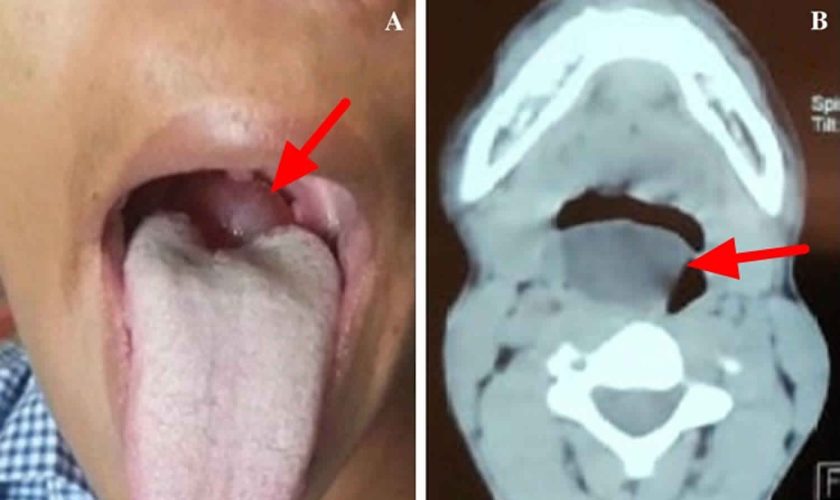
close

Larynx

Conditions & DiseasesLarynxPharynx

Vallecular cysts

vallecular-cyst

Vallecular cysts

Vallecular cysts also called epiglottic mucus retention cysts or base of tongue cysts, are benign retention cysts of the minor salivary glands that are typically present at birth in the tongue base of affected infants 1. The commonest site for vallecular cysts is the lingual surface of epiglottis accounting for 10.5% to 20.1% of all laryngeal cysts 2. Vallecular cysts distort the epiglottis when they increase in size and eventually fill the vallecula. The vallecula is the depression behind the root of the tongue between the median and lateral epiglottic folds on each side 3.

The incidence of vallecular cysts on laryngoscopy has been reported as 1 in 1,250 to 1 in 4,200, but the true incidence is difficult to estimate 4.

Vallecular cyst is a rare cause of upper airway obstruction in infants and children and are typically not associated with other anomalies or syndromes. Presentation like acute stridor with near fatal respiratory distress is extremely rare. In infants and children, vallecular cysts present most commonly with stridor and feeding difficulty but may cause life-threatening airway obstruction 5. In adults, most vallecular cysts are asymptomatic but may present with sensation of a lump in the throat (globus), voice change, difficulty swallowing (dysphagia), painful swallowing (odynophagia) or shortness of breath (dyspnea) 6. Vallecular cysts may also be discovered during administration of anesthesia, where they may obscure the view of the glottis and cause difficult endotracheal intubation 7. In adults, vallecular cysts are more common but less dangerous. The peak incidence is in the fifth decade of life, and the majority of cysts occur in men 6.

Some believe that the vallecular cyst develops because of an obstruction of a minor salivary gland when the duct of a mucous gland or lingual tonsillar crypt becomes obstructed and dilates 8, while others believe that the vallecular cyst is a variant of a thyroglossal duct cyst. Vallecular cysts have therefore been classified as ductal cysts, retention cysts, and lymphoepithelial cysts and are caused by inflammation, irritation, or trauma 6.

Infants with vallecular cysts are considered to be at risk of airway obstruction and death 8. Therefore, all such cysts in infants and children should be removed surgically, with marsupialization via carbon dioxide laser (CO2 laser) or electrocautery being the most commonly used method 9. Vallecular cyst is commonly managed using microlaryngoscope and specialized instruments.

Figure 1. Vallecular cyst

vallecular cyst

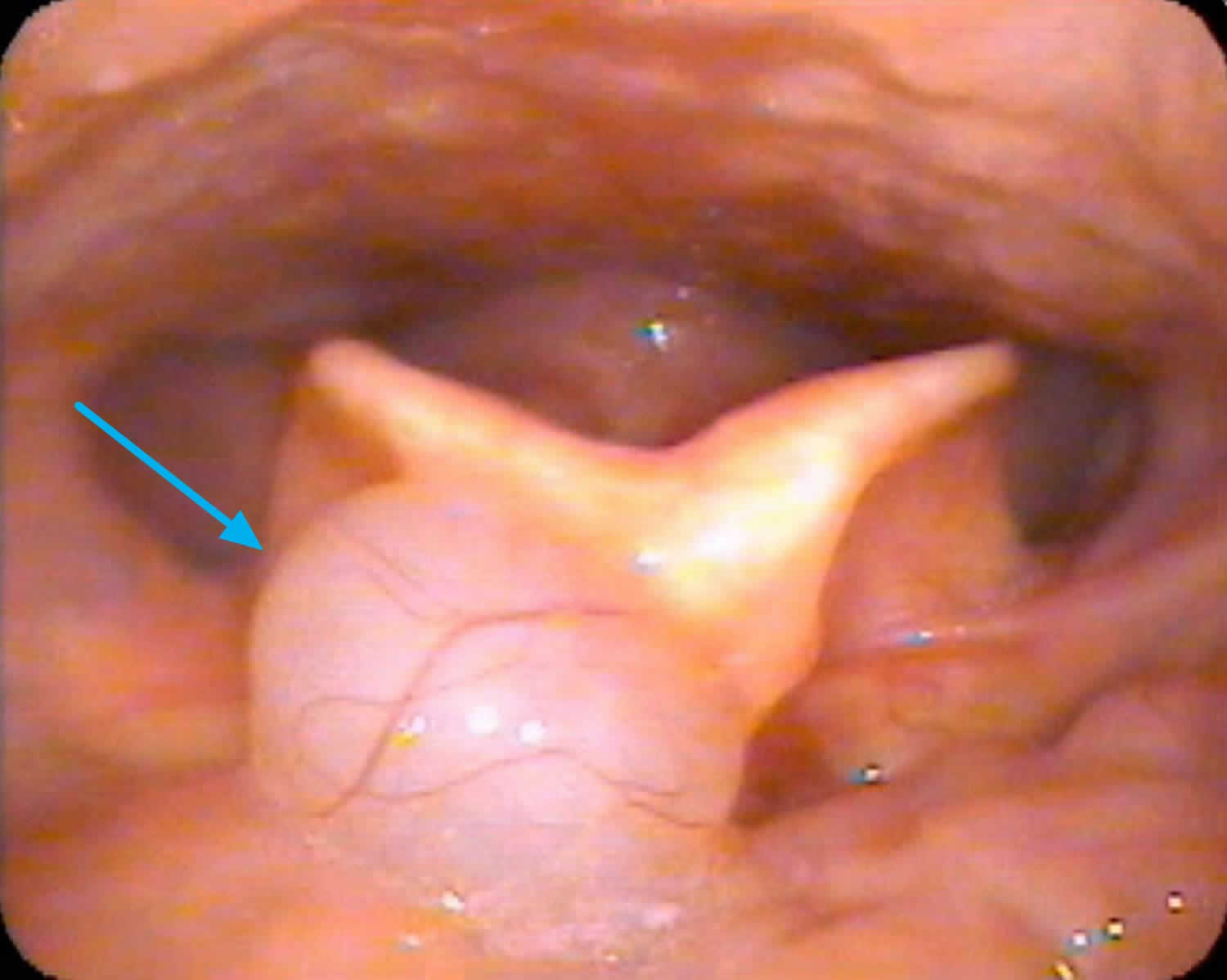
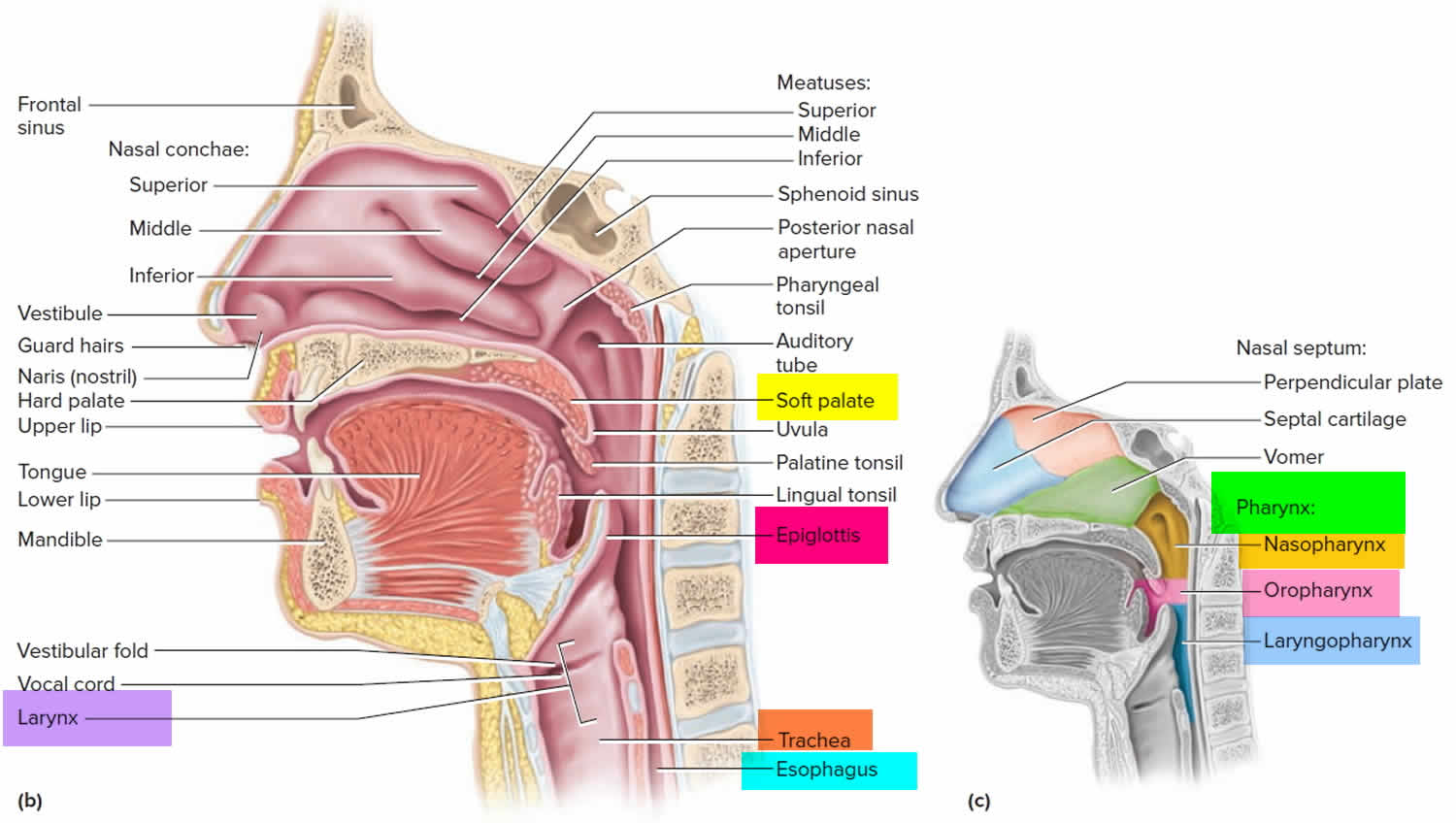
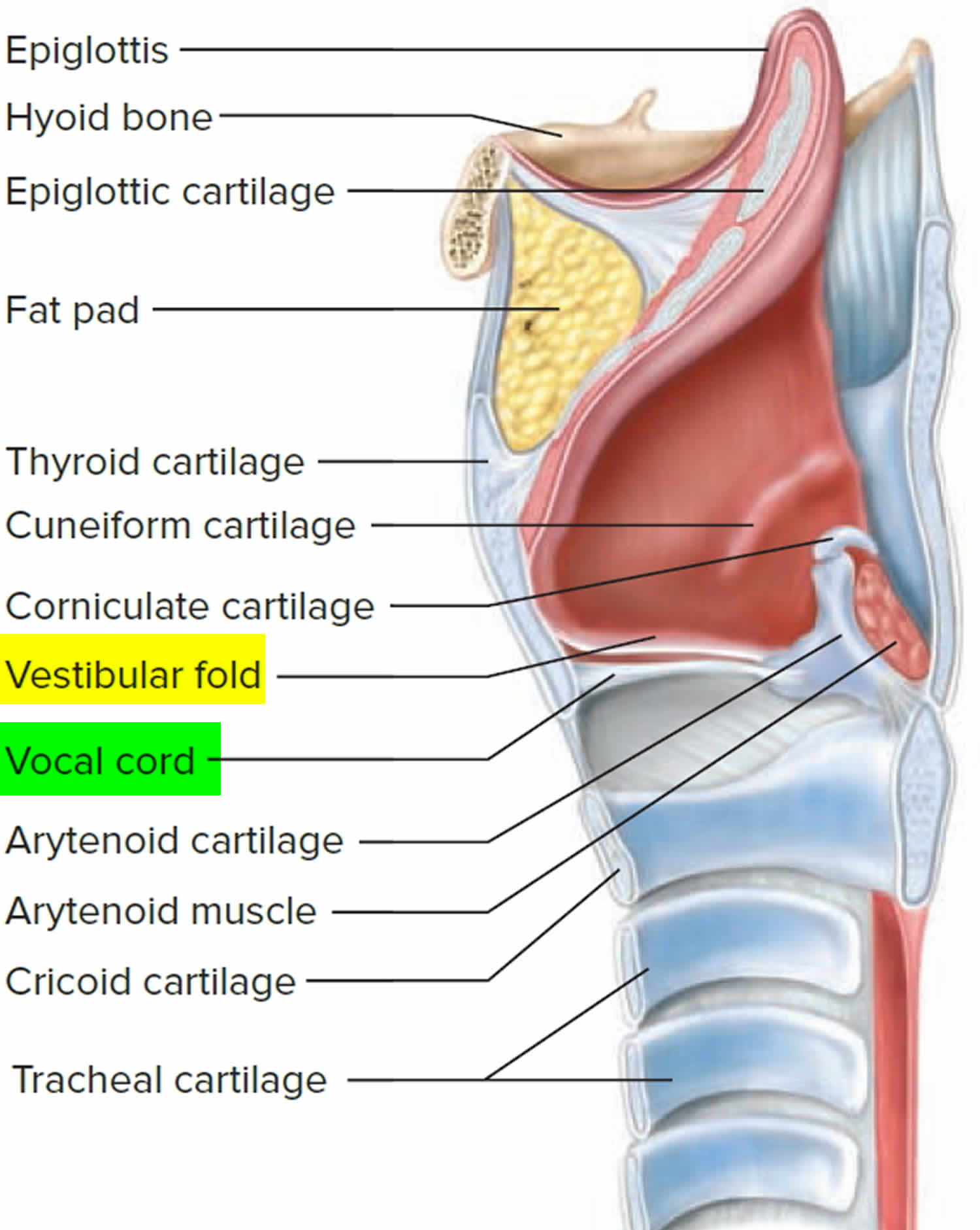
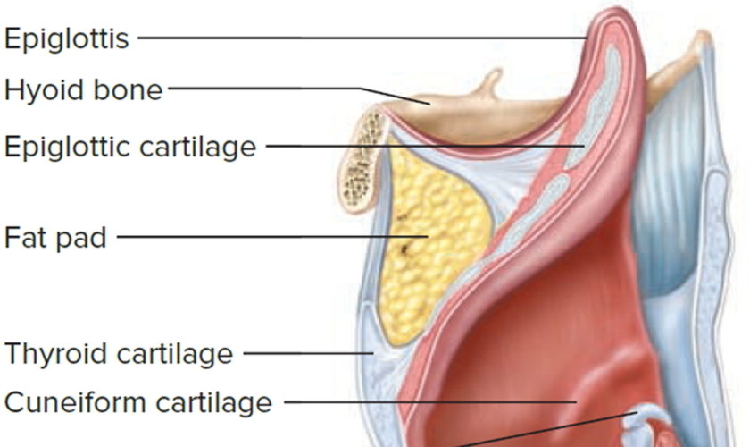
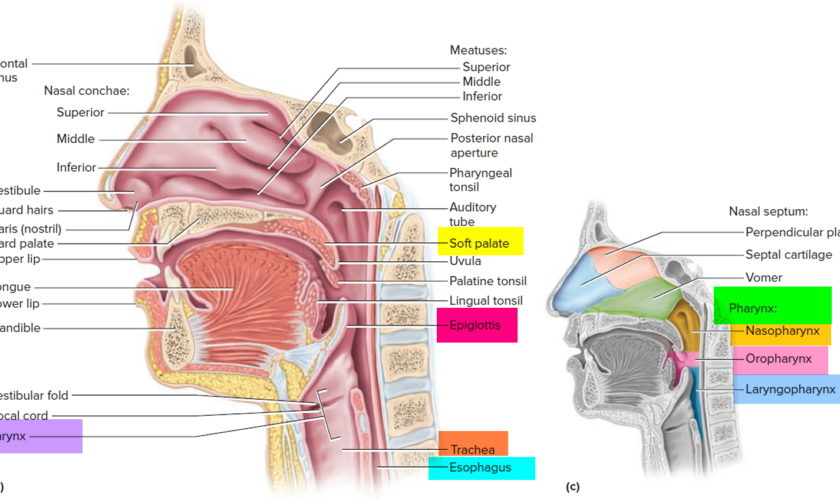
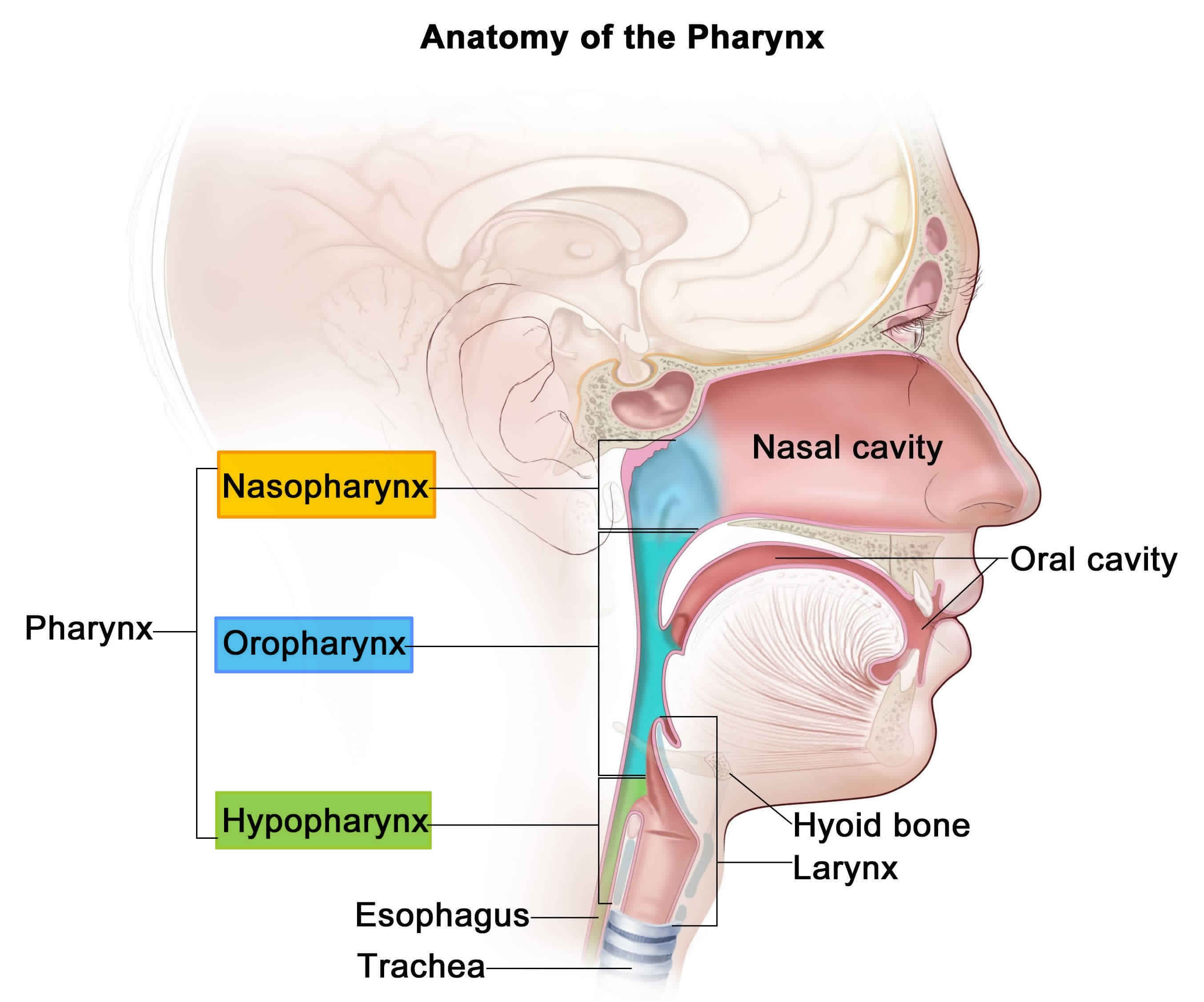
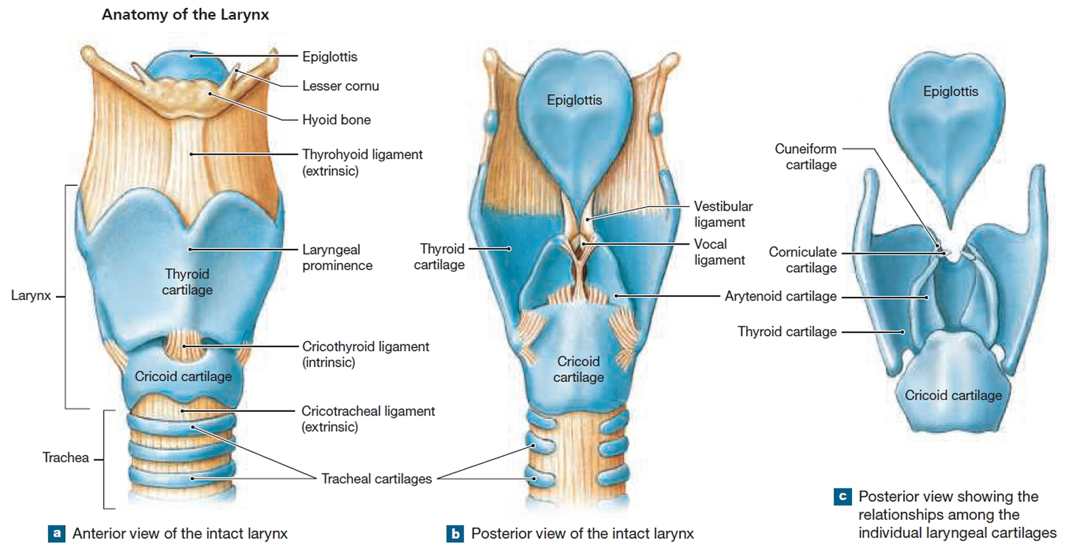
Figure 2. Pharynx and larynx anatomy

pharynx and larynx anatomy

larynx

Vallecular cyst causes

Vallecular cysts are thought to be secondary cysts formed from either ductal obstruction of mucous glands or cystic tongue lesions developed from misplaced embryonic remnants of the foregut 10.

Vallecular cysts commonly arise from the lingual surface of the epiglottis and are unilocular cysts containing clear sterile fluid arising from the lingular surface of the epiglottis 11.

Histologically vallecular cysts are lined by non-keratinizing squamous or respiratory epithelium with mucous glands with an external lining of squamous epithelium 12.

Vallecular cyst symptoms

Vallecular cysts may present with diverse symptoms affecting the voice, airway, and swallowing. Patients with vallecular cysts often have similar symptoms/signs as those with laryngomalacia.

  • Inspiratory stridor is usually present at birth (noisy inhale)
  • Feeding difficulties
  • Minimal, moderate or severe respiratory distress

Vallecular cysts can cause feeding difficulties due to upper airway obstruction and pressure at the laryngeal inlet 12.

Nearly two-thirds of vallecular cysts are asymptomatic and are diagnosed incidentally on routine laryngeal examination 6.

Vallecular cyst diagnosis

If the vallecular cysts are very small, diagnosis may be delayed until the child is older and begins to complain of swallowing difficulties. In the majority of patients, vallecular cyst is large enough to bring the patient to the attention of the otolaryngologist (ear, nose and throat specialist) who can then confirm the diagnosis using flexible laryngoscopy. Imaging (CT scans, X-rays, etc.) is not required for patients with vallecular cysts.

Antenatal diagnosis of vallecular cyst has been reported by Cuillier et al. 11 in a 28 week gestation fetus with polyhydramnios using MRI following a suspicion on ultrasound imaging. In this case, polyhydramnios was secondary to partial obstruction of the esophagus due to mass effect. Vallecular cyst was noted at birth to be filling the oral cavity and needed cyst aspiration followed by endotracheal intubation due to airway obstruction. Prenatal diagnosis of a significant vallecular cyst gives the window of opportunity for parental counseling and multidisciplinary planning for intervention after birth. In suspected cases with severe airway obstruction diagnosed prenatally, an ex-utero intrapartum treatment (EXIT) procedure may be planned 13.

Aero-digestive evaluation

Infants with vallecular cysts need to be evaluated for both airway and feeding issues. Management of the airway often requires a combination of supportive, medical and surgical care. Feeding and swallowing issues are common in children with vallecular cysts and often need to be addressed by speech pathologists and gastroenterology specialists.

Vallecular cyst treatment

Surgical removal is the treatment of choice for vallecular cyst 14. Surgery is performed endoscopically. Once the airway is secured with an endotracheal tube, this may be performed either by aspiration, marsupialization  (deroofing) or excision using either microlaryngeal instruments or a laser 15. Marsupialization via coblation has been used to treat vallecular cyst. Coblation involves the use of radiofrequency and normal saline to create an isoelectric field of sodium and chloride ions moving at high speeds that have sufficient energy to breakdown tissues 16. This modality has the advantages of a minimally invasive technique with reduced thermal damage, bleeding, tissue damage and postoperative recovery time 17. In general, simple aspiration of vallecular cyst is avoided due to the high risk of recurrence 18. Most reports show low recurrence rate of vallecular cyst after marsupialization. Li Y et al. 12 reported recurrence of vallecular cyst in 15% of cases in their center following marsupialization.

Patients generally do very well after surgery and most often resume normal diet with no breathing issues. Occasionally, patients may require some support for secondary laryngomalacia or reflux until the airway grows sufficiently. Recurrence of the cyst is very rare following treatment.

Vallecular cyst treatment options

Surgical treatment for vallecular cysts in infants includes aspiration, marsupialization (deroofing) and excision. The surgical approach is transoral under direct vision with or without a microlaryngoscope or using a microlaryngoscope with a camera assembly 19. The various tools used for this purpose include direct electrocautery, carbon dioxide laser (CO2 laser) or microlarygngoscopic instruments.

The use of the carbon dioxide laser (CO2 laser) for surgery of the vocal fold is a subject of controversy. Many prefer to avoid it, for although the cutting beam is reasonably precise, it is hypothesized that the tissue reaction is somewhat unpredictable, probably because of the emitted heat. The alternative is microscopic instruments. Although they are more technically difficult to use, they offer equivalent accuracy and perhaps less potential for inadvertent damage and scarring.

Suzuki et al. 19 reported a negligible recurrence rate after marsupialization of vallecular cysts as compared to complete excision. Complete excision is more invasive and there is the possibility of bleeding and postoperative residual scarring. Hence, marsupialization is the preferred treatment of vallecular cysts.

The same study recommends that aspiration should be attempted only as an initial maneuver in cases of difficult intubation and not as definitive treatment due to high rates of recurrence.

Da Vinci robot-assisted excision of a vallecular cyst was reported recently by McLeod et al. 20 and further research is needed to explore this modality of treatment.

Excision of cyst using a tonsillar snare has also been reported as an easy and cost-effective method of treatment 21.

Conventional laparoscopic instruments used were a 4 mm 0-degree telescope; 3 mm hook electrocautery and Maryland forceps. These give very good vision during surgery. The use of long instruments (approximately 33 cm) permits easy accessibility and maneuverability to the deep oral cavity and avoids undue overlapping and fighting between instruments. The instruments are insulated along their length which prevents thermal injury to other structures in the oral cavity. Pediatric surgeons are used to operating with conventional laparoscopic instruments as against specialized instruments like microdebrider, microscissors, etc.

References
  1. Romak JJ, Olsen SM, Koch CA, Ekbom DC. Bilateral vallecular cysts as a cause of Dysphagia: case report and literature review. Int J Otolaryngol. 2010;2010:697583. doi:10.1155/2010/697583 https://www.ncbi.nlm.nih.gov/pmc/articles/PMC3005807
  2. Parelkar SV, Patel JL, Sanghvi BV, et al. An Unusual Presentation of Vallecular Cyst with near Fatal Respiratory Distress and Management Using Conventional Laparoscopic Instruments. J Surg Tech Case Rep. 2012;4(2):118-120. doi:10.4103/2006-8808.110257 https://www.ncbi.nlm.nih.gov/pmc/articles/PMC3673355
  3. De A, Don DM, Magee W 3rd, Ward SL. Vallecular cyst as a cause of obstructive sleep apnea in an infant. J Clin Sleep Med. 2013;9(8):825-826. Published 2013 Aug 15. doi:10.5664/jcsm.2932 https://www.ncbi.nlm.nih.gov/pmc/articles/PMC3716675
  4. Mason DG, Wark KJ. Unexpected difficult intubation. Asymptomatic epiglottic cysts as a cause of upper airway obstruction during anaesthesia. Anaesthesia. 1987;42(4):407–410.
  5. Berger G, Averbuch E, Zilka K, Berger R, Ophir D. Adult vallecular cyst: thirteen-year experience. Otolaryngology. 2008;138(3):321–327.
  6. Arens C, Glanz H, Kleinsasser O. Clinical and morphological aspects of laryngeal cysts. European Archives of Oto-Rhino-Laryngology. 1997;254(9-10):430–436.
  7. Rivo J, Matot I. Asymptomatic vallecular cyst: airway management considerations. Journal of Clinical Anesthesia. 2001;13(5):383–386.
  8. Gutiérrez JP, Berkowitz RG, Robertson CF. Vallecular cysts in newborns and young infants. Pediatric Pulmonology. 1999;27(4):282–285.
  9. Leuin S, Cunningham M, Volk MS, Hartnick C. Transhyoid approach to excision of recurrent vallecular pseudocysts. Laryngoscope. 2008;118(1):124–127.
  10. A.K. Lahiri, K.K. Somashekar, B. Wittkop, et al. Large vallecular masses; differential diagnosis and imaging features J Clin Imaging Sci, 8 (2018), p. 26, 10.4103/jcis.JCIS_15_18
  11. F. Cuillier, S. Samperiz, R. Testud, et al. Antenatal diagnosis and management of a vallecular cyst. Ultrasound Obstet Gynecol, 20 (2002), pp. 623-626, 10.1046/j.1469-0705.2002.00860.x
  12. Y. Li, A.L. Irace, N.D. Dombrowski, et al. Vallecular cyst in the pediatric population: evaluation and management. Int J Pediatr Otorhinolaryngol, 113 (2018), pp. 198-203, 10.1016/j.ijporl.2018.07.040
  13. P.A. Jayagopi, S. Chandran, B. Sriram, K.T. Chang. Ex-utero intrapartum treatment (EXIT) procedure for giant fetal epignathus. Indian Pediatr, 52 (10) (2015), pp. 893-895, 10.1007/s13312-015-0740-9
  14. Congenital vallecular cyst causing severe inspiratory stridor in a newborn. Journal of Pediatric Surgery Case Reports Volume 59, August 2020, 101460 https://doi.org/10.1016/j.epsc.2020.101460
  15. A.F. AlAbdulla. Congenital vallecular cyst causing airway compromise in a 2-month-old girl. Case Rep Med, 2015 (2015), Article 975859, 10.1155/2015/975859
  16. Z. Wang, Y. Zhang, Y. Ye, et al. Clinical efficacy of low-temperature radiofrequency ablation of pharyngolaryngeal cyst in 84 Chinese infants. Medicine (Baltim), 96 (44) (2017), Article e8237, 10.1097/MD.0000000000008237
  17. S. Gogia, S.K. Agarwal, A. Agarwal. Vallecular cyst in neonates: case series—a clinicosurgical insight vol. 2014, Case Rep Otolaryngol (2014), p. 764860, 10.1155/2014/764860
  18. M. Raftopulos, M. Soma, D. Lowinger, et al. Vallecular cysts: a differential diagnosis to consider for neonatal stridor and failure to thrive. JRSM Short Rep, 4 (4) (2013), p. 29, 10.1177/2042533313476689
  19. Suzuki J, Hashimoto S, Watanabe K, Takahashi K. Congenital vallecular cyst in an infant: Case report and review of 52 recent cases. J Laryngol Otol. 2011;125:1199–203.
  20. McLeod IK, Melder PC. Da Vinci robot-assisted excision of a vallecular cyst: A case report. Ear Nose Throat J. 2005;84:170–2.
  21. Bhandary S. Innovative Surgical Technique in the Management of Vallecular Cyst. Online J Health Allied Sci. 2003;3:2.
read more
LarynxRespiratory System

What is the larynx ?

larynx

The Larynx

The larynx is the upper end of the lower airway. It is continuous with the trachea below and the pharynx posterosuperiorly.

The larynx is a cartilaginous chamber about 4 cm (1.5 in.) long. Its primary function is to keep food and drink out of the airway, but it evolved the additional role of sound production (phonation) in many animals; hence, we colloquially think of it as the “voice box.”

Figure 1. Larynx and pharynx anatomy

anatomy of the pharynx and larynx

Figure 2. Larynx anatomy

anatomy-of-the-larynx


larynx

The superior opening of the larynx is guarded by a flap of tissue called the epiglottis. At rest, the epiglottis stands almost vertically. During swallowing, however, extrinsic muscles of the larynx pull the larynx upward toward the epiglottis, the tongue pushes the epiglottis downward to meet it, and the epiglottis closes the airway and directs food and drink into the esophagus behind it. The vestibular folds of the larynx, play a greater role in keeping food and drink out of the airway, however.

The framework of the larynx consists of nine cartilages. The first three are solitary and relatively large. The most superior one, the epiglottic cartilage, is a spoon-shaped supportive plate in the epiglottis. The largest, the thyroid cartilage, is named for its shieldlike shape. It broadly covers the anterior and lateral aspects of the larynx. The “Adam’s apple” is an anterior peak of the thyroid cartilage called the laryngeal prominence. Testosterone stimulates the growth of this prominence, which is therefore larger in males than in females. Inferior to the thyroid cartilage is a ringlike cricoid cartilage. The thyroid and cricoid cartilages essentially constitute the “box” of the voice box.

The remaining cartilages are smaller and occur in three pairs. Posterior to the thyroid cartilage are the two arytenoid cartilages and attached to their upper ends is a pair of little horns, the corniculate cartilages. The arytenoid and corniculate cartilages function in speech, as explained shortly. A pair of cuneiform cartilages supports the soft tissues between the arytenoids and the epiglottis.

A group of fibrous ligaments binds the cartilages of the larynx together and forms a suspension system for the upper airway. A broad sheet called the thyrohyoid ligament suspends the larynx from the hyoid bone above, and below, the cricotracheal ligament suspends the trachea from the cricoid cartilage. These are collectively called the extrinsic ligaments because they link the larynx to other organs. The intrinsic ligaments are contained entirely within the larynx and link its nine cartilages to each other; they include ligaments of the vocal cords and vestibular folds.

Figure 3. Cricothyroid ligament (cricovocal membrane, cricothyroid membrane)

trachea - airway in the neck

The cricothyroid ligament (cricothyroid membrane) is attached to the arch of cricoid cartilage and extends superiorly to end in a free upper margin within the space enclosed by the thyroid cartilage.

On each side, this upper free margin attaches :

  • anteriorly to the thyroid cartilage, and
  • posteriorly to the vocal processes of the arytenoid cartilages.

The free margin between these two points of attachment is thickened to form the vocal ligament, which is under the vocal fold (true vocal cord) of the larynx. The cricothyroid ligament is also thickened anteriorly in the midline to form a distinct median cricothyroid ligament, which spans the distance between the arch of cricoid cartilage and the inferior thyroid notch and adjacent deep surface of the thyroid cartilage up to the attachment of the vocal ligaments.

  • In emergency situations, when the airway is blocked above the level of the vocal folds, the median cricothyroid ligament can be perforated to establish an airway. Except for small vessels and the occasional presence of a pyramidal lobe of the thyroid gland, normally there are few structures between the median cricothyroid ligament and skin.

In infants, the larynx is relatively high in the throat and the epiglottis touches the soft palate. This creates a more or less continuous airway from the nasal cavity to the larynx and allows an infant to breathe continually while swallowing. The epiglottis deflects milk away from the airstream, like rain running off a tent while it remains dry inside. By age 2, the root of the tongue becomes more muscular and forces the larynx to descend to a lower position. It then becomes impossible to breathe and swallow at the same time without choking.

The walls of the larynx are quite muscular. The superficial extrinsic muscles connect the larynx to the hyoid bone and elevate the larynx during swallowing. Also called the infrahyoid group. The deeper intrinsic muscles control the vocal cords by pulling on the corniculate and arytenoid cartilages, causing the cartilages to pivot. Depending on their direction of rotation, the arytenoid cartilages abduct or adduct the vocal cords.

The interior wall of the larynx has two folds on each side that stretch from the thyroid cartilage in front to the arytenoid cartilages in back. The superior vestibular folds play no role in speech but close the larynx during swallowing. They are supported by the vestibular ligaments. The inferior vocal cords (vocal folds) produce sound when air passes between them. They contain the vocal ligaments and are covered with stratified squamous epithelium, best suited to endure vibration and contact between the cords. The vocal cords and the opening between them are collectively called the glottis.

Sound Production

Air passing through the glottis vibrates the vocal folds and produces sound waves. Air forced between the adducted vocal cords vibrates them, producing a high-pitched sound when the cords are relatively taut and a lower pitched sound when they are more slack. Children have slender, short vocal folds, so their voices are high-pitched. At puberty the larynx of a male enlarges more than that of a female. In adult males, the vocal cords are usually longer and thicker, vibrate more slowly, and produce lower-pitched sounds than in females. Loudness is determined by the force of the air passing between the vocal cords.

Although the vocal cords alone produce sound, they do not produce intelligible speech; some anatomists have likened their sound to a hunter’s duck call. Amplification and echoing of the sound occur within the pharynx, oral cavity, nasal cavity, and paranasal sinuses. The crude sounds from the larynx are formed into words by actions of the pharynx, oral cavity, voluntary movements of the tongue, and lips.

The Pharynx

The pharynx, is a common passageway shared by both the digestive and respiratory systems. The pharynx connects the nose, mouth, and throat. The digestive and respiratory systems share the pharynx. It extends between the posterior nasal apertures and the entrances to the trachea and esophagus. The curving superior and posterior walls are attached to the axial skeleton, but the lateral walls are flexible and muscular. The pharynx is a muscular funnel extending about 13 cm (5 in.) from the posterior nasal apertures to the larynx. The pharynx is attached above to the base of the skull and is continuous below, approximately at the level of vertebra CVI (cervical vertebrum C6), with the top of the esophagus. The walls of the pharynx are attached anteriorly to the margins of the nasal cavities, oral cavity, and larynx. Muscles of the pharynx play necessary roles in swallowing and speech.

The pharynx is subdivided into three regions (Figure 1):

  1. the Nasopharynx,
  2. the Oropharynx, and
  3. the Laryngopharynx.

The Nasopharynx

The posterior apertures (choanae) of the nasal cavities open into the nasopharynx above the soft palate. The nasopharynx receives the auditory (eustachian) tubes from the middle ears and houses the pharyngeal tonsil. The nasopharynx passes only air and is lined by pseudostratified columnar epithelium. Inhaled air turns 90° downward as it passes through the nasopharynx. Relatively large particles (>10 μm) generally cannot make the turn because of their inertia. They collide with the wall of the nasopharynx and stick to the mucosa near the tonsil, which is well positioned to respond to airborne pathogens.

The Oropharynx

The oropharynx extends between the soft palate and the base of the tongue at the level of the hyoid bone. Like the posterior and inferior portions of the nasopharynx, the posterior portion of the oral cavity communicates directly with the oropharynx. The epithelium changes from a pseudostratified ciliated columnar epithelium to a nonkeratinized (mucosal type) stratified squamous epithelium at the boundary between the nasopharynx and oropharynx. The posterior margin of the soft palate supports the dangling uvula and two pairs of muscular pharyngeal arches, the posterior arch and the anterior arch.

The Laryngopharynx

The narrow laryngopharynx includes the region of the pharynx lying between the hyoid bone and the entrance to the esophagus. Like the oropharynx, the laryngopharynx is lined with a stratified squamous epithelium that resists abrasion, chemicals, and pathogens.

Larynx cancer

Larynx or throat cancer is cancer of the vocal cords, larynx (voice box), or other areas of the throat 1.

Alternative Names:

  • Vocal cord cancer;
  • Throat cancer;
  • Laryngeal cancer;
  • Cancer of the glottis;
  • Cancer of oropharynx or hypopharynx.

Causes of larynx cancer

People who smoke or use tobacco are at risk of developing throat cancer. Drinking too much alcohol over a long time also increases risk. Smoking and drinking alcohol combined lead to an increased risk for throat cancer.

Most throat cancers develop in adults older than 50. Men are more likely than women to develop throat cancer.

Prevention of larynx cancer

Do not smoke or use other tobacco. Limit or avoid alcohol use.

Figure 3. Larynx cancer

larynx cancer

Symptoms of larynx cancer

Symptoms of throat cancer include any of the following 1:

  • Abnormal (high-pitched) breathing sounds
  • Cough
  • Coughing up blood
  • Difficulty swallowing
  • Hoarseness that does not get better in 3 to 4 weeks
  • Neck or ear pain
  • Sore throat that does not get better in 2 to 3 weeks, even with antibiotics
  • Swelling or lumps in the neck
  • Weight loss not due to dieting

Exams and Tests for larynx cancer

The health care provider will perform a physical exam. This may show a lump on the outside of the neck.

The provider may look in your throat or nose using a flexible tube with a small camera at the end.

Other tests that may be ordered include:

  • Biopsy of suspected tumor
  • Chest x-ray
  • CT scan of chest
  • CT scan of head and neck
  • MRI of the head or neck
  • PET (positron emission tomography) scan:  PET scan is an imaging test that allows your doctor to check for diseases in your body. The scan uses a special dye that has radioactive tracers. These tracers are injected into a vein in your arm. Your organs and tissues then absorb the tracer.

Treatment for larynx cancer

The goal of treatment is to completely remove the cancer and prevent it from spreading to other parts of the body.

When the tumor is small, either surgery or radiation therapy alone can be used to remove the tumor.

When the tumor is larger or has spread to lymph nodes in the neck, a combination of radiation and chemotherapy is often used to save the voice box (vocal cords). If this is not possible, the voice box is removed. This surgery is called laryngectomy.

Outlook (Prognosis) for larynx cancer

Throat cancers may be cured when detected early. If the cancer has spread (metastasized) to surrounding tissues or lymph nodes in the neck, about half of patients can be cured. If the cancer has spread to parts of the body outside the head and neck, the cancer is not curable. Treatment is aimed at prolonging and improving quality of life.

After treatment, therapy is needed to help with speech and swallowing. If the person is not able to swallow, a feeding tube will be needed.

Possible Complications of larynx cancer

Complications of this type of cancer may include:

  • Airway obstruction
  • Difficulty swallowing
  • Disfigurement of the neck or face
  • Hardening of the skin of the neck
  • Loss of voice and speaking ability
  • Spread of the cancer to other body areas (metastasis).
References
  1. Throat or larynx cancer. Medline Plus, U.S. National Library of Medicine. https://medlineplus.gov/ency/article/001042.htm
read more
LarynxRespiratory System

What is the pharynx ?

anatomy of the pharynx and larynx

What is the pharynx and the larynx

The pharynx, is a common passageway shared by both the digestive and respiratory systems. The pharynx is a muscular funnel extending about 13 cm (5 in.) from the posterior nasal apertures to the larynx. The pharynx is attached above to the base of the skull and is continuous below, approximately at the level of vertebra CVI (cervical vertebrum C6), with the top of the esophagus. The walls of the pharynx are attached anteriorly to the margins of the nasal cavities, oral cavity, and larynx. Muscles of the pharynx play necessary roles in swallowing and speech.

The pharynx is subdivided into three regions (Figure 1):

  1. the Nasopharynx,
  2. the Oropharynx, and
  3. the Laryngopharynx.

Figure 1. Pharynx and larynx anatomy

anatomy of the pharynx and larynx

The pharynx connects the nose, mouth, and throat. The digestive and respiratory systems share the pharynx. It extends between the posterior nasal apertures and the entrances to the trachea and esophagus. The curving superior and posterior walls are attached to the axial skeleton, but the lateral walls are flexible and muscular.

The Nasopharynx

Nasopharynx is the upper part of the throat (pharynx) that lies behind the nose. The nasopharynx a box-like chamber about 1½ inches on each edge. Nasopharynx lies just above the soft part of the roof of the mouth (soft palate) and just in back of the nasal passages. The posterior apertures (choanae) of the nasal cavities open into the nasopharynx above the soft palate. The nasopharynx receives the auditory (eustachian) tubes from the middle ears and houses the pharyngeal tonsil. The nasopharynx passes only air and is lined by pseudostratified columnar epithelium. Inhaled air turns 90° downward as it passes through the nasopharynx. Relatively large particles (>10 μm) generally cannot make the turn because of their inertia. They collide with the wall of the nasopharynx and stick to the mucosa near the tonsil, which is well positioned to respond to airborne pathogens.

The nasopharynx serves as a passageway for air traveling from the nose to the throat (and then on to the lungs).

The Oropharynx

The oropharynx extends between the soft palate and the base of the tongue at the level of the hyoid bone. Like the posterior and inferior portions of the nasopharynx, the posterior portion of the oral cavity communicates directly with the oropharynx. The epithelium changes from a pseudostratified ciliated columnar epithelium to a nonkeratinized (mucosal type) stratified squamous epithelium at the boundary between the nasopharynx and oropharynx. The posterior margin of the soft palate supports the dangling uvula and two pairs of muscular pharyngeal arches, the posterior arch and the anterior arch.

The Laryngopharynx

The narrow laryngopharynx includes the region of the pharynx lying between the hyoid bone and the entrance to the esophagus. Like the oropharynx, the laryngopharynx is lined with a stratified squamous epithelium that resists abrasion, chemicals, and pathogens.

The Larynx

The larynx is the upper end of the lower airway. It is continuous with the trachea below and the pharynx posterosuperiorly.

The larynx is a cartilaginous chamber about 4 cm (1.5 in.) long. Its primary function is to keep food and drink out of the airway, but it evolved the additional role of sound production (phonation) in many animals; hence, we colloquially think of it as the “voice box.”

Figure 2. Larynx anatomy

anatomy of the larynx

larynx

The superior opening of the larynx is guarded by a flap of tissue called the epiglottis. At rest, the epiglottis stands almost vertically. During swallowing, however, extrinsic muscles of the larynx pull the larynx upward toward the epiglottis, the tongue pushes the epiglottis downward to meet it, and the epiglottis closes the airway and directs food and drink into the esophagus behind it. The vestibular folds of the larynx, play a greater role in keeping food and drink out of the airway,
however.

The framework of the larynx consists of nine cartilages. The first three are solitary and relatively large. The most superior one, the epiglottic cartilage, is a spoon-shaped supportive plate in the epiglottis. The largest, the thyroid cartilage, is named for its shieldlike shape. It broadly covers the anterior and lateral aspects of the larynx. The “Adam’s apple” is an anterior peak of the thyroid cartilage called the laryngeal prominence. Testosterone stimulates the growth of this prominence, which is therefore larger in males than in females. Inferior to the thyroid cartilage is a ringlike cricoid cartilage. The thyroid and cricoid cartilages
essentially constitute the “box” of the voice box.

In infants, the larynx is relatively high in the throat and the epiglottis touches the soft palate. This creates a more or less continuous airway from the nasal cavity to the larynx and allows an infant to breathe continually while swallowing. The epiglottis deflects milk away from the airstream, like rain running off a tent while it remains dry inside. By age 2, the root of the tongue becomes more muscular and forces the larynx to descend to a lower position. It then becomes impossible to breathe and swallow at the same time without choking.

The walls of the larynx are quite muscular. The superficial extrinsic muscles connect the larynx to the hyoid bone and elevate the larynx during swallowing. Also called the infrahyoid group. The deeper intrinsic muscles control the vocal cords by pulling on the corniculate and arytenoid cartilages, causing the cartilages to pivot. Depending on their direction of rotation, the arytenoid cartilages abduct or adduct the vocal cords.

The interior wall of the larynx has two folds on each side that stretch from the thyroid cartilage in front to the arytenoid cartilages in back. The superior vestibular folds play no role in speech but close the larynx during swallowing. They are supported by the vestibular ligaments. The inferior vocal cords (vocal folds) produce sound when air passes between them. They contain the vocal ligaments and are covered with stratified squamous epithelium, best suited to endure vibration and contact between the cords. The vocal cords and the opening between them are collectively called the glottis.

Sound Production

Air passing through the glottis vibrates the vocal folds and produces sound waves. Air forced between the adducted vocal cords vibrates them, producing a high-pitched sound when the cords are relatively taut and a lower pitched sound when they are more slack. Children have slender, short vocal folds, so their voices are high-pitched. At puberty the larynx of a male enlarges more than that of a female. In adult males, the vocal cords are usually longer and thicker, vibrate more slowly, and produce lower-pitched sounds than in females. Loudness is determined by the force of the air passing between the vocal cords.

Although the vocal cords alone produce sound, they do not produce intelligible speech; some anatomists have likened their sound to a hunter’s duck call. Amplification and echoing of the sound occur within the pharynx, oral cavity, nasal cavity, and paranasal sinuses. The crude sounds from the larynx are formed into words by actions of the pharynx, oral cavity, voluntary movements of the tongue, and lips.

read more
Health Jade