close

Skin

Conditions & DiseasesSkin

Skin cancer in children

Skin cancer in children

Skin cancer in children

Skin cancer is very rare in children. Skin cancer is a type of cancer that grows in the cells of the skin. Skin cancer can spread to and damage nearby tissue and spread to other parts of the body.

There are 3 main types of skin cancer:

  1. Basal cell carcinoma (BCC). The majority of skin cancers are basal cell carcinoma. It’s a very treatable cancer. It starts in the basal cell layer of the skin (epidermis) and grows very slowly. The cancer usually appears as a small, shiny bump or nodule on the skin. It occurs mainly on areas exposed to the sun, such as the head, neck, arms, hands, and face. It more often occurs among people with light-colored eyes, hair, and skin.
  2. Squamous cell carcinoma (SCC). This cancer is less common. It grows faster than basal cell carcinoma, but it’s also very treatable. Squamous cell carcinoma may appear as nodules or red, scaly patches of skin, and may be found on the face, ears, lips, and mouth. It can spread to other parts of the body, but this is rare. This type of skin cancer is most often found in people with light skin.
  3. Melanoma. This type of skin cancer is a small portion of all skin cancers, but it causes the most deaths. It starts in the melanocyte cells that make pigment in the skin. It may begin as a mole that turns into cancer. This cancer may spread quickly. Melanoma most often appears on fair-skinned people, but is found in people of all skin types.

The term non-melanoma skin cancer refers to all types of skin cancer apart from melanoma. BCC and SCC are also called keratinocyte cancer.

Each subtype of skin cancer has unique characteristics.

Skin cancer most commonly affects older adults. Your risk goes up as you get older.

Skin cancer in children key points:

  • Skin cancer is rare in children.
  • Skin cancer is more common in people with light skin, light-colored eyes, and blond or red hair.
  • Bring your child to see a doctor if you see any unusual changes in your child’s skin.
  • Follow the ABCDE rule to tell the difference between a normal mole and melanoma.
  • Biopsy is used to diagnose skin cancer.
  • Skin cancer can be treated with surgery, medicine, and radiation.
  • Staying out of the sun is the best way to prevent skin cancer.

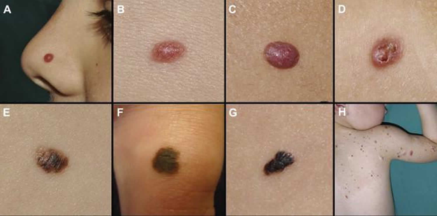
Figure 1. Melanoma in children

Melanoma in children

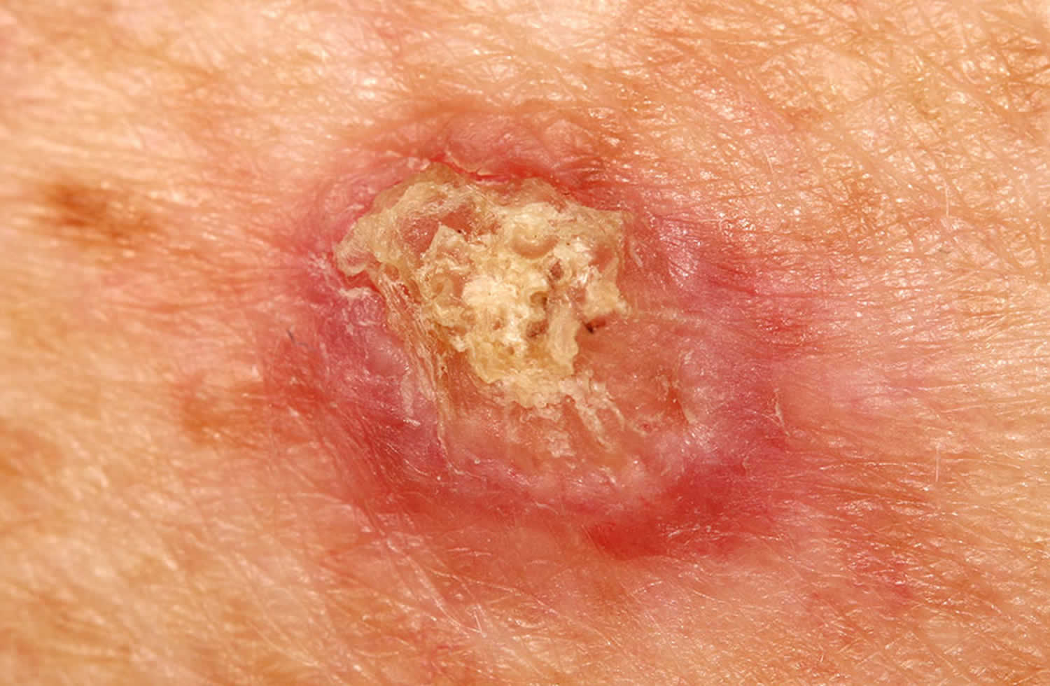
Figure 2. Squamous cell carcinoma in children

Squamous cell carcinoma in children

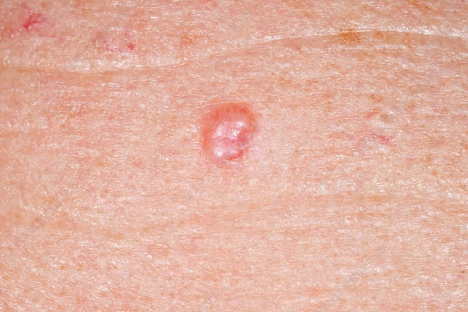
Figure 3. Basal cell carcinoma in children

Basal cell carcinoma in children

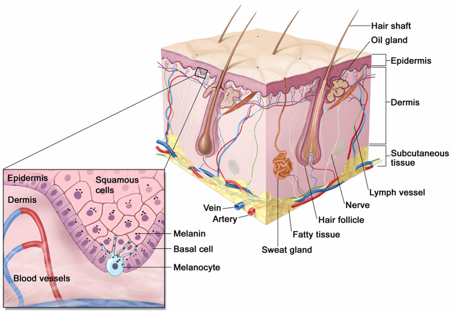
Figure 4. Skin anatomy

Skin anatomy

Basal cell carcinoma in children

Basal cell carcinoma (BCC) is a common, locally invasive, keratinocyte cancer also known as non-melanoma cancer. Basal cell carcinoma is very rarely a threat to life. Basal cell carcinoma (BCC) is the most common form of skin cancer. Basal cell carcinoma is also known as rodent ulcer and basalioma. Patients with basal cell carcinoma often develop multiple primary tumors over time.

Basal cell carcinoma main characteristics are:

  • Slowly growing plaque or nodule
  • Skin coloured, pink or pigmented
  • Varies in size from a few millimetres to several centimetres in diameter
  • Spontaneous bleeding or ulceration

A tiny proportion of basal cell carcinomas grow rapidly, invade deeply, and/or metastasise to local lymph nodes.

The cause of basal cell carcinoma is multifactorial:

  • Most often, there are DNA mutations in the patched (PTCH) tumour suppressor gene, part of hedgehog signalling pathway
  • These may be triggered by exposure to ultraviolet radiation
  • Various spontaneous and inherited gene defects predispose to basal cell carcinoma

Risk factors for basal cell carcinoma include:

  • Age and sex: basal cell carcinomas are particularly prevalent in elderly males. However, they also affect females and younger adults
  • Previous basal cell carcinoma or other form of skin cancer (squamous cell carcinoma, melanoma)
  • Sun damage (photoaging, actinic keratosis)
  • Repeated prior episodes of sunburn
  • Fair skin, blue eyes and blond or red hair—note; basal cell carcinoma can also affect darker skin types
  • Previous cutaneous injury, thermal burn, disease (eg cutaneous lupus, sebaceous nevus)
  • Inherited syndromes: basal cell carcinoma is a particular problem for families with basal cell naevus syndrome (Gorlin syndrome), Bazex-Dupré-Christol syndrome, Rombo syndrome, Oley syndrome and xeroderma pigmentosum
  • Other risk factors include ionizing radiation, exposure to arsenic, and immune suppression due to disease or medicines

Types of basal cell carcinoma

There are several distinct clinical types of basal cell carcinoma, and over 20 histological growth patterns of basal cell carcinoma.

Nodular basal cell carcinoma

  • Also known as nodulocystic carcinoma
  • Most common type of facial basal cell carcinoma
  • Shiny or pearly nodule with a smooth surface
  • May have central depression or ulceration, so its edges appear rolled
  • Blood vessels cross its surface
  • Cystic variant is soft, with jelly-like contents
  • Micronodular, microcystic and infiltrative types are potentially aggressive subtypes

Superficial basal cell carcinoma

  • Most common type in younger adults
  • Most common type on upper trunk and shoulders
  • Slightly scaly, irregular plaque
  • Thin, translucent rolled border
  • Multiple microerosions

Morphoeic basal cell carcinoma

  • Also known as morpheic, morphoeiform or sclerosing basal cell carcinoma
  • Usually found in mid-facial sites
  • Waxy, scar-like plaque with indistinct borders
  • Wide and deep subclinical extension
  • May infiltrate cutaneous nerves (perineural spread)

Basosquamous carcinoma

  • Also known as basisquamous carcinoma and mixed basal-squamous cell carcinoma
  • Mixed basal cell carcinoma (basal cell carcinoma) and squamous cell carcinoma (SCC)
  • Infiltrative growth pattern
  • Potentially more aggressive than other forms of basal cell carcinoma

Primary basal cell carcinoma treatment

The treatment for a basal cell carcinoma depends on its type, size and location, the number to be treated, patient factors, and the preference or expertise of the doctor. Most basal cell carcinomas are treated surgically. Long-term follow-up is recommended to check for new lesions and recurrence; the latter may be unnecessary if histology has reported wide clear margins.

Excision biopsy

Excision means the lesion is cut out and the skin stitched up.

  • Most appropriate treatment for nodular, infiltrative and morphoeic basal cell carcinomas
  • Should include 3 to 5 mm margin of normal skin around the tumor
  • Very large lesions may require flap or skin graft to repair the defect
  • Pathologist will report deep and lateral margins
  • Further surgery is recommended for lesions that are incompletely excised

Mohs micrographically controlled excision

Mohs micrographically controlled surgery involves examining carefully marked excised tissue under the microscope, layer by layer, to ensure complete excision.

  • Very high cure rates achieved by trained Mohs surgeons
  • Used in high-risk areas of the face around eyes, lips and nose
  • Suitable for ill-defined, morphoeic, infiltrative and recurrent subtypes
  • Large defects are repaired by flap or skin graft

Superficial skin surgery

Superficial skin surgery comprises shave, curettage, and electrocautery. It is a rapid technique using local anaesthesia and does not require sutures.

  • Suitable for small, well-defined nodular or superficial basal cell carcinomas
  • Lesions are usually located on trunk or limbs
  • Wound is left open to heal by secondary intention
  • Moist wound dressings lead to healing within a few weeks
  • Eventual scar quality variable

Cryotherapy

Cryotherapy is the treatment of a superficial skin lesion by freezing it, usually with liquid nitrogen.

  • Suitable for small superficial basal cell carcinomas on covered areas of trunk and limbs
  • Best avoided for basal cell carcinomas on head and neck, and distal to knees
  • Double freeze-thaw technique
  • Results in a blister that crusts over and heals within several weeks.
  • Leaves permanent white mark

Photodynamic therapy

Photodynamic therapy (PDT) refers to a technique in which basal cell carcinoma is treated with a photosensitising chemical, and exposed to light several hours later.

  • Topical photosensitisers include aminolevulinic acid lotion and methyl aminolevulinate cream
  • Suitable for low-risk small, superficial basal cell carcinomas
  • Best avoided if tumor in site at high risk of recurrence
  • Results in inflammatory reaction, maximal 3–4 days after procedure
  • Treatment repeated 7 days after initial treatment
  • Excellent cosmetic results

Imiquimod cream

Imiquimod is an immune response modifier.

  • Best used for superficial basal cell carcinomas less than 2 cm diameter
  • Applied three to five times each week, for 6–16 weeks
  • Results in a variable inflammatory reaction, maximal at three weeks
  • Minimal scarring is usual

Fluorouracil cream

5-Fluorouracil cream is a topical cytotoxic agent.

  • Used to treat small superficial basal cell carcinomas
  • Requires prolonged course, eg twice daily for 6–12 weeks
  • Causes inflammatory reaction
  • Has high recurrence rates

Radiotherapy

Radiotherapy or X-ray treatment can be used to treat primary basal cell carcinomas or as adjunctive treatment if margins are incomplete.

  • Mainly used if surgery is not suitable
  • Best avoided in young patients and in genetic conditions predisposing to skin cancer
  • Best cosmetic results achieved using multiple fractions
  • Typically, patient attends once-weekly for several weeks
  • Causes inflammatory reaction followed by scar
  • Risk of radiodermatitis, late recurrence, and new tumors

Advanced or metastatic basal cell carcinoma treatment

Locally advanced primary, recurrent or metastatic basal cell carcinoma requires multidisciplinary consultation. Often a combination of treatments is used.

  • Surgery
  • Radiotherapy
  • Targeted therapy

Targeted therapy refers to the hedgehog signalling pathway inhibitors, vismodegib and sonidegib. These drugs have some important risks and side effects.

Basal cell carcinoma prognosis

Most basal cell carcinomas are cured by treatment. Cure is most likely if treatment is undertaken when the lesion is small.

Death from basal and squamous cell skin cancers is uncommon. It’s thought that about 2,000 people in the US die each year from these cancers, and that this rate has been dropping in recent years. Most people who die from these cancers are elderly and may not have seen a doctor until the cancer had already grown quite large. Other people more likely to die of these cancers are those whose immune system is suppressed, such as people who have had organ transplants.

About 50% of people with basal cell carcinoma develop a second one within 3 years of the first. They are also at increased risk of other skin cancers, especially melanoma. Regular self-skin examinations and long-term annual skin checks by an experienced health professional are recommended.

Squamous cell carcinoma in children

Cutaneous squamous cell carcinoma (SCC) is a common type of keratinocyte cancer, or non-melanoma skin cancer. It is derived from cells within the epidermis that make keratin — the horny protein that makes up skin, hair and nails.

Cutaneous squamous cell carcinoma is an invasive disease, referring to cancer cells that have grown beyond the epidermis. Squamous cell carcinoma can sometimes metastasise (spread) and may prove fatal.

Cutaneous squamous cell carcinomas present as enlarging scaly or crusted lumps. They usually arise within pre-existing actinic keratosis or intraepidermal carcinoma.

  • They grow over weeks to months
  • They may ulcerate
  • They are often tender or painful
  • Located on sun-exposed sites, particularly the face, lips, ears, hands, forearms and lower legs
  • Size varies from a few millimetres to several centimetres in diameter.

Types of cutaneous squamous cell carcinoma

Distinct clinical types of invasive cutaneous squamous cell carcinoma include:

  • Cutaneous horn — the horn is due to excessive production of keratin
  • Keratoacanthoma — a rapidly growing keratinising nodule that may resolve without treatment
  • Carcinoma cuniculatum (‘verrucous carcinoma’), a slow-growing, warty tumour on the sole of the foot.
  • Multiple eruptive squamous cell carcinoma/keratoacanthoma-like lesions arising in syndromes, such as multiple self-healing squamous epitheliomas of Ferguson-Smith and Grzybowski syndrome

The histopathologist may classify a tumor as well differentiated, moderately well differentiated, poorly differentiated or anaplastic cutaneous squamous cell carcinoma. There are other variants.

Causes cutaneous squamous cell carcinoma

More than 90% of cases of squamous cell carcinoma are associated with numerous DNA mutations in multiple somatic genes. Mutations in the p53 tumour suppressor gene are caused by exposure to ultraviolet radiation (UV), especially UVB (known as signature 7). Other signature mutations relate to cigarette smoking, ageing and immune suppression (eg, to drugs such as azathioprine). Mutations in signalling pathways affect the epidermal growth factor receptor, RAS, Fyn, and p16INK4a signalling.

Beta-genus human papillomaviruses (wart virus) are thought to play a role in squamous cell carcinoma arising in immune-suppressed populations. β-HPV and HPV subtypes 5, 8, 17, 20, 24, and 38 have also been associated with an increased risk of cutaneous squamous cell carcinoma in immunocompetent individuals.

Risk factors for cutaneous squamous cell carcinoma include:

  • Age and sex: squamous cell carcinomas are particularly prevalent in elderly males. However, they also affect females and younger adults.
  • Previous squamous cell carcinoma or another form of skin cancer (basal cell carcinoma, melanoma) are a strong predictor for further skin cancers.
  • Actinic keratosis
  • Outdoor occupation or recreation
  • Smoking
  • Fair skin, blue eyes and blond or red hair
  • Previous cutaneous injury, thermal burn, disease (eg cutaneous lupus, epidermolysis bullosa, leg ulcer)
  • Inherited syndromes: squamous cell carcinoma is a particular problem for families with xeroderma pigmentosum and albinism
  • Other risk factors include ionising radiation, exposure to arsenic, and immune suppression due to disease (eg chronic lymphocytic leukaemia) or medicines. Organ transplant recipients have a massively increased risk of developing squamous cell carcinoma.

Cutaneous squamous cell carcinoma treatment

Cutaneous squamous cell carcinoma is nearly always treated surgically. Most cases are excised with a 3–10 mm margin of normal tissue around a visible tumor. A flap or skin graft may be needed to repair the defect.

Other methods of removal include:

  • Shave, curettage, and electrocautery for low-risk tumours on trunk and limbs
  • Aggressive cryotherapy for very small, thin, low-risk tumors
  • Mohs micrographic surgery for large facial lesions with indistinct margins or recurrent tumours
  • Radiotherapy for an inoperable tumour, patients unsuitable for surgery, or as adjuvant

Advanced or metastatic squamous cell carcinoma treatment

Locally advanced primary, recurrent or metastatic squamous cell carcinoma requires multidisciplinary consultation. Often a combination of treatments is used.

  • Surgery
  • Radiotherapy
  • Cemiplimab
  • Experimental targeted therapy using epidermal growth factor receptor inhibitors

The exact number of people who develop or die from basal and squamous cell skin cancers each year isn’t known for sure. Death from basal and squamous cell skin cancers is uncommon. It’s thought that about 2,000 people in the US die each year from these cancers, and that this rate has been dropping in recent years. Most people who die from these cancers are elderly and may not have seen a doctor until the cancer had already grown quite large. Other people more likely to die of these cancers are those whose immune system is suppressed, such as people who have had organ transplants.

Cutaneous squamous cell carcinoma prognosis

Most squamous cell carcinomas are cured by treatment. A cure is most likely if treatment is undertaken when the lesion is small. The risk of recurrence or disease-associated death is greater for tumours that are > 20 mm in diameter and/or > 2 mm in thickness at the time of surgical excision.

About 50% of people at high risk of squamous cell carcinoma develop a second one within 5 years of the first. They are also at increased risk of other skin cancers, especially melanoma. Regular self-skin examinations and long-term annual skin checks by an experienced health professional are recommended.

Melanoma in children

Melanoma is a skin cancer that arises from melanocytes (pigment-producing cells). Childhood melanoma usually refers to melanoma diagnosed in individuals under the age of 18 years.

Cutaneous melanoma in children is rare, and extremely rare before puberty 1. Melanoma comprises 3% of all pediatric cancers 2.

Melanoma arising in children has been classified into the following types 3:

  • Melanoma present at birth (congenital melanoma)
  • Melanoma developing in congenital melanocytic nevus (brown birthmark)
  • Melanoma arising in patients with dysplastic or atypical nevi (most often superficial spreading melanoma arising de novo)
  • Malignant blue nevus
  • Nodular melanoma (40-50% of malignant melanoma in children)
  • Spitzoid melanoma

Risk factors for childhood melanoma include 4:

  • Giant congenital nevus
  • Fitzpatrick skin phototypes I-II (i.e. fair skin that burns easily and tans poorly, freckles)
  • Immunodeficiency or immunosuppression
  • History of retinoblastoma
  • Familial atypical naevi (dysplastic naevus syndrome)
  • Many moles
  • Xeroderma pigmentosum (a very rare disorder with extreme sensitivity to sunlight)

Like the adult population, melanoma mainly affects Caucasian children and is associated with sun exposure. There is a slight female preponderance 1.

Melanoma in a congenital melanocytic nevus

Small congenital nevi arise in 1 in 100 births. Melanoma is a rare complication of small to medium congenital nevi. It tends to appear on the edge of the birthmark and is recognized by change within the mole and the ABCDE criteria.

The risk of melanoma is higher in larger congenital nevi 5. Melanoma arises in about 4% of children 10 years or younger that have a giant congenital melanocytic nevus >40 cm in diameter. Giant congenital melanocytic nevi are very rare, arising in 1 in 20,000 births. In giant congenital melanocytic nevi:

  • Melanoma may arise within the centre of the melanoma
  • It tends to arise within deeper dermal naevus cells rather than within superficial naevus cells
  • The melanoma may also arise within the central nervous system due to neurocutaneous melanocytosis
  • The risk of melanoma is greater in giant naevi that cross the midline of the spine and in children with satellite naevi
  • Prophylactic removal of the naevi does not appear to reduce the risk of melanoma

These melanomas can be difficult to detect early. Excision may also be difficult or impossible.

Melanoma in children aged 11 and older

Melanoma in older children appears similar to melanoma in adults; it presents as a growing lesion that looks different from the child’s other lesions. Most are pigmented. About 60% have the ABCDE criteria.

  • A: Asymmetry
  • B: Border irregularity
  • C: Color variation
  • D: Diameter >6 mm
  • E: Evolving

Melanoma in children aged 10 or younger

Superficial spreading melanoma is less common in younger children and melanoma has the ABCDE criteria in 40% of cases.

Melanoma in young children is more commonly amelanotic (red coloured), nodular, and tends to be thicker at diagnosis than in older children and adults.

Cordoro et al 6 have suggested adding additional ABCD detection criteria for skin lesions in children:

  • A: Amelanotic (the lesion is skin colored or red)
  • B: Bleeding, Bump
  • C: Color uniformity
  • D: De novo, any Diameter

Childhood melanoma treatment

Treatment of childhood melanoma is the same as in adults.

  • Lesions that are suspicious for melanoma are completely removed by initial diagnostic excision biopsy, usually with a 2-mm clinical margin.
  • If melanoma is confirmed, a second surgical procedure is undertaken to remove a wider margin of normal skin. This is called wide local excision. The size of the margin depends on the Breslow thickness of the melanoma.
  • If the melanoma has thickness >1 mm or other features of concern, sentinel node biopsy may be offered. However, its role in the pediatric population is not well established.
  • Follow-up is arranged to look for recurrence and new lesions of concern 7.

Metastatic melanoma or advanced melanoma is melanoma that has spread to lymph nodes or elsewhere in the body. Treatment is individualized but may include surgery, radiotherapy, chemotherapy or targeted therapy.

Childhood melanoma prognosis

Prognosis of melanoma depends on the stage of melanoma, ie whether it has spread beyond its original site in the skin. Spread of melanoma to lymph nodes and elsewhere is more likely in thicker tumors (measured by Breslow thickness at the time of removal of a primary tumor).

Survival rates are similar in older children and adults 2. However, melanomas in children under 11 years of age appear to have a less aggressive behavior than those detected in adults 1.

What causes skin cancer in kids?

The common forms of skin cancer listed above are related to exposure to ultraviolet (UV) radiation from sunlight or tanning beds or lamps and the effects of ageing. Skin cancer is more common in people with light skin, light-colored eyes, and blond or red hair.

Other risk factors include:

  • Smoking (especially for squamous cell carcinoma)
  • Human papillomavirus infection (genital warts), particularly for mucosal sites such as oral mucosa, lips and genitals
  • Immune suppression, for example in patients who have received an organ transplant and are on azathioprine and ciclosporin
  • Human immunodeficiency virus infection (HIV)
  • Exposure to ionizing radiation or radiation therapy in the past
  • Exposure to certain chemicals, such as arsenic and coal tar
  • Longstanding skin diseases such as lichen sclerosus, lupus erythematosus, linear porokeratosis or cutaneous tuberculosis
  • A longstanding wound or scar, for example, from a thermal burn (a Marjolin ulcer).
  • Age.
  • Time spent in the sun
  • History of sunburns
  • Actinic keratoses or Bowen disease. These are rough or scaly red or brown patches on the skin.
  • Family history of skin cancer
  • Having many freckles
  • Having many moles
  • Having skin cancer in the past
  • Having atypical moles (dysplastic nevi). These large, oddly shaped moles run in families.
  • Taking a medicine that suppresses the immune system.

Some skin cancers are due to genetic conditions, such as:

  • Albinism
  • Basal cell nevus syndrome (Gorlin syndrome)
  • Bazex–Dupré–Christol syndrome
  • Bloom syndrome
  • Brooke-Spiegler syndrome
  • Cowden syndrome
  • Dyskeratosis congenita
  • Epidermolysis bullosa
  • Epidermodysplasia verruciformis
  • Familial atypical mole-melanoma syndrome (FAMM)
  • Premature ageing syndromes (progeria)
  • Rothmund-Thomson syndrome
  • Torré-Muir syndrome
  • Xeroderma pigmentosum (XP).

Skin cancer in children prevention

The American Academy of Dermatology and the Skin Cancer Foundation advise you to:

  • Limit how much sun your child gets between the hours of 10 a.m. and 4 p.m.
  • Use broad-spectrum sunscreen with an SPF 30 or higher that protects against both UVA and UVB rays. Put it on the skin of children older than 6 months of age who are exposed to the sun.
  • Reapply sunscreen every 2 hours, even on cloudy days. Reapply after swimming.
  • Use extra caution near water, snow, and sand. They reflect the damaging rays of the sun. This can increase the chance of sunburn.
  • Make sure your child wears clothing that covers the body and shades the face. Hats should provide shade for both the face, ears, and back of the neck. Wearing sunglasses will reduce the amount of rays reaching the eye and protect the lids of the eyes, as well as the lens.
  • Don’t let your child use or be around sunlamps or tanning beds.

The American Academy of Pediatrics approves of the use of sunscreen on babies younger than 6 months old if adequate clothing and shade are not available. You should still try to keep your baby out of the sun. Dress the baby in lightweight clothing that covers most surface areas of skin. But you also may use a small amount of sunscreen on the baby’s face and back of the hands.

Skin cancer in children symptoms

Skin cancers generally appear as a lump or nodule, an ulcer, or a changing lesion.

Symptoms of basal cell carcinoma (BCC) appear on areas exposed to the sun, such as the head, face, neck, arms, and hands. The symptoms can include:

  • A small, raised bump that is shiny or pearly, and may have small blood vessels
  • A small, flat spot that is scaly, irregularly shaped, and pale, pink, or red
  • A spot that bleeds easily, then heals and appears to go away, then bleeds again in a few weeks
  • A growth with raised edges, a lower area in the center, and brown, blue, or black areas

Symptoms of squamous cell carcinoma (SCC) appear on areas exposed to the sun, such as the head, face, neck, arms, and hands. They can also appear on other parts of the body, such as skin in the genital area. The symptoms can include:

  • A rough or scaly bump that grows quickly
  • A wart-like growth that may bleed or crust over.
  • Flat, red patches on the skin that are irregularly shaped, and may or may not bleed

Symptoms of melanoma include a change in a mole, or a new mole that has ABCDE traits such as:

  • Asymmetry. One half of the mole does not match the other half.
  • Border irregularity. The edges of the mole are ragged or irregular.
  • Color. The mole has different colors in it. It may be tan, brown, black, red, or other colors. Or it may have areas that appear to have lost color.
  • Diameter. The mole is bigger than 6 millimeters across, about the size of a pencil eraser. But some melanomas can be smaller.
  • Evolving. A mole changes in size, shape, or color.

Other symptoms of melanoma can include a mole that:

  • Itches or hurts
  • Oozes, bleeds, or becomes crusty
  • Turns red or swells
  • Looks different from your child’s other moles.

Complications of skin cancer in children

Skin cancer can usually be treated and cured before complications occur. Signs of advanced, aggressive or neglected skin cancer may include:

  • Ulceration
  • Bleeding
  • Spread of a tumor to lymph glands and other organs such as liver and brain (metastasis).

Possible complications depend on the type and stage of skin cancer. Melanoma is more likely to cause complications. And the more advanced the cancer, the more likely there will be complications.

Complications may result from treatment, such as:

  • Loss of large areas of skin and underlying tissue
  • Scarring
  • Problems with the area healing
  • Infection in the area
  • Damage to nerves
  • Return of the skin cancer after treatment

Melanoma may spread to organs throughout the body and cause death.

Skin cancer in children diagnosis

Skin cancers are generally diagnosed clinically by a dermatologist or family doctor, when learning of an enlarging, crusting or bleeding lesion. The lesion will be inspected carefully, and ideally, a full skin examination will also be conducted. Dermatoscopy (a special magnifying light) may be used to confirm the diagnosis, to detect early skin cancers, and to exclude benign lesions.

Your doctor will examine your child’s skin. Tell your doctor:

  • When you first noticed the skin problem
  • If it oozes fluid or bleeds, or gets crusty
  • If it’s changed in size, color, or shape
  • If your child has pain or itching

Tell your doctor if your child has had skin cancer in the past, and if other your family members have had skin cancer.

Your child’s doctor will likely take a small piece of tissue (biopsy) from a mole or other skin mark that may look like cancer to confirm the diagnosis. The tissue is sent to a lab. A doctor called a histopathologist looks at the tissue under a microscope. He or she may do other tests to see if cancer cells are in the sample. It can take a few days for the report to be issued, or longer if special tests are required. The biopsy results will likely be ready in a few days or a week. Your child’s doctor will tell you the results. He or she will talk with you about other tests that may be needed if cancer is found.

Complete excision is usually undertaken to make a diagnosis if melanoma is suspected, as a partial biopsy can be misleading in melanocytic tumors. Genetic testing for melanoma and blood-based melanoma detection may be available in some centers.

Skin cancer in children treatment

Early treatment of skin cancer usually cures it. The majority of skin cancers are treated surgically, using a local anesthetic to numb the skin. Surgical techniques include:

  • Excision biopsy
    • Simple excision: This is done to cut the cancer from the skin, along with some of the healthy tissue around it. Your child is given a local anesthetic. Then, the doctor uses a scalpel to remove the tumor from the skin. The doctor may also remove some of the normal skin around the tumor. This is called a margin. Stitches or a bandage strip may be used to close the wound. The tissue that was removed is sent to a lab for testing. If the report shows that not all the cancer was removed, your child will likely need another procedure to remove the rest of the cancer.
    • Shave excision: This method is used for cancer that is only in the top layers of the skin. Your child is given a local anesthetic. Then, the doctor uses a small blade to shave off the tumor. The goal is to remove the tumor at its base.
  • Mohs surgery: This procedure removes the cancer and a small amount of normal tissue. It’s done on sensitive areas, such as the face. During Mohs surgery, your child is given a local anesthetic to numb the area being treated. The cancer is removed from the skin one layer at a time. Each layer is checked under a microscope for cancer. If cancer cells are seen, another layer of skin is removed. Layers are removed until the doctor doesn’t see any more cancer. The procedure may take several hours, depending on how many layers need to be removed. After this surgery, the cancer is fully removed and the wound can be repaired.

Treatment options for superficial skin cancers include:

  • Minor surgery including curettage and diathermy/cautery and electrosurgery: This procedure removes tissue and burns (cauterizes) the area. Your child is given a local anesthetic to numb the area. The doctor then uses a sharp spoon-shaped tool called a curette to remove the cancer. This is called curettage. After curettage, the doctor passes an electric needle over the surface of the scraped area to stop bleeding, and destroy any other cancer cells. After it heals, a flat white scar may remain.
  • Cryotherapy: This method uses cold to destroy the cancer cells. This method is best for very small cancers near the skin’s surface. The doctor uses a device that sprays liquid nitrogen onto the tumor. This freezes the cells and destroys them. The dead skin then falls off. Your child may have some swelling and blistering in the area after treatment. A white scar is usually left behind. The procedure may need to be repeated.
  • Topical chemotherapy such as fluorouracil cream, imiquimod cream or ingenol mebutate gel. This kind of medicine is only used if the cancer is just in the top layers of the skin. The medicine is applied several times a week for a few weeks.
  • Photodynamic therapy (photosensitising cream plus light)
  • Radiotherapy (x-ray treatment): This is treatment with high-energy X-rays. Electron beam radiation is often used for skin cancer. This type of radiation doesn’t go deeper than the skin. This helps limit side effects. The radiation damages the cancer cells and stops them from growing. Radiation therapy is a local therapy. This means that it affects the cancer cells only in the treated area.
  • Lasers

Treatment for advanced or metastatic basal cell carcinoma may include targeted therapies vismodegib and sonidegib.

Treatment for advanced and metastatic melanoma may include:

  • Systemic immunotherapy using ipilimumab or checkpoint inhibitors pembrolizumab or nivolumab
  • Topical and intralesional immunotherapy for melanoma metastases
  • Targeted therapy against BRAF mutations using vemurafenib or dabrafenib or MEK inhibition with trametinib. The goal of targeted therapy is to shrink advanced melanoma tumors. This type of therapy is done with medicines that target specific parts of melanoma cells. For example, medicines called BRAF inhibitors target cells with a change in the BRAF gene. This gene is found in about half of all melanomas.
  • Combination medications, such as cometinib.

Patients with skin cancer may be at increased risk of developing other skin cancers. They may be advised to:

  • Practice careful sun protection, including the regular application of sunscreens
  • Learn and practice self-skin examination
  • Have regular skin checks
  • Undergo digital dermatoscopic surveillance (mole mapping), especially if they have many moles or atypical moles
  • Seek medical attention if they notice any changing or enlarging skin lesions
  • Take nicotinamide (vitamin B3) to reduce the numbers of squamous cell carcinomas.

Living with skin cancer in kids

If your child has skin cancer, you can help him or her during treatment in these ways:

  • Your child may have trouble eating. A dietitian or nutritionist may be able to help.
  • Your child may be very tired. He or she will need to learn to balance rest and activity.
  • Get emotional support for your child. Counselors and support groups can help.
  • Keep all follow-up appointments.
  • Keep your child out of the sun.

After treatment, check your child’s skin every month or as often as advised.

References
  1. Paradela S, Fonseca E, Pita-Fernández S, et al. Prognostic factors for melanoma in children and adolescents: a clinicopathologic, single-center study of 137 Patients. Cancer. 2010;116(18):4334-4344. doi:10.1002/cncr.25222
  2. Han D, Zager JS, Han G, et al. The unique clinical characteristics of melanoma diagnosed in children. Ann Surg Oncol. 2012;19(12):3888-3895. doi:10.1245/s10434-012-2554-5
  3. Mehregan AH, Mehregan DA. Malignant melanoma in childhood. Cancer. 1993;71(12):4096-4103. doi:10.1002/1097-0142(19930615)71:12<4096::aid-cncr2820711248>3.0.co;2-z
  4. Paradela S, Fonseca E, Prieto VG. Melanoma in children. Arch Pathol Lab Med. 2011;135(3):307-316. doi:10.1043/2009-0503-RA.1
  5. Vourc’h-Jourdain M, Martin L, Barbarot S; aRED. Large congenital melanocytic nevi: therapeutic management and melanoma risk: a systematic review. J Am Acad Dermatol. 2013;68(3):493-8.e14. doi:10.1016/j.jaad.2012.09.039
  6. Cordoro KM, Gupta D, Frieden IJ, McCalmont T, Kashani-Sabet M. Pediatric melanoma: results of a large cohort study and proposal for modified ABCD detection criteria for children. J Am Acad Dermatol. 2013;68(6):913-925. doi:10.1016/j.jaad.2012.12.953
  7. Rao BN, Hayes FA, Pratt CB, et al. Malignant melanoma in children: its management and prognosis. J Pediatr Surg. 1990;25(2):198-203. doi:10.1016/0022-3468(90)90402-u
read more
Conditions & DiseasesInfectious DiseaseSkinSkin System

How to get rid of uneven skin tone

Vitiligo

What causes uneven skin tone

Pigmentation of skin depends on the amount and type of melanin, degree of skin vascularity, presence of carotene, and thickness of the stratum corneum 1.

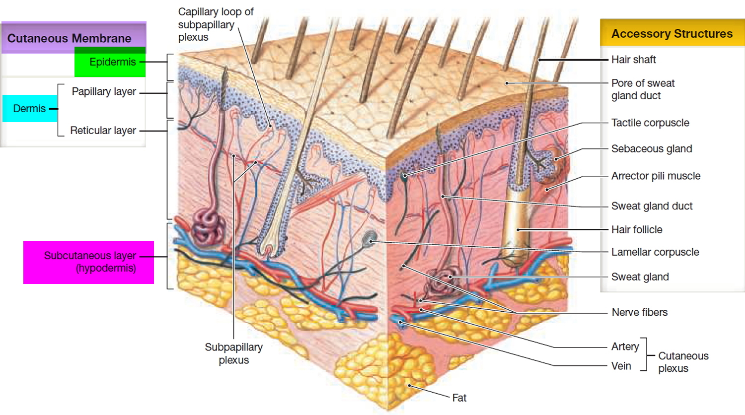
The skin consists of two layers:

  1. a stratified squamous epithelium called the Epidermis and
  2. a deeper connective tissue layer called the Dermis (Figure 1).

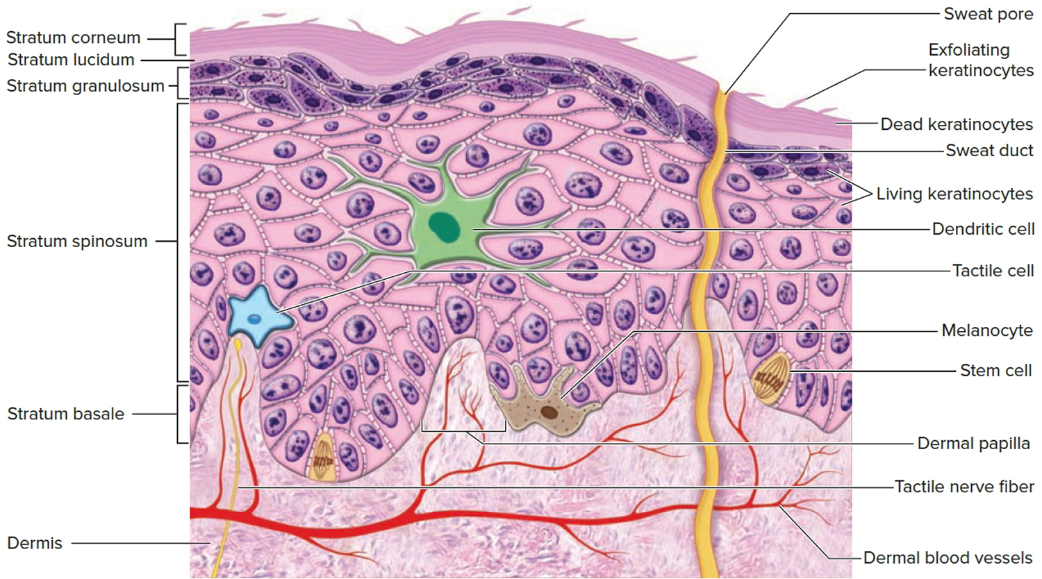
The brown tones of the skin result from the pigment-producing cells called melanocytes. Melanocytes are scattered among the basal cells of the stratum basale. They have numerous cytoplasmic processes that inject melanin—a black, yellow-brown, or brown pigment—into the basal cells in this layer and into the keratinocytes of more superficial layers. The ratio of melanocytes to stem cells ranges between 1:4 and 1:20 depending on the region examined. Melanocytes are most abundant in the cheeks, forehead, nipples, and genital region.

Differences in skin color result from varying levels of melanocyte activity, not varying numbers of melanocytes. Albinism is an inherited disorder characterized by deficient melanin production; individuals with this condition have a normal distribution of melanocytes, but the cells cannot produce melanin. It affects approximately one person in 10,000.

Figure 1. Skin structure

skin structure and layers

Figure 2. Structure and skin cells of the Epidermis

structure of epidermis

Skin hyperpigmentation usually results from an increased number, or activity, of melanocytes. Epidermal increases in melanin usually enhance with a Wood lamp, whereas dermal increases do not. Some disorders, such as melasma, may have dermal and epidermal changes and can be classified as mixed 2.

  • Hypopigmentation of skin may result from a reduction of melanocytes or from an inability of the melanocytes to produce melanin or properly transport melanosomes. Causes of hyper- and hypopigmentation are discussed in this article and are listed in Table 1.
  • Certain skin pigmentation disorders are more common in certain skin types. The most commonly used system for identifying skin types is the Fitzpatrick system (Table 2) 3.

Table 1. Causes of uneven skin tone (hyper- and hypopigmentation)

Hyperpigmentation

  • Postinflammatory hyperpigmentation (acne, psoriasis, atopic and contact dermatitis, lichen planus, trauma, drugs, and fixed-drug eruptions)
  • Melasma
  • Solar lentigines
  • Ephelides (freckles)
  • Café-au-lait macules
  • Nevi
  • Melanoma and precursors

Hypopigmentation

  • Acquired (common)
  • Vitiligo
  • Pityriasis alba
  • Tinea versicolor
  • Postinflammatory hypopigmentation
  • Congenital (uncommon)
  • Albinism
  • Piebaldism
  • Tuberous sclerosis
  • Hypomelanosis of Ito
[Source 1]

Table 2. Skin Type Classification

Skin typeSkin colorCharacteristics

I

White; very fair; red or blond hair; blue eyes; freckles

Always burns, never tans

II

White; fair; red or blond hair; blue, hazel, or green eyes

Usually burns, tans with difficulty

III

Cream white; fair with any eye or hair color; very common

Sometimes mild burn, gradually tans

IV

Brown; typically Mediterranean skin

Rarely burns, tans with ease

V

Dark brown; Middle-Eastern skin types

Very rarely burns, tans very easily

VI

Black

Never burns, tans very easily


[Source 3]

Table 3. Summary of common uneven skin tone disorders seen by doctors

Common uneven skin tone disorders and their treatments
Disorder Description Location Cause
Postinflammatory hyperpigmentatin Irregular, darkly-pigmented macules or patches Previous sites of injury or inflammation Trauma, inflammation
Treatment : Hydroquinone (Eldoquin Forte), azelaic acid (Azelex), retinoids, chemical peels, laser therapy; combination therapy is most effective
——————————————————–
Disorder Description Location Cause
Melasma Pigmented, well-defined macules; light brown, brown, or gray in color Face (63 percent centrofacial, 21 percent malar, 16 percent mandibular), forearms Pregnancy, oral contraceptives, phenytoin (Dilantin), idiopathic
Treatment : Sunscreen; combinations of: hydroquinone, retinoids, glycolic acid peels, topical steroids; laser therapy, intense pulsed light therapy for dermal lesions
——————————————————–
Disorder Description Location Cause
Solar lentigines 1- to 3-cm macules, well-circumscribed, light yellow to dark brown, variegated color Face, hands, forearms, chest, back, shins Acute, chronic ultraviolet light exposure
Treatment : Hydroquinone, retinoids, chemical peels, laser therapy, cryotherapy
——————————————————–
Disorder Description Location Cause
Ephelides 1- to 2-mm, shaply defined macules, red to tan to light brown in color Childhood onset, face, neck, chest, arms, legs Sun exposure in susceptible persons (i.e., skin types I to II)
Treatment : None needed; fades in winter months
——————————————————–
Disorder Description Location Cause
Café-au-lait macules Tan to brown patches, 1 to 20 cm, epidermal, present at birth or early childhood Usually on trunk, but possible anywhere Increased melanin in melanocytes, basal keratinocytes
Treatment : Laser therapy, surgical excision; cosmetic treatment
——————————————————–
Disorder Description Location Cause
Vitiligo Unpigmented macules and patches, sharply defined, 5 to 50 mm, coalescent Face, hands, forearms, neck, genitalia, body folds, periorificial Unknown, possibly immune-mediated
Treatment : Sunscreens, concealers, dyes, topical steroids, oral psoralens with psoralen ultraviolet A-range, narrow-band ultraviolet-B therapy, depigmentation, grafting

Hyperpigmentation Disorders

Postinflammatory Hyperpigmentation

Postinflammatory hyperpigmentation is a common consequence of an injury or inflammation to dark skin (Fitzpatrick types IV to VI see Table 2 above), resulting in lesions that can persist for months or years. This can be psychologically devastating to some people. Postinflammatory hyperpigmentation may also occur after laser therapy for other pigmented skin lesions, and may be transient or long lasting. A typical example can be seen in Figure 3.

Figure 3. Postinflammatory hyperpigmentation of distal right leg following resolution of dermatitis eruption

hypopigmentation leg

Postinflammatory hyperpigmentation presents as irregular, darkly pigmented macules and patches at sites of previous injury or inflammation. Treatment is often difficult, requiring prolonged courses of therapy and excellent patient compliance.

Available methods of treatment for postinflammatory hyperpigmentation include hydroquinone 3% or 4% (Eldoquin Forte) twice daily, azelaic acid 20% cream (Azelex) twice daily, salicylic or glycolic acid peels, retinoids, and laser therapy. However, monotherapy often produces unsatisfactory results. In one study, the addition of serial glycolic acid peels to a hydroquinone 2%/glycolic acid 10% combination twice daily and tretinoin 0.05% (Retin-A) at bedtime resulted in faster lightening without significant adverse effects 4. Additionally, retinoids such as tazarotene 0.1% cream (Tazorac) are well-tolerated and somewhat effective at reducing hyper-pigmentation and disease severity 5.

Pretreatment with topical therapies has been studied in patients with skin types I to III undergoing carbon dioxide laser resurfacing. No conclusive benefit was noted in one limited trial involving patients at the lowest risk for postin-flammatory hyperpigmentation 6. At present, no preventative measures have proven beneficial in any skin type.

Melasma

Melasma is a progressive, macular, nonscaling hypermelanosis of sun-exposed areas of the skin, primarily on the face and dorsal forearms. It is usually associated with pregnancy, oral contraceptives, or anticonvulsants (e.g., phenytoin [Dilantin]), or it may be idiopathic. Melasma affects women nine times more often than men, and it is more prominent in patients with skin types IV to VI (e.g., Asian, Middle Eastern, South American). It is usually asymptomatic, but it is often cosmetically distressing to the patient. Melasma typically presents in one of three patterns of distribution: centrofacial (63 percent), malar (21 percent), and mandibular (16 percent). It is usually, but not always, bilateral (Figure 4).

Figure 4. Melasma

Melasma

Three types of melasma exist: epidermal, dermal, and mixed. Epidermal melasma tends to be light brown, enhancing under Wood lamp examination. Dermal melasma is usually grayish in color and nonenhancing. Mixed types are dark brown with variable enhancement.

Topical treatment with hydroquinone 3% or 4%, glycolic acid 10% peel, azelaic acid 20% cream, and retinoids (e.g., tretinoin 0.05% or 0.1% cream; adapalene 0.1% or 0.3% gel [Differin]) all have some effectiveness. Combination products with hydroquinone and retinoids, glycolic acid, or topical steroids seem to be somewhat more effective. Typically, treatment must be continued indefinitely to maintain effect 7. In one drug-company-sponsored study, a triple-combination treatment of fluocinonide 0.01%/hydroquinone 4%/tretinoin 0.05% cream (Tri-Luma) showed significantly greater effectiveness at improving dyspigmentation than treatment with any two of these ingredients combined, with mild side effects 8. Epidermal and mixed types are not often responsive to laser therapies and frequently result in significant postinflammatory hyperpigmentation; therefore, their use cannot be recommended. However, several small studies suggest that dermal or refractory/mixed-type melasmas may be effectively treated with laser therapy or by a combination of intense pulsed-light therapy and hydroquinone with sunscreen 9.

Prevention of melasma involves decreasing exposure of susceptible skin to ultraviolet (UV) rays. Opaque sunblocks with titanium dioxide or zinc oxide are most effective. There are transparent sunscreens containing these agents as well (e.g., Blue Lizard Sunscreen Sensitive SPF 30+, Neutrogena Sensitive Skin Sunblock 30+, Sol-bar Zinc Sunscreen SPF 38). Melasma that is induced by pregnancy or oral contraceptive use tends to fade within several months after delivery or medication cessation, so watchful waiting should be encouraged in these instances whenever possible.

Solar Lentigines

Solar lentigines (i.e., liver spots) are macular, 1- to 3-cm, hyperpigmented, well-circumscribed lesions on sun-exposed surfaces of the skin. They vary in color from light yellow to dark brown, and they often have a variegated appearance. The face, hands, forearms, chest, back and shins are the most common locations, erupting after acute or chronic UV exposure. White or Asian persons are most likely to develop solar lentigines, especially those with skin types I to III and a tendency to freckle (Figure 5).

Figure 5. Solar lentigines

Solar lentigines

Solar lentigines result from a local proliferation of basal melanocytes and a subsequent increase in melanization, differing from freckles, which result from increased melanin production. Systemic disorders presenting with multiple lentigines may include Peutz-Jeghers syndrome (gastrointestinal hamartomas; buccal, lip, perioral, or digital macules; onset at birth or early childhood), LEOPARD syndrome (multiple lentigines, electrocardiogram abnormalities, ocular hypertelorism, pulmonic stenosis, abnormal genitalia, retarded growth, and sensorineural deafness), and LAMB syndrome (multiple lentigines, atrial and/or mucocutaneous myxomas, myxoid neurofibromas, ephelides, and blue nevi). Solar lentigines must be differentiated from premalignant lesions, such as pigmented actinic keratoses or lentigo maligna. Pigmented lesions with rapid growth or change, associated symptoms (e.g., pain, itching, easy or recurrent bleeding, poor healing), atypical lesions, or those with features suspicious for melanoma should be biopsied. Full thickness excisional biopsy or punch biopsy (for large lesions or those on the face or cosmetically sensitive area) is an acceptable method of biopsy in these instances. Solar lentigines can be distinguished clinically from flat seborrheic dermatoses or pigmented actinic keratoses by the absence of epidermal hyperkeratosis. Biopsy may facilitate diagnosis in uncertain cases.

Treatment of solar lentigines consists of ablative therapies (e.g., chemical peels, cryotherapy, laser therapy) or topical therapies (e.g., hydroquinone, retinoids), and is summarized in Table 4. Chemical peels with 30% to 35% trichloroacetic acid (Trichlor) solution or brief (i.e., less than 10 seconds) cryotherapy with liquid nitrogen have resulted in significant lightening of lentigines, but data are limited on long-term improvements, and recurrences are common. Additionally, cryotherapy can be painful, and prolonged treatment is associated with hypopigmentation 10. Laser therapy for solar lentigines has shown benefit in at least one small, randomized controlled trial, with effectiveness superior to liquid nitrogen cryotherapy. The frequency-doubled Q-switched neodymium-doped yttrium aluminum garnet (ND: YAG) laser produced the best cosmetic results and was tolerated best. Postinflammatory hyperpigmentation is a known complication of laser therapy, and must be considered when determining the best treatment option for each patient 11.

Table 4. Treatment of Solar Lentigines

TreatmentType/doseSide effects

Chemical peels

30% to 35% trichloroacetic acid (Trichlor)

Transient stinging, burning, pain

Cryotherapy

Liquid nitrogen

Pain, hypopigmentation with prolonged exposure

Laser therapy

Neodymium-doped yttrium aluminum garnet (ND: YAG) laser

Pain, postinflammatory hyperpigmentation, redness, textural changes, hypopigmentation

Hydroquinone (Eldoquin Forte)

3% to 4% topical

Hypersensitivity, acne, ochronosis

Mequinol/tretinoin (Solage)

2% mequinol/0.01% tretinoin topical solution

Redness, dryness, itching, sensitivity

Retinoids

Tazarotene 0.1% cream (Tazorac); adapalene 0.1% or 0.3% gel (Differin)

Redness, dryness, itching, sensitivity

[Source 1]

Topical therapies for solar lentigines are also available. Hydroquinone has been available for more than 30 years and is moderately effective. Adverse effects to hydroquinone include hypersensitivity, acneiform eruptions, and, rarely, ochronosis (i.e., blotchy hyperpigmentation) 12. Additionally, the lightening effects of hydroquinone are slow (months), and relapse with medication discontinuation is the rule. More recently, a combination of mequinol/tretinoin (Solage) has been shown to be safe and effective in treating solar lentigines, and shows promise for prolonged maintenance 13. Retinoids such as tazarotene 0.1% cream and adapalene 0.1% or 0.3% gel may reduce the appearance of solar lentigines, but evidence is limited 14.

Prevention of solar lentigines depends on limiting sun exposure, using sunscreen regularly (especially in patients with fair skin [types I to III] and those prone to freckling), and preventing sunburns, especially after 20 years of age.

Ephelides

Ephelides (i.e., freckles) are small, 1- to 2-mm, sharply defined macular lesions of uniform color, most often found on the face, neck, chest, and arms. Color may vary from red to tan to light brown, and they may vary in number from a few to hundreds. Onset is usually in childhood after sun exposure. They are asymptomatic. Treatment of these lesions is not usually necessary, as they tend to fade during winter months. Cosmetically, undesired lesions can be treated similarly to lentigines (i.e., cryotherapy, hydroquinone, azelaic acid, glycolic acid peels, and laser therapy). These lesions should be differentiated from juvenile lentigines (2 to 10 mm) and solar lentigines (2 to 20 mm), which usually arrive later in life.

Café-au-lait macules

Café-au-lait macules are tan or brown macules ranging in size from 1 to 20 cm, which are present at birth or occur early in life. They are epidermal in origin, representing an increase in melanin in melanocytes and basal keratinocytes. They may be found on any body part, but often are located on the trunk (Figure 6). Ten to 30 percent of the population has an isolated café-au-lait macule 15.

Café-au-lait macules are asymptomatic and require treatment for cosmesis only. Laser therapies and surgical excision are effective. More than six café-au-lait lesions (5 mm or larger, prepubertal; and 15 mm or larger, postpubertal) should raise suspicion for an underlying systemic disorder such as tuberous sclerosis, neurofibromatosis, Albright syndrome, or Fanconi anemia 3.

Figure 6. Café-au-lait macules

Café-au-lait macules

Hypopigmented Lesions

Vitiligo

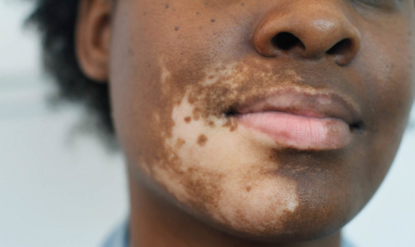
Vitiligo is a disfiguring skin disease resulting in loss of pigmentation. It results from an immune-mediated destruction of melanocytes. It presents with well-defined milky-white patches of skin (leukoderma). Vitiligo can be cosmetically very disabling, particularly in people with dark skin. Its exact cause is unknown. Vitiligo affects all skin types and is generally considered a cosmetic condition, but it can cause significant psychological distress, particularly to some black patients.

Lesions in vitiligo consist of unpigmented, sharply defined macules ranging in size from 5 to 50 mm. Some will have a rim of hyperpigmentation or erythema. Common sites of involvement include the face, neck, dorsal hands, genitalia, body folds, and axillae (Figure 7). Perioral, periorbital, periumbilical, and perianal lesions also occur.

Four types of vitiligo exist:

  • Generalized vitiligo involves greater than 10 percent of the body surface area.
  • Acral/acrofacial vitiligo typically involves the face and distal extremities (i.e., the so-called “tip/lip” pattern).
  • Localized vitiligo tends to involve a smaller body surface area and is generally stable in nature.
  • Segmental (i.e., single dermatome or extremity) vitiligo is more often present in children and has a poorer prognosis for treatment.

Who gets vitiligo ?

Vitiligo is found in 0.5 -1 percent of the general population and occurs in all races, affecting males and females equally. Family history of vitiligo is established in 25 to 30 percent of patients 15. Onset is often insidious, but is frequently related to a recent stress, illness, or trauma (e.g., sunburn). Peak onset is in the second and third decades of life, with 50 percent occurring before 20 years of age 15.

Even though most people with vitiligo are in good general health, they face a greater risk of having autoimmune diseases such as diabetes, thyroid disease (in 20% of patients over 20 years with vitiligo), pernicious anaemia (B12 deficiency), Addison disease (adrenal gland disease), systemic lupus erythematosus, rheumatoid arthritis, psoriasis, and alopecia areata (round patches of hair loss).

A vitiligo-like leukoderma may occur in patients with metastatic melanoma. It can also be induced by certain drugs, such as immune checkpoint inhibitors (pembrolizumab, nivolumab) and BRAF inhibitors (vemurafenib, dabrafenib) used to treat metastatic melanoma. Vitiligo is also 3 times more common in haematology patients that have had allogeneic bone marrow and stem-cell transplants, than in the normal population.

Figure 7. Vitiligo

Vitiligo

What causes vitiligo ?

Vitiligo is due to loss or destruction of melanocytes, which are the cells that produce melanin. Melanin determines the color of your skin, hair, and eyes. If melanocytes cannot form melanin or if their number decreases, skin color becomes progressively lighter.

The exact cause of vitiligo is unknown. It is thought to be a systemic autoimmune disorder, associated with deregulated innate immune response, although this has been disputed for segmental vitiligo. There is a genetic susceptibility and vitiligo is a component of some rare syndromes. The gene encoding the melanocyte enzyme tyrosinase, TYR, is likely involved.

There are three theories on the cause of vitiligo:

  • The pigment cells are injured by abnormally functioning nerve cells.
  • There may be an autoimmune reaction against the pigment cells.
  • Autotoxic theory – the pigment cells self-destruct.

Current investigations are evaluating the pattern of cytokines (messenger proteins), particularly Interferon (IFN)-γ, and the role of the hair follicle in repigmentation.

How is vitiligo diagnosed ?

Vitiligo is normally a clinical diagnosis, and no tests are necessary to make the diagnosis. The white patches may be seen more easily under Wood lamp examination (black light).

Occasionally skin biopsy may be recommended, particularly in early or inflammatory vitiligo, when a lymphocytic infiltration may be observed. Melanocytes and epidermal pigment are absent in established vitiligo patches.

Blood tests to assess other potential autoimmune diseases or polyglandular syndromes may be arranged, such as thyroid function, B12 levels and autoantibody screen.

Clinical photographs are useful to document the extent of vitiligo for monitoring. Serial digital images may be arranged on follow-up. The extent of vitiligo may be scored according to the body surface area affected by depigmentation.

How is vitiligo treated ?

Treatment of vitiligo is currently unsatisfactory. Repigmentation treatment is most successful on face and trunk; hands, feet and areas with white hair respond poorly. Compared to longstanding patches, new ones are more likely to respond to medical therapy.

When successful repigmentation occurs, melanocyte stem cells in the bulb at the base of the hair follicle are activated and migrate to the skin surface. They appear as perifollicular brown macules.

General measures

Minimise skin injury: wear protective clothing

  • A cut, a graze, a scratch may lead to a new patch of vitiligo

Cosmetic camouflage can disguise vitiligo. Options include:

  • Make-up, dyes and stains
  • Waterproof products
  • Dihydroxyacetone-containing products “tan without sun”
  • Micropigmentation or tattooing for stable vitiligo

Sun protection: stay indoors when sunlight is at its peak, cover up with sun protective clothing and apply SPF 50+ sunscreen to exposed skin.

  • White skin can only burn on exposure to ultraviolet radiation (UVR); it cannot tan
  • Sunburn may cause vitiligo to spread
  • Tanning of normal skin makes vitiligo patches appear more obvious
Topical treatments

Topical treatments for vitiligo include:

  • Corticosteroid creams. These can be used for vitiligo on trunk and limbs for up to 3 months. Potent steroids should be avoided on thin-skinned areas of face (especially eyelids), neck, armpits and groin.
  • Calcineurin inhibitors (pimecrolimus cream and tacrolimus ointment. These can be used for vitiligo affecting eyelids, face, neck, armpits and groin.
  • Experimental treatment with topical ruxolitinib, a janus kinase inhibitor, shows great promise for facial vitiligo.
Phototherapy

Phototherapy refers to treatment with ultraviolet (UV) radiation. Options include:

  • Whole-body or localised broadband or narrowband (311 nm) UVB
  • Excimer laser UVB (308 nm) or targeted UVB for small areas of vitiligo
  • Oral, topical, or bathwater photochemotherapy (PUVA)

Phototherapy probably works in vitiligo by 2 mechanisms.

  • Immune suppression—preventing destruction of the melanocytes
  • Stimulation of cytokines (growth factors)

Treatment is usually given twice weekly for a trial period of 3–4 months. If repigmentation is observed, treatment is continued until repigmentation is complete or for a maximum of 1–2 years.

  • Phototherapy is unsuitable for very fair skinned people.
  • The treatment intensity aims for the vitiligo skin to be a light “carnation” pink
  • If repigmentation is observed, treatment is continued until repigmentation is complete or for a maximum of 1–2 years.
  • Treatment times are generally brief. The aim is to cause the treated skin to appear very slightly pink the following day.
  • It is important to avoid burning (red, blistered, peeling, itchy or painful skin), as this could cause the vitiligo to get worse.

A meta-analysis included 35 unique studies reporting outcome after phototherapy for generalised vitiligo. Marked or clinically useful response was achieved in 36% after 12 months of NBUVB and in 62% after 12 months of PUVA. Face and neck responded better than trunk, which responded better than extremities. It was not very effective on hands and feet.

Systemic therapy

Systemic treatments for vitiligo include:

  • Oral minocycline, a tetracycline antibiotic with anti-inflammatory properties
  • Mini-pulses of oral steroids for 3 to 6 months, eg dexamethasone 2.5 mg, 2 days per week
  • Subcutaneous afamelanotide

It is anticipated that monoclonal antibody biologic agents will be developed to treat vitiligo.

Surgical treatment of stable vitiligo

Surgical treatment for stable and segmental vitiligo requires removal of the top layer of vitiligo skin (by shaving, dermabrasion, sandpapering or laser) and replacement with pigmented skin removed from another site.

Techniques include:

  • Non-cultured melanocyte-keratinocyte cell suspension transplantation.
  • Punch grafting
  • Blister grafts, formed by suction or cryotherapy
  • Split skin grafting
  • Cultured autografts of melanocytes grown in tissue culture
Depigmentation therapy

Depigmentation therapy, using monobenzyl ether of hydroquinone, may be considered in severely affected, dark skinned individuals.

Cyotherapy and laser treatment (eg 755 nm Q-switched alexandrite or 694 nm Q-switched ruby) have also been used successfully to depigment small areas of vitiligo.

Other Hypopigmentation Disorders

Other disorders commonly associated with hypopigmentation include pityriasis alba, tinea versicolor, postinflammatory hypomelanosis (i.e., loss of melanin), atopic dermatitis, psoriasis, and guttate parapsoriasis. Additionally, it may also result from dermabrasion, chemical peels, and intralesional steroid therapy.

References
  1. Common Pigmentation Disorders. Am Fam Physician. 2009 Jan 15;79(2):109-116. http://www.aafp.org/afp/2009/0115/p109.html
  2. Stulberg DL, Clark N, Tovey D. Common hyperpigmentation disorders in adults: Part I. Diagnostic approach, café-au-lait macules, diffuse hyper-pigmentation, sun exposure, and phototoxic reactions. Am Fam Physician. 2003;68(10):1955–1960.
  3. Fitzpatrick TB. Fitzpatrick’s Dermatology in General Medicine. 4th ed. New York, NY: McGraw-Hill; 1993:966–968,1694,1984.
  4. Burns RL, Prevost-Blank PL, Lawry MA, Lawry TB, Faria DT, Fivenson DP. Glycolic acid peels for postinflammatory hyperpigmentation in black patients. A comparative study. Dermatol Surg. 1997;23(3):171–175.
  5. Grimes P, Callender V. Tazarotene cream for postinflammatory hyper-pigmentation and acne vulgaris in darker skin: a double-blind, randomized, vehicle-controlled study. Cutis. 2006;77(1):45–50.
  6. West TB, Alster TS. Effect of pretreatment on the incidence of hyperpigmentation following cutaneous CO2 laser resurfacing. Dermatol Surg. 1999;25(1):15–17.
  7. Espinal-Perez LE, Moncada B, Castanedo-Cazares JP. A double-blind randomized trial of 5% ascorbic acid vs. 4% hydroquinone in melasma. Int J Dermatol. 2004;43(8):604–607.
  8. Torok H, Taylor S, Baumann L, et al. A large 12-month extension study of an 8-week trial to evaluate the safety and efficacy of triple combination (TC) cream in melasma patients previously treated with TC cream or one of its dyads. J Drugs Dermatol. 2005;4(5):592–597.
  9. Wang CC, Hui CY, Sue YM, Wong WR, Hong HS. Intense pulsed light for the treatment of refractory melasma in Asian persons. Dermatol Surg. 2004;30(9):1196–1200.
  10. Lugo-Janer A, Lugo-Somolinos A, Sanchez JL. Comparison of trichloroacetic acid solution and cryosurgery in the treatment of solar lentigines. Int J Dermatol. 2003;42(10):829–831.
  11. Wang CC, Sue YM, Yang CH, Chen CK. A comparison of Q-switched alexandrite laser and intense pulsed light for the treatment of freckles and lentigines in Asian persons: a randomized, physician-blinded, split-face comparative trial. J Am Acad Dermatol. 2006;54(5):804–810.
  12. Draelos ZD. Novel approach to the treatment of hyperpigmented photo-damaged skin: 4% hydroquinone/0.3% retinol versus tretinoin 0.05% emollient cream. Dermatol Surg. 2005;31(7 pt 2):799–804.
  13. Jarratt M. Mequinol 2%/tretinoin 0.01% solution: an effective and safe alternative to hydroquinone 3% in the treatment of solar lentigines. Cutis. 2004;74(5):319–322.
  14. Kang S, Kreuger GG, Tanghetti EA, et al., for the Tazarotene Cream in Photodamage Study Group. A multicenter, randomized, double-blind trial of tazarotene 0.1% cream in the treatment of photodamage. J Am Acad Dermatol. 2005;52(2):268–274.
  15. Fathman EM, Habif TP. Skin Disease: Diagnosis and Treatment. 1st ed. St. Louis, Mo.: Mosby; 2001:58,184–186,308–311,469.
read more
Conditions & DiseasesSkinSkin System

How to get rid of chapped lips

chapped lips

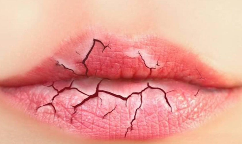
What causes chapped lips

Most people get chapped lips from time to time and dry chapped lips can occur in cold and even warm weather. To avoid chapped lips, use a lip balm with sunscreen when outside in any weather. To soothe dry chapped lips treat with beeswax and phenol (such as Carmex).

If chapping is severe and doesn’t respond to treatment at home, consult your doctor. Rarely, persistent chapped lips may indicate an underlying problem.

Here are some recommendations for preventing chapped lips

There’s much you can do to treat — and prevent — chapped lips. Consider these tips:

  • Protect your lips. Before going out in cold, dry weather, apply a lubricating lip cream, lipstick or lip balm that contains sunscreen — and then cover your lips with a scarf. Reapply often while outdoors.
  • Avoid excessive sun exposure.
  • Avoid licking your lips. Saliva evaporates quickly, leaving lips drier than before you licked them. If you tend to lick your lips, avoid flavored lip balm — which may tempt you to lick your lips even more.
  • Try to keep the air in your home humid.
  • Use a sunscreen on your lips when outside in sunny weather.
  • Stay hydrated. Drink plenty of fluids, and moisten the air in your home with a humidifier.
  • Avoid allergens. Avoid contact with irritants or allergens, such as fragrances or dyes, in cosmetics or skin care products.
  • Breathe through your nose. Breathing through your mouth can cause your lips to dry out.

Here are some recommendations for soothing chapped and sore lips:

  • Drink additional fluids in winter months.
  • Treat chapped lips with beeswax and petrolatum (Vaseline).

See your health care provider if you have sores that will not heal on your lips.

read more
NailsSkinSkin System

Ingrown toenail

ingrown toenail

Ingrown toenail

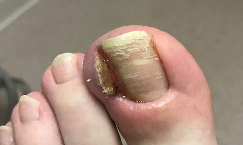
Toenails and fingernails are clear, hard derivatives of the stratum corneum. They are composed of very thin, dead, scaly cells, densely packed together and filled with parallel fibers of hard keratin. An ingrown toenail or onychocryptosis, occurs when the edge of the nail grows down and into the skin of the toe. There may be pain, redness, and swelling around the nail.

An ingrown toenail may occur at any age and is the mostly commonly encountered toenail problem likely to be seen in general practice 1.

The epidemiology of ingrown toenail (onychocryptosis) is difficult to determine as it is often considered to be a minor medical problem and as such has been some-what neglected in the literature. The few studies that have been conducted suggest a slightly higher male-to-female ratio, particularly in the 14–25 age group 2, but it can affect patients of any age. There are multiple reasons why an ingrown toenail will develop, including improper nail cutting technique, tight-fitting footwear, trauma, anatomical factors such as thickening of the nail plate, pincer-shaped toenail, pressure from abutting digits caused by hallux valgus or lesser toe deformities, the presence of a subungual exostosis and, occasionally, the use of isotretinoin in the treatment of severe acne 3, 4, 5.

Causes of ingrown toenail

An ingrown toenail can result from a number of things, but poorly fitting shoes and toenails that are not trimmed properly are the most common causes. The skin along the edge of a toenail may become red and infected. The great toe is usually affected, but any toenail can become ingrown.

Ingrown toenails may occur when extra pressure is placed on your toe. Most commonly, this pressure is caused by shoes that are too tight or too loose. If you walk often or participate in athletics, a shoe that is even a little tight can cause this problem. Some deformities of the foot or toes can also place extra pressure on the toe.

Nails that are not trimmed properly can also cause ingrown toenails.

  • When your toenails are trimmed too short or the edges are rounded rather than cut straight across, the nail may curl downward and grow into the skin.
  • Poor eyesight and physical inability to reach the toe easily, as well as having thick nails, can make improper trimming of the nails more likely.
  • Picking or tearing at the corners of the nails can also cause an ingrown toenail.

Some people are born with nails that are curved and tend to grow downward. Others have toenails that are too large for their toes. Stubbing your toe or other injuries can also lead to an ingrown toenail.

Prevention of ingrown toenail

Wear shoes that fit properly. Shoes that you wear every day should have plenty of room around your toes. Shoes that you wear for walking briskly or for running should have plenty of room also, but not be too loose.

When trimming your toenails:

  • Considering briefly soaking your foot in warm water to soften the nail.
  • Use a clean, sharp nail trimmer.
  • Trim toenails straight across the top. Do not taper or round the corners or trim too short.
  • Do not pick or tear at the nails.

Keep the feet clean and dry. People with diabetes should have routine foot exams and nail care.

Figure 1. Ingrown toenail

ingrown toenail

Figure 2. Nail anatomy

nail anatomy

toenail

How to treat an ingrown toenail

Often the medial or lateral nail edge of an incurved toenail may press into the nail sulcus without actual skin penetration, which leads to the formation of a painful hyperkeratotic lesion. Should the nail plate penetrate through the thin skin of the nail sulcus, infection and hypergranulation tissue will ensue and the toenail is then best described as ingrown (Figure 1) 6. Either condition may become symptomatic and require definitive surgical management. Some doctors suggest that, ideally, all presentations of ingrown toenail should be managed conservatively in the first instance 7.

Conservative treatment

Careful trimming of the affected distal nail corner is the preferred conservative management of an incurved toenail followed by application of a suitable antiseptic dressing. Referral to a podiatrist to carefully trim the nail edge and debride any hyperkeratotic lesion may be necessary. Traditional conservative treatments have included the use of astringent soaks such as hypertonic solution of Epsom salts or the application of 25% silver nitrate solution may be beneficial but are rarely used today. Encouraging patients to carefully trim their toenails to avoid creating sharp spicules and to place small inserts of cotton wool beneath the nail corners to protect the skin of the nail fold may also be helpful 8.

Patients presenting with an ingrown toenail that has penetrated the nail sulcus usually require a partial avulsion of the offending border of toenail to allow for drainage and the excision of any hypergranulation tissue 9. Local anesthesia should first be attained via a digital ring block injection of the hallux proximal to the area of erythema with plain local anaesthetic solution. Using small sterile nail nippers, the distal corner section of the involved nail edge is removed taking care not to leave any sharp projections. Care must be exercised not to extend the nail excision more proximal than necessary to avoid the nail matrix area. Hypergranulation tissue should be excised if present. Application of a haemostatic dressing such as Gelatamp may also be necessary. Oral antibiotics are sometimes required but are usually not necessary, providing the offending nail section and any hypergranulation tissue are removed. Home care then consists of soaking twice daily in Epsom salt or saline solution and using a topical antiseptic dressing until healed.

Home remedies for ingrown toenail

  • If you have diabetes, nerve damage in the leg or foot, poor blood circulation to your foot, or an infection around the nail, go to the doctor right away. Do NOT try to treat this problem at home.

To treat an ingrown nail at home:

  • Soak the foot in warm water 3 to 4 times a day if possible. Keep the toe dry, otherwise.
  • Gently massage over the inflamed skin.
  • Place a small piece of cotton or dental floss under the nail. Wet the cotton with water or antiseptic.

You may trim the toenail one time, if needed. When trimming your toenails:

  • Consider briefly soaking your foot in warm water to soften the nail.
  • Use a clean, sharp trimmer.
  • Trim toenails straight across the top. Do not taper or round the corners or trim too short. Do not try to cut out the ingrown portion of the nail yourself. This will only make the problem worse.

Consider wearing sandals until the problem has gone away. Over-the-counter medications that are placed over the ingrown toenail may help some with the pain but do not treat the problem.

If this does not work and the ingrown nail gets worse, see your family doctor, a foot specialist (podiatrist) or a skin specialist (dermatologist).

If your ingrown nail does not heal or keeps coming back, your doctor may remove part of the nail.

  • Numbing medicine is first injected into the toe.
  • Using scissors, your doctor then cuts along the edge of the nail where the skin is growing over. This portion of the nail is then removed. This is called a partial nail avulsion.
  • It will take 2 to 4 months for the nail to regrow.

Sometimes your doctor will use a chemical, electrical current, or another small surgical cut to destroy or remove the area from which a new nail may grow.

If the toe is infected, your doctor may prescribe antibiotics.

Ingrown Toenail Surgery – Winograd technique

If conservative care proves unsatisfactory or if the patient presents with a recurrent ingrown toenail then surgical treatment is indicated. Sharp nail resection and phenol-ablation techniques have been reported in the literature as safe and effective methods for treating ingrown toenails 10, 11, 12.

The Winograd technique is perhaps the most commonly performed sharp procedure 10 and is often used when a hypertrophied nailfold is in need of excision. Winograd, a US podiatrist first described the technique in 1929 13. A large case series analysis published by Güler et al 10 in 2014, which followed 239 patients who underwent a Winograd procedure for ingrown toenails, reported that 96.3% (n = 230) were satisfied with the procedure and 231 patients (96.6%) were satisfied with the cosmetic result 10. Another recent analysis performed by Kayalar et al 11 of 224 patients who underwent the Winograd procedure found a high success rate of 91.2% (n = 202), with revisional surgery only required in 7.1% 
(n = 16) at a minimum 10-month follow-up 11.

The local anesthetic preferred for the procedure is either bupivacaine 0.5% or ropivacaine 0.75% plain. Instruments and materials required are:

  • nonstick antiseptic gauze (eg Bactigras paraffin-wax gauze)
  • 4.0 non-absorbable suture
  • suture forceps
  • scalpel handle
  • English anvil nail splitter
  • small curette
  • straight haemostat
  • #15 blade
  • sterile saline
  • digit tourniquet (eg Tourni-cot ring)

The procedure is illustrated in Figure 3 and outlined below:

  • Prepare the toe using alcohol skin wipes and inject 3–4 ml of bupivcaine 0.5% or ropivacaine 0.75% plain as digital ring block.
  • Use standard sterile technique after applying a suitable alcoholic chlorhexidine or povidone-iodine liquid solution to prepare the foot.
  • Use a digit tourniquet to exsanguinate the toe.
  • Use the nail splitter to split 2–3 mm of the affected side of the nail longitudinally (Figure 3A)
  • Make two skin incisions:
    + The first is made deeply through the split nail to include the nail bed extending proximally to include the dorsal skin over the nail matrix (Figure 3B)
    + The final incision is to excise the lateral nailfold, connecting the proximal and distal ends of the initial incision (Figure 3C, D).
  • Excise the nail section, bed and matrix tissue in total (Figure 3E) and the gently curette to remove any remaining matrix tissue (Figure 3F).
  • Flush with normal saline.
  • Close the wound with simple interrupted sutures (Figure 3F, H).
  • Remove the tourniquet and allow the toe to bleed for a minute or two with local pressure to control the reactive hyperaemia often encountered after release of the tourniquet.
  • Dress the toe with a suitable antiseptic dressing, sterile gauze and crepe bandage.

The patient should be advised of the following the post-operative protocol:

  • Rest and elevate the foot.
  • Use oral analgesics as needed.
  • Keep the dressing dry until post-operative review.
  • Advise of increasing pain/discomfort (infection).
  • The suture will be removed in 5–7 days.

Figure 3. Ingrown Toenail Surgery – Winograd technique

ingrown toenail surgery

Ingrown Toenail Surgery – Phenol Matrixectomy

Phenol matrixectomy is an alternative procedure often favoured by podiatrists and was first described by Boll, a US podiatrist, in 1945 14 and popularised by Bell, a UK podiatrist, in 1977 15. The procedure differs from the Winograd technique in that the nail matrix is destroyed via chemical ablation rather than being surgically excised. Phenol BP is caustic to tissues and, although the optimum application time has not been accurately determined, a histological study on fresh cadaveric specimens suggested at least 60 seconds is necessary for the phenol to penetrate matrix tissue 16. Phenol matrixectomy has been reported to offer certain advantages over the more traditional surgical approach, including lower levels of post-operative pain and infection, and has comparable success rates 17. A recently published randomised controlled trial of 123 patients with ingrown toenails concluded that the phenolic ablation technique produced less recurrence and spicule formation than the sharps nail resection (n =117 at 12 months follow-up) and no significant difference in post-operative infection rates 12. Another large retrospective case study of 197 phenolic ablations performed reported a 98.5% (n = 194) success rate at mean follow-up of 36 months with no severe complications observed 18.

The local anaesthetic preferred for this procedure is lignocaine 1–2% plain. Instruments and materials required are the same as those listed above for the Winograd technique plus:

  • Betadine ointment
  • Beaver handle with #62 Beaver blade
  • sterile cotton-tipped applicators
  • liquefied phenol BP.

No sutures are needed. The procedure is illustrated in Figure 4 and outlined below:

  • Prepare the toe using alcohol skin wipes and inject 3–4 ml of lignocaine 1–2% plain as digital ring block.
  • Use standard sterile technique after preparing the foot.
  • Use a digit tourniquet to exsanguinate the toe.
  • Use a nail splitter to split 2–3 mm of the affected side of nail longitudinally (Figure 4A) and complete to the proximal edge with the #62 Beaver blade (Figure 4B).
  • Perform avulsion of the nail by grasping the sectioned nail with a haemostat and using a gentle distraction technique to rotate it towards the midline of the nail plate and ease the nail free of the nail bed (Figure 4C, D).
  • Gently curette the surgical site to remove any remaining matrix tissue.
  • Apply liquid phenol BP directly to the site of the germinal nail matrix using small cotton wool applicator tips or the corner of gauze soaked phenol in 3 x 20-second applications (Figure 4E). Application of phenol should be carefully confined to the immediate surgical area, avoiding excess phenol contact with surrounding skin as this may result in unnecessary tissue injury. It is important to ensure that the nail sulcus and matrix area are dried immediately before applying phenol as the chemical is quickly neutralised by body fluid.
  • Flush the site using chlorhexidine solution (Figure 4F).
  • Remove the tourniquet and use local pressure to control any bleeding, which is normally minimal if any.
  • Dress the toe with Betadine ointment, sterile gauze and crepe bandage.

Advise the patient of the following post-operative protocol:

  • Postoperative analgesics are rarely required.
  • Keep the toe dry overnight.
  • Clean the site daily after showering using Epsom salts dissolved in water and to re-dress with Betadine ointment and a simple toe dressing until fully healed (2–3 weeks).
  • Monitor for signs of increasing pain/discomfort (infection).
  • The wound will be reviewed at 1 week post-operative to evaluate healing, at which time the wound should be debrided of any material inhibiting free drainage of the healing tissue.

Figure 4. Ingrown Toenail Surgery – Phenol-ablation technique

ingrown toenail surgery - phenol matrixectomy

References
  1. Murtagh J. Patient education. Ingrowing toenails. Aust Fam Physician 1993;22:206.
  2. Langford DT, Burke C, Robertson K. Risk factors in onychocryptosis. Br J Surg 1989;76:45–48.
  3. Shaikh FM, Jafri M, Giri SK, Keane R. Efficacy of wedge resection with phenolization in the treatment of ingrowing toenails. J Am Podiatr Med Assoc 2008;98:118–22.
  4. Ikard RW. Onychocryptosis. J Am Coll Surg 1998;187:96–102.
  5. Baran R, Roujeau JC. New millennium, new nail problems. Dermatol Ther 2002;15:64–70.
  6. Heifetz CJ. Ingrown toenail: A clinical study. Am J Surg 1937;38:298–315.
  7. Ingrown toenails: the role of the GP. Australian Family Physician Volume 44, No.3, 2015 Pages 102-105. https://www.racgp.org.au/afp/2015/march/ingrown-toenails-the-role-of-the-gp/
  8. Ilfeld FW. Ingrown toenail treated with cotton collodion insert. Foot Ankle 1991;11:312–13.
  9. Crislip TW, Boberg JS. Nail Surgery. In: Southerland J, Alder D, Boberg J, et al, editors. McGlamry’s Comprehensive textbook of foot and ankle surgery. 4th edn. Vol 1. Philadelphia: Lippincott Williams & Wilkins, 2013;109–16.
  10. Güler O, Mahirogulları M, Mutlu S, Çerçi H, Seker A, Mutlu H. An evaluation of partial matrix excision with Winograd method for the surgical treatment of ingrown toenails. JAREM 2014;4:7–11.
  11. Kayalar M, Bal E, Toros T, Özaksar K, Gürbüz Y, Ademoglu Y. Results of partial matrixectomy for chronic ingrown toenail. Foot Ankle Int 2011;32:888–95.
  12. Bos AM, van Tilburg MW, van Sorge AA, Klinkenbijl JH. Randomized clinical trial of surgical technique and local antibiotics for ingrowing toenail. J Br Surg 2007;94:292–96.
  13. Winograd AM. A modification in the technique of operation for ingrown toenail. J Am Podiatr Med Assoc. 2007 Jul-Aug;97(4):274-7.
  14. Boll O. Surgical correction of ingrowing toenails. J Natl Assoc Chiropod 1945;35:8.
  15. Bell DRC. Advanced techniques. Chiropodist 1977;32:238–43.
  16. Boberg JS, Frederiksen MS, Harton FM. Scientific analysis of phenol nail surgery. J Am Podiatr Med Assoc 2002;92:575–79.
  17. Herold N, Houshian S, Riegels-Nielsen P. A prospective comparison of wedge matrix resection with nail matrix phenolization for the treatment of ingrown toenail. J Foot Ankle Surg 2001;40:390–95.
  18. Vaccari S, Dika E, Balestri R, Rech G, Piraccini BM, Fanti PA. Partial excision of matrix and phenolic ablation for the treatment of ingrowing toenail: A 36-month follow-up of 197 treated patients. Dermatol Surg 2010;36:1288–93.
read more
SkinSkin System

Skin tags

skin tags

What are skin tags

A cutaneous skin tag also known as acrochordons or fibroepithelial polyps, is an extremely common skin growth, reportedly 46% of the general population, increasing in frequency with patient age. Skin tags are small, and typically pedunculated benign neoplasms 1. Most of the time, skin tags are harmless 2. Skin tags consist of hyperplastic soft dermis and epidermis and are usually skin colored or brownish (Figure 1). They are generally 2 to 5 mm in size, although they may become larger. The most common locations are in skin folds (e.g., neck, armpits, groin), where skin irritation can be a causative factor.

Skin tags may bother patients because of associated symptoms such as itching, pain, and rubbing against clothes or simply because of their appearance. There may be a familial predisposition for developing these lesions, as well as the established association with obesity and insulin resistance.

Diagnosis is based on the appearance and location of lesions. They must be differentiated from neurofibromas, seborrheic keratoses, and pedunculated nevi. There have been rare case reports of skin tags that were found to be basal or squamous cell carcinomas. Treatment consists of cryosurgery, electrodesiccation, or simple scissor or shave excision. Electrodesiccation causes less hypopigmentation than cryotherapy and is the preferred treatment in nonwhite patients. An ear speculum placed over a small lesion may be helpful in directing the freeze pattern during cryosurgery.

What causes skin tags

A cutaneous tag usually occurs in older adults. They are thought to occur from skin rubbing against skin.

Skin tags are strongly associated with obesity and insulin resistance. Along with many other types of common benign skin lesions found on a skin exam of most adult patients (benign melanocytic nevi, seborrheic keratoses, cherry angiomas), they develop increasingly with age, and despite their benign nature, they can be of great concern and impact a patient’s quality of life. Other common benign lesions can mimic acrochordons, including benign melanocytic nevi and neurofibromas 3.

Skin tags occur in 25% to 46% of adults and increase with age and during pregnancy 4. Studies 5, 6 have found that skin tags are associated with the metabolic syndrome (obesity, dyslipidemia, hypertension, insulin resistance, and elevated C-reactive protein levels). This suggests they may be viewed as cutaneous clues for cardiovascular disease.

One inherited syndrome is frequently discussed in the context of skin tags. This syndrome, Birt-Hogg-Dube syndrome, is a rare autosomal dominant genodermatosis characterized by multiple fibrofolliculomas and trichodiscomas that may be indistinguishable from acrochordons 3. Birt-Hogg-Dube syndrome is caused by a mutation in the FLCN gene that produces folliculin, a tumor suppressor protein. Reported associations with Birt-Hogg-Dube syndrome include spontaneous pneumothorax and renal cell carcinoma (especially chromophobe and hybrid oncocytic carcinomas) 3.

In addition, one variant of basal cell carcinoma may present with clinical overlap with skin tags 3. The fibroepithelioma of Pinkus is a subtype of basal cell carcinoma that is classically reported as a pink acrochordon-like lesion (pedunculated papule) appearing on the lower back. This was first described by Hermann Pinkus in 1953 as a premalignant fibroepithelial tumor 3.

Figure 1. Skin tags

skin tags

Figure 2. Skin structure

skin structure and layers

Skin Tags Signs and Symptoms

The skin tag sticks out of the skin and may have a short, narrow stalk connecting it to the surface of the skin. Some skin tags are as long as a half an inch (1 centimeter). Most skin tags are the same color as skin, or a little darker.

Occasionally, skin tag lesions may be hyperkeratotic or filiform (a warty appearance), and may, in fact, have a concurrent wart (verruca vulgaris). Around the neck and axilla, acrochordons may present concurrently with acanthosis nigricans, the velvety brown plaques that may also occur in these same areas. The parallel presentation of both acrochordons and acanthosis nigricans is not surprising as both are commonly associated with obesity and insulin resistance and diabetes. Along these lines, acanthosis nigricans may have a component of papillomatous changes apparent on histopathology.

In most cases, a skin tag is painless and does not grow or change. However, it may become irritated from rubbing by clothing or other materials.

Places where skin tags occur include:

  • Neck
  • Underarms
  • Middle of the body, or under folds of skin
  • Eyelids
  • Inner thighs
  • Other body areas

Skin tags diagnosis

Your health care provider can diagnose this condition by looking at your skin. Skin tags typically do not need to be examined with histopathology. Given the general lack of clinical concern when presented with these lesions on the exam, no laboratory, radiographic, or other tests are typically necessary to investigate patients with skin tags.

Sometimes a skin biopsy is done. When sampled, pathology reveals a papillary dermis composed of loosely arranged collagen fibers and dilated capillaries and lymphatic vessels, without appendageal structures (hair follicles, sweat glands). It is still possible, however, unlikely, to have a concerning lesion present clinically as an skin tag or as a component of an skin tag. This includes such neoplasms as malignant melanoma, squamous cell carcinoma, or basal cell carcinoma (notably, the basal cell carcinoma subtype fibroepithelioma of Pinkus, which may present as an acrochordon-like pink papule in the lower back).

These cases are exceedingly rare, however, and it would not be considered cost-effective to regularly evaluate acrochordon-like lesions with pathology unless there is a particular indication. These indications include clinical suspicion for one of these more concerning lesions (dysplastic or atypical nevi, malignant melanoma, basal cell carcinoma, squamous cell carcinoma), presentation of the acrochordon-like growth as a new or changing lesion, or concern for Birt-Hogg-Dube or similar syndrome.

How to get rid of skin tags

Treatment is often not needed because skin tags are benign. If irritated or cosmetically undesired, skin tags may be treated with any destructive modality, but they are most commonly treated by snip excision (with scissors) or liquid nitrogen cryotherapy. Other options may include shave excision, electrocautery, and ligation (tying a string or suture around the lesion).

  • Do not cut it yourself, because it can bleed a lot.

See your doctor if the skin tag changes, or if you want it removed.

Your doctor may recommend treatment if the skin tag is irritating, or you don’t like how it looks.

Treatment may include:

  • Surgery to remove it
  • Freezing it (cryotherapy)
  • Burning it off (cauterization)
  • Tying string or dental floss around it to cut off the blood supply so that it will eventually fall off.

Outlook (Prognosis) for skin tags

A skin tag is most often harmless (benign). It may become irritated if clothing rubs against it. In most cases, the growth usually does not grow back after it is removed. However, new skin tags may form on other parts of the body.

References
  1. Diagnosing Common Benign Skin Tumors. Am Fam Physician. 2015 Oct 1;92(7):601-607. http://www.aafp.org/afp/2015/1001/p601.html
  2. Cutaneous skin tag. Medline Plus. https://medlineplus.gov/ency/article/000848.htm
  3. Lipoff J, Chatterjee K. Acrochordon. [Updated 2017 Aug 23]. In: StatPearls [Internet]. Treasure Island (FL): StatPearls Publishing; 2017 Jun-. Available from: https://www.ncbi.nlm.nih.gov/books/NBK448169/
  4. Banik R, Lubach D. Skin tags: localization and frequencies according to sex and age. Dermatologica. 1987;174(4):180–183.
  5. Tamega AA, Aranha AM, Guiotoku MM, Miot LD, Miot HA. Association between skin tags and insulin resistance [in Portuguese]. An Bras Dermatol. 2010;85(1):25–31.
  6. Sari R, Akman A, Alpsoy E, Balci MK. The metabolic profile in patients with skin tags. Clin Exp Med. 2010;10(3):193–197.
read more
SkinSkin System

Dermis

dermis

What is dermis

The dermis, the second major region of the skin, is a strong, flexible connective tissue. The cells of the dermis are typical of any connective tissue proper: fibroblasts, macrophages, mast cells, and scattered white blood cells. The fiber types—collagen, elastic, and reticular—also are typical.

The function of the dermis is to bind the entire body together like a body stocking. It is your “hide” and corresponds to animal hides used to make leather products.

The dermis is also the site where all the accessory structures of the skin – your hair, nails, and a variety of multicellular exocrine glands originate. These structures are located in the dermis and protrude through the epidermis to the surface.

The dermis has two regions:

  1. the Papillary Dermis and
  2. the Reticular Dermis.

Figure 1. Dermis layers

dermis layers

Papillary layer of Dermis

The papillary dermis, the superficial 20% of the dermis, is areolar connective tissue containing very thin collagen and elastic fibers. It includes the dermal papillae (“nipples”), fingerlike projections that extend into the overlying epidermis. These projections of the dermal papillae into the epidermis increase the surface area for exchange of gases, nutrients, and waste products between these layers. Recall that the epidermis is avascular and depends on the diffusion of these materials from the underlying dermis.

The inter-digitation of these layers also strengthens the dermal-epidermal junction and thus reduces blister formation.

On the palms of the hands and the soles of the feet, the dermal papillae lie atop larger mounds called dermal ridges. These elevate the overlying epidermis into epidermal ridges or friction ridges, which create fingerprints, palm-prints, and footprints. Epidermal ridges increase friction and enhance the gripping ability of the hands and feet.

Patterns of these ridges are genetically determined and unique to each person. Because sweat pores open along the crests of the friction ridges, they leave distinct fingerprints on almost anything they touch. Thus, fingerprints are “sweat films.”

Reticular layer of Dermis

The deeper reticular dermis, which accounts for about 80% of the thickness of the dermis, is dense irregular connective tissue. Its extracellular matrix contains thick bundles of interlacing collagen and elastic fibers that run in many different planes. However, most run parallel to the skin surface. The reticular layer is named for its networks of collagen fibers (reticulum = network); the name does not imply any special abundance of reticular fibers. Separations or less dense regions between the collagen bundles form the cleavage lines or tension lines of the skin.

These invisible lines occur over the entire body: They run longitudinally in the skin of the limbs and head and in circular patterns around the neck and trunk. A knowledge of cleavage lines is important to surgeons. Incisions made parallel to these lines tend to gape less and heal more readily than incisions made across cleavage lines.

Figure 2. Cleavage or tension lines of the skin

cleavage lines of skin

The collagen fibers of the dermis give skin its strength and resilience. Thus, many jabs and scrapes do not penetrate this tough layer. Furthermore, elastic fibers in the dermis provide the skin with stretch-recoil properties. Extreme stretching of the skin, as occurs in obesity and pregnancy, can tear the collagen in the dermis. Such dermal tearing results in silvery white scars called striae (“streaks”), which is commonly known as “stretch marks.” The dermis is also the receptive site for the pigments used in tattoos.

From the deep part of the dermis arise the skin surface markings called flexure lines. Observe, for example, the deep skin creases on your palm. These result from
a continual folding of the skin, often over joints, where the dermis attaches tightly to underlying structures. Flexure lines are also visible on the wrists, soles, fingers, and toes.

The dermis is richly supplied with nerve fiber and blood vessels. The dermal blood vessels consist of two vascular plexuses (a plexus is a network of converging and diverging vessels). The deep dermal plexus is located between the hypodermis and the dermis. It nourishes the hypodermis and the structures located within the deeper portions of the dermis. The more superficial subpapillary plexus, located just below the dermal papillae, supplies the more superficial dermal structures, the dermal papillae, and the epidermis.

Dermal blood vessels do more than just nourish the dermis and overlying epidermis; they also perform a critical role in temperature regulation. These vessels are so extensive that they can hold 5% of all blood in the body. When internal organs need more blood or more heat, nerves stimulate the dermal vessels to constrict, shunting more blood into the general circulation and making it available to the internal organs. By contrast, on hot days the dermal vessels engorge with warm blood, cooling the body by radiating heat away from it.

Below the dermis is another connective tissue layer, the hypodermis, which is not part of the skin but is customarily studied in conjunction with it. Most of the skin is 1 to 2 mm thick, but it ranges from less than 0.5 mm on the eyelids to 6 mm between the shoulder blades. The difference is due mainly to variation in thickness of the dermis, although skin is classified as thick or thin based on the relative thickness of the epidermis alone.

The Epidermis

The skin consists of two main parts, the most superficial part of the skin is the epidermis. The epidermis is a thinner portion of the skin, which is composed of
epithelial tissue. While the epidermis is avascular, the dermis is vascular. For this reason, if you cut the epidermis there is no bleeding, but if the cut penetrates to the dermis there is bleeding.

The epidermis is composed of keratinized stratified squamous epithelium. It contains five principal types of cells: stem cells, keratinocytes, melanocytes, Merkel cells (Tactile cells) and Dendritic cells (Langerhans cells). About 90% of epidermal cells are keratinocytes, which are arranged in four or five layers and produce the protein keratin. Keratin is a tough, fibrous protein that helps protect the skin and underlying tissues from abrasions, heat, microbes, and chemicals.

Keratinocytes also produce lamellar granules, which release a water-repellent sealant that decreases water entry and loss and inhibits the entry of foreign materials.

About 8% of the epidermal cells are melanocytes, which produce the pigment melanin. Their long, slender projections extend between the keratinocytes and transfer melanin granules to them. Melanin is a yellowred or brown-black pigment that contributes to skin color and absorbs damaging ultraviolet (UV) light. Once inside keratinocytes, the melanin granules cluster to form a protective veil over the nucleus, on the side toward the skin surface. In this way, they shield the nuclear DNA from damage by UV light. Although their melanin granules effectively protect keratinocytes, melanocytes themselves are particularly susceptible to damage by UV light.

Intraepidermal macrophages or Langerhans cells (Dendritic cells) arise from red bone marrow and migrate to the epidermis, where they constitute a small fraction of the epidermal cells. They participate in immune responses mounted against microbes that invade the skin, and are easily damaged by UV light. Their role in the immune response is to help other cells of the immune system recognize an invading microbe and destroy it.

Tactile epithelial cells, or Merkel cells, are the least numerous of the epidermal cells. They are located in the deepest layer of the epidermis, where they contact the flattened process of a sensory neuron (nerve cell), a structure called a tactile disc or Merkel disc. Tactile epithelial cells and their associated tactile discs detect touch sensations.

Several distinct layers of keratinocytes in various stages of development form the epidermis. In most regions of the body the epidermis has four strata or layers —stratum basale, stratum spinosum, stratum granulosum, and a thin stratum corneum. This is called thin skin. Where exposure to friction is greatest, such as in the fingertips, palms, and soles, the epidermis has five layers—stratum basale, stratum spinosum, stratum granulosum, stratum lucidum, and a thick stratum corneum. This is called thick skin.

read more
SkinSkin System

Epidermis

epidermis

What is epidermis

The epidermis is a keratinized stratified squamous epithelium. That is, the epidermis outermost layer consists of dead cells packed with the tough protein keratin. Like other epithelia, the epidermis lacks blood vessels and depends on the diffusion of nutrients from the underlying connective tissue. It has sparse nerve endings for touch and pain, but most sensations of the skin are due to nerve endings in the dermis.

Figure 1. Skin structure

skin structure and layers

There are 5 cell types in the epidermis: stem cells, keratinocytes, melanocytes, Merkel cells (Tactile cells) and Dendritic cells (Langerhans cells).

Cells of the Epidermis

The epidermis is composed of five types of cells (Figure 2):

  1. Stem cells are undifferentiated cells that divide and give rise to the keratinocytes described next. They are found only in the deepest layer of the epidermis, called the stratum basale.
  2. Keratinocytes are the great majority of epidermal cells. They are named for their role in synthesizing keratin. In ordinary histological specimens, nearly all visible epidermal cells are keratinocytes.
  3. Melanocytes also occur only in the stratum basale, amid the stem cells and deepest keratinocytes. They synthesize the brown to black pigment melanin. They have branching processes that spread among the keratinocytes and continually shed melanin-containing fragments (melanosomes) from their tips. The keratinocytes phagocytize these and gather the melanin granules on the “sunny side” of their nucleus. Like a parasol, the dark granules shield the DNA from ultraviolet rays.
  4. Merkel cells (Tactile cells), relatively few in number, are receptors for touch. They, too, are found in the basal layer of the epidermis and are associated with an underlying dermal nerve fiber. The tactile cell and its nerve fiber are collectively called a tactile disc.
  5. Langerhans cells (Dendritic cells) are found in two layers of the epidermis called the stratum spinosum and stratum granulosum (described in the next section). They are immune cells that originate in the bone marrow but migrate to the epidermis and epithelia of the oral cavity, esophagus, and vagina. The epidermis has as many as 800 dendritic cells per square millimeter. They stand guard against toxins, microbes, and other pathogens that penetrate into the skin. When they detect such invaders, they alert the immune system so the body can defend itself.

Figure 2. 5 layers of epidermis

layers of epidermis

Layers of the Epidermis

The epidermis of thick skin has five layers. Beginning at the basal lamina and traveling superficially toward the epithelial surface, we find the stratum basale, stratum spinosum, stratum granulosum, stratum lucidum, and stratum corneum. Refer to Figure 2 as we describe the layers in a section of thick skin.

Stratum Basale

The deepest epidermal layer is the stratum basale or stratum germinativum. This single layer of cells is firmly attached to the basal lamina, which separates the epidermis from the loose connective tissue of the adjacent dermis. Large stem cells, termed basal cells, dominate the stratum basale. As basal cells undergo mitosis, new keratinocytes are formed and move into the more superficial layers of the epidermis. This upward migration of cells replaces more superficial keratinocytes that are shed at the epithelial surface.

The brown tones of the skin result from the pigment-producing cells called melanocytes. Melanocytes are scattered among the basal cells of the stratum basale. They have numerous cytoplasmic processes that inject melanin—a black, yellow-brown, or brown pigment—into the basal cells in this layer and into the keratinocytes of more superficial layers. The ratio of melanocytes to stem cells ranges between 1:4 and 1:20 depending on the region examined. Melanocytes are most abundant in the cheeks, forehead, nipples, and genital region.

Differences in skin color result from varying levels of melanocyte activity, not varying numbers of melanocytes. Albinism is an inherited disorder characterized by deficient melanin production; individuals with this condition have a normal distribution of melanocytes, but the cells cannot produce melanin. It affects approximately one person in 10,000.

Skin surfaces that lack hair contain specialized epithelial cells known as Merkel cells (tactile cells). These cells are found among the cells of the stratum basale and are most abundant in skin where sensory perception is most acute, such as fingertips and lips. Merkel cells are sensitive to touch and, when compressed, release chemicals that stimulate sensory nerve endings, providing information about objects touching the skin. There are many other kinds of touch receptors,
but they are located in the dermis and will be introduced in later sections.

Stratum Spinosum

Each time a basal cell divides, one of the daughter cells is pushed into the next, more superficial layer, the stratum spinosum. The stratum spinosum is several cells thick. Each keratinocyte in the stratum spinosum contains bundles of protein filaments that extend from one side of the cell to the other. These bundles, called tonofibrils, begin and end at a desmosome (macula adherens) that connects the keratinocyte to its neighbors. The tonofibrils act as cross braces, strengthening and supporting the cell junctions. This interlocking network of desmosomes and tonofibrils ties all the cells in the stratum spinosum together.

The deepest cells within the stratum spinosum are mitotically active and continue to divide, making the epithelium thicker. Melanocytes are common in this layer, as are Langerhans cells (also termed dendritic cells). Langerhans cells, which account for 3–8 percent of the cells in the epidermis, are most common in the superficial portion of the stratum spinosum. These cells play an important role in triggering an immune response against epidermal cancer cells and pathogens that have penetrated the superficial layers of the epidermis.

Stratum Granulosum

Superficial to the stratum spinosum is the stratum granulosum (granular layer). This is the most superficial layer of the epidermis in which all the cells still possess a nucleus. The stratum granulosum consists of keratinocytes that have moved out of the stratum spinosum. By the time cells reach this layer, they have begun to manufacture large quantities of the proteins keratohyalin and keratin. Keratohyalin accumulates in electron dense keratohyalin granules. These granules form an intracellular matrix that surrounds the keratin filaments. Cells of this layer also contain membrane-bound granules that release their contents by exocytosis, which forms sheets of a lipid-rich substance that begins to coat the cells of the stratum granulosum. In more superficial layers, this substance forms a complete water resistant layer around the cells that protects the epidermis, but also prevents the diffusion of nutrients and wastes into and out of the cells. As a result, cells in the more superficial layers of the epidermis die.

Environmental factors often influence the rate at which keratinocytes synthesize keratohyalin and keratin. Increased friction against the skin, for example, stimulates increased synthesis, thickening the skin and forming a callus (also termed a clavus).

In humans, keratin forms the basic structural component of hair and nails. It is a very versatile material, however, and it also forms the claws of dogs and cats, the horns of cattle and rhinos, the feathers of birds, the scales of snakes, the baleen of whales, and a variety of other interesting epidermal structures.

Stratum Lucidum

The stratum lucidum is a thin zone superficial to the stratum granulosum, seen only in thick skin. Here, the keratinocytes are densely packed with a clear protein named eleidin. The cells have no nuclei or other organelles. This zone has a pale, featureless appearance with indistinct cell boundaries.

Stratum Corneum

The stratum corneum is the most superficial layer of both thick and thin skin. It consists of numerous layers of flattened, dead cells that possess a thickened plasma membrane. These dehydrated cells lack organelles and a nucleus, but still contain many keratin filaments. Because the interconnections established in the stratum spinosum remain intact, the cells of this layer are usually shed in large groups or sheets, rather than individually.

An epithelium containing large amounts of keratin is termed a keratinized or cornified epithelium.

Normally, the stratum corneum is relatively dry, which makes the surface unsuitable for the growth of many microorganisms. Maintenance of this barrier involves coating the surface with the secretions of sebaceous and sweat glands (discussed in a later section). The process of keratinization occurs everywhere on exposed skin surfaces except over the anterior surface of the eyes. Although the stratum corneum is water resistant, it is not waterproof. Water from the interstitial fluids slowly penetrates the surface and evaporates into the surrounding air. This process, called insensible perspiration, accounts for a loss of roughly 500 ml (about 1 pint) of water per day.

It takes 15–30 days for a cell to move superficially from the stratum basale to the stratum corneum. The dead cells in the exposed stratum corneum layer usually remain for two weeks before they are shed or washed away. Thus, the deeper portions of the epithelium—and all underlying tissues—are always protected by a barrier composed of dead, durable, and expendable cells.

The Life History of a Keratinocyte

Dead cells constantly flake off the skin surface. Because you constantly lose these epidermal cells, they must be continually replaced. Keratinocytes are produced deep in the epidermis by the mitosis of stem cells in the stratum basale. Some of the deepest keratinocytes in the stratum spinosum also continue dividing. Mitosis requires an abundant supply of oxygen and nutrients, which these deep cells acquire from the blood vessels in the nearby dermis.

Once the epidermal cells migrate more than two or three cells away from the dermis, their mitosis ceases. As new keratinocytes form, they push the older ones toward the surface. In 30 to 40 days, a keratinocyte makes its way to the surface and flakes off. This migration is slower in old age and faster in skin that has been injured or stressed. Injured epidermis regenerates more rapidly than any other tissue in the body. Mechanical stress from manual labor or tight shoes accelerates keratinocyte multiplication and results in calluses or corns, thick accumulations of dead keratinocytes on the hands or feet.

As keratinocytes are shoved upward by the dividing cells below, they flatten and produce more keratin filaments and lipid-filled membrane-coating vesicles. In the stratum granulosum, four important developments occur: (1) Keratohyalin granules release a protein called filaggrin that binds the cytoskeletal keratin filaments together into coarse, tough bundles. (2) The cells produce a tough layer of envelope proteins just beneath the plasma membrane, resulting in a nearly indestructible protein sac around the keratin bundles. (3) Membrane-coating vesicles release a lipid mixture that spreads out over the cell surface and waterproofs it. (4) Finally, as these barriers cut the keratinocytes off from the supply of nutrients from below, their organelles degenerate and the cells die, leaving just the tough waterproof sac enclosing coarse bundles of keratin. These processes, along with the tight junctions between keratinocytes, result in an epidermal water barrier that is crucial to the retention of body water.

Thin and Thick Skin

Most of the body is covered by thin skin, which has only four layers because the stratum lucidum is typically absent. In thin skin, the epidermis is a mere 0.08 mm thick and the stratum corneum is only a few cell layers deep. Thick skin, found only on the palms of the hands and soles of the feet, contains all five layers and may be covered by 30 or more layers of keratinized cells. As a result, the epidermis in these locations is up to six times thicker than the epidermis covering the general body surface.

Dermal Ridges

The stratum basale of the epidermis forms dermal ridges (also known as friction ridges) that extend into the dermis, increasing the area of contact between the two regions. Projections from the dermis toward the epidermis, called dermal papillae (singular, papilla), extend between adjacent ridges (Figure 1 and 2).

The contours of the skin surface follow the ridge patterns, which vary from small conical pegs (in thin skin) to the complex whorls seen on the thick skin of the palms and soles. Ridges on the palms and soles increase the surface area of the skin and promote friction, ensuring a secure grip. Ridge shapes are genetically determined: Those of each person are unique and do not change during a lifetime. Ridge patterns on the fingertips can therefore identify individuals.

Epidermis function


The skin is much more than a container for the body. It has a variety of very important functions that go well beyond appearance, as you shall see here.

  1. Resistance to trauma and infection. The skin suffers the most physical injuries to the body, but it resists and recovers from trauma better than other organs do. The epidermal cells are packed with the tough protein keratin and linked by strong desmosomes that give this epithelium its durability. Few infectious organisms can penetrate the intact skin. Bacteria and fungi colonize the surface, but their numbers are kept in check by its relative dryness, its slight acidity (pH 4–6), and defensive antimicrobial peptides called dermcidin and defensins. The protective acidic film is called the acid mantle.
  2. Other barrier functions. The skin is important as a barrier to water. It prevents the body from absorbing excess water when you are swimming or bathing, but even more importantly, it prevents the body from losing excess water. The epidermis is also a barrier to ultraviolet (UV) rays, blocking much of this cancer causing radiation from reaching deeper tissue layers; and it is a barrier to many potentially harmful chemicals. It is, however, permeable to several drugs and poisons.
  3. Vitamin D synthesis. The skin carries out the first step in the synthesis of vitamin D, which is needed for bone development and maintenance. The liver and kidneys complete the process.
  4. Sensation. The skin is our most extensive sense organ. It is equipped with a variety of nerve endings that react to heat, cold, touch, texture, pressure, vibration, and tissue injury. These sensory receptors are especially abundant on the face, palms, fingers, soles, nipples, and genitals. There are relatively few on the back and in skin overlying joints such as the knees and elbows.
  5. Thermoregulation. The skin receives 10 times as much blood flow as it needs for its own maintenance, and is richly supplied with nerve endings called thermoreceptors, which monitor the body surface temperature. All of this relates to its great importance in regulating body temperature. In response to chilling, the body retains heat by constricting blood vessels of the dermis (cutaneous vasoconstriction), keeping warm blood deeper in the body. In response to overheating, it loses excess heat by dilating those vessels (cutaneous vasodilation), allowing more blood to flow close to the surface and lose heat through the skin. If this is insufficient to restore normal temperature, sweat glands secrete perspiration. The evaporation of sweat can have a powerful cooling effect. Thus, the skin plays roles in both warming and cooling the body.
  6. Nonverbal communication. The skin is an important means of nonverbal communication. Humans, like most other primates, have much more expressive faces than other mammals. Complex skeletal muscles insert in the dermis and pull on the skin to create subtle and varied facial expressions. The general appearance of the skin, hair, and nails is also important to social acceptance and to a person’s self-image and emotional state—whether the ravages of adolescent acne, the presence of a birthmark or scar, or just a “bad hair day.”

The Dermis

Beneath the epidermis is a connective tissue layer, the dermis. It ranges from 0.2 mm thick in the eyelids to about 4 mm thick in the palms and soles. It is composed mainly of collagen, but also contains elastic and reticular fibers, fibroblasts, and the other cells typical of fibrous connective tissue. It is well supplied with blood vessels, cutaneous glands, and nerve endings. The hair follicles and nail roots are embedded in the dermis. In the face, skeletal muscles attach to dermal collagen fibers and produce such expressions as a smile, a wrinkle of the forehead, or the lifting of an eyebrow.

The boundary between the epidermis and dermis is histologically conspicuous and usually wavy. The upward waves are fingerlike extensions of the dermis called dermal papillae and the downward epidermal waves between the papillae are called epidermal ridges. The dermal and epidermal boundaries thus interlock like corrugated cardboard, an arrangement that resists slippage of the epidermis across the dermis. If you look closely at your hand and wrist, you will see delicate furrows that divide the skin into tiny rectangular to rhomboidal areas. The dermal papillae produce the raised areas between the furrows. On the fingertips, this wavy boundary forms the friction ridges that produce fingerprints. In highly sensitive areas such as the lips and genitals, exceptionally tall dermal papillae allow blood capillaries and nerve fibers to reach close to the surface. This imparts a redder color and more sensitivity to touch in such areas.

read more
SkinSkin System

Skin

skin

The Skin

The skin (integument) is the body’s largest and heaviest organ. In adults, the skin covers an area of 1.5 to 2.0 m2 and accounts for about 15% of the body weight.

The skin consists of two layers:

  1. a stratified squamous epithelium called the Epidermis and
  2. a deeper connective tissue layer called the Dermis (Figure 1).

Figure 1. Skin structure

skin structure and layers

Below the dermis is another connective tissue layer, the hypodermis, which is not part of the skin but is customarily studied in conjunction with it. Most of the skin is 1 to 2 mm thick, but it ranges from less than 0.5 mm on the eyelids to 6 mm between the shoulder blades. The difference is due mainly to variation in thickness of the dermis, although skin is classified as thick or thin based on the relative thickness of the epidermis alone.

Thick skin covers the palms, soles, and corresponding surfaces of the fingers and toes. Its epidermis alone is about 0.5 mm thick, due to a very thick surface layer of dead cells called the stratum corneum (see Figure 2). Thick skin has sweat glands but no hair follicles or sebaceous (oil) glands. The rest of the body is covered with thin skin, which has an epidermis about 0.1 mm thick, with a thin stratum corneum. It possesses hair follicles, sebaceous glands, and sweat glands.

The accessory structures include hair, nails, and a variety of multicellular exocrine glands. These structures are located in the dermis and protrude through the epidermis to the surface.

Figure 2. Structure and skin cells of the Epidermis

structure of epidermis

Functions of the skin

The skin is much more than a container for the body. It has a variety of very important functions that go well beyond appearance, as you shall see here.

  1. Resistance to trauma and infection. The skin suffers the most physical injuries to the body, but it resists and recovers from trauma better than other organs do. The epidermal cells are packed with the tough protein keratin and linked by strong desmosomes that give this epithelium its durability. Few infectious organisms can penetrate the intact skin. Bacteria and fungi colonize the surface, but their numbers are kept in check by its relative dryness, its slight acidity (pH 4–6), and defensive antimicrobial peptides called dermcidin and defensins. The protective acidic film is called the acid mantle.
  2. Other barrier functions. The skin is important as a barrier to water. It prevents the body from absorbing excess water when you are swimming or bathing, but even more importantly, it prevents the body from losing excess water. The epidermis is also a barrier to ultraviolet (UV) rays, blocking much of this cancer causing radiation from reaching deeper tissue layers; and it is a barrier to many potentially harmful chemicals. It is, however, permeable to several drugs and poisons.
  3. Vitamin D synthesis. The skin carries out the first step in the synthesis of vitamin D, which is needed for bone development and maintenance. The liver and kidneys complete the process.
  4. Sensation. The skin is our most extensive sense organ. It is equipped with a variety of nerve endings that react to heat, cold, touch, texture, pressure, vibration, and tissue injury. These sensory receptors are especially abundant on the face, palms, fingers, soles, nipples, and genitals. There are relatively few on the back and in skin overlying joints such as the knees and elbows.
  5. Thermoregulation. The skin receives 10 times as much blood flow as it needs for its own maintenance, and is richly supplied with nerve endings called thermoreceptors, which monitor the body surface temperature. All of this relates to its great importance in regulating body temperature. In response to chilling, the body retains heat by constricting blood vessels of the dermis (cutaneous vasoconstriction), keeping warm blood deeper in the body. In response to overheating, it loses excess heat by dilating those vessels (cutaneous vasodilation), allowing more blood to flow close to the surface and lose heat through the skin. If this is insufficient to restore normal temperature, sweat glands secrete perspiration. The evaporation of sweat can have a powerful cooling effect. Thus, the skin plays roles in both warming and cooling the body.
  6. Nonverbal communication. The skin is an important means of nonverbal communication. Humans, like most other primates, have much more expressive faces than other mammals. Complex skeletal muscles insert in the dermis and pull on the skin to create subtle and varied facial expressions. The general appearance of the skin, hair, and nails is also important to social acceptance and to a person’s self-image and emotional state—whether the ravages of adolescent acne, the presence of a birthmark or scar, or just a “bad hair day.”

The Epidermis

The epidermis is a keratinized stratified squamous epithelium. That is, its surface consists of dead cells packed with the tough protein keratin. Like other epithelia, the epidermis lacks blood vessels and depends on the diffusion of nutrients from the underlying connective tissue. It has sparse nerve endings for touch and pain, but most sensations of the skin are due to nerve endings in the dermis.

There are 5 cell types in the epidermis: stem cells, keratinocytes, melanocytes, Merkel cells (Tactile cells) and Dendritic cells (Langerhans cells).

Cells of the Epidermis

The epidermis is composed of five types of cells (Figure 2):

  1. Stem cells are undifferentiated cells that divide and give rise to the keratinocytes described next. They are found only in the deepest layer of the epidermis, called the stratum basale.
  2. Keratinocytes are the great majority of epidermal cells. They are named for their role in synthesizing keratin. In ordinary histological specimens, nearly all visible epidermal cells are keratinocytes.
  3. Melanocytes also occur only in the stratum basale, amid the stem cells and deepest keratinocytes. They synthesize the brown to black pigment melanin. They have branching processes that spread among the keratinocytes and continually shed melanin-containing fragments (melanosomes) from their tips. The keratinocytes phagocytize these and gather the melanin granules on the “sunny side” of their nucleus. Like a parasol, the dark granules shield the DNA from ultraviolet rays.
  4. Merkel cells (Tactile cells), relatively few in number, are receptors for touch. They, too, are found in the basal layer of the epidermis and are associated with an underlying dermal nerve fiber. The tactile cell and its nerve fiber are collectively called a tactile disc.
  5. Langerhans cells (Dendritic cells) are found in two layers of the epidermis called the stratum spinosum and stratum granulosum (described in the next section). They are immune cells that originate in the bone marrow but migrate to the epidermis and epithelia of the oral cavity, esophagus, and vagina. The epidermis has as many as 800 dendritic cells per square millimeter. They stand guard against toxins, microbes, and other pathogens that penetrate into the skin. When they detect such invaders, they alert the immune system so the body can defend itself.

Layers of the Epidermis

The epidermis of thick skin has five layers. Beginning at the basal lamina and traveling superficially toward the epithelial surface, we find the stratum basale, stratum spinosum, stratum granulosum, stratum lucidum, and stratum corneum. Refer to Figure 2 as we describe the layers in a section of thick skin.

Stratum Basale

The deepest epidermal layer is the stratum basale or stratum germinativum. This single layer of cells is firmly attached to the basal lamina, which separates the epidermis from the loose connective tissue of the adjacent dermis. Large stem cells, termed basal cells, dominate the stratum basale. As basal cells undergo mitosis, new keratinocytes are formed and move into the more superficial layers of the epidermis. This upward migration of cells replaces more superficial keratinocytes that are shed at the epithelial surface.

The brown tones of the skin result from the pigment-producing cells called melanocytes. Melanocytes are scattered among the basal cells of the stratum basale. They have numerous cytoplasmic processes that inject melanin—a black, yellow-brown, or brown pigment—into the basal cells in this layer and into the keratinocytes of more superficial layers. The ratio of melanocytes to stem cells ranges between 1:4 and 1:20 depending on the region examined. Melanocytes are most abundant in the cheeks, forehead, nipples, and genital region.

Differences in skin color result from varying levels of melanocyte activity, not varying numbers of melanocytes. Albinism is an inherited disorder characterized by deficient melanin production; individuals with this condition have a normal distribution of melanocytes, but the cells cannot produce melanin. It affects approximately one person in 10,000.

Skin surfaces that lack hair contain specialized epithelial cells known as Merkel cells (tactile cells). These cells are found among the cells of the stratum basale and are most abundant in skin where sensory perception is most acute, such as fingertips and lips. Merkel cells are sensitive to touch and, when compressed, release chemicals that stimulate sensory nerve endings, providing information about objects touching the skin. There are many other kinds of touch receptors,
but they are located in the dermis and will be introduced in later sections.

Stratum Spinosum

Each time a basal cell divides, one of the daughter cells is pushed into the next, more superficial layer, the stratum spinosum. The stratum spinosum is several cells thick. Each keratinocyte in the stratum spinosum contains bundles of protein filaments that extend from one side of the cell to the other. These bundles, called tonofibrils, begin and end at a desmosome (macula adherens) that connects the keratinocyte to its neighbors. The tonofibrils act as cross braces, strengthening and supporting the cell junctions. This interlocking network of desmosomes and tonofibrils ties all the cells in the stratum spinosum together.

The deepest cells within the stratum spinosum are mitotically active and continue to divide, making the epithelium thicker. Melanocytes are common in this layer, as are Langerhans cells (also termed dendritic cells). Langerhans cells, which account for 3–8 percent of the cells in the epidermis, are most common in the superficial portion of the stratum spinosum. These cells play an important role in triggering an immune response against epidermal cancer cells and pathogens that have penetrated the superficial layers of the epidermis.

Stratum Granulosum

Superficial to the stratum spinosum is the stratum granulosum (granular layer). This is the most superficial layer of the epidermis in which all the cells still possess a nucleus. The stratum granulosum consists of keratinocytes that have moved out of the stratum spinosum. By the time cells reach this layer, they have begun to manufacture large quantities of the proteins keratohyalin and keratin. Keratohyalin accumulates in electron dense keratohyalin granules. These granules form an intracellular matrix that surrounds the keratin filaments. Cells of this layer also contain membrane-bound granules that release their contents by exocytosis, which forms sheets of a lipid-rich substance that begins to coat the cells of the stratum granulosum. In more superficial layers, this substance forms a complete water resistant layer around the cells that protects the epidermis, but also prevents the diffusion of nutrients and wastes into and out of the cells. As a result, cells in the more superficial layers of the epidermis die.

Environmental factors often influence the rate at which keratinocytes synthesize keratohyalin and keratin. Increased friction against the skin, for example, stimulates increased synthesis, thickening the skin and forming a callus (also termed a clavus).

In humans, keratin forms the basic structural component of hair and nails. It is a very versatile material, however, and it also forms the claws of dogs and cats, the horns of cattle and rhinos, the feathers of birds, the scales of snakes, the baleen of whales, and a variety of other interesting epidermal structures.

Stratum Lucidum

The stratum lucidum is a thin zone superficial to the stratum granulosum, seen only in thick skin. Here, the keratinocytes are densely packed with a clear protein named eleidin. The cells have no nuclei or other organelles. This zone has a pale, featureless appearance with indistinct cell boundaries.

Stratum Corneum

The stratum corneum is the most superficial layer of both thick and thin skin. It consists of numerous layers of flattened, dead cells that possess a thickened plasma membrane. These dehydrated cells lack organelles and a nucleus, but still contain many keratin filaments. Because the interconnections established in the stratum spinosum remain intact, the cells of this layer are usually shed in large groups or sheets, rather than individually.

An epithelium containing large amounts of keratin is termed a keratinized or cornified epithelium.

Normally, the stratum corneum is relatively dry, which makes the surface unsuitable for the growth of many microorganisms. Maintenance of this barrier involves coating the surface with the secretions of sebaceous and sweat glands (discussed in a later section). The process of keratinization occurs everywhere on exposed skin surfaces except over the anterior surface of the eyes. Although the stratum corneum is water resistant, it is not waterproof. Water from the interstitial fluids slowly penetrates the surface and evaporates into the surrounding air. This process, called insensible perspiration, accounts for a loss of roughly 500 ml (about 1 pint) of water per day.

It takes 15–30 days for a cell to move superficially from the stratum basale to the stratum corneum. The dead cells in the exposed stratum corneum layer usually remain for two weeks before they are shed or washed away. Thus, the deeper portions of the epithelium—and all underlying tissues—are always protected by a barrier composed of dead, durable, and expendable cells.

The Life History of a Keratinocyte

Dead cells constantly flake off the skin surface. Because you constantly lose these epidermal cells, they must be continually replaced. Keratinocytes are produced deep in the epidermis by the mitosis of stem cells in the stratum basale. Some of the deepest keratinocytes in the stratum spinosum also continue dividing. Mitosis requires an abundant supply of oxygen and nutrients, which these deep cells acquire from the blood vessels in the nearby dermis.

Once the epidermal cells migrate more than two or three cells away from the dermis, their mitosis ceases. As new keratinocytes form, they push the older ones toward the surface. In 30 to 40 days, a keratinocyte makes its way to the surface and flakes off. This migration is slower in old age and faster in skin that has been injured or stressed. Injured epidermis regenerates more rapidly than any other tissue in the body. Mechanical stress from manual labor or tight shoes accelerates keratinocyte multiplication and results in calluses or corns, thick accumulations of dead keratinocytes on the hands or feet.

As keratinocytes are shoved upward by the dividing cells below, they flatten and produce more keratin filaments and lipid-filled membrane-coating vesicles. In the stratum granulosum, four important developments occur: (1) Keratohyalin granules release a protein called filaggrin that binds the cytoskeletal keratin filaments together into coarse, tough bundles. (2) The cells produce a tough layer of envelope proteins just beneath the plasma membrane, resulting in a nearly indestructible protein sac around the keratin bundles. (3) Membrane-coating vesicles release a lipid mixture that spreads out over the cell surface and waterproofs it. (4) Finally, as these barriers cut the keratinocytes off from the supply of nutrients from below, their organelles degenerate and the cells die, leaving just the tough waterproof sac enclosing coarse bundles of keratin. These processes, along with the tight junctions between keratinocytes, result in an epidermal water barrier that is crucial to the retention of body water.

Thin and Thick Skin

Most of the body is covered by thin skin, which has only four layers because the stratum lucidum is typically absent. In thin skin, the epidermis is a mere 0.08 mm thick and the stratum corneum is only a few cell layers deep. Thick skin, found only on the palms of the hands and soles of the feet, contains all five layers and may be covered by 30 or more layers of keratinized cells. As a result, the epidermis in these locations is up to six times thicker than the epidermis covering the general body surface.

Dermal Ridges

The stratum basale of the epidermis forms dermal ridges (also known as friction ridges) that extend into the dermis, increasing the area of contact between the two regions. Projections from the dermis toward the epidermis, called dermal papillae (singular, papilla), extend between adjacent ridges (Figure 1 and 2).

The contours of the skin surface follow the ridge patterns, which vary from small conical pegs (in thin skin) to the complex whorls seen on the thick skin of the palms and soles. Ridges on the palms and soles increase the surface area of the skin and promote friction, ensuring a secure grip. Ridge shapes are genetically determined: Those of each person are unique and do not change during a lifetime. Ridge patterns on the fingertips can therefore identify individuals.

The Dermis

Beneath the epidermis is a connective tissue layer, the dermis. It ranges from 0.2 mm thick in the eyelids to about 4 mm thick in the palms and soles. It is composed mainly of collagen, but also contains elastic and reticular fibers, fibroblasts, and the other cells typical of fibrous connective tissue. It is well supplied with blood vessels, cutaneous glands, and nerve endings. The hair follicles and nail roots are embedded in the dermis. In the face, skeletal muscles attach to dermal collagen fibers and produce such expressions as a smile, a wrinkle of the forehead, or the lifting of an eyebrow.

The boundary between the epidermis and dermis is histologically conspicuous and usually wavy. The upward waves are fingerlike extensions of the dermis called dermal papillae and the downward epidermal waves between the papillae are called epidermal ridges. The dermal and epidermal boundaries thus interlock like corrugated cardboard, an arrangement that resists slippage of the epidermis across the dermis. If you look closely at your hand and wrist, you will see delicate furrows that divide the skin into tiny rectangular to rhomboidal areas. The dermal papillae produce the raised areas between the furrows. On the fingertips, this wavy boundary forms the friction ridges that produce fingerprints. In highly sensitive areas such as the lips and genitals, exceptionally tall dermal papillae allow blood capillaries and nerve fibers to reach close to the surface. This imparts a redder color and more sensitivity to touch in such areas.

There are two zones of dermis called the papillary and reticular layers:

  1. The papillary layer is a thin zone of areolar tissue in and near the dermal papillae. This loosely organized tissue allows for mobility of leukocytes and other defenses against organisms introduced through breaks in the epidermis. This layer is especially rich in small blood vessels. The reticular layer of the dermis is deeper and much thicker. It consists of dense irregular connective tissue. The boundary between the papillary and reticular layers is often vague.
  2. In the reticular layer, the collagen forms thicker bundles with less room for ground substance, and there are often small clusters of adipocytes. Stretching of the skin in obesity and pregnancy can tear the collagen fibers and produce striae or stretch marks. These occur especially in areas most stretched by weight gain: the thighs, buttocks, abdomen, and breasts.

There are extensive plexuses of blood vessels at the dermal–epidermal boundary, in mid-dermis, and between the dermis and hypodermis. When dermal blood vessels are damaged by such causes as burns and friction from tight shoes, serous fluid can seep out of the vessels and accumulate as a blister, separating the epidermis from the dermis until the fluid is either reabsorbed or expelled by rupture of the blister.

The Hypodermis

Beneath the skin is a layer called the hypodermis9 or subcutaneous tissue. The boundary between the dermis and hypodermis is indistinct, but the hypodermis generally has more areolar and adipose tissue. It pads the body and binds the skin to the underlying tissues. Drugs are introduced into the hypodermis by injection because the subcutaneous tissue is highly vascular and absorbs them quickly.

Subcutaneous fat is hypodermis composed predominantly of adipose tissue. It serves as an energy reservoir and thermal insulation. It is not uniformly distributed; for example, it is virtually absent from the scalp but relatively abundant in the abdomen, hips, thighs, and female breasts. The subcutaneous fat averages about 8% thicker in women than in men, and varies with age. Infants and elderly people have less subcutaneous fat than other people and are therefore more sensitive to cold.

Skin Color

The color of the epidermis is due to a combination of factors: circulation in the dermis and variable quantities of two epidermal pigments, carotene and melanin.

Dermal Blood Supply

Blood contains red blood cells that carry the protein hemoglobin. When hemoglobin is bound to oxygen, it has a bright red color, giving blood vessels in the dermis a reddish tint that is most visible in fair-skinned people. When those vessels dilate, as during inflammation, the red tones become much more pronounced.

When circulation in the dermis is temporarily reduced, the skin becomes relatively pale. A frightened Caucasian may “turn white” because of a sudden drop in blood flow to the skin. During a sustained reduction in oxygen content in the blood, hemoglobin becomes a darker red. Seen from the surface, the skin takes on a bluish coloration called cyanosis. In people of any skin color, cyanosis is most apparent in areas of thin skin, such as the lips or beneath the nails. It can be a response to extreme cold or the result of circulatory or respiratory disorders, such as heart failure or severe asthma.

Epidermal Pigments

Two pigments determine skin color: carotene and melanin.

Carotene is an orange-yellow pigment also found in various orange- or yellow-colored vegetables, such as carrots, corn, and squashes. It can be converted to vitamin A, which is required for epithelial maintenance and the synthesis of photoreceptor pigments in the eye. Carotene normally accumulates in the subcutaneous fat as well as inside keratinocytes, becoming especially evident in the dehydrated cells of the stratum corneum.

Melanin is produced and stored in melanocytes of thin skin, and the amount produced is genetically determined. The black, yellow-brown, or brown melanin forms in intracellular vesicles called melanosomes. These vesicles, which are transferred to keratinocytes, color the keratinocytes temporarily, until lysosomes destroy the melanosomes. As a result, the cells in more superficial layers of the epidermis have less melanin, and therefore are lighter in color, than cells within the deeper layers of the epithelium. In light-skinned individuals, melanosome transfer occurs in the stratum basale and stratum spinosum, and the cells of more superficial layers lose their pigmentation. In dark-skinned individuals, the melanosomes are larger and the transfer may occur in the stratum granulosum as well, making the pigmentation darker and more persistent.

Little or no melanin is produced in the thick skin of the palms of the hands and soles of the feet. Melanin produced in thick skin is difficult to see because of the thickness of the stratum corneum.

Melanin pigments help protect the underlying dermis and also prevent skin damage by absorbing ultraviolet (UV) radiation in sunlight. A little ultraviolet radiation is necessary because the skin requires it to form vitamin D. The small intestine needs vitamin D to absorb calcium and phosphorus; inadequate vitamin D impairs bone maintenance and growth. However, too much UV radiation may damage chromosomes and cause widespread tissue damage similar to that caused by mild to moderate burns.

Within each keratinocyte, the melanosomes are most abundant around the cell’s nucleus, helping absorb the UV radiation before it can damage the nuclear DNA. Melanocytes respond to UV exposure by synthesizing and transferring more melanin. The skin “tans,” but this response is not quick enough to prevent a sunburn on the first day at the beach; it takes about 10 days. Anyone can get a sunburn, but dark-skinned individuals have greater initial protection against the effects of UV radiation. Repeated UV exposure sufficient to stimulate tanning will result in long-term skin damage in the dermis and epidermis. In the dermis, damage to fibrocytes causes abnormal connective tissue structure and premature wrinkling. In the epidermis, chromosomal damage in basal cells or melanocytes can lead to skin cancer.

Blood Supply to the Skin

Arteries and veins supplying the skin form an interconnected network at the junction between the reticular layer of the dermis and the subcutaneous layer. This network is called the cutaneous plexus. Branches of the arteries supply the adipose tissue of the subcutaneous layer as well as more superficial tissues of the skin. As small arteries travel toward the epidermis, branches supply the hair follicles, sweat glands, and other structures in the dermis.

Upon reaching the papillary layer, these small arteries enter another branching network, the subpapillary plexus. From there, capillary loops follow the contours of the epidermal–dermal boundary. These capillaries empty into a network of delicate veins that rejoin the papillary plexus. From there, larger veins drain into a network of veins in the deeper cutaneous plexus. Circulation to the skin must be tightly regulated. Skin plays a key role in thermoregulation, the control of body temperature. When body temperature increases, increased circulation to the skin permits the loss of excess heat. When body temperature decreases, reduced circulation to the skin promotes retention of body heat.

Total blood volume in the body is relatively constant. Thus, increased blood flow to the skin means a decreased blood flow to some other organ(s). The nervous, cardiovascular, and endocrine systems interact to regulate blood flow to the skin while maintaining adequate blood flow to other organs and systems.

Nerve Supply to the Skin

Nerve fibers in the skin control blood flow, adjust gland secretion rates, and monitor sensory receptors in the dermis and the deeper layers of the epidermis – the presence of Merkel cells in the deeper layers of the epidermis. These cells are touch receptors monitored by sensory nerve endings known as tactile discs. The epidermis also contains sensory nerves that are believed to respond to pain and temperature. The dermis contains similar receptors as well as other, more specialized receptors.

read more
Conditions & DiseasesSkinSkin System

Pustular psoriasis

Pustular psoriasis

What is pustular psoriasis

Pustular psoriasis is a severe form of a skin disorder called psoriasis. Pustular psoriasis is characterized by white pustules (blisters of noninfectious pus) surrounded by red skin 1. The pus consists of white blood cells. It is not an infection, nor is it contagious.Pustular psoriasis and other forms of psoriasis are caused by immune-mediated skin disease that causes itchy or sore patches of thick, raised, red skin with silvery scales to appear on the skin 2. Inflammation is a normal immune system response to injury and foreign invaders (such as bacteria). However, when inflammation is abnormal and uncontrolled, it can damage the body’s tissues and organs. Individuals with pustular psoriasis have repeated episodes in which large areas of skin become red and inflamed and develop small pus-filled blisters (pustules). The skin problems can be accompanied by fever, extreme tiredness (fatigue), muscle weakness, an increased number of white blood cells, and other signs of inflammation throughout the body (systemic inflammation). The inflammation problems subside and reappear often. Episodes can be triggered by infection, exposure to or withdrawal from certain medications, menstruation, or pregnancy, although the trigger is often unknown. Generalised pustular psoriasis can be life-threatening if not treated.

  • People with generalised pustular psoriasis should be referred immediately for same-day specialist assessment and treatment.

While many affected individuals have features only of generalised pustular psoriasis (called generalised pustular psoriasis alone), some develop features of another skin condition called plaque psoriasis (psoriasis vulgaris), either before or after generalised pustular psoriasis appears. Plaque psoriasis, the most common form of psoriasis, is characterized by red, scaly patches of skin (plaques) on parts of the body.

Types of Pustular Psoriasis

Von Zumbusch pustular psoriasis

Von Zumbusch pustular psoriasis can appear abruptly on the skin. It is characterized by widespread areas of reddened skin, which become painful and tender. Within hours, the pustules appear. Over the next 24 to 48 hours, the pustules dry, leaving the skin with a glazed and smooth appearance. Children rarely develop Von Zumbusch pustular psoriasis, but when it does happen it is often the first psoriasis flare and may have a better outcome than in adults. Von Zumbusch pustular psoriasis is associated with fever, chills, severe itching, dehydration, a rapid pulse rate, exhaustion, anemia, weight loss and muscle weakness.

  • Von Zumbusch pustular psoriasis can be life-threatening and requires immediate medical care. People with von Zumbusch pustular psoriasis often need to be hospitalized for rehydration and start topical and systemic treatment, which typically includes antibiotics.

Palmoplantar pustular psoriasis

Palmoplantar pustular psoriasis causes pustules on the palms of the hand and soles of the feet. It commonly affects the base of the thumb and the sides of the heels. Pustules initially appear in a studded pattern on top of red plaques of skin, but then turn brown, peel and become crusted. Palmoplantar pustular psoriasis is usually cyclical, with new crops of pustules followed by periods of low activity.

Acropustulosis

Acropustulosis (acrodermatitis continua of Hallopeau) is a rare type of psoriasis characterized by skin lesions on the ends of the fingers and sometimes on the toes. The eruption occasionally starts after an injury to the skin or infection. The lesions can be painful and disabling, and cause deformity of the nails. Occasionally bone changes occur in severe cases.

Figure 1. Palmoplantar pustular psoriasis

Pustular psoriasis

Symptoms of Pustular psoriasis

Pustular psoriasis is primarily seen in adults. It may be limited to certain areas of the body — for example, the hands and feet. Generalized pustular psoriasis also can cover most of the body. It tends to go in a cycle with reddening of the skin followed by pustules and scaling.

Triggers for Pustular psoriasis

A number of factors may trigger pustular psoriasis, including:

  • Internal medications
  • Irritating topical agents
  • Overexposure to UV light
  • Pregnancy
  • Systemic steroids
  • Infections
  • Emotional stress
  • Sudden withdrawal of systemic medications or potent topical steroids.

Treatment of pustular psoriasis

It is not unusual for doctors to combine or rotate treatments for pustular psoriasis due to the potential side effects of systemic medications and phototherapy. More than one study indicates a combination of acitretin (brand name Soriatane) and methotrexate can send pustular psoriasis into rapid remission and eventual clear the skin; however these medications do not need to be combined to be effective for pustular psoriasis. Treatments for specific types of pustular psoriasis include:

  • Generalized pustular psoriasis: The goal of treatment is to prevent infection and fluid loss, stabilize the body’s temperature and restore the skin’s chemical balance. Acitretin, cyclosporine, methotrexate, oral PUVA (the light-sensitizing drug psoralen plus ultraviolet light A) and TNF-alpha blockers, such as infliximab, are often prescribed.
  • Localized pustular psoriasis: This form can be stubborn to treat. Topical treatments are usually prescribed first. Your doctor may prescribe PUVA, ultraviolet light B (UVB), acitretin, methotrexate or cyclosporine.
  • Von Zumbusch pustular psoriasis: Treatment often consists of acitretin, cyclosporine or methotrexate. Some doctors may prescribe oral steroids for those who do not respond to other treatments or who have become very ill, but their use is controversial because sudden withdrawal of steroids can trigger von Zumbusch pustular psoriasis. PUVA may be used once the severe stage of pustule development and redness has passed.
  • Palmoplantar pustular psoriasis: Because PP often is stubborn to treat, doctors usually prescribe topical treatments first, and then consider other options, including PUVA, UVB, acitretin, methotrexate or cyclosporine.
  • Acropustulosis: Traditionally this form of pustular psoriasis has been hard to treat. Occlusion of topical preparations may help some people. Some people have had success using systemic medications to clear lesions and restore the nails.

Prognosis

Psoriasis is a chronic (long-lasting) disease of the immune system. It cannot be cured. This means that most people have psoriasis for life. Pustular psoriasis flare-ups that may be painful and disabling.

References
  1. Pustular Psoriasis. National Psoriasis Foundation. https://www.psoriasis.org/about-psoriasis/types/pustular
  2. About Psoriasis. National Psoriasis Foundation. https://www.psoriasis.org/about-psoriasis
read more
Health Jade