close

Kidneys

BladderKidneysProceduresUretersUrethraUrinary System

Uroflowmetry

Uroflowmetry

Uroflowmetry

Uroflowmetry is a simple, diagnostic screening procedure used to measures the flow rate of urine over time (urine speed and urine volume). Uroflowmetry tracks how fast urine flows, how much flows out, and how long it takes. It’s a diagnostic test to assess how well your urinary tract functions. Your doctor may suggest uroflowmetry if you have trouble urinating, or have a slow stream. Uroflowmetry test is noninvasive (the skin is not pierced), and may be used to assess bladder and sphincter function. Uroflowmetry measurements are performed in a health care provider’s office; no anesthesia is needed.

By measuring the average and top rates of urine flow, this test can show an obstruction in your urinary tract such as an enlarged prostate. When combined with the cystometrogram, uroflowmetry can help find problems like a weak bladder.

For uroflowmetry test, you should arrive at the doctor’s office with a fairly full bladder. If possible, do not urinate for a few hours before the test.

You will be asked to urinate privately into a special toilet that has a container for collecting the urine and a scale or a funnel connected to the electronic uroflowmeter. The equipment creates a graph that shows changes in urine flow rate from second to second so your doctor can see when the flow rate is the highest and how many seconds it takes to get there. This records information about your urine flow on a flow chart. The flow rate is calculated as milliliters (ml) of urine passed per second. Both average and top flow rates are measured.

Results of this test will be abnormal if the bladder muscles are weak or urine flow is blocked. Another approach to measuring flow rate is to record the time it takes to urinate into a special container that accurately measures the volume of urine.

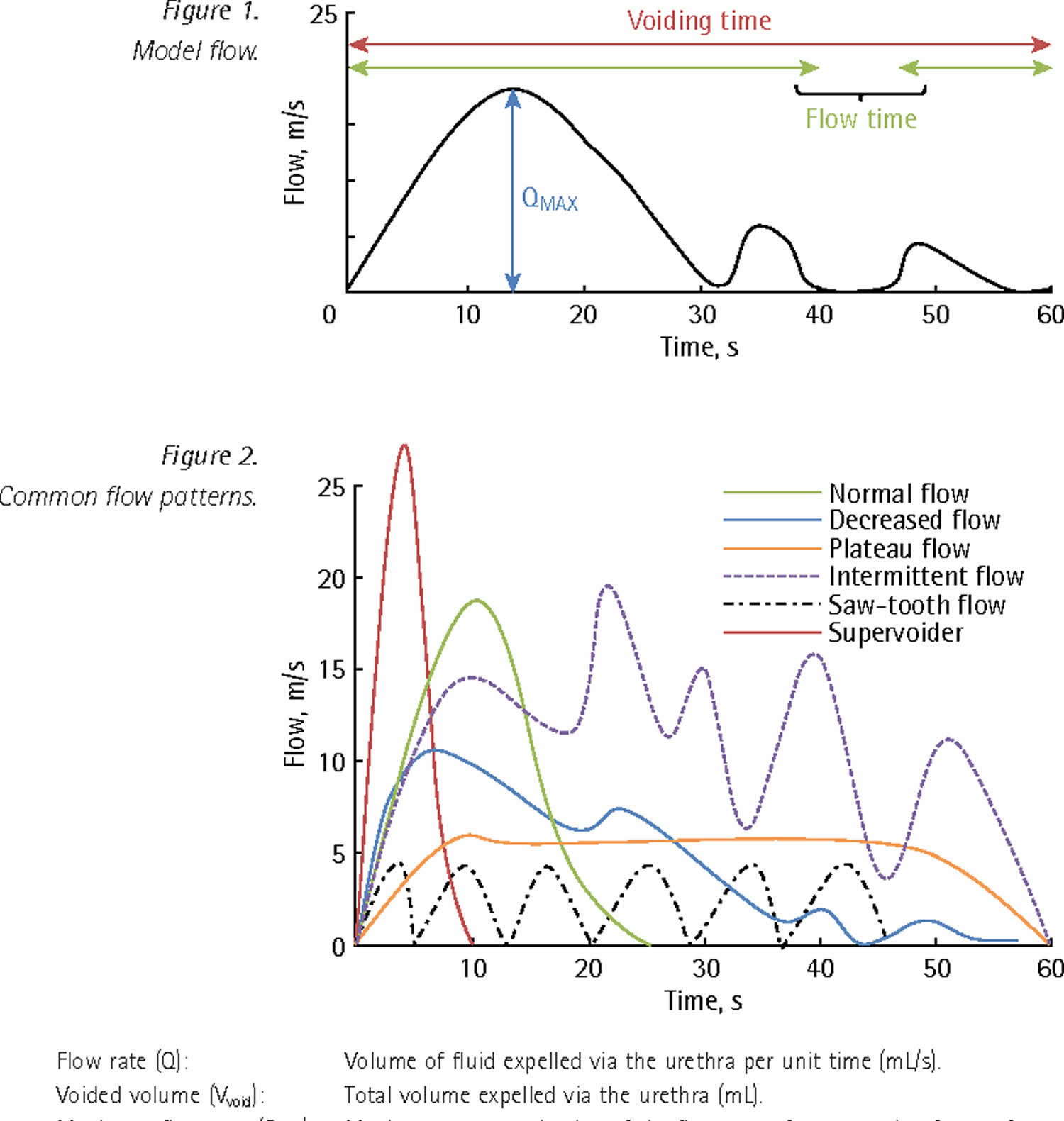
Common urine flow patterns:

  • Flow rate (Q): Volume of fluid expelled via the urethra per unit time (mL/s).
  • Voided volume (Vvoid): Total volume expelled via the urethra (mL).
  • Average flow rate (Qave): Voided volume divided by the flow time.
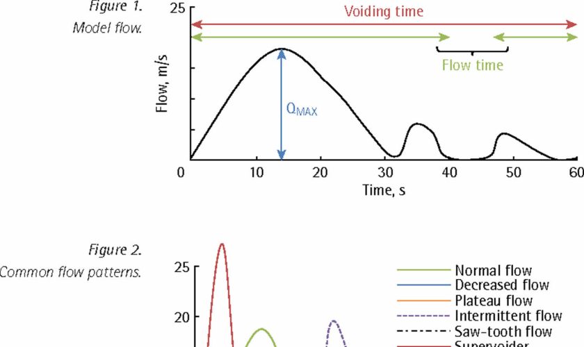
  • Maximum flow rate (Qmax): Maximum measured value of the flow rate after correction for artefacts.
  • Voiding time: Total duration of micturition (second).
  • Flow time: Time over which measurable flow actually occurs.
  • Time to maximum flow: Elapsed time from onset of flow to maximum flow.

The fastest flow rate, also known as maximum flow rate (Qmax), is used to understand if a block or obstruction is severe.

Your doctor will know your test results right away. Average results are based on your age and sex.

  • Typically, urine flow rate from 10 ml to 21 ml per second. Women range closer to 15 ml to 18 ml per second.
  • A slow or low flow rate may mean there is an obstruction at the bladder neck or in the urethra, an enlarged prostate, or a weak bladder.
  • A fast or high flow rate may mean there are weak muscles around the urethra, or urinary incontinence problems.

You may be asked to take other tests to fully learn what’s going on for treatment. Your urologist will create a treatment plan based on test results and your health history.

Facts about urine:

  • Adults pass about a quart and a half of urine each day, depending on the fluids and foods consumed.
  • The volume of urine formed at night is about half that formed in the daytime.
  • Normal urine is sterile. It contains fluids, salts, and waste products, but it is free of bacteria, viruses, and fungi.
  • The tissues of the bladder are isolated from urine and toxic substances by a coating that discourages bacteria from attaching and growing on the bladder wall.

Figure 1. Uroflowmetry

Uroflowmetry

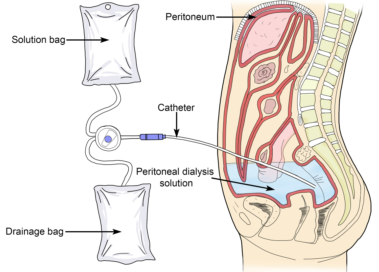
How does the urinary system work?

The body takes nutrients from food and converts them to energy. After the body has taken the food components that it needs, waste products are left behind in the bowel and in the blood.

The urinary system helps the body to eliminate liquid waste called urea and keeps the chemicals, such as potassium and sodium, and water in balance. Urea is produced when foods containing protein, such as meat, poultry, and certain vegetables, are broken down in the body. Urea is carried in the bloodstream to the kidneys, where it is removed along with water and other wastes in the form of urine.

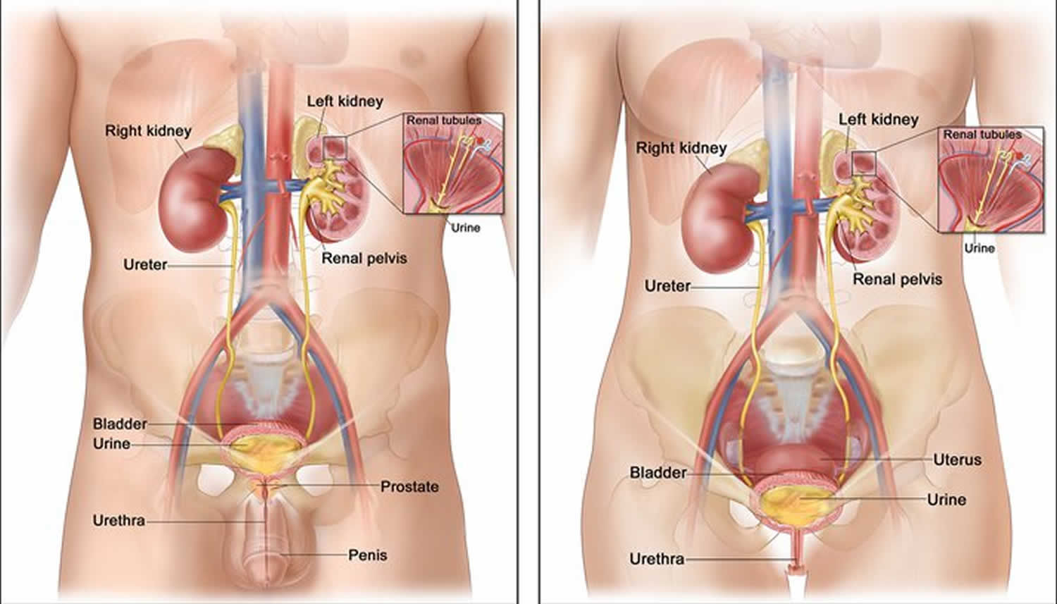
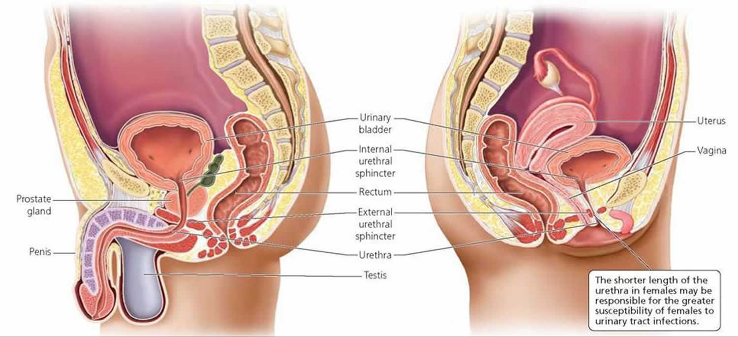
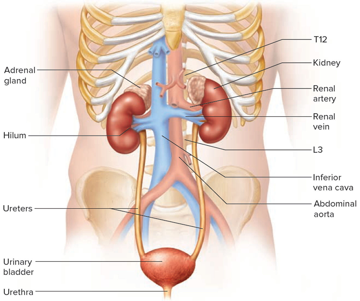
Urinary system parts and their functions:

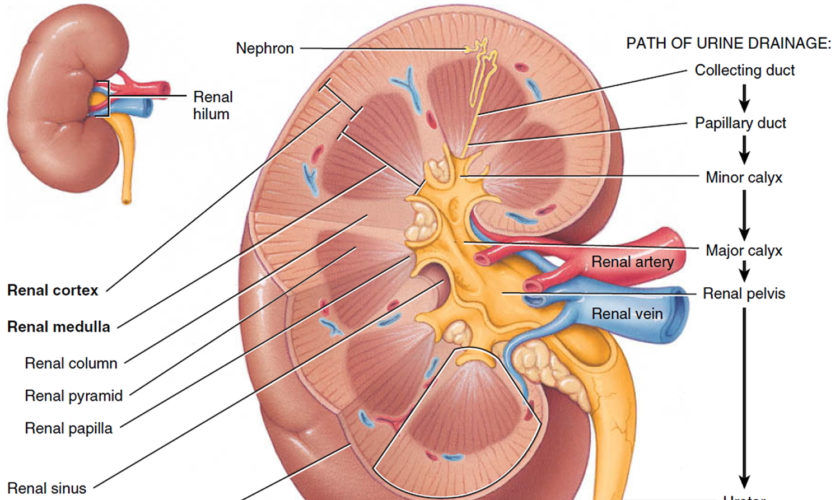
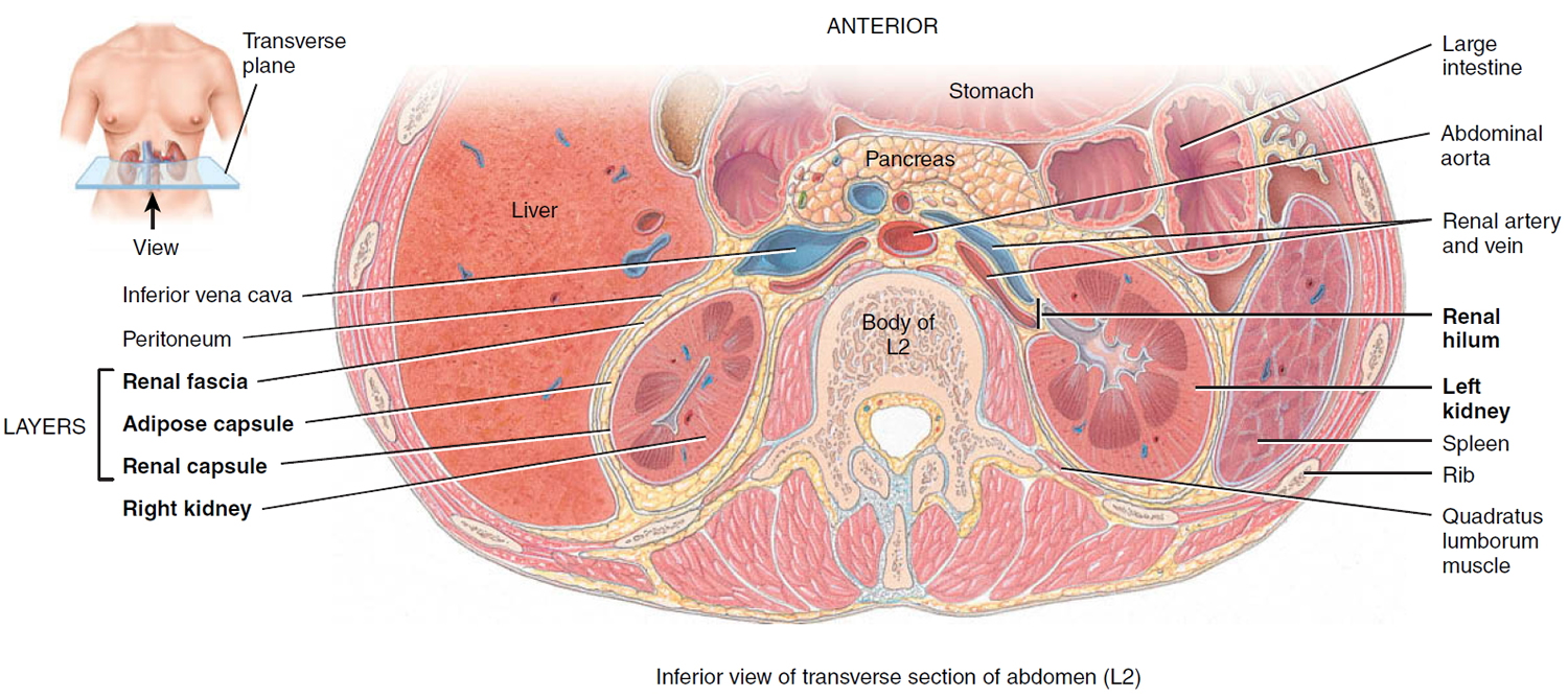
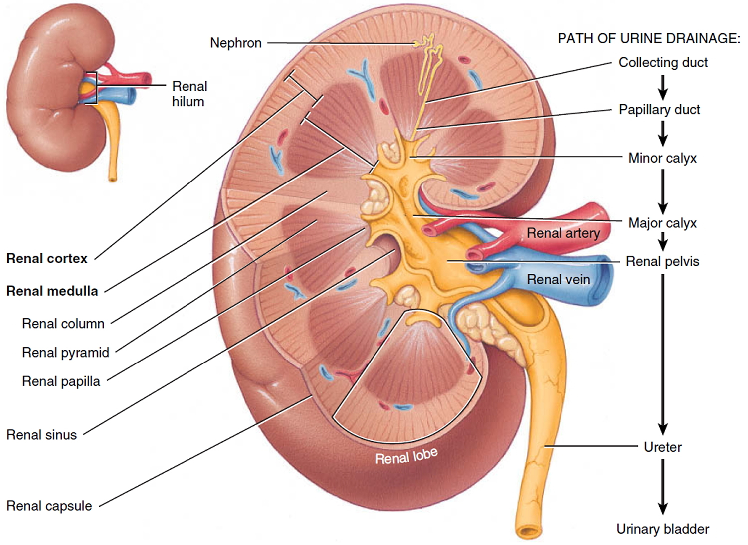
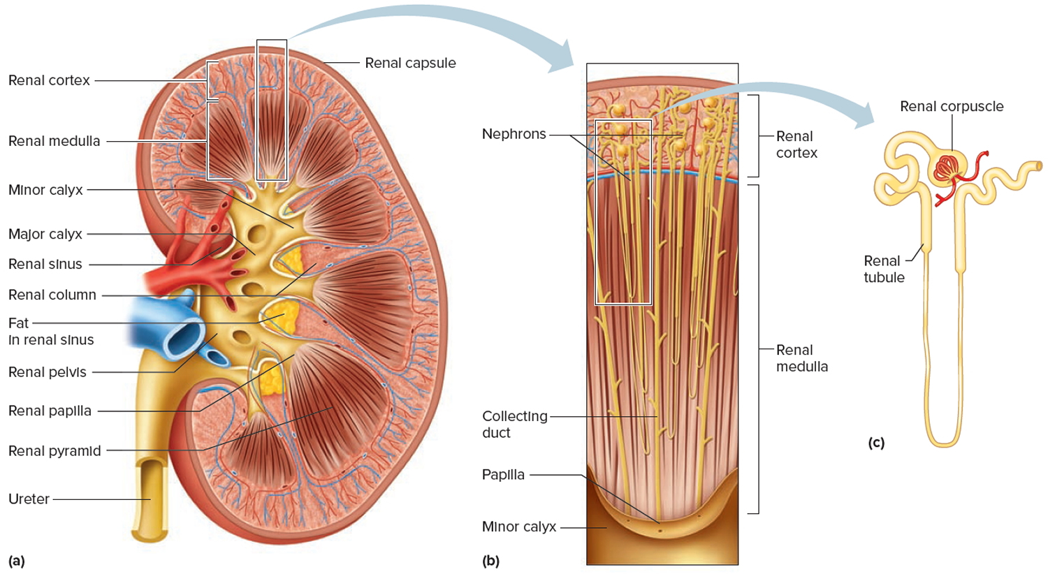
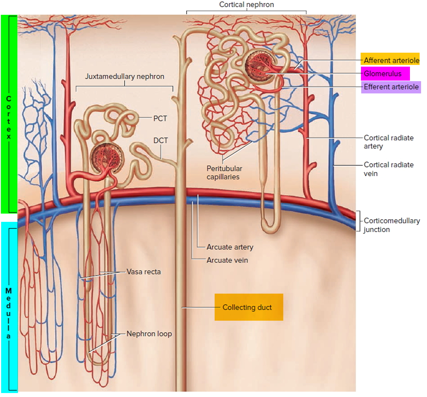
  • Two kidneys. This pair of purplish-brown organs is located below the ribs toward the middle of the back. Their function is to remove liquid waste from the blood in the form of urine, keep a stable balance of salts and other substances in the blood, and produce erythropoietin, a hormone that aids the formation of red blood cells. The kidneys also help to regulate blood pressure. The kidneys remove urea from the blood through tiny filtering units called nephrons. Each nephron consists of a ball formed of small blood capillaries, called a glomerulus, and a small tube called a renal tubule. Urea, together with water and other waste substances, forms the urine as it passes through the nephrons and down the renal tubules of the kidney.
  • Two ureters. These narrow tubes that carry urine from the kidneys to the bladder. Muscles in the ureter walls continually tighten and relax forcing urine downward, away from the kidneys. If urine backs up, or is allowed to stand still, a kidney infection can develop. About every 10 to 15 seconds, small amounts of urine are emptied into the bladder from the ureters.
  • Bladder. This triangle-shaped, hollow organ is located in the lower abdomen. It is held in place by ligaments that are attached to other organs and the pelvic bones. The bladder’s walls relax and expand to store urine, and contract and flatten to empty urine through the urethra. The typical healthy adult bladder can store up to two cups of urine for two to five hours.
  • Two sphincter muscles. These circular muscles help keep urine from leaking by closing tightly like a rubber band around the opening of the bladder
  • Nerves in the bladder. The nerves alert a person when it is time to urinate, or empty the bladder
  • Urethra. This tube allows urine to pass outside the body

Figure 2. Urinary system and anatomy

Urinary system anatomy

urinary tract system

Reasons for the uroflowmetry test

Uroflowmetry is a quick, simple diagnostic screening test that provides valuable feedback about the health of the lower urinary tract. It is commonly performed to determine if there is obstruction to normal urine outflow. Medical conditions that can alter the normal flow of urine include, but are not limited to, the following:

  • Benign prostatic hypertrophy. A benign enlargement of the prostate gland that usually occurs in men over age 50. Enlargement of the prostate interferes with normal passage of urine from the bladder. If left untreated, the enlarged prostate can obstruct the bladder completely.
  • Cancer of the prostate, or bladder tumor.
  • Urinary incontinence. Involuntary release of urine from the bladder.
  • Urinary blockage. Obstruction of the urinary tract can occur for many reasons along any part of the urinary tract from kidneys to urethra. Urinary obstruction can lead to a backflow of urine causing infection, scarring, or kidney failure if untreated.
  • Neurogenic bladder dysfunction. Improper function of the bladder due to an alteration in the nervous system, such as a spinal cord lesion or injury.
  • Frequent urinary tract infections.

Uroflowmetry may be performed in conjunction with other diagnostic procedures, such as cystometry, cystography, retrograde cystography, and cystoscopy.

There may be other reasons for your doctor to recommend uroflowmetry.

Urine flow rate test

Uroflowmetry is performed by having a person urinate into a special funnel that is connected to a measuring instrument. The measuring instrument calculates the amount of urine, rate of flow in seconds, and length of time until completion of the void. This information is converted into a graph and interpreted by a doctor. The information helps evaluate function of the lower urinary tract or help determine if there is an obstruction of normal urine outflow.

During normal urination, the initial urine stream starts slowly, but almost immediately speeds up until the bladder is nearly empty. The urine flow then slows again until the bladder is empty. In persons with a urinary tract obstruction, this pattern of flow is altered, and increases and decreases more gradually. The uroflowmeter graphs this information, taking into account the person’s gender and age. Depending on the results of the procedure, other tests may be recommended by your doctor.

Other related procedures that may be used to diagnose urinary outflow obstruction or lower urinary tract dysfunction include cystometry, cystography, retrograde cystography, and cystoscopy.

Before the urology flow rate test

  • Your doctor will explain the procedure to you and offer you the opportunity to ask any questions that you might have about the procedure.
  • Generally, no prior preparation, such as fasting or sedation, is required.
  • You may be instructed to drink about four glasses of water several hours before the test is performed to ensure that your bladder is full. In addition, you should not empty your bladder before arriving for the procedure.
  • If you are pregnant or suspect that you are pregnant, you should notify your doctor.
  • Notify your doctor of all medications (prescription and over-the-counter) and herbal supplements that you are taking.
  • Based on your medical condition, your doctor may request other specific preparation.

During the urology flow rate test

Uroflowmetry may be performed on an outpatient basis or as part of your stay in the hospital. Procedures may vary depending on your condition and your doctor’s practices.

Generally, uroflowmetry follows this process:

  1. You will be taken into the procedure area and instructed how to use the uroflowmetry device.
  2. When you are ready to urinate, you will press the flowmeter start button and count for five seconds before beginning urination.
  3. You will begin to urinate into the funnel device that is attached to the regular commode. The flowmeter will record information as you are urinating.
  4. You should not push or strain as you urinate. You should remain as still as possible.
  5. When you have finished urinating, you will count for five seconds and press the flowmeter button again.
  6. You should not put any toilet paper into the funnel device.
  7. The procedure will be concluded at this point. Depending on your specific medical condition, you may be asked to perform the test on several consecutive days.

After the urology flow rate test

Generally, there is no special type of care following uroflowmetry. However, your doctor may give you additional or alternate instructions after the procedure, depending on your particular situation.

Uroflowmetry normal flow

There is great variation in uroflowmetry parameters even in the non‐symptomatic population 1, although flow curves are generally repeatable for the same patient. In particular, there are no definitive ‘normal’ ranges for maximum flow rate (Qmax), although it decreases with age and voided volume (but not in a directly proportional manner). Males aged <40 years usually have a Qmax of >25 mL/s, and females usually have a Qmax of 5–10 mL/s more than males at a given bladder volume. Beware the ‘normal flow’ that in fact represents the effect of a compensatory increase in the voiding pressure generated by the detrusor in patients with bladder outlet obstruction 2.

Decreased urine flow

This is the most common abnormal flow trace seen in practice and is represented by a dampened curve with decreased Qmax and prolonged flow time. A significantly decreased Qmax (generally accepted as <15 mL/s) cannot be used to distinguish between BOO in men, outflow obstruction in women, and impaired detrusor contractility 6; in appropriate cases, formal multichannel urodynamic studies with concomitant measurements of flow and detrusor pressures are important to delineate between these conditions.

Despite the limitations, Qmax remains the single best non‐invasive urodynamic test to detect possible lower urinary tract obstruction. The test is also useful in some clinical situations to guide further evaluation to predict outcome after surgery and for preoperative counseling:

  • Males with a Qmax above the threshold value of 15 mL/s (or 12 mL/s) 3 may have a poorer outcome after prostate surgery for presumed bladder outlet obstruction 4 and these men should be considered for formal urodynamics to arrive at a definite diagnosis and decrease treatment failures.
  • Females undergoing mid‐urethral sling surgery with a Qmax of <15 mL/s at preoperative uroflowmetry are more likely to fail a trial of void after sling surgery 5.

Plateau urine flow

A long flow time, associated with a poor flow is typical of a stricture in the lower urinary tract. Another commonly encountered scenario is the patient with post‐radical prostatectomy incontinence. One should suspect an anastomotic stricture if this flow curve pattern is seen in the office during initial postoperative assessment. The patient should be considered for a cystoscopy with a view to treat the stricture as the next step in management, rather than referral for a formal urodynamic study as difficult catheterisation is commonly encountered.

Intermittent urine flow

This may be seen in patients who void with some abdominal straining due to bladder outlet obstruction or a poorly contractile detrusor, and is often superimposed on a decreased or plateauing curve pattern.

‘Saw‐tooth’ urine flow

Often pathogneumonic of detrusor‐sphincter‐dyssynergia, this curve should prompt urgent pressure‐flow studies to investigate high intravesical pressures that might damage the upper tracts.

‘Super‐voider’

This is seen after surgery for bladder outlet obstruction (e.g. TURP or urethroplasty), in patients with decreased urethral resistance (e.g. intrinsic urethral sphincter deficiency), or occasionally in those with detrusor overactivity. It may be considered ‘normal’ if there are no symptoms or signs to suggest underlying pathology, and is sometimes seen in young healthy female patients who may have a Qmax exceeding 40 mL/s.

‘Kicking the bucket’, and other artefacts

Urologists must be wary of artefacts and always compare the automated printout reading with the curve and clinical context. Smooth muscle physiology suggests that there should not be any abrupt spikes on a trace. A patient who accidentally kicks the flowmeter can appear to have a ‘normal’ Qmax. Other artefacts created by abdominal straining, squeezing the prepuce, or even variations in the direction of the urinary stream (within the funnel of the uroflowmeter) are common and urologists must recognise these.

Uroflowmetry procedure risks

Because uroflowmetry is a noninvasive procedure, it is safe for most persons. The test is usually done in privacy to ensure that the person voids in a natural setting.

There may be risks depending on your specific medical condition. Be sure to discuss any concerns with your doctor prior to the procedure.

Certain factors or conditions may interfere with the accuracy of uroflowmetry. These factors include, but are not limited to, the following:

  • Straining with urination
  • Body movement during urination
  • Certain medications that affect bladder and sphincter muscle tone
References
  1. Wyndaele JJ. Normality in urodynamics studied in healthy adults. J Urology 1999; 161: 899–902.
  2. Jarvis, T.R., Chan, L. and Tse, V. (2012), PRACTICAL UROFLOWMETRY. BJU Int, 110: 28-29. doi:10.1111/bju.11617
  3. McLoughlin J, Gill KP, Abel PD, Williams G. Symptoms versus flow rates versus urodynamics in the selection of patients for prostatectomy. Br J Urol 1990; 66: 303–305.
  4. Jensen KM, Jorgensen JB, Mogensen P. Urodynamics in prostatism. I. Prognostic value of uroflowmetry. Scand J Urol Nephrol 1988; 22: 109–117.
  5. Wheeler TL, Richter HE, Greer WJ, Bowling CB, Redden DT, Varner RE. Predictors of success with postoperative voiding trials after a mid‐urethral sling procedure. J Urol 2008; 179: 600–604.
read more
Conditions & DiseasesKidneysUrinary System

Kidney failure

kidney failure

What is kidney failure

Kidney failure means your kidneys no longer work well enough to do their job 1. Healthy kidneys clean your blood by removing excess fluid, minerals, and wastes. They also make hormones that keep your bones strong and your blood healthy.

Glomerular filtration rate (GFR) is the best measure of overall kidney function in health and disease 2. The normal level of glomerular filtration rate (GFR) varies according to age, sex, and body size. Normal glomerular filtration rate (GFR) in young adults is approximately 120 to 130 mL/min per 1.73 m² and declines with age 3. A glomerular filtration rate (GFR) level less than 60 mL/min per 1.73 m² represents loss of half or more of the adult level of normal kidney function. Below this level, the prevalence of complications of chronic kidney disease increases. Although the age-related decline in GFR has been considered part of normal aging, decreased GFR in the elderly is an independent predictor of adverse outcomes, such as death and cardiovascular disease 4. In addition, decreased GFR in the elderly requires adjustment in drug dosages, as in other patients with chronic kidney disease 5. Therefore, the definition of chronic kidney disease is the same, regardless of age. Because GFR declines with age, the prevalence of chronic kidney disease increases with age; approximately 17% of persons older than 60 years of age have an estimated GFR less than 60 mL/min per 1.73 m² 6.

The National Kidney Foundation Practice Guidelines define kidney failure as either:

  1. GFR less than 15 mL/min per 1.73 m², which is accompanied in most cases by signs and symptoms of uremia, or
  2. A need to start kidney replacement therapy (dialysis or transplantation).

Having kidney failure means that:

  • 85-90% of your kidney function is gone
  • your kidneys don’t work well enough to keep you alive

There is no cure for kidney failure, but with treatment it is possible to live a long life.

  • Approximately 98% of patients with kidney failure in the United States begin dialysis when their GFR is less than 15 mL/min per 1.73 m² 7. Kidney failure is not synonymous with end-stage renal disease (ESRD). End-stage renal disease is an administrative term in the United States. It indicates that a patient is treated with dialysis or transplantation, which is the condition for payment for health care by the Medicare end-stage renal disease Program. The classification of end-stage renal disease does not include patients with kidney failure who are not treated with dialysis and transplantation. Thus, although the term end-stage renal disease provides a simple operational classification of patients according to treatment, it does not precisely define a specific level of kidney function.
  • The level of kidney function, regardless of diagnosis, determines the stage of chronic kidney disease according to the Kidney Disease Outcomes Quality Initiative chronic kidney disease classification.

Every day, your kidneys filter about 120 to 150 quarts of blood to produce about 1 to 2 quarts of urine, composed of wastes and extra fluid. Kidney failure means your kidneys are not filtering as well as they should. When your kidneys fail, harmful wastes and extra salt and fluid buildup in your body. You then need treatment to replace the work your damaged kidneys have stopped doing.

Your kidneys filter wastes and extra fluid from your blood to keep you healthy. The wastes and extra fluid become urine that is stored in your bladder until you urinate. When your kidneys fail, dialysis can take over a small part of the work your damaged kidneys can no longer do.

The treatments for kidney failure are:

  • Hemodialysis
  • Peritoneal dialysis
  • Kidney transplant

These treatments can help you stay well.

Left untreated, kidney failure will lead to coma, seizures, and death.

You can make treatments work better by:

  • sticking to your treatment schedule
  • taking all medicines your doctor prescribes
  • following a special diet that keeps wastes from building up in your blood
  • being active most days of the week.

What causes kidney failure

Kidneys can become damaged from a physical injury or a disease like diabetes, high blood pressure, or other disorders.

Diabetes is the most common cause of kidney failure. High blood pressure is the second most common cause of kidney failure.

Other problems that can cause kidney failure include 8:

  • Autoimmune diseases, such as lupus and IgA nephropathy
  • Genetic diseases (diseases you are born with), such as polycystic kidney disease
  • Nephrotic syndrome
  • Urinary tract problems

Sometimes the kidneys can stop working very suddenly (within two days). This type of kidney failure is called acute kidney injury or acute renal failure. Common causes of acute renal failure include:

  • Heart attack
  • Illegal drug use and drug abuse
  • Not enough blood flowing to the kidneys
  • Urinary tract problems

This type of kidney failure is not always permanent. Your kidneys may go back to normal or almost normal with treatment and if you do not have other serious health problems.

Having one of the health problems that can lead to kidney failure does not mean that you will definitely have kidney failure.

Kidney failure does not happen overnight. It is the end result of a gradual loss of kidney function. In fact, some people do not even know they have kidney disease until their kidneys fail. Why not? Because people with early kidney disease may not have any symptoms. Symptoms usually show up late in the progression of the disease.

What are signs and symptoms of kidney failure

Healthy kidneys remove wastes and extra fluid from your blood. But when your kidneys fail, wastes and extra fluid can build up in your blood and make you feel sick. You may have some of the following symptoms:

  • nausea
  • little or no urination
  • headaches
  • drowsiness
  • trouble sleeping
  • loss of appetite
  • weight loss
  • weakness
  • fatigue, or feeling tired
  • generalized itching or numbness
  • weight loss
  • muscle cramps (especially in the legs)
  • high blood pressure
  • edema—swelling, usually in the legs, feet, or ankles and less often in the hands or face
  • anemia (a low blood count)
  • trouble sleeping
  • darkened skin
  • dry skin
  • trouble concentrating
  • vomiting

When these conditions occur, you need treatment to replace the work your damaged kidneys have stopped doing. Left untreated, kidney failure will lead to coma, seizures, and death.

Once you begin treatment for kidney failure, your symptoms will improve and you will begin to feel much better.

Stages of kidney failure

Chronic kidney disease progresses in stages.

The Glomerular filtration rate (GFR) provides an estimate of how much blood is filtered by the kidneys each minute. In normal kidneys GFR > 60 ml/minute. A GFR of less than 60 ml/minute/1.73m² may mean you have kidney disease

Table 1. Reference Table for Population Mean eGFR from NHANES III

Age (years)Mean eGFR (mL/min/1.73 m²)
20–29116
30–39107
40–4999
50–5993
60–6985
7075
[Source 9]

Each chronic kidney disease patient is classified into one of the following 5 stages of CKD based on their estimated glomerular filtration rate (eGFR) and the level of proteinuria, because management and prognosis varies according to the progression of damage.

  • CKD Stage 1: Kidney damage with normal or increased eGFR (above 90 mL/min/1.73 m²), but other tests have detected signs of kidney damage
  • CKD Stage 2: Mild reduction in eGFR (60-89 mL/min/1.73 m²), with other signs of kidney damage
  • CKD Stage 3: Moderate reduction in eGFR (30-59 mL/min/1.73 m²)
    • Stage 3a (G3a) – an eGFR of 45 to 59ml/min/1.73 m²
    • Stage 3b (G3b) – an eGFR of 30 to 44ml/min/1.73 m²
  • CKD Stage 4: Severe reduction in eGFR (15-29 mL/min/1.73 m²)
  • CKD Stage 5 (End Stage Kidney Disease or ESRD): Kidney failure (eGFR less than 15 mL/min/1.73 m²), meaning the kidneys have lost almost all of their function and require dialysis

Alongside your eGFR, your urine albumin:creatinine ratio (ACR) can help give a more accurate picture of how well your kidneys are working.

Your albumin:creatinine ratio (ACR) result is given as a stage from 1 to 3:

  • A1 – an ACR of less than 3mg/mmol
  • A2 – an ACR of 3 to 30mg/mmol
  • A3 – an ACR of more than 30mg/mmol

For both eGFR and ACR, a higher stage indicates more severe kidney disease.

Stages-of-Chronic-Kidney-Disease

How will kidney failure affect your life?

Kidney failure will affect your life in many ways. You may find you cannot do all the things you used to do at home or at work. You may have less energy and may feel depressed. Physical problems may include:

  • ankle or belly swelling
  • stomach sickness
  • throwing up
  • loss of appetite
  • feeling tired
  • weakness
  • confusion
  • headaches

Having kidney failure does not have to take over your life. Having kidney failure does not have to mean giving up hobbies, work, social activities, or time with family.

Can you continue to work with kidney failure?

Yes, many people with kidney failure continue to work. Your employer may give you lighter physical jobs or schedule your work hours around your hemodialysis sessions. If you are on peritoneal dialysis, you will need space and time to change the dialysis solution in the middle of the work day. Most employers are happy to make these changes.

As a result of the Americans with Disabilities Act, an employer cannot fire you because you are on dialysis or had a kidney transplant. The law requires an employer to make reasonable adjustments to the workplace for a person with a disability. If your employer is not willing to meet your needs, your dialysis clinic’s renal social worker may be able to help find a way to satisfy both you and your employer. As a last resort, you may need to file a complaint with the Equal Employment Opportunity Commission. Your renal social worker may be able to help you with this complaint, or you may need the help of a lawyer. Many times, just the mention of legal action is enough to cause an employer to make reasonable changes in the workplace.

Can you be active with kidney failure?

Yes. Physical activity is an important part of staying healthy when you have kidney failure. Being active makes your muscles, bones, and heart stronger. Physical activity also makes your blood circulate faster so your body gets more oxygen. Your body needs oxygen to use the energy from food. If you are on dialysis, physical activity can help more wastes move into your blood for dialysis to remove them.

You will find that physical activity can also improve your mood and give you a sense of well-being.

Talk with your doctor before you start an exercise routine. Start slow, with easier activities such as walking at a normal pace or gardening. Work up to harder activities such as walking briskly or swimming. Aim for at least 30 minutes of exercise most days of the week.

Kidney failure treatment

You have three treatment options to choose from to filter your blood. A fourth option offers care without replacing the work of the kidneys. None of these treatments helps the kidneys get better. However, they all can help you feel better.

  1. Hemodialysis uses a machine to move your blood through a filter outside your body, removing wastes.
  2. Peritoneal dialysis uses the lining of your belly to filter your blood inside your body, removing wastes.
  3. Kidney transplantation is surgery to place a healthy kidney from a person who has just died or a living person, usually a family member, into your body to take over the job of filtering your blood.
  4. Conservative management is the choice not to treat kidney failure with dialysis or a transplant. Instead, the focus is on using medicines to keep you comfortable, preserving kidney function through diet, and treating the problems of kidney failure, such as anemia—a shortage of red blood cells that can make you tired—and weak bones.

How does Hemodialysis work?

Purpose of Hemodialysis

The purpose of hemodialysis is to filter your blood. This type of dialysis uses a machine to remove harmful wastes and extra fluid, as your kidneys did when they were healthy. Hemodialysis helps control blood pressure and balance important minerals, such as potassium, sodium, calcium, and bicarbonate, in your blood. Hemodialysis is not a cure for kidney failure; however, it can help you feel better and live longer.

How Hemodialysis Works

Before you can begin dialysis, a surgeon will create a vascular access, usually in your arm. A vascular access lets high volumes of blood flow continuously during hemodialysis treatments to filter the largest possible amounts of blood per treatment.

Hemodialysis uses a machine to move your blood through a filter, called a dialyzer, outside your body. A pump on the hemodialysis machine draws your blood through a needle into a tube, a few ounces at a time. Your blood then travels through the tube, which takes it to the dialyzer. Inside the dialyzer, your blood flows through thin fibers that filter out wastes and extra fluid. After the dialyzer filters your blood, another tube carries your blood back to your body. You can do hemodialysis at a dialysis center or in your home.

Hemodialysis can replace part of your kidney function. You will also need dietary changes, medicines, and limits on water and other liquids you drink and get from food. Your dietary changes, the number of medicines you need, and limits on liquid will depend on where you receive your treatments—at a dialysis center or at home—and how often you receive treatments—three or more times a week.

Pros and Cons of Hemodialysis

The pros and cons of hemodialysis differ for each person. What may be bad for one person may be good for another. Following is a list of the general pros and cons of dialysis center and home hemodialysis.

Dialysis Center Hemodialysis

Pros

  • Dialysis centers are widely available.
  • Trained health care providers are with you at all times and help administer the treatment.
  • You can get to know other people with kidney failure who also need hemodialysis.
  • You don’t have to have a trained partner or keep equipment in your home.

Cons

  • The center arranges everyone’s treatments and allows few exceptions to the schedule.
  • You need to travel to the center for treatment.
  • This treatment has the strictest diet and limits on liquids because the longer time between treatments means wastes and extra fluid can build up in your body.
  • You may have more frequent ups and downs in how you feel from day to day because of the longer time between sessions.
  • Feeling better after a treatment may take a few hours.
Home Hemodialysis

Pros

  • You can do the treatment at the times you choose; however, you should follow your doctor’s orders about how many times a week you need treatment.
  • You don’t have to travel to a dialysis center.
  • You gain a sense of control over your treatment.
  • You will have fewer ups and downs in how you feel from day to day because of more frequent sessions.
  • You can do your treatments at times that will let you work outside the home.
  • You will have a more manageable diet and fewer limits on liquids because the shorter time between sessions prevents the buildup of wastes and extra fluid.
  • You can take along a hemodialysis machine when traveling.
  • You can spend more time with your loved ones because you don’t have to go to the dialysis center three times a week.

Cons

  • Not all dialysis centers offer home hemodialysis training and support.
  • You and a family member or friend will have to set aside a week or more at the beginning for training.
  • Helping with treatments may be stressful for your family or friend.
  • You need space for storing the hemodialysis machine and supplies at home.
  • You will need to learn to put dialysis needles into your vascular access.
  • Medicare and private insurance companies may limit the number of treatments they will pay for when you use home hemodialysis. Few people can afford the costs for additional treatments.

Figure 1. Hemodialysis

hemodialysis for kidney failure

hemodialysis unit for kidney failure

How does Peritoneal Dialysis work ?

Purpose of Peritoneal Dialysis

The purpose of peritoneal dialysis is to filter wastes and extra fluid from your body. This type of dialysis uses the lining of your belly—the space in your body that holds your stomach, bowels, and liver—to filter your blood. This lining, called the peritoneum, acts to do the work of your kidneys.

How Peritoneal Dialysis Works

A doctor will place a soft tube, called a catheter, in your belly a few weeks before you start treatment. The catheter stays in your belly permanently. When you start peritoneal dialysis, you will empty a kind of salty water, called dialysis solution, from a plastic bag through the catheter into your belly. When the bag is empty, you can disconnect your catheter from the bag so you can move around and do your normal activities. While the dialysis solution is inside your belly, it soaks up wastes and extra fluid from your body. After a few hours, you drain the used dialysis solution through another tube into a drain bag. You can throw away the used dialysis solution, now filled with wastes and extra fluid, in a toilet or tub. Then you start over with a fresh bag of dialysis solution. The process of emptying the used dialysis solution and refilling your belly with fresh solution is called an exchange. The process goes on continuously, so you always have dialysis solution in your belly soaking up wastes and extra fluid from your body.

Figure 2. Peritoneal dialysis

peritoneal dialysis for kidney failure

Types of Peritoneal Dialysis

Two types of peritoneal dialysis are available. After you have learned about the types of peritoneal dialysis, you can choose the type that best fits your life. If one schedule or type of peritoneal dialysis does not suit you, talk with your doctor about trying the other type.

Continuous ambulatory peritoneal dialysis does not require a machine and you can do it in any clean, well-lit place. The time period that the dialysis solution is in your belly is the dwell time. With continuous ambulatory peritoneal dialysis, the dialysis solution stays in your belly for a dwell time of 4 to 6 hours, or more. The process of draining the used dialysis solution and replacing it with fresh solution takes about 30 to 40 minutes. Most people change the dialysis solution at least four times a day and sleep with solution in their belly at night. With continuous ambulatory peritoneal dialysis, you do not have to wake up and perform dialysis tasks during the night.

Continuous cycler-assisted peritoneal dialysis uses a machine called a cycler to fill and empty your belly three to five times during the night while you sleep. In the morning, you begin one exchange with a dwell time that lasts the entire day. You may do an additional exchange in the middle of the afternoon without the cycler to increase the amount of waste removed and to reduce the amount of fluid left behind in your body.

You may need a combination of continuous ambulatory peritoneal dialysis and continuous cycler-assisted peritoneal dialysis if you weigh more than 175 pounds or if your peritoneum filters wastes slowly. For example, some people use a cycler at night and perform one exchange during the day. Others do four exchanges during the day and use a minicycler to perform one or more exchanges during the night. You’ll work with your health care team to find the best schedule for you.

Pros and Cons of Peritoneal Dialysis

Each type of peritoneal dialysis has pros and cons.

Continuous Ambulatory Peritoneal Dialysis

Pros

  • You can do continuous ambulatory peritoneal dialysis alone.
  • You can do continuous ambulatory peritoneal dialysis at the times you choose, as long as you perform the required number of exchanges each day.
  • You can do continuous ambulatory peritoneal dialysis in many locations.
  • You can travel as long as you bring dialysis bags with you or have them delivered to your destination.
  • You don’t need a machine for continuous ambulatory peritoneal dialysis.
  • You gain a sense of control over your treatment.

Cons

  • Continuous ambulatory peritoneal dialysis can disrupt your daily schedule.
  • Continuous ambulatory peritoneal dialysis is a continuous treatment, and you should do all exchanges 7 days a week.
  • Boxes of dialysis solution will take up space in your home.

Continuous Cycler-assisted Peritoneal Dialysis

Pros

  • You can do exchanges at night, while you sleep.
  • You may not have to perform exchanges during the day.

Cons

  • You need a machine.
  • Your connection to the cycler limits your movement at night.

Is dialysis a cure for kidney failure ?

No. Hemodialysis and peritoneal dialysis help you feel better and live longer; however, they do not cure kidney failure. Although people with kidney failure are now living longer than ever, over the years kidney disease can cause other problems, such as heart disease, bone disease, arthritis, nerve damage, infertility, and malnutrition. These problems won’t go away with dialysis; however, doctors now have new and better ways to prevent or treat them. You should discuss these problems and their treatments with your doctor.

How Kidney Transplantation Works

The transplant process has many steps.

The first step is to talk with your health care provider about whether you are a candidate for a transplant. Transplantation is not for everyone. Your health care provider may tell you that you are not healthy enough for surgery or that you have a condition that would make transplantation unlikely to succeed. If you are a good candidate for a transplant, your health care provider will refer you to a transplant center.

Medical, Psychological, and Social Evaluation at a Transplant Center

The next step is a thorough physical, psychological, and social evaluation at the transplant center, where you will meet members of your transplant team. Your pretransplant evaluation may require several visits to the transplant center over the course of weeks or even months.

You will need to have blood tests as well as other tests to check your heart and other organs. Your blood type and other matching factors help determine whether your body will accept an available donor kidney.

Your transplant team will make sure you are healthy enough for surgery. Some medical conditions or illnesses could make transplantation less likely to succeed.

In addition, your team will make sure you can understand and follow the schedule for taking the medicines needed after surgery. Team members need to be sure that you are mentally prepared for the responsibilities of caring for a transplanted kidney.

If a family member or friend wants to donate a kidney, that person will need a health exam to test whether the kidney is a good match.

Who is on your transplant team ?

Your transplant team has many members, including your

  • surgeon—the doctor who places the kidney in your body.
  • nephrologist—a doctor who specializes in kidney health. The nephrologist may work in partnership with a nurse practitioner or a physician’s assistant.
  • transplant coordinator—a specially trained nurse who will be your point of contact, arrange your appointments, and educate you before and after the transplant.
  • social worker—a person who is trained to help people solve problems in their daily lives, such as finding employment, affordable housing, or daycare.
  • dietitian—a person who is an expert in food and nutrition. Dietitians teach people about the foods they should eat and how to plan healthy meals.

Placement on the Waiting List

If your medical evaluation shows you are a good candidate for a transplant, your transplant center will submit your name to be placed on the national waiting list for a kidney from a deceased donor. The Organ Procurement and Transplantation Network has a computer network that links all regional organ-gathering organizations—known as organ procurement organizations—and transplant centers. The United Network for Organ Sharing (UNOS), a private, nonprofit organization, runs the Organ Procurement and Transplantation Network under a contract with the Federal Government. When UNOS officially adds you to the waiting list, UNOS will notify you and your transplant team.

UNOS allows you to register with multiple transplant centers to increase your chances of receiving a kidney. Each transplant center usually requires a separate medical evaluation.

Waiting Period

UNOS gives preference to people who have been on the waiting list the longest. However, other factors—such as your age, where you live, and your blood type—may make your wait longer or shorter. Wait times can range from a few months to several years.

If you have a living donor, you do not need to be placed on the waiting list and can schedule the surgery when it is convenient for you and your donor.

While you are on the waiting list, notify the transplant center of changes in your health. Also, let the transplant center know if you move or change phone numbers. The center will need to find you immediately when a kidney becomes available.

While you wait for a kidney, you will have blood drawn once a month. The sample will be sent to the transplant center. The center must have a recent sample of your blood for comparison with any kidney that becomes available.

Organ procurement organizations identify potential organs for transplant and coordinate with the national network. When a deceased donor kidney becomes available, the organ procurement organization notifies UNOS and creates a computer-generated list of suitable recipients.

Whether you are receiving your kidney from a deceased donor or a living donor, the transplant team considers three factors in matching kidneys with potential recipients. These matching factors help predict whether your body’s immune system—which protects your body from infection by identifying and destroying bacteria, viruses, and other potentially harmful foreign substances—will accept or reject the new kidney.

  • Blood type. Your blood type—A, B, AB, or O—must be compatible with the donor’s. Blood type is the most important matching factor. Some transplant centers have developed techniques for transplanting kidneys that are not matched by blood type.
  • Human leukocyte antigens. These six antigens are proteins that help your immune system tell the difference between your own body’s tissues and foreign substances. You may still receive a kidney if the antigens do not completely match, as long as your blood type is compatible with the organ donor’s blood type and other tests show no problems with matching.
  • Cross-matching antigens. The cross-match is the last test performed before a kidney transplant can take place. A lab technician mixes a small sample of your blood with a sample of the organ donor’s blood in a tube to see if the mixture causes a reaction. If no reaction occurs—called a negative cross-match—the transplant can proceed.

Kidney Transplant Surgery

If you are on a waiting list for a deceased donor kidney, you must go to the hospital as soon as you receive notification that a kidney is available. If a family member or friend is donating the kidney you will receive, you will schedule the operation in advance. Your transplant team will operate on you and your donor at the same time, usually in side-by-side rooms. One surgeon will perform the nephrectomy—the removal of the kidney from the donor—while another prepares you for placement of the donated kidney. In some centers, the same surgeon performs both operations. You will receive general anesthesia to make you sleep during the operation. The surgery usually takes 3 or 4 hours.

Surgeons—place most transplanted kidneys in the lower front part of your abdomen. The kidney is connected to an artery, which brings unfiltered blood into the kidney, and a vein, which takes filtered blood out of the kidney. The surgeon also transplants the ureter from the donor to let urine from the new kidney flow to your bladder. Unless your damaged kidneys cause problems such as infection, they can remain in their normal position. The transplanted kidney takes over the job of filtering your blood. Your body normally attacks anything it sees as foreign, so to keep your body from attacking the kidney you need to take medicines called immunosuppressants for as long as the transplanted kidney functions.

Recovery from Kidney Transplant Surgery

After surgery, you will probably feel sore and groggy when you wake up. However, many people who have a transplant report feeling much better immediately after surgery. Even if you wake up feeling great, you will typically need to stay in the hospital for several days to recover from surgery, and longer if you have any complications. You will have regular follow-up visits after leaving the hospital.

If you have a living donor, the donor will probably also stay in the hospital for several days. However, a new technique for removing a kidney for donation uses a smaller incision and may make it possible for the donor to leave the hospital in 2 to 3 days.

Pros and Cons of Kidney Transplantation

Following is a list of the pros and cons of kidney transplantation.

Kidney Transplantation

Pros

  • A transplanted kidney works like a healthy kidney.
  • If you have a living donor, you can choose the time of your operation.
  • You may feel healthier and have an improved quality of life.
  • You have fewer dietary restrictions.
  • You won’t need dialysis.
  • People who receive a donated kidney have a greater chance of living a longer life than those who stay on dialysis.

Cons

  • Transplantation requires surgery.
  • You will go through extensive medical testing at the transplant clinic.
  • You may need to wait years for a deceased donor kidney.
  • Your body may reject the new kidney, so one transplant may not last a lifetime.
  • You’ll need to take immunosuppressants, which may cause other health problems, for as long as the transplanted kidney functions.

What do you need to know about care after your kidney transplant ?

You need to know how to keep your body from rejecting your new kidney. Your immune system will sense that your new kidney is foreign. To keep your body from rejecting it, you will have to take medicines, called immunosuppressants, that turn off, or suppress, your immune system response. You may have to take two or more immunosuppressants, as well as medicines such as antibiotics to protect against infections. Your transplant team will teach you what each medicine is for and when to take each one. Be sure you understand the instructions for taking your medicines before you leave the hospital.

What are signs that your body is rejecting your new kidney ?

Often, rejection begins before any signs appear. The signs of rejection include indications that your kidney is not working as well as it should—for example, high blood pressure or swelling because your kidney is not getting rid of extra salt and fluid in your body. Advances in immunosuppressants have made other signs of rejection—such as fever, soreness in the lower abdomen where the new kidney is, and a decrease in the amount of urine you make—rare. If you have any of these symptoms, tell your transplant team. You will receive stronger doses of your immunosuppressants and additional medicines to help keep your body from rejecting your new kidney.

Even if you do everything you should, your body may still reject the new kidney, and you may need to go on dialysis. Unless your transplant team determines that you are no longer a good candidate for transplantation, you can go back on the waiting list for another kidney.

How do you know your new kidney is working properly ?

Blood tests help you know your new kidney is working. Before you leave the hospital, you will schedule an appointment with your transplant team at the transplant center. At that appointment, a health care provider will draw blood to be tested. The tests show how well your kidneys are removing wastes from your blood. At first, you may return to the transplant center every 2 weeks, then every month. Eventually, you will need to return to the transplant center only once every 6 months or once every year, after your transplant team has determined that your kidney is doing its job.

Your blood tests may show that your kidney is not removing wastes from your blood as well as it should. You may have other signs that your body is rejecting your new kidney. If these problems occur, your transplant surgeon or nephrologist may order a kidney biopsy. Biopsy is a procedure that involves taking a small piece of tissue for examination under a microscope. Your transplant surgeon or nephrologist performs the biopsy in the transplant center or a hospital. The health care provider will give you light sedation and local anesthetic; however, in some cases, a patient may require general anesthesia. A pathologist—a doctor who specializes in diagnosing diseases—examines the tissue in a lab. The test can show whether your body is rejecting your new kidney.

What are the side effects of immunosuppressants ?

Some immunosuppressants may change your appearance. Your face may get fuller; you may gain weight or develop acne or facial hair. Not all people have these problems, and those who do can use diet, makeup, and hair removal to minimize changes in appearance.

Immunosuppressants weaken your immune system, which can lead to infections. In some people over long periods of time, a weakened immune system can increase their risk of developing cancer. Some immunosuppressants cause cataracts, diabetes, extra stomach acid, high blood pressure, and bone disease. When used over time, these medicines may also cause liver or kidney damage in some people. Your transplant team will order regular tests to monitor the levels of immunosuppressants in your blood and to measure your liver and kidney function.

What financial help is available to pay for a kidney transplant ?

United States citizens who have kidney failure are eligible to receive Medicare, the Federal Government insurance program. Treatment for kidney failure costs a lot; however, Medicare pays much of the cost, usually up to 80 percent. Often, private insurance pays the rest. For people who are not eligible for Medicare or who still need help with the portion Medicare does not cover, states have Medicaid programs that provide funds for health care based on financial need. Your social worker can help you locate resources for financial help.

What help is available to pay for kidney transplant medicines ?

Through patient-assistance programs, prescription drug companies give discounts to people who can show they cannot afford the cost of their prescribed medicines. Social workers can help patients complete applications to these programs.

The Partnership for Prescription Assistance has a website that directs patients, caregivers, and doctors to more than 275 public and private patient-assistance programs, including more than 150 programs offered by pharmaceutical companies. The website www.pparx.org features tools to help a person determine which programs might be available.

NeedyMeds is a nonprofit organization that helps people find appropriate patient-assistance programs. The NeedyMeds website—www.needymeds.org —provides a directory of patient-assistance programs that can be searched by a medicine’s brand or generic name or by a program or company name. Applications for these programs are usually available online.

Eating, Diet, and Nutrition for Kidney Transplant Patients

The diet for transplant patients has more choices than the diet for dialysis patients, although you may still have to cut back on some foods. Your diet will probably change as your medicines, test results, weight, and blood pressure change.

  • You may need to count calories. Your medicines may give you a bigger appetite and cause you to gain weight.
  • You may have to eat less sodium. Your medicines may cause your body to retain sodium, leading to high blood pressure.

Your transplant center’s dietitian can help you understand the reasons for dietary limits, recognize foods you should avoid, and plan healthy and tasty meals.

What is conservative management for kidney failure ?

Conservative management for kidney failure is the choice to say no to or stop dialysis treatments. For many people, dialysis not only extends life, it also improves the quality of life. For others who have serious conditions in addition to kidney failure, dialysis may seem like a burden that only prolongs suffering. If you have serious conditions in addition to kidney failure, dialysis may not prolong your life or improve the quality of your life.

You have the right to say no to or stop dialysis. You may want to speak with your doctor, spouse, family, counselor, or renal social worker, who helps people with kidney disease, to help you make this decision.

If you stop dialysis treatments or say you do not want to begin them, you may live for a few weeks or for several months, depending on your health and your remaining kidney function. You may choose to receive care from a hospice—a facility or home program designed to meet the physical and emotional needs of the terminally ill—during this time. Hospice care focuses on relief of pain and other symptoms. Whether or not you choose to use a hospice, your doctor can give you medicines to make you more comfortable. Your doctor can also give you medicines to treat the problems of kidney failure, such as anemia or weak bones. You may restart dialysis treatment if you change your mind.

Advance Directives

An advance directive is a statement or document in which you give instructions either to withhold certain treatments, such as dialysis, or to provide them, depending on your wishes and the specific circumstances. Even if you are happy with your quality of life on dialysis, you should think about circumstances that might make you want to stop dialysis treatments. At some point in a medical crisis, you might lose the ability to tell your health care team and loved ones what you want. Advance directives may include

  • a living will
  • a durable power of attorney for health care decisions
  • a do not resuscitate (DNR) order—a legal form that tells your health care team you do not want cardiopulmonary resuscitation (CPR) or other life-sustaining treatment if your heart were to stop or if you were to stop breathing.

A living will is a document that details the conditions under which you would want to refuse treatment. You may state that you want your health care team to use all available means to sustain your life, or you may direct that you be withdrawn from dialysis if you fall into a coma from which you most likely won’t wake up. In addition to dialysis, you may choose or refuse the following life-sustaining treatments:

  • CPR
  • feedings through a tube in your stomach
  • mechanical or artificial means to help you breathe
  • medicines to treat infections
  • surgery
  • receiving blood

Refusing to have CPR is the same as a do not resuscitate (DNR) order. If you choose to have a do not resuscitate (DNR) order, your doctor will place the order in your medical chart.

A durable power of attorney for health care decisions or a health care proxy is a document you use to assign a person to make health care decisions for you in the event you cannot make them for yourself. Make sure the person you name understands your values and will follow your instructions.

Each state has its own laws on advance directives. You can obtain a form for an advance medical directive that’s valid in your state from the National Hospice and Palliative Care Organization.

Kidney failure diet

Your dialysis center has a renal dietitian to help you plan your meals. A renal dietitian has special training in caring for the food and nutrition needs of people with kidney disease. Work with a registered dietitian to develop a meal plan that includes foods that you enjoy eating while maintaining your kidney health.

Use this information to help you learn how to eat right to feel right on hemodialysis. Read one section at a time. Then, review with your renal dietitian the sections marked “Talk with Your Renal Dietitian.”

Keep a copy of this information handy to remind yourself of foods you can eat and foods to avoid.

You will need to carefully plan your meals and keep track of the amount of liquids you eat and drink. It helps to limit or avoid foods and beverages that have lots of

  • potassium
  • phosphorus
  • sodium—for example, vegetable juice and sports drinks

The first steps to eating right 10.

Step 1: Choose and prepare foods with less salt and sodium

Why? To help control your blood pressure. Your diet should contain less than 2,300 milligrams of sodium each day 10.

  • Buy fresh food often. Sodium (a part of salt) is added to many prepared or packaged foods you buy at the supermarket or at restaurants.
  • Cook foods from scratch instead of eating prepared foods, “fast” foods, frozen dinners, and canned foods that are higher in sodium. When you prepare your own food, you control what goes into it.
  • Use spices, herbs, and sodium-free seasonings in place of salt.
  • Check for sodium on the Nutrition Facts label of food packages. A Daily Value of 20 percent or more means the food is high in sodium.
  • Try lower-sodium versions of frozen dinners and other convenience foods.
  • Rinse canned vegetables, beans, meats, and fish with water before eating.

Look for food labels with words like sodium free or salt free; or low, reduced, or no salt or sodium; or unsalted or lightly salted.

Why is knowing about sodium important for someone with advanced chronic kidney disease ?

Too much sodium in a person’s diet can be harmful because it causes blood to hold fluid. People with chronic kidney disease need to be careful not to let too much fluid build up in their bodies. The extra fluid raises blood pressure and puts a strain on the heart and kidneys. A dietitian can help people find ways to reduce the amount of sodium in their diet. Nutrition labels provide information about the sodium content in food. The U.S. Food and Drug Administration advises that healthy people should limit their daily sodium intake to no more than 2,300 milligrams (mg), the amount found in 1 teaspoon of table salt. People who are at risk for a heart attack or stroke because of a condition such as high blood pressure or kidney disease should limit their daily sodium intake to no more than 1,500 mg. Choosing sodium-free or low-sodium food products will help them reach that goal.

Sodium is found in ordinary table salt and many salty seasonings such as soy sauce and teriyaki sauce. Canned foods, some frozen foods, and most processed meats have large amounts of salt. Snack foods such as chips and crackers are also high in salt.

Alternative seasonings such as lemon juice, salt-free seasoning mixes, and hot pepper sauce can help people reduce their salt intake. People with advanced chronic kidney disease should avoid salt substitutes that use potassium, such as AlsoSalt or Nu-Salt, because chronic kidney disease limits the body’s ability to eliminate potassium from the blood. The table below provides some high-sodium foods and suggestions for low-sodium alternatives that are healthier for people with any level of chronic kidney disease who have high blood pressure.

High-sodium FoodsLow-sodium Alternatives
  • Salt
  • Regular canned vegetables
  • Hot dogs and canned meat
  • Packaged rice with sauce
  • Packaged noodles with sauce
  • Frozen vegetables with sauce
  • Frozen prepared meals
  • Canned soup
  • Regular tomato sauce
  • Snack foods
  • Salt-free herb seasonings
  • Low-sodium canned foods
  • Frozen vegetables without sauce
  • Fresh, cooked meat
  • Plain rice without sauce
  • Plain noodles without sauce
  • Fresh vegetables without sauce
  • Homemade soup with fresh ingredients
  • Reduced-sodium tomato sauce
  • Unsalted pretzels
  • Unsalted popcorn

Why is knowing about potassium important for someone with advanced chronic kidney disease ?

Keeping the proper level of potassium in the blood is essential. Potassium keeps the heart beating regularly and muscles working right. Problems can occur when blood potassium levels are either too low or too high. Damaged kidneys allow potassium to build up in the blood, causing serious heart problems. Potassium is found in many fruits and vegetables, such as bananas, potatoes, avocados, and melons. People with advanced chronic kidney disease may need to avoid some fruits and vegetables. Blood tests can indicate when potassium levels have climbed above normal range. A renal dietitian can help people with advanced chronic kidney disease find ways to limit the amount of potassium they eat. The potassium content of potatoes and other vegetables can be reduced by boiling them in water. The following table gives examples of some high-potassium foods and suggestions for low-potassium alternatives for people with advanced chronic kidney disease.

High-potassium FoodsLow-potassium Alternatives
  • Oranges and orange juice
  • Melons
  • Apricots
  • Bananas
  • Potatoes
  • Tomatoes
  • Sweet potatoes
  • Cooked spinach
  • Cooked broccoli
  • Beans (baked, kidney, lima, pinto)
  • Apples and apple juice
  • Cranberries and cranberry juice
  • Canned pears
  • Strawberries, blueberries, raspberries
  • Plums
  • Pineapple
  • Cabbage
  • Boiled Cauliflower

Why is knowing about phosphorus important for someone with advanced chronic kidney disease ?

Damaged kidneys allow phosphorus, a mineral found in many foods, to build up in the blood. Too much phosphorus in the blood pulls calcium from the bones, making the bones weak and likely to break. Too much phosphorus may also make skin itch. Foods such as milk and cheese, dried beans, peas, colas, canned iced teas and lemonade, nuts, and peanut butter are high in phosphorus. A renal dietitian can help people with advanced chronic kidney disease learn how to limit phosphorus in their diet.

As chronic kidney disease progresses, a person may need to take a phosphate binder such as sevelamer hydrochloride (Renagel), lanthanum carbonate (Fosrenol), calcium acetate (PhosLo), or calcium carbonate (Tums) to control the phosphorus in the blood. These medications act like sponges to soak up, or bind, phosphorus while it is in the stomach. Because it is bound, the phosphorus does not get into the blood. Instead, it is removed from the body in the stool.

The table below lists some high-phosphorus foods and suggestions for low-phosphorus alternatives that are healthier for people with advanced chronic kidney disease.

High-phosphorus FoodsLow-phosphorus Alternatives
  • Dairy foods (milk, cheese, yogurt)
  • Beans (baked, kidney, lima, pinto)
  • Nuts and peanut butter
  • Processed meats (hot dogs, canned meat)
  • Cola
  • Canned iced teas and lemonade
  • Bran cereals
  • Egg yolks
  • Liquid non-dairy creamer
  • Sherbet
  • Cooked rice
  • Rice, wheat, and corn cereals
  • Popcorn
  • Peas
  • Lemon-lime soda
  • Root beer
  • Powdered iced tea and lemonade mixes

Step 2: Eat the right amount and the right types of protein

Why? To help protect your kidneys. When your body uses protein, it produces waste. Your kidneys remove this waste. Eating more protein than you need may make your kidneys work harder.

  • Eat small portions of protein foods.
  • Protein is found in foods from plants and animals. Most people eat both types of protein. Talk to your dietitian about how to choose the right combination of protein foods for you.

When kidney function declines to the point where dialysis becomes necessary, patients should include more protein in their diet because dialysis removes large amounts of protein from the blood.

Animal-protein foods:

  • Chicken
  • Fish
  • Meat
  • Eggs
  • Dairy

A cooked portion of chicken, fish, or meat is about 2 to 3 ounces or about the size of a deck of cards. A portion of dairy foods is ½ cup of milk or yogurt, or one slice of cheese.

Plant-protein foods:

  • Beans
  • Nuts
  • Grains

A portion of cooked beans is about ½ cup, and a portion of nuts is ¼ cup. A portion of bread is a single slice, and a portion of cooked rice or cooked noodles is ½ cup.

Step 3: Choose foods that are healthy for your heart

Why? To help keep fat from building up in your blood vessels, heart, and kidneys. To help keep fat from building up in your blood vessels, heart, and kidneys.

  • Grill, broil, bake, roast, or stir-fry foods, instead of deep frying.
  • Cook with nonstick cooking spray or a small amount of olive oil instead of butter.
  • Trim fat from meat and remove skin from poultry before eating.
  • Try to limit saturated and trans fats. Read the food label.

Heart-healthy foods:

  • Lean cuts of meat, such as loin or round
  • Poultry without the skin
  • Fish
  • Beans
  • Vegetables
  • Fruits
  • Low-fat or fat-free milk, yogurt, and cheese

Limit alcohol

Drink alcohol only in moderation: no more than one drink per day if you are a woman, and no more than two if you are a man. Drinking too much alcohol can damage the liver, heart, and brain and cause serious health problems. Ask your health care provider how much alcohol you can drink safely.

A renal dietitian can help people learn about the amount and sources of protein in their diet. Animal protein in egg whites, cheese, chicken, fish, and red meats contain more of the essential nutrients a body needs. With careful meal planning, a well-balanced vegetarian diet can also provide these nutrients. A renal dietitian can help people with advanced chronic kidney disease make small adjustments in their eating habits that can result in significant protein reduction. For example, people can lower their protein intake by making sandwiches using thinner slices of meat and adding lettuce, cucumber slices, apple slices, and other garnishes. The following table lists some higher-protein foods and suggestions for lower-protein alternatives that are better choices for people with chronic kidney disease trying to limit their protein intake.

Why is it important to keep track of how much liquid you eat or drink ?

You may feel better if you keep track of and limit how much liquid you eat and drink. Excess fluid can build up in your body and may cause

  • swelling and weight gain between dialysis sessions
  • changes in your blood pressure
  • your heart to work harder, which can lead to serious heart trouble
  • a buildup of fluid in your lungs, making it hard for you to breathe

Hemodialysis removes extra fluid from your body. However, hemodialysis can remove only so much fluid at a time safely. If you come to your hemodialysis with too much fluid in your body, your treatment may make you feel ill. You may get muscle cramps or have a sudden drop in blood pressure that causes you to feel dizzy or sick to your stomach.

Your health care provider can help you figure out how much liquid is right for you.

One way to limit how much liquid you have is to limit the salt in the foods you eat. Salt makes you thirsty, so you drink more. Avoid salty foods such as chips and pretzels.

Your renal dietitian will give you other tips to help you limit how much liquid you consume while making sure you don’t feel too thirsty.

What foods count as liquid and why ?

Foods that are liquid at room temperature, such as soup, contain water. Gelatin, pudding, ice cream, and other foods that include a lot of liquid in the recipe also count. Most fruits and vegetables contain water, such as melons, grapes, apples, oranges, tomatoes, lettuce, and celery. When you count up how much liquid you have in a day, be sure to count these foods.

What is your dry weight ?

Your dry weight is your weight after a hemodialysis session has removed all extra fluid from your body. Controlling your liquid intake helps you stay at your proper dry weight. If you let too much fluid build up between sessions, it is harder to achieve your dry weight. Your health care provider can help you figure out what dry weight is right for you.

My dry weight goal: _____________.

Should you take vitamin and mineral supplements ?

You may not get enough vitamins and minerals in your diet because you have to avoid so many foods. Hemodialysis also removes some vitamins from your body. Your health care provider may prescribe a vitamin and mineral supplement designed specifically for people with kidney failure.

Warning: Do not take nutritional supplements you can buy over the counter. These supplements may contain vitamins or minerals that are harmful to you. For safety reasons, talk with your health care provider before using probiotics, dietary supplements, or any other medicine together with or in place of the treatment your health care provider prescribes.

What is acute kidney failure

Acute kidney failure occurs when your kidneys suddenly become unable to filter waste products from your blood 11. When your kidneys lose their filtering ability, dangerous levels of wastes may accumulate, and your blood’s chemical makeup may get out of balance.

Acute kidney failure — also called acute renal failure or acute kidney injury — develops rapidly over a few hours or a few days 11. Acute kidney failure is most common in people who are already hospitalized, particularly in critically ill people who need intensive care.

Acute kidney failure can be fatal and requires intensive treatment 11. However, acute kidney failure may be reversible 11. If you’re otherwise in good health, you may recover normal or nearly normal kidney function 11.

What are Signs and Symptoms of Acute Kidney Failure

Signs and symptoms of acute kidney failure may include:

  • Decreased urine output, although occasionally urine output remains normal
  • Fluid retention, causing swelling in your legs, ankles or feet
  • Drowsiness
  • Shortness of breath
  • Fatigue
  • Confusion
  • Nausea
  • Seizures or coma in severe cases
  • Chest pain or pressure

Sometimes acute kidney failure causes no signs or symptoms and is detected through lab tests done for another reason.

Causes of Acute kidney failure

Acute kidney failure can occur when:

  • You have a condition that slows blood flow to your kidneys
  • You experience direct damage to your kidneys
  • Your kidneys’ urine drainage tubes (ureters) become blocked and wastes can’t leave your body through your urine

Impaired blood flow to the kidneys

Diseases and conditions that may slow blood flow to the kidneys and lead to kidney failure include:

  • Blood or fluid loss
  • Blood pressure medications
  • Heart attack
  • Heart disease
  • Infection
  • Liver failure
  • Use of aspirin, ibuprofen (Advil, Motrin IB, others), naproxen (Aleve, others) or related drugs
  • Severe allergic reaction (anaphylaxis)
  • Severe burns
  • Severe dehydration

Damage to the kidneys

These diseases, conditions and agents may damage the kidneys and lead to acute kidney failure:

  • Blood clots in the veins and arteries in and around the kidneys
  • Cholesterol deposits that block blood flow in the kidneys
  • Glomerulonephritis, inflammation of the tiny filters in the kidneys (glomeruli)
  • Hemolytic uremic syndrome, a condition that results from premature destruction of red blood cells
  • Infection
  • Lupus, an immune system disorder causing glomerulonephritis
  • Medications, such as certain chemotherapy drugs, antibiotics, dyes used during imaging tests and zoledronic acid (Reclast, Zometa), used to treat osteoporosis and high blood calcium levels (hypercalcemia)
  • Multiple myeloma, a cancer of the plasma cells
  • Scleroderma, a group of rare diseases affecting the skin and connective tissues
  • Thrombotic thrombocytopenic purpura, a rare blood disorder
  • Toxins, such as alcohol, heavy metals and cocaine
  • Vasculitis, an inflammation of blood vessels

Urine blockage in the kidneys

Diseases and conditions that block the passage of urine out of the body (urinary obstructions) and can lead to acute kidney failure include:

  • Bladder cancer
  • Blood clots in the urinary tract
  • Cervical cancer
  • Colon cancer
  • Enlarged prostate
  • Kidney stones
  • Nerve damage involving the nerves that control the bladder
  • Prostate cancer

Risk factors for Acute kidney failure

Acute kidney failure almost always occurs in connection with another medical condition or event. Conditions that can increase your risk of acute kidney failure include:

  • Being hospitalized, especially for a serious condition that requires intensive care
  • Advanced age
  • Blockages in the blood vessels in your arms or legs (peripheral artery disease)
  • Diabetes
  • High blood pressure
  • Heart failure
  • Kidney diseases
  • Liver diseases.

How to Prevent having an Acute Kidney Failure ?

Acute kidney failure is often difficult to predict or prevent. But you may reduce your risk by taking care of your kidneys. Try to:

  • Pay attention to labels when taking over-the-counter (OTC) pain medications. Follow the instructions for OTC pain medications, such as aspirin, acetaminophen (Tylenol, others) and ibuprofen (Advil, Motrin IB, others). Taking too much of these medications may increase your risk of acute kidney failure. This is especially true if you have pre-existing kidney disease, diabetes or high blood pressure.
  • Work with your doctor to manage kidney problems. If you have kidney disease or another condition that increases your risk of acute kidney failure, such as diabetes or high blood pressure, stay on track with treatment goals and follow your doctor’s recommendations to manage your condition.
  • Make a healthy lifestyle a priority. Be active; eat a sensible, balanced diet; and drink alcohol only in moderation — if at all.

Complications of acute kidney failure

Potential complications of acute kidney failure include:

  • Fluid buildup. Acute kidney failure may lead to a buildup of fluid in your lungs, which can cause shortness of breath.
  • Chest pain. If the lining that covers your heart (pericardium) becomes inflamed, you may experience chest pain.
  • Muscle weakness. When your body’s fluids and electrolytes — your body’s blood chemistry — are out of balance, muscle weakness can result. Elevated levels of potassium in your blood are particularly dangerous.
  • Permanent kidney damage. Occasionally, acute kidney failure causes permanent loss of kidney function, or end-stage renal disease. People with end-stage renal disease require either permanent dialysis — a mechanical filtration process used to remove toxins and wastes from the body — or a kidney transplant to survive.
  • Death. Acute kidney failure can lead to loss of kidney function and, ultimately, death. The risk of death is higher in people who had kidney problems before acute kidney failure.

Acute kidney failure diagnosis

If your signs and symptoms suggest that you have acute kidney failure, your doctor may recommend certain tests and procedures to verify your diagnosis. These may include:

  • Urine output measurements. The amount of urine you excrete in a day may help your doctor determine the cause of your kidney failure.
  • Urine tests. Analyzing a sample of your urine, a procedure called urinalysis, may reveal abnormalities that suggest kidney failure.
  • Blood tests. A sample of your blood may reveal rapidly rising levels of urea and creatinine — two substances used to measure kidney function.
  • Imaging tests. Imaging tests such as ultrasound and computerized tomography may be used to help your doctor see your kidneys.
  • Removing a sample of kidney tissue for testing. In some situations, your doctor may recommend a kidney biopsy to remove a small sample of kidney tissue for lab testing. Your doctor inserts a needle through your skin and into your kidney to remove the sample.

Treatment for acute kidney failure

Treatment for acute kidney failure typically requires a hospital stay. Most people with acute kidney failure are already hospitalized. How long you’ll stay in the hospital depends on the reason for your acute kidney failure and how quickly your kidneys recover.

In some cases, you may be able to recover at home.

Treating the underlying cause of your kidney failure

Treatment for acute kidney failure involves identifying the illness or injury that originally damaged your kidneys. Your treatment options depend on what’s causing your kidney failure.

Treating complications until your kidneys recover

Your doctor will also work to prevent complications and allow your kidneys time to heal. Treatments that help prevent complications include:

  • Treatments to balance the amount of fluids in your blood. If your acute kidney failure is caused by a lack of fluids in your blood, your doctor may recommend intravenous (IV) fluids. In other cases, acute kidney failure may cause you to have too much fluid, leading to swelling in your arms and legs. In these cases, your doctor may recommend medications (diuretics) to cause your body to expel extra fluids.
  • Medications to control blood potassium. If your kidneys aren’t properly filtering potassium from your blood, your doctor may prescribe calcium, glucose or sodium polystyrene sulfonate (Kayexalate, Kionex) to prevent the accumulation of high levels of potassium in your blood. Too much potassium in the blood can cause dangerous irregular heartbeats (arrhythmias) and muscle weakness.
  • Medications to restore blood calcium levels. If the levels of calcium in your blood drop too low, your doctor may recommend an infusion of calcium.
  • Dialysis to remove toxins from your blood. If toxins build up in your blood, you may need temporary hemodialysis — often referred to simply as dialysis — to help remove toxins and excess fluids from your body while your kidneys heal. Dialysis may also help remove excess potassium from your body. During dialysis, a machine pumps blood out of your body through an artificial kidney (dialyzer) that filters out waste. The blood is then returned to your body.

Home remedies for acute kidney failure

During your recovery from acute kidney failure, your doctor may recommend a special diet to help support your kidneys and limit the work they must do. Your doctor may refer you to a dietitian who can analyze your current diet and suggest ways to make your diet easier on your kidneys.

Depending on your situation, your dietitian may recommend that you:

  • Choose lower potassium foods. Your dietitian may recommend that you choose lower potassium foods. High-potassium foods include bananas, oranges, potatoes, spinach and tomatoes. Examples of low-potassium foods include apples, cabbage, green beans, grapes and strawberries.
  • Avoid products with added salt. Lower the amount of sodium you eat each day by avoiding products with added salt, including many convenience foods, such as frozen dinners, canned soups and fast foods. Other foods with added salt include salty snack foods, canned vegetables, and processed meats and cheeses.
  • Limit phosphorus. Phosphorus is a mineral found in foods, such as milk, cheese, dried beans, nuts and peanut butter. Too much phosphorus in your blood can weaken your bones and cause skin itchiness. Your dietitian can give you specific recommendations on phosphorus and how to limit it in your particular situation.

As your kidneys recover, you may no longer need to eat a special diet, although healthy eating remains important.

References
  1. Kidney Failure. National Institute of Diabetes and Digestive and Kidney Diseases. https://www.niddk.nih.gov/health-information/kidney-disease/kidney-failure
  2. Smith HW The Kidney: Structure and Function in Health and DiseaseNew YorkOxford Univ Pr, 1951, pg. 520-74
  3. Lindeman RD, TobinJ, ShockNW. Longitudinal studies on the rate of decline in renal function with age, J Am Geriatr Soc, 1985, vol. 33, pg. 278-85
  4. ManjunathG, TighiouartH, CoreshJ, MacleodB, SalemDN, GriffithJL. Level of kidney function as a risk factor for cardiovascular outcomes in the elderly, Kidney Int, 2003, vol. 63, pg. 1121-1129
  5. Aronoff GR, Berns JS, BrierME, GolperTA, MorrisonG, SingerI. Drug Prescribing in Renal Failure: Dosing Guidelines for AdultsPhiladelphiaAmerican College of Physicians, 2002
  6. CoreshJ, AstorBC, GreeneT, EknoyanG, LeveyAS. Prevalence of chronic kidney disease and decreased kidney function in the adult US population: Third National Health and Nutrition Examination Survey, Am J Kidney Dis, 2003, vol. 41, pg. 1-12
  7. ObradorGT, AroraPKauszAT, RuthazerR, PereiraBJ, LeveyAS. Level of renal function at the initiation of dialysis in the U.S. end-stage renal disease population, Kidney Int, 1999, vol. 56, pg. 2227-35
  8. Kidney failure. American Kidney Fund. http://www.kidneyfund.org/kidney-disease/kidney-failure/
  9. http://nkdep.nih.gov/professionals/gfr_calculators/gfr_faq.htm
  10. Eating Right for Chronic Kidney Disease. National Institute of Diabetes and Digestive and Kidney Diseases. https://www.niddk.nih.gov/health-information/kidney-disease/chronic-kidney-disease-ckd/eating-nutrition
  11. Acute kidney failure. Mayo Clinic. https://www.mayoclinic.org/diseases-conditions/kidney-failure/basics/definition/con-20024029
read more
12 Body SystemsKidneys

Kidney

kidney anatomy

Kidney

The paired kidneys are reddish, kidney bean–shaped organs located just above the waist between the peritoneum and the posterior wall of the abdomen. Because their position is posterior to the peritoneum of the abdominal cavity, the organs are said to be retroperitoneal (Figure 1). The kidneys are located between the levels of the last thoracic vertebrae T12 and third lumbar (L3) vertebrae, a position where they are partially protected by ribs 11 and 12. If these lower ribs are fractured, they can puncture the kidneys and cause significant, even life-threatening damage. The right kidney is slightly lower than the left (see Figure 1) because the liver occupies considerable space on the right side superior to the kidney.

A typical adult kidney is 10–12 cm (4–5 in.) long, 5–7 cm (2–3 in.) wide, and 3 cm (1 in.) thick—about the size of a bar of bath soap—and weighs about 135–150 g (4.5–5 oz). The concave medial border of each kidney faces the vertebral column (see Figure 2). Near the center of the concave border is an indentation called the renal hilum, through which the ureter emerges from the kidney along with blood vessels, lymphatic vessels, and nerves.

Three layers of tissue surround each kidney. The deep layer, the renal capsule, is a smooth, transparent sheet of dense irregular connective tissue that is continuous with the outer coat of the ureter. It serves as a barrier against trauma and helps maintain the shape of the kidney. The middle layer, the adipose capsule, is a mass of fatty tissue surrounding the renal capsule. It also protects the kidney from trauma and holds it firmly in place within the abdominal cavity. The superficial layer, the renal fascia, is another thin layer of dense irregular connective tissue that anchors the kidney to the surrounding structures and to the abdominal wall. On the anterior surface of the kidneys, the renal fascia is deep to the peritoneum.

Figure 1. Kidney location

kidney location

Figure 2. Kidney location (transverse section)

kidney location cross section

Kidney Anatomy

A frontal section through the kidney reveals two distinct regions: a superficial, light red region called the renal cortex and a deep, darker reddish-brown inner region called the renal medulla (medulla = inner portion) (Figure 3). The renal medulla consists of several cone-shaped renal pyramids. The base (wider end) of each pyramid faces the renal cortex, and its apex (narrower end), called a renal papilla, points toward the renal hilum. The renal cortex is the smooth-textured area extending from the renal capsule to the bases of the renal pyramids and into the spaces between them. It is divided into an outer cortical zone and an inner juxtamedullary zone. Those portions of the renal cortex that extend between renal pyramids are called renal columns.

Together, the renal cortex and renal pyramids of the renal medulla constitute the parenchyma or functional portion of the kidney. Within the parenchyma are the functional units of the kidney—about 1 million microscopic structures called nephrons. Filtrate (filtered fluid) formed by the nephrons drains into large papillary ducts, which extend through the renal papillae of the pyramids. The papillary ducts drain into cuplike structures called minor and major calyces. Each kidney has 8 to 18 minor calyces and 2 or 3 major calyces. A minor calyx receives filtrate from the papillary ducts of one renal papilla and delivers it to a major calyx. Once the filtrate enters the calyces it becomes urine because no further reabsorption can occur. The reason for this is that the simple epithelium of the nephron and ducts becomes transitional epithelium in the calyces. From the major calyces, urine drains into a single large cavity called the renal pelvis and then out through the ureter to the urinary bladder.

The hilum expands into a cavity within the kidney called the renal sinus, which contains part of the renal pelvis, the calyces, and branches of the renal blood vessels and nerves. Adipose tissue helps stabilize the position of these structures in the renal sinus.

Figure 3. Kidney anatomy

kidney anatomy

Figure 4. Kidney structure

kidney structure

Blood and Nerve Supply of the Kidneys

Because the kidneys remove wastes from the blood and regulate its volume and ionic composition, it is not surprising that they are abundantly supplied with blood vessels. Although the kidneys constitute less than 0.5% of total body mass, they receive 20–25% of the resting amount of blood pumped by the heart per minute via the right and left renal arteries (Figure 4 and 5). In adults, renal blood flow, the blood flow through both kidneys, is about 1200 mL per minute.

Within the kidney, the renal artery divides into several segmental arteries, which supply different segments (areas) of the kidney. Each segmental artery gives off several branches that enter the parenchyma and pass through the renal columns between the renal lobes as the interlobar arteries. A renal lobe consists of a renal pyramid, some of the renal column on either side of the renal pyramid, and the renal cortex at the base of the renal pyramid. At the bases of the renal pyramids, the interlobar arteries arch between the renal medulla and cortex; here they are known as the arcuate arteries.

Divisions of the arcuate arteries produce a series of cortical radiate (interlobular) arteries. These arteries radiate outward and enter the renal cortex. Here, they give off branches called afferent arterioles. Each nephron receives one afferent arteriole, which divides into a tangled, ball-shaped capillary network called the glomerulus. The glomerular capillaries then reunite to form an efferent arteriole that carries blood out of the glomerulus. Glomerular capillaries are unique among capillaries in the body because they are positioned between two arterioles, rather than between an arteriole and a venule. Because they are capillary networks and they also play an important role in urine formation, the glomeruli are considered part of both the cardiovascular and the urinary systems.

The efferent arterioles divide to form the peritubular capillaries, which surround tubular parts of the nephron in the renal cortex. Extending from some efferent arterioles are long, loop-shaped capillaries called vasa recta that supply tubular portions of the nephron in the renal medulla. The peritubular capillaries eventually reunite to form cortical radiate (interlobular) veins, which also receive blood from the vasa recta. Then the blood drains through the arcuate veins to the interlobar veins running between the renal pyramids. Blood leaves the kidney through a single renal vein that exits at the renal hilum and carries venous blood to the inferior vena cava.

Many renal nerves originate in the renal ganglion and pass through the renal plexus into the kidneys along with the renal arteries. Renal nerves are part of the sympathetic division of the autonomic nervous system. Most are vasomotor nerves that regulate the flow of blood through the kidney by causing vasodilation or vasoconstriction of renal arterioles.

Ultrafiltration of plasma is the main function of the glomeruli

Filtration is based on size and charge.

  • Small solutes cross readily.
  • Larger substances are generally restricted.
  • Negatively charged molecules are restricted.

Volume of ultrafiltrate = 135–180 liters(L)/day

  • 99% water reabsorbed 1–1.5 L urine excreted
  • Glomerular filtration rate (GFR) provides an estimate of how much blood is filtered by the kidneys each minute. In normal kidneys GFR > 60 ml/minute. A GFR of less than 60 ml/minute/1.73m² may mean you have kidney disease
  • The formula used to estimate GFR uses serum creatinine, age, gender, and race.
  • eGFR (mL/min/1.73 m2) = 175 x (serum creatinine)–1.154 x (Age)–0.203 x (0.742 if female) x (1.212 if African American)
  • Kidney failure is an eGFR < 15 ml/minute. Most people below this level need dialysis or a kidney transplant. Talk with your health care provider about your treatment options.
  • eGFR is not reliable for patients with rapidly changing creatinine levels, extremes in muscle mass and body size, or altered diet patterns.
  • For a free Glomerular Filtration Rate (GFR) Calculators please go here: https://www.niddk.nih.gov/health-information/communication-programs/nkdep/laboratory-evaluation/glomerular-filtration-rate-calculators

Table 1. Reference Table for Population Mean eGFR from NHANES III

Age (years)Mean eGFR (mL/min/1.73 m²)
20–29116
30–39107
40–4999
50–5993
60–6985
7075
[Source 1 ]

Figure 5. Microcirculation of the kidney

kidney microcirculation

Note: DCT = distal convoluted tubule; PCT = proximal convoluted tubule

Kidney function

The primary function of the kidneys is to help maintain homeostasis by regulating the composition (including pH) and the volume of the extracellular fluid. The kidneys accomplish this by removing metabolic wastes from the blood and combining the wastes with excess water and electrolytes to form urine, which they then excrete.

Kidneys maintain homeostasis

  1. Regulatory function
    • Control composition and volume of blood
    • Maintain stable concentrations of inorganic anions such as sodium (Na), potassium (K), and calcium (Ca)
    • Maintain acid-base balance
  2. Excretory function
    • Produce urine
    • Remove metabolic wastes including nitrogenous waste

Kidneys blood filtration and urine production

  • Filtration: Glomeruli generate ultrafiltrate of the plasma.
  • Reabsorption: Tubules selectively reabsorb substances from the ultrafiltrate.
  • Secretion: Tubules secrete substances into the urine.

Examples:

  • Potassium is reabsorbed from and secreted into the urine by the tubules.
  • Sodium is generally reabsorbed by the tubules.
  • Organic acids are secreted into the urine.
  • Albumin is generally reabsorbed within the tubules.

Damaged kidneys allow albumin to cross the filtration barrier into the urine

  • Increased glomerular permeability allows albumin (and other proteins) to cross the glomerulus into the urine.
  • Higher levels of protein within the tubule may exacerbate kidney damage by exceeding tubules’ ability to reabsorb the proteins.
  • An elevated urine albumin-to-creatinine ratio (UACR) is used to identify damaged kidneys. Urine albumin-to-creatinine ratio (UACR) results are used for screening, diagnosing, and treating chronic kidney disease. Forty percent of people are identified with chronic kidney disease on the basis of urine albumin alone.

The kidneys have several other important functions:

  • Produce Erythropoietin which stimulates marrow production of red blood cells.
  • Playing a role in the activation of vitamin D [activate 25(OH)D to 1,25 (OH)2D (active vitamin D)].
  • Helping to maintain blood volume and blood pressure by secreting the enzyme Renin.
  • Metabolize drugs and endogenous substances (e.g., insulin).

In patients with kidney failure:

  • Kidneys cannot maintain homeostasis.
  • Kidney failure is associated with fluid, electrolyte, and hormonal imbalances and metabolic abnormalities.
  • End stage kidney failure means the patient is on dialysis or has a kidney transplant.

Kidney problems

For about one-third of older people, kidney (also called renal) function remains steady throughout life. But for the rest of us, kidney function gradually starts to decline around age 35, sometimes worsening quickly in later years with increasing structural and hormonal changes. Your kidneys are normally more than capable of meeting the body’s demands, so there is a built-in reserve of kidney function, even as you age. Older kidneys, however, may not be as resilient as younger ones if they have been stressed. The result may be a higher risk of fluid imbalances, build-up of waste products, and other serious consequences in later years. Doses of medications must also be reduced if kidney function has declined, since your body can accumulate “overdose” levels if your kidneys cannot get rid of drugs efficiently.

Because the kidneys are important in regulating a variety of bodily functions, you may eventually develop problems requiring medical attention if your kidneys are not working well. These problems from kidney disease may include:

  • Fluid and electrolyte imbalance – for example, having too much or too little sodium, potassium, or water in your body
  • Build-up of waste products in your body—for example, urea or acids
  • Loss of protein through your kidneys
  • High blood pressure from too much fluid in your body
  • Anemia, or low blood counts
  • Brittle bones

If these problems become severe enough or don’t recover, you may end up needing dialysis – a procedure that uses a machine to wash out your blood to make up for the loss of kidney function.

Other kidney problems include:

  • Kidney Cancer
  • Kidney Cysts
  • Kidney Stones
  • Kidney Infections

Your doctor can do blood and urine tests to check if you have kidney disease. If your kidneys fail, you will need dialysis or a kidney transplant.

Symptoms of kidney disease

Kidney disease is called a ‘silent disease’ as there are often few or no symptoms. In fact, you can lose up to 90 per cent of your kidneys’ functionality before seeing any symptoms 2. Some signs and symptoms include:

  • a change in the frequency and quantity of urine passed, especially at night (usually an increase at first)
  • blood in the urine (hematuria)
  • foaming urine
  • puffiness around the eyes and ankles (edema)
  • pain in the back (under the lower ribs, where the kidneys are located)
  • pain or burning when passing urine.

If your kidneys begin to fail, there is a build-up of waste products and extra fluid in the blood, as well as other problems, gradually leading to:

  • tiredness and inability to concentrate
  • generally feeling unwell
  • loss of appetite
  • nausea and vomiting
  • shortness of breath.

Risk factors for kidney disease

You are more ‘at risk’ of developing chronic kidney disease if you:

  • have high blood pressure
  • have diabetes
  • have established heart problems (heart failure or past heart attack) or have had a stroke
  • are obese
  • are over 60 years of age
  • have a family history of kidney failure
  • smoke
  • have a history of acute kidney injury.

High blood pressure and kidney disease

High blood pressure (hypertension) is increased pressure inside the arteries that carry blood from your heart to all parts of your body. Untreated, high blood pressure can damage your kidneys.

Also, high blood pressure can develop as a result of kidney disease or renal artery stenosis (narrowing of the main artery to one or both kidneys). Your kidneys control the amount of fluid in your blood vessels and produce a hormone called renin that helps to control blood pressure.

Diabetes and kidney disease

About 20 to 30 per cent of people with diabetes develop a type of kidney disease called diabetic nephropathy. This is a serious disease and may worsen other diabetic complications such as nerve and eye damage, as well as increasing the risk of cardiovascular (heart) disease. Diabetic nephropathy is the main cause of kidney failure (also known as ‘end-stage kidney disease’ or ESKD).

Kidney disease and cardiovascular risks

Cardiovascular disease is the most common cause of death in people with chronic (ongoing) kidney disease. Compared to the general population, people with chronic kidney disease are two to three times more likely to have cardiovascular (heart and blood vessel) problems such as:

  • angina (heart pain)
  • heart attack
  • stroke
  • heart failure.

This increased risk is partly caused by factors common to both chronic kidney disease and cardiovascular disease, such as high blood pressure. However, researchers are discovering that chronic kidney disease is, in itself, an important risk factor for the development of cardiovascular disease, and a history of cardiovascular disease is a risk factor for the development of chronic kidney disease.

The kidneys regulate water and salts, remove certain wastes and make various hormones. Kidney disease increases the risk of cardiovascular disease in many ways, including:

  • high blood pressure – the kidneys help to regulate blood pressure by producing a hormone called renin. They also help to regulate the amount of salt and fluid in the body
  • heart strain – holding excess fluid in the body puts strain on the heart and increases the risk of complications such as left ventricular hypertrophy (enlarged left heart chamber), which can cause heart failure
  • stiff arteries – kidneys make a hormone that helps to regulate the use of calcium throughout the body. A person with chronic kidney disease may develop calcified (stiffened) arteries and heart valves, perhaps caused by hormones not being produced efficiently
  • increased blood fats (hyperlipidemia) – some people with chronic kidney disease have increased levels of low-density lipoprotein (LDL) “bad” cholesterol, which may be caused by disturbed hormone levels. High levels of LDL “bad” cholesterol is a known risk factor in the development of cardiovascular disease
  • blood clots – the blood of people with some types of chronic kidney disease, and those with kidney failure, is prone to clotting. A clot (thrombus) lodged within a blood vessel may cut off the blood supply. This increases the risk of many complications, including heart attack and stroke. A clot in one of the kidney arteries may cause high blood pressure.

Diagnosis of kidney disease

Early diagnosis and optimal management can often prevent kidney damage from becoming worse and reduce the risk of kidney failure.

Chronic kidney disease often has very few symptoms, or only general symptoms, such as tiredness, headaches and feeling sick. The doctor may begin by reviewing your medical history and performing a physical examination.

The diagnostic tests for kidney disease chosen by your doctor depend on factors including your symptoms, age, medical history, lifestyle and general health. Tests for kidney disease include:

  • urine tests
  • blood tests
  • imaging
  • kidney biopsy.

Urine tests for kidney disease

Damaged or inflamed kidneys ‘leak’ substances such as blood or protein into the urine. The preferred test for detecting protein in the urine is a urine albumin-to-creatinine ratio (urine ACR) test, which shows the amount of albumin (a type of protein) in the urine.

A urine albumin-to-creatinine ratio (urine ACR) test should be done at least once a year if the person has diabetes or high blood pressure, and every two years if the person has any of the other identified risk factors for developing chronic kidney disease.

A urine albumin-to-creatinine ratio (urine ACR) test is performed by sending a sample of your urine to a laboratory for analysis.

Blood tests for kidney disease

The best measure of kidney function is the glomerular filtration rate (GFR), which can be estimated from a blood test that checks the blood for creatinine (a waste product made by muscle tissue). A normal result is higher than 90 mL/min/1.73 m2. If the result is persistently less than 60 mL/min/1.73 m2 for at least three months, this confirms that the person has chronic kidney disease.

Blood tests can reveal other abnormalities of kidney function, such as:

  • high levels of acids (acidosis)
  • anemia (insufficient red blood cells or hemoglobin, the protein in red blood cells that transports oxygen)
  • high levels of potassium (hyperkalemia)
  • low levels of salt (hyponatremia)
  • changes to the levels of calcium and phosphate.

Imaging tests for kidney disease

Tests that create various pictures or images may include:

  • x-rays – to check the size of the kidneys and look for kidney stones
  • cystogram – a bladder x-ray
  • voiding cystourethrogram – where the bladder is x-rayed before and after urination
  • ultrasound – sound waves are ‘bounced’ off the kidneys to create a picture. Ultrasound may be used to check the size of the kidneys. Kidney stones and blood vessel blockages may be visible on ultrasound
  • computed tomography (CT) – x-rays and digital computer technology are used to create an image of the urinary tract, including the kidneys
  • magnetic resonance imaging (MRI) – a strong magnetic field and radio waves are used to create a three-dimensional image of the urinary tract, including the kidneys.

Biopsy for kidney disease

A biopsy means that a small piece of tissue is taken for testing in a laboratory. Biopsies used in the investigation of kidney disease may include:

  • Kidney biopsy – the doctor inserts a special needle into the back, under local anaesthesia, to obtain a small sample of kidney tissue. A kidney biopsy can confirm a diagnosis of chronic kidney disease.
  • Bladder biopsy – the doctor inserts a thin tube (cystoscope) into the bladder via the urethra. This allows the doctor to view the inside of the bladder and check for abnormalities. This procedure is called a cystoscopy. The doctor may take a biopsy of bladder tissue for examination in a laboratory.

Your doctor may arrange other tests, depending on the suspected cause of your kidney disorder.

Prevention of kidney disease

Medication and changes to lifestyle, along with an early referral to a kidney specialist (nephrologist), can prevent or delay kidney failure.

Healthy lifestyle choices to keep your kidneys functioning well include:

  • Eat lots of fruit and vegetables including legumes (peas or beans), and grain-based food such as bread, pasta, noodles and rice.
  • Eat lean meat such as chicken and fish each week.
  • Eat only small amounts of salty or fatty food.
  • Drink plenty of water instead of other drinks. Minimise consumption of sugary soft drinks.
  • Maintain a healthy weight.
  • Stay fit. Do at least 30 minutes of physical activity that increases your heart rate on five or more days of the week, including walking, lawn mowing, bike riding, swimming or gentle aerobics.
  • If you don’t smoke, don’t start. If you do, quit. Ask your doctor for help with quitting.
  • Limit your alcohol to no more than two small drinks per day if you are male, or one small drink per day if you are female.
  • Have your blood pressure checked regularly.
  • Do things that help you relax and reduce your stress levels.

A range of medication is available for high blood pressure. Different blood pressure medications work in different ways, so it is not unusual for more than one type to be prescribed. The dose may change according to your needs.

Treatment for kidney disease

If detected early enough, the progress of kidney disease can be slowed and sometimes even prevented. In the early stages, changes to diet and medication can help to increase the life of your kidneys.

If kidney function is reduced to less than 10 per cent of normal, the loss of function must be replaced by dialysis or a kidney transplant. Dialysis is a treatment for kidney failure that removes waste products and extra water from the blood by filtering it through a special membrane (fine filter).

Types of kidney diseases

Most kidney diseases attack the nephrons 3. This damage may leave kidneys unable to remove wastes. Causes can include genetic problems, injuries, or medicines. You have a higher risk of kidney disease if you have diabetes, high blood pressure, or a close family member with kidney disease.

Acute kidney injury

Acute kidney injury is sudden damage to the kidneys. In many cases it will be short term and your kidney function can continue to recover over time 4; however, long-term outcomes can vary from:

  • full recovery and normal kidney function
  • partial recovery with lower levels of kidney function, but no dialysis needed
  • permanent kidney damage that requires dialysis.

People who have a history of acute kidney injury have a higher risk of chronic kidney disease (see below Chronic kidney disease).

Causes of acute kidney injury

The main causes are:

  • reduced blood supply to the kidneys (for example as a result of major surgery or a heart attack)
  • damage to the actual kidney tissue caused by a drug, severe infection or radioactive dye
  • obstruction to urine leaving the kidney (for example because of kidney stones or an enlarged prostate).

People who have chronic kidney disease are also at increased risk of acute kidney injury.

Diagnosis for acute kidney injury

The kidney damage usually occurs quite quickly over a matter of days (compared to the months or years for chronic kidney disease to develop).

This leads to reduced output of urine, a sudden rise in toxins in the body, as well as a rapid build up of fluid.

Acute kidney injury may be diagnosed if you have a sharp increase in the levels of creatinine in your blood, or if your urine output is significantly decreased.

Treating acute kidney injury

The goals of treatment are to:

  • find and treat the cause of the acute kidney injury
  • use medications to support the kidneys
  • closely monitor the urine output and creatinine levels to check for toxins and assess kidney function.

Severe acute kidney injury may require dialysis treatment for one to two weeks while the kidneys recover.

Prognosis (outcome) of acute kidney injury

After acute kidney injury, long-term outcomes can vary from:

  • full recovery and normal kidney function
  • partial recovery with lower levels of kidney function, but no dialysis needed
  • permanent kidney damage that requires dialysis.

After an acute kidney injury your kidney function can continue to recover over time. People who have a history of acute kidney injury have a higher risk of chronic kidney disease.

It is recommended that you have a Kidney Health Check performed by your doctor every year for the first three years following an acute kidney injury.

The Kidney Health Check has three tests:

  1. A blood test to find out the level of waste products in your blood and calculate what’s called your estimated glomerular filtration rate (eGFR).
  2. A urine test to check for albumin (a type of protein) or blood in your urine.
  3. A blood pressure test, as kidney disease causes high blood pressure and high blood pressure causes kidney disease.

If kidney disease is suspected, your doctor may also organize a renal ultrasound scan. This test is taken to show the size of your kidneys, locate kidney stones or tumours, and find any problems in the structure of your kidneys and urinary tract.

Following your Kidney Health Check and depending on your circumstances, other tests and procedures may be required.

  • If you have one or more risk factors for chronic kidney disease it is recommended that you see your doctor for a Kidney Health Check every two years.
  • If you have diabetes or high blood pressure it is recommended that you have a Kidney Health Check every year.

These tests may be carried out by your doctor or you may be referred to a kidney specialist (nephrologist).

Chronic kidney disease

Chronic kidney disease also known as CKD or chronic kidney failure, is a long-term condition (chronic or more than 3 months) where your kidneys are damaged and lose their ability to filter waste and fluid out of your blood. Chronic kidney disease is called “chronic” because the damage to your kidneys happens slowly over a long period of time. Advanced chronic kidney disease can cause dangerous levels of fluid, electrolytes, toxins and wastes to build up in your body and harm your health. A person is said to have chronic kidney disease (CKD) if they have abnormalities of kidney function or structure present for more than 3 months. If the kidney damage is severe, your kidneys may stop working. This is called kidney failure and it means you will need dialysis or a kidney transplant. The medical definition of CKD includes all individuals with markers of kidney damage or those with an estimated glomerular filtration rate (eGFR) of less than 60 mL/min/1.73m² on at least 2 occasions 90 days apart (with or without markers of kidney damage) 5. A normal eGFR is above 90mL/min/1.73m². Markers of kidney disease may include: albuminuria (albumin:creatinine ratio [ACR] > 3 mg/mmol), presence of blood in urine (hematuria) (or presumed or confirmed renal origin), electrolyte abnormalities due to tubular disorders, renal histological abnormalities, structural abnormalities detected by imaging (e.g. polycystic kidneys, reflux nephropathy) or a history of kidney transplantation 5.

Chronic kidney disease is a common condition often associated with getting older. Chronic kidney disease can affect anyone, but it’s more common in people who are black or of south Asian origin.

CKD can get worse over time and eventually the kidneys may stop working altogether or end-stage renal disease (ESRD) is when your kidneys have stopped working well enough for you to survive without dialysis or a kidney transplant, but this is uncommon. Many people with CKD are able to live long lives with the condition. In the U.S., 37 million people have CKD 6. That is more than 1 in 7 adults.

Many people with chronic kidney disease (CKD) will not have symptoms because it does not usually cause problems until it reaches an advanced stage. CKD may only be diagnosed if you have a blood or urine test for another reason and the results show a possible problem with your kidneys.

The diagnosis of CKD requires the following:

  • Decline of kidney function for 3 months or more AND
  • Evidence of kidney damage (e.g. albuminuria or abnormal biopsy) OR
  • GFR <60 mL/min/1.73 m²

At a more advanced kidney disease stage, symptoms can include:

  • tiredness
  • swollen ankles, feet or hands
  • shortness of breath
  • feeling sick
  • blood in your pee (urine)

Damage to your kidneys cannot be reversed. But if doctors find CKD early, there are ways you can keep the damage from getting worse, such as following a kidney-friendly eating plan, being active and taking certain medicines. If you have a medical condition that increases your risk of kidney disease, your doctor may monitor your blood pressure and kidney function with urine and blood tests during office visits.

Treatment for chronic kidney disease focuses on slowing the progression of kidney damage, usually by controlling the cause. But, even controlling the cause might not keep kidney damage from progressing. Chronic kidney disease can progress to end-stage kidney failure, which is fatal without artificial filtering (dialysis) or a kidney transplant.

CKD stages

CKD stages

Each chronic kidney disease patient is classified into one of the following 5 stages of CKD based on their estimated glomerular filtration rate (eGFR) and the level of proteinuria, because management and prognosis varies according to the progression of damage.

  • CKD Stage 1: Kidney damage with normal or increased eGFR (above 90 mL/min/1.73 m²), but other tests have detected signs of kidney damage
  • CKD Stage 2: Mild reduction in eGFR (60-89 mL/min/1.73 m²), with other signs of kidney damage
  • CKD Stage 3: Moderate reduction in eGFR (30-59 mL/min/1.73 m²)
    • Stage 3a (G3a) – an eGFR of 45 to 59ml/min/1.73 m²
    • Stage 3b (G3b) – an eGFR of 30 to 44ml/min/1.73 m²
  • CKD Stage 4: Severe reduction in eGFR (15-29 mL/min/1.73 m²)
  • CKD Stage 5 (End Stage Kidney Disease or ESRD): Kidney failure (eGFR less than 15 mL/min/1.73 m²), meaning the kidneys have lost almost all of their function and require dialysis

Alongside your eGFR, your urine albumin:creatinine ratio (ACR) can help give a more accurate picture of how well your kidneys are working.

Your albumin:creatinine ratio (ACR) result is given as a stage from 1 to 3:

  • A1 – an ACR of less than 3mg/mmol
  • A2 – an ACR of 3 to 30mg/mmol
  • A3 – an ACR of more than 30mg/mmol

For both eGFR and ACR, a higher stage indicates more severe kidney disease.

Figure 6. CKD stages

Stages-of-Chronic-Kidney-Disease

Chronic kidney disease causes

Chronic kidney disease is usually caused by other disease or condition that impairs kidney function, causing kidney damage to worsen over several months or years such as a disease like diabetes or high blood pressure. Diabetes means that your blood sugar is too high and diabetes is the most common cause of kidney failure. High blood pressure is the second most common cause of kidney failure. Chronic kidney disease is often the result of a combination of different problems.

CKD can be caused by:

You can help prevent CKD by making healthy lifestyle changes and ensuring any underlying conditions you have are well controlled.

Risk factors for developing CKD

Risk factors are things that give you a higher chance of having a condition, such as kidney disease. Having one of these risk factors does not mean that you will get kidney disease. But if you do, and you find and treat kidney disease early, you may be able to prevent it from getting worse.

Risk factors that can increase your risk of chronic kidney disease include:

  • Diabetes. Diabetes is the leading risk factor for kidney disease and the most common cause of kidney failure. High blood sugar from diabetes damages your kidneys and lowers their ability to filter waste and fluid from your blood. Over time, this causes kidney disease. If you have diabetes, healthy eating, being active and taking medicine can help slow or avoid damage to your kidneys.
  • High blood pressure. High blood pressure is a leading risk factor for kidney disease and the second most common cause of kidney failure, after diabetes. When you have high blood pressure, the force of your blood flowing through the tiny blood vessels in your kidneys can cause damage. High blood pressure can also be a symptom of kidney disease. Keeping your blood pressure under control can help prevent kidney disease or keep it from getting worse.
  • Heart (cardiovascular) disease
  • Smoking
  • Obesity
  • Being Black, Native American or Asian American
  • Family history of kidney disease
  • Abnormal kidney structure
  • Older age
  • Frequent use of medications that can damage the kidneys.

Chronic kidney disease prevention

To reduce your risk of developing kidney disease:

  • Follow instructions on over-the-counter medications. When using nonprescription pain relievers, such as aspirin, ibuprofen (Advil, Motrin IB, others) and acetaminophen (Tylenol, others), follow the instructions on the package. Taking too many pain relievers for a long time could lead to kidney damage.
  • Maintain a healthy weight. If you’re at a healthy weight, maintain it by being physically active most days of the week. If you need to lose weight, talk with your doctor about strategies for healthy weight loss.
  • Don’t smoke. Cigarette smoking can damage your kidneys and make existing kidney damage worse. If you’re a smoker, talk to your doctor about strategies for quitting. Support groups, counseling and medications can all help you to stop.
  • Manage your medical conditions with your doctor’s help. If you have diseases or conditions that increase your risk of kidney disease, work with your doctor to control them. Ask your doctor about tests to look for signs of kidney damage.

Chronic kidney disease signs and symptoms

Signs and symptoms of chronic kidney disease develop over time if kidney damage progresses slowly. In the early stages of chronic kidney disease, you might have few signs or symptoms. You might not realize that you have kidney disease until the condition is advanced. This is because your kidneys have a greater capacity to do their job than is needed to keep you healthy. For example, you can donate one kidney and remain healthy. You can also have kidney damage without any symptoms because, despite the damage, your kidneys are still doing enough work to keep you feeling well. For many people, the only way to know if you have kidney disease is to get your kidneys checked with blood and urine tests.

Loss of kidney function can cause a buildup of fluid or body waste or electrolyte problems. Depending on how severe it is, loss of kidney function can cause:

  • Nausea
  • Vomiting
  • Loss of appetite
  • Fatigue and weakness
  • Sleep problems
  • Urinating more or less
  • Decreased mental sharpness
  • Muscle cramps
  • Swelling of feet and ankles
  • Dry, itchy skin
  • High blood pressure (hypertension) that’s difficult to control
  • Shortness of breath, if fluid builds up in the lungs
  • Chest pain, if fluid builds up around the lining of the heart

Signs and symptoms of kidney disease are often nonspecific. This means they can also be caused by other illnesses. Because your kidneys are able to make up for lost function, you might not develop signs and symptoms until irreversible damage has occurred.

Make an appointment with your doctor if you have signs or symptoms of kidney disease. Early detection might help prevent kidney disease from progressing to kidney failure.

Early stages of CKD

Kidney disease does not tend to cause symptoms when it’s at an early stage. This is because the body is usually able to cope with a significant reduction in kidney function. Kidney disease is often only diagnosed at this stage if a routine test for another condition, such as a blood or urine test, detects a possible problem. If it’s found at an early stage, medicine and regular tests to monitor it may help stop it becoming more advanced.

Later stages of CKD

A number of symptoms can develop if kidney disease is not found early or it gets worse despite treatment.

Later stages of CKD symptoms can include:

  • weight loss and poor appetite
  • swollen ankles, feet or hands – as a result of water retention (edema)
  • shortness of breath
  • tiredness
  • blood in your pee (urine)
  • an increased need to pee – particularly at night
  • difficulty sleeping (insomnia)
  • itchy skin
  • muscle cramps
  • feeling sick
  • headaches
  • erectile dysfunction in men

People with late stage CKD can also develop anemia, bone disease, and malnutrition. This stage of CKD is known as kidney failure, end-stage renal disease or established renal failure. It may eventually require treatment with dialysis or a kidney transplant.

Chronic kidney disease complications

Chronic kidney disease can affect almost every part of your body. Potential complications include:

  • Fluid retention, which could lead to swelling in your arms and legs, high blood pressure, or fluid in your lungs (pulmonary edema)
  • A sudden rise in potassium levels in your blood (hyperkalemia), which could impair your heart’s function and can be life-threatening
  • Anemia
  • Heart disease. If you have kidney disease, it increases your chances of having a stroke or heart attack.
  • High blood pressure can be both a cause and a result of kidney disease. High blood pressure damages your kidneys, and damaged kidneys don’t work as well to help control your blood pressure.
  • Weak bones and an increased risk of bone fractures
  • Decreased sex drive, erectile dysfunction or reduced fertility
  • Damage to your central nervous system, which can cause difficulty concentrating, personality changes or seizures
  • Decreased immune response, which makes you more vulnerable to infection
  • Pericarditis, an inflammation of the saclike membrane that envelops your heart (pericardium)
  • Pregnancy complications that carry risks for the mother and the developing fetus
  • Irreversible damage to your kidneys (end-stage kidney disease), eventually requiring either dialysis or a kidney transplant for survival

If you have CKD, you also have a higher chance of having a sudden change in kidney function caused by illness, injury, or certain medicines. This is called acute kidney injury (AKI).

Chronic kidney disease diagnosis

As a first step toward diagnosis of kidney disease, your doctor discusses your personal and family history with you. Among other things, your doctor might ask questions about whether you’ve been diagnosed with high blood pressure, if you’ve taken a medication that might affect kidney function, if you’ve noticed changes in your urinary habits and whether you have family members who have kidney disease.

Next, your doctor performs a physical exam, checking for signs of problems with your heart or blood vessels, and conducts a neurological exam.

For kidney disease diagnosis, you might also need certain tests and procedures to determine how severe your kidney disease is (stage).

Chronic kidney disease (CKD) can be diagnosed with blood and urine tests. In many cases, CKD is only found when a routine blood or urine test you have for another problem shows that your kidneys may not be working normally.

Blood test

The main test for kidney disease is a blood test. The test measures the levels of a waste product called creatinine and urea in your blood. Your doctor uses your blood test results, plus your age, size, gender and ethnic group to calculate how many milliliters (mL) of waste your kidneys should be able to filter in a minute. This calculation is known as your estimated glomerular filtration rate (eGFR). Healthy kidneys should be able to filter more than 90mL/min. You may have CKD if your rate is lower than this.

Your test results can be used to determine how damaged your kidneys are, known as the stage of CKD.

This can help your doctor decide the best treatment for you and how often you should have tests to monitor your condition.

The Glomerular filtration rate (GFR) provides an estimate of how much blood is filtered by the kidneys each minute.

Table 1. Reference Table for Population Mean eGFR from NHANES III

Age (years)Mean eGFR (mL/min/1.73 m²)
20–29116
30–39107
40–4999
50–5993
60–6985
7075
[Source 1]

Each chronic kidney disease patient is classified into one of the following 5 stages of CKD based on their estimated glomerular filtration rate (eGFR) and the level of proteinuria, because management and prognosis varies according to the progression of damage.

  • CKD Stage 1: Kidney damage with normal or increased eGFR (above 90 mL/min/1.73 m²), but other tests have detected signs of kidney damage
  • CKD Stage 2: Mild reduction in eGFR (60-89 mL/min/1.73 m²), with other signs of kidney damage
  • CKD Stage 3: Moderate reduction in eGFR (30-59 mL/min/1.73 m²)
    • Stage 3a (G3a) – an eGFR of 45 to 59ml/min/1.73 m²
    • Stage 3b (G3b) – an eGFR of 30 to 44ml/min/1.73 m²
  • CKD Stage 4: Severe reduction in eGFR (15-29 mL/min/1.73 m²)
  • CKD Stage 5 (End Stage Kidney Disease or ESRD): Kidney failure (eGFR less than 15 mL/min/1.73 m²), meaning the kidneys have lost almost all of their function and require dialysis

Alongside your eGFR, your urine albumin:creatinine ratio (ACR) can help give a more accurate picture of how well your kidneys are working.

Urine test

A urine test is also done to:

  • check the levels of substances called albumin and creatinine in your urine – known as the albumin:creatinine ratio, or ACR
  • check for blood or protein in your urine. Healthy kidneys usually almost completely prevent certain substances in the blood from entering the urine. Kidney damage may become noticeable when blood or protein are found in urine. Examples include blood proteins like albumin. Blood and protein are only found in significant amounts in urine if the kidneys are damaged. The amount of protein in urine is taken as a sign of how bad the damage is.

Alongside your eGFR, your urine albumin:creatinine ratio (ACR) can help give a more accurate picture of how well your kidneys are working.

Your albumin:creatinine ratio (ACR) result is given as a stage from 1 to 3:

  • A1 – an ACR of less than 3 mg/mmol
  • A2 – an ACR of 3 to 30 mg/mmol
  • A3 – an ACR of more than 30 mg/mmol

For both eGFR and ACR, a higher stage indicates more severe kidney disease.

Other tests

Sometimes other tests are also used to assess the level of damage to your kidneys.

These may include:

  • an ultrasound scan, MRI scan or CT scan – to see what the kidneys look like and check whether there are any blockages
  • a kidney biopsy – a small sample of kidney tissue is removed using a needle and the cells are examined under a microscope for signs of damage. Kidney biopsy is often done with local anesthesia using a long, thin needle that’s inserted through your skin and into your kidney. The biopsy sample is sent to a lab for testing to help determine what’s causing your kidney problem.

Chronic kidney disease treatment

There’s no cure for chronic kidney disease (CKD). Depending on the cause, some types of kidney disease can be treated. Your treatment will depend on the stage of your CKD. Treatment usually consists of measures to help control signs and symptoms, reduce complications, and slow progression of the kidney disease. If your kidneys become severely damaged, you might need treatment for end-stage kidney disease.

The main treatments for chronic renal failure are:

  • Treating the cause of your kidney disease
  • Lifestyle changes – to help you stay as healthy as possible
  • Medicine – to control associated problems, such as high blood pressure and high cholesterol
  • Dialysis – treatment to replicate some of the kidney’s functions, which may be necessary in advanced (stage 5) CKD
  • Kidney transplant – this may also be necessary in advanced (stage 5) CKD

Treating the cause of your kidney disease

Your doctor will work to slow or control the cause of your kidney disease. Treatment options vary depending on the cause. But kidney damage can continue to worsen even when an underlying condition, such as diabetes mellitus or high blood pressure, has been controlled.

Lifestyle changes for people with chronic kidney disease

The following lifestyle measures are usually recommended for people with kidney disease:

  • stop smoking if you smoke
  • eat a healthy, balanced diet
  • restrict your salt intake to less than 6g a day – that’s around 1 teaspoon
  • do regular exercise – aim to do at least 150 minutes a week
  • manage your alcohol intake so you drink no more than the recommended limit of 14 units of alcohol a week
  • lose weight if you’re overweight or obese
  • avoid over-the-counter non-steroidal anti-inflammatory drugs (NSAIDs), such as ibuprofen, except when advised to by a medical professional – these medicines can harm your kidneys if you have kidney disease

Treating complications

Kidney disease complications can be controlled to make you more comfortable. Treatments might include:

  • High blood pressure medications. People with kidney disease can have worsening high blood pressure. Your doctor might recommend medications to lower your blood pressure — commonly angiotensin-converting enzyme (ACE) inhibitors or angiotensin II receptor blockers — and to preserve kidney function. High blood pressure medications can initially decrease kidney function and change electrolyte levels, so you might need frequent blood tests to monitor your condition. Your doctor may also recommend a water pill (diuretic) and a low-salt diet.
  • Medications to relieve swelling. People with chronic kidney disease often retain fluids. This can lead to swelling in the legs as well as high blood pressure. Medications called diuretics can help maintain the balance of fluids in your body.
  • Medications to treat anemia. Supplements of the hormone erythropoietin (EPO), sometimes with added iron, help produce more red blood cells. This might relieve fatigue and weakness associated with anemia.
  • Medications to lower cholesterol levels. Your doctor might recommend medications called statins to lower your cholesterol. People with chronic kidney disease often have high levels of bad cholesterol, which can increase the risk of heart disease.
  • Medications to protect your bones. Hyperphosphatemia is a frequent complication of CKD due to a decreased filtered load of phosphorous. This leads to increased secretion of a parathyroid hormone (PTH) and causes secondary hyperparathyroidism. Hyperparathyroidism results in the normalization of phosphorous and calcium but at the expense of bone. This results in renal osteodystrophy. Therefore, phosphorus binders along with dietary restriction of phosphorus are used to treat secondary hyperparathyroidism. Calcium and vitamin D supplements can help prevent weak bones and lower your risk of fracture. You might also take medication known as a phosphate binder to lower the amount of phosphate in your blood and protect your blood vessels from damage by calcium deposits (calcification).
  • A lower protein diet to minimize waste products in your blood. As your body processes protein from foods, it creates waste products that your kidneys must filter from your blood. To reduce the amount of work your kidneys must do, your doctor might recommend eating less protein. A registered dietitian can suggest ways to lower your protein intake while still eating a healthy diet.

Your doctor might recommend regular follow-up testing to see whether your kidney disease remains stable or progresses.

Treatment for end-stage kidney disease

If your kidneys can’t keep up with waste and fluid clearance on their own and you develop complete or near-complete kidney failure, you have end-stage kidney disease. At that point, you need dialysis or a kidney transplant.

  • Dialysis. Dialysis artificially removes waste products and extra fluid from your blood when your kidneys can no longer do this. In hemodialysis, a machine filters waste and excess fluids from your blood. In peritoneal dialysis, a thin tube inserted into your abdomen fills your abdominal cavity with a dialysis solution that absorbs waste and excess fluids. After a time, the dialysis solution drains from your body, carrying the waste with it.
  • Kidney transplant. A kidney transplant involves surgically placing a healthy kidney from a donor into your body. Transplanted kidneys can come from deceased or living donors. After a transplant, you’ll need to take medications for the rest of your life to keep your body from rejecting the new organ. You don’t need to be on dialysis to have a kidney transplant.

For some who choose not to have dialysis or a kidney transplant, a third option is to treat your kidney failure with conservative measures. Conservative measures likely will include symptom management, advance care planning and care to keep you comfortable (palliative care).

Kidney Cancer

Kidney cancer is a disease that starts in the kidneys 8. It happens when healthy cells in one or both kidneys grow out of control and form a lump (called a tumor).

Types of kidney cancer

Renal cell carcinoma

Renal cell carcinoma, also known as renal cell cancer or renal cell adenocarcinoma, is by far the most common type of kidney cancer. About 9 out of 10 kidney cancers are renal cell carcinomas 9.

Although renal cell carcinoma usually grows as a single tumor within a kidney, sometimes there are 2 or more tumors in one kidney or even tumors in both kidneys at the same time.

There are several subtypes of renal cell carcinoma, based mainly on how the cancer cells look under a microscope. Knowing the subtype of renal cell carcinoma can be a factor in deciding treatment and can also help your doctor determine if your cancer might be due to an inherited genetic syndrome.

Clear cell renal cell carcinoma: this is the most common form of renal cell carcinoma. About 7 out of 10 people with renal cell carcinoma have this kind of cancer. When seen under a microscope, the cells that make up clear cell renal cell carcinoma look very pale or clear.

Papillary renal cell carcinoma: this is the second most common subtype – about 1 in 10 renal cell carcinomas are of this type. These cancers form little finger-like projections (called papillae) in some, if not most, of the tumor. Some doctors call these cancers chromophilic because the cells take in certain dyes and look pink under the microscope.

Chromophobe renal cell carcinoma: this subtype accounts for about 5% (5 cases in 100) of renal cell carcinomas. The cells of these cancers are also pale, like the clear cells, but are much larger and have certain other features that can be recognized when looked at with a microscope .

Rare types of renal cell carcinoma: these subtypes are very rare, each making up less than 1% of renal cell carcinomas:

  • Collecting duct renal cell carcinoma
  • Multilocular cystic renal cell carcinoma
  • Medullary carcinoma
  • Mucinous tubular and spindle cell carcinoma
  • Neuroblastoma-associated renal cell carcinoma

Unclassified renal cell carcinoma: rarely, renal cell cancers are labeled as unclassified because the way they look doesn’t fit into any of the other categories or because there is more than one type of cell present.

Other types of kidney cancers

Other types of kidney cancers include transitional cell carcinomas, Wilms tumors, and renal sarcomas.

Transitional cell carcinoma: of every 100 cancers in the kidney, about 5 to 10 are transitional cell carcinomas, also known as urothelial carcinomas.

Transitional cell carcinomas don’t start in the kidney itself, but in the lining of the renal pelvis (where the ureters meet the kidneys). This lining is made up of cells called transitional cells that look like the cells that line the ureters and bladder. Cancers that develop from these cells look like other urothelial carcinomas, such as bladder cancer, under the microscope. Like bladder cancer, these cancers are often linked to cigarette smoking and being exposed to certain cancer-causing chemicals in the workplace.

People with transitional cell carcinoma often have the same signs and symptoms as people with renal cell cancer − blood in the urine and, sometimes, back pain.

Wilms tumor (nephroblastoma): Wilms tumors almost always occur in children. This type of cancer is very rare among adults.

Renal sarcoma: renal sarcomas are a rare type of kidney cancer that begin in the blood vessels or connective tissue of the kidney. They make up less than 1% of all kidney cancers.

Benign (non-cancerous) kidney tumors

Some kidney tumors are benign (non-cancerous). This means they do not metastasize (spread) to other parts of the body, although they can still grow and cause problems.

Benign kidney tumors can be treated by removing or destroying them, using many of the same treatments that are also used for kidney cancers, such as surgery, radiofrequency ablation, and arterial embolization. The choice of treatment depends on many factors, such as the size of the tumor and if it is causing any symptoms, the number of tumors, whether tumors are in both kidneys, and the person’s general health.

Renal adenoma: renal adenomas are the most common benign kidney tumors. They are small, slow-growing tumors that are often found on imaging tests (such as CT scans) when the doctor is looking for something else. Seen with a microscope, they look a lot like low-grade (slow growing) renal cell carcinomas.

In rare cases, tumors first thought to be renal adenomas turn out to be small renal cell carcinomas. Because they are hard to tell apart, suspected adenomas are often treated like renal cell cancers.

Oncocytoma: oncocytomas are benign kidney tumors that can sometimes grow quite large. As with renal adenomas, it can sometimes be hard to tell them apart from kidney cancers. Oncocytomas do not normally spread to other organs, so surgery often cures them.

Angiomyolipoma: angiomyolipomas are rare. They often develop in people with tuberous sclerosis, a genetic condition that also affects the heart, eyes, brain, lungs, and skin. These tumors are made up of different types of connective tissues (blood vessels, smooth muscles, and fat). If they aren’t causing any symptoms, they can often just be watched closely. If they start causing problems (like pain or bleeding), they may need to be treated.

The American Cancer Society’s most recent estimates for kidney cancer in the United States are for 2017 10:

  • About 63,990 new cases of kidney cancer (40,610 in men and 23,380 in women) will occur.
  • About 14,400 people (9,470 men and 4,930 women) will die from this disease.

These numbers include all types of kidney and renal pelvis cancers.

Most people with kidney cancer are older. The average age of people when they are diagnosed is 64. Kidney cancer is very uncommon in people younger than age 45.

Kidney cancer is among the 10 most common cancers in both men and women. Overall, the lifetime risk for developing kidney cancer is about 1 in 63 (1.6%). This risk is higher in men than in women.

For reasons that are not totally clear, the rate of new kidney cancers has been rising since the 1990s, although this seems to have leveled off in the past few years. Part of this rise was probably due to the use of newer imaging tests such as CT scans, which picked up some cancers that might never have been found otherwise. The death rates for these cancers have gone down slightly since the middle of the 1990s.

Studies show there is a link between kidney cancer and kidney disease. Some studies show that people with kidney disease may have a higher risk for kidney cancer. On the other hand, about one-third of the 300,000 kidney cancer survivors in the United States have or will develop kidney disease 11. Some reasons are:

  • Long-term dialysis. Some studies show that people on long-term dialysis have an increased risk for kidney cancer. Experts believe this risk is due to kidney disease rather than dialysis.
  • Surgery on the kidney (called “nephrectomy”). Your risk for kidney disease is higher if all (rather than part) of the kidney must be removed due to cancer. If the tumor is small, it is better to remove only the tumor, but not the whole kidney. This lessens your chance of developing kidney disease. However, removing all of the kidney is often better for your survival if the tumor is large or centrally located.
  • Immunosuppressant medicines. Some anti-rejection medicines that must be taken by kidney transplant recipients to prevent rejection can increase your risk for kidney cancer. However, taking your immunosuppressant medicine is important if you have a transplant. Without it, your body will reject your new kidney.

Remember, not everyone with kidney cancer will get kidney disease. Likewise, not everyone who has kidney disease or a transplant will get kidney cancer. Ask your healthcare provider what you can do to lessen your risk.

What treatments are used to treat kidney cancer?

There are several ways to treat kidney cancer, depending on its type and stage.

Local treatments: Some treatments are called local therapies, meaning they treat the tumor without affecting the rest of the body. Types of local therapy used for kidney cancer include:

  • Surgery
  • Ablation and other local therapies
  • Active surveillance
  • Radiation therapy

These treatments are more likely to be useful for earlier stage (less advanced) cancers, although they might also be used in some other situations.

Systemic treatments: Kidney cancer can also be treated using drugs, which can be given by mouth or directly into the bloodstream. These are called systemic therapies because they can reach cancer cells almost anywhere in the body. Depending on the type of kidney cancer, several different types of drugs might be used, including:

  • Targeted therapy
  • Immunotherapy (biologic therapy)
  • Chemotherapy

Depending on the stage of the cancer and other factors, different types of treatment may be combined at the same time or used after one another.

Some of these treatments can also be used as palliative treatment when all the cancer cannot be removed. Palliative treatment is meant to relieve symptoms, such as pain, but it is not expected to cure the cancer.

Kidney Stones

A kidney stone is a solid piece of material that forms in the kidney from substances in the urine 12. It may be as small as a grain of sand or as large as a pearl. Most kidney stones pass out of the body without help from a doctor. But sometimes a stone will not go away. It may get stuck in the urinary tract, block the flow of urine and cause great pain.

Depending on your situation, you may need nothing more than to take pain medication and drink lots of water to pass a kidney stone. In other instances — for example, if stones become lodged in the urinary tract, are associated with a urinary infection or cause complications — surgery may be needed.

The following may be signs of kidney stones that need a doctor’s help:

  • Extreme pain in your back or side below the ribs that will not go away
  • Pink, red or brown urine
  • Fever and chills if an infection is present
  • Nausea and vomiting
  • Urine that smells bad or looks cloudy
  • A burning feeling when you urinate
  • Pain that radiates to the lower abdomen and groin
  • Pain that comes in waves and fluctuates in intensity
  • Pain on urination
  • Persistent need to urinate
  • Urinating more often than usual
  • Urinating small amounts

Your doctor will diagnose a kidney stone with urine, blood, and imaging tests.

If you have a stone that won’t pass on its own, you may need treatment. It can be done with shock waves; with a scope inserted through the tube that carries urine out of the body, called the urethra; or with surgery.

Your doctor may recommend preventive treatment to reduce your risk of recurrent kidney stones if you’re at increased risk of developing them again.

Kidney Infections

Kidney infection (pyelonephritis) is a type of urinary tract infection (UTI) that generally begins in your urethra or bladder and travels to one or both of your kidneys 13. Most kidney infections are caused by bacteria or viruses that first infect your lower urinary tract, usually your bladder 14. Then, the infection moves upstream to one or both of your kidneys, which are part of the upper urinary tract.

In some cases, you can get a kidney infection after surgery if bacteria enter your body during the procedure and travel through your blood to the kidneys.

Your body has ways to defend against infections in the urinary tract. For example, urine normally flows one way from your kidneys to your bladder. Viruses or bacteria that enter are flushed out by urinating. This one-way flow of urine usually prevents an infection in your urinary tract.

Sometimes your body’s defenses fail and bacteria or viruses cause a urinary tract infection (UTI) in the bladder. If you have symptoms of a bladder infection, see a health care professional. You may need treatment to prevent the infection from spreading to your kidneys. Kidney infections are often very painful and can cause serious health problems.

A kidney infection requires prompt medical attention. If not treated properly, a kidney infection can permanently damage your kidneys or the bacteria can spread to your bloodstream and cause a life-threatening infection.

Kidney infection treatment, which usually includes antibiotics, might require hospitalization.

Who is more likely to develop a kidney infection ?

You are more likely to develop a kidney infection if you:

  • Are a woman 15
  • have a urinary tract infection (UTI) in the bladder.
  • had a urinary tract infection (UTI) during the past 12 months.
  • are pregnant. Scientists think that hormonal changes and shifts in the position of the urinary tract during pregnancy make it easier for bacteria to travel to the kidneys and cause infection.
  • have a problem in your urinary tract that blocks or changes the normal flow of urine. The flow of urine may be blocked if you have a defect in the structure of your urinary tract, such as a narrowed urethra, an enlarged prostate, or a kidney stone.
  • have vesicoureteral reflux, which is when urine can back up, or reflux, into one or both kidneys. Health care professionals most commonly diagnose vesicoureteral reflux in children 16.
  • have diabetes or problems with your body’s immune, or natural defense, system.
  • have a spinal cord injury or nerve damage around the bladder.
  • have trouble emptying your bladder completely, called urinary retention.

What are the complications of kidney infections ?

In rare cases, kidney infections may cause:

  • high blood pressure
  • kidney failure
  • permanent kidney scars, called renal scarring, which can lead to chronic kidney disease

Your chance of a complication is slightly greater if you have:

  • kidney disease from other causes
  • a problem with the structure of your urinary tract
  • repeated episodes of kidney infection

Complications from a kidney infection are rare if a health care professional prescribes antibiotics to treat your infection.

Kidney Cysts

Kidney cysts are round pouches of fluid that form on or in the kidneys. Kidney cysts can be associated with serious disorders that may impair kidney function 17. But more commonly, kidney cysts are a type called simple kidney cysts — noncancerous cysts that rarely cause complications.

It’s not clear what causes simple kidney cysts. Typically, only one cyst occurs on the surface of a kidney, but multiple cysts can affect one or both kidneys. However, simple kidney cysts aren’t the same as the cysts that develop when a person has polycystic kidney disease, which is a genetic disorder. Simple kidney cysts do not enlarge the kidneys, replace their normal structure, or cause reduced kidney function like cysts do in people with polycystic kidney disease.

Simple kidney cysts are often detected during an imaging test performed for another condition. Simple kidney cysts that don’t cause signs or symptoms usually don’t require treatment.

Simple kidney cysts are more common as people age. An estimated 25 percent of people 40 years of age and 50 percent of people 50 years of age have simple kidney cysts 18.

What causes simple kidney cysts ?

The cause of simple kidney cysts is not fully understood. Obstruction of tubules—tiny structures within the kidneys that collect urine—or deficiency of blood supply to the kidneys may play a role. Diverticula—sacs that form on the tubules—may detach and become simple kidney cysts. The role of genetic factors in the development of simple kidney cysts has not been studied.

What are the symptoms of simple kidney cysts ?

Simple kidney cysts usually do not cause symptoms or harm the kidneys. In some cases, however, pain can occur between the ribs and hips when cysts enlarge and press on other organs. Sometimes cysts become infected, causing fever, pain, and tenderness. Simple kidney cysts are not thought to affect kidney function, but one study found an association between the presence of cysts and reduced kidney function in hospitalized people younger than 60 years of age 18. Some studies have found a relationship between simple kidney cysts and high blood pressure. For example, high blood pressure has improved in some people after a large cyst was drained. However, this relationship is not well understood 19.

How are simple kidney cysts diagnosed ?

Most simple kidney cysts are found during imaging tests done for other reasons. When a cyst is found, the following imaging tests can be used to determine whether it is a simple kidney cyst or another, more serious condition. These imaging tests are performed at an outpatient center or hospital by a specially trained technician, and the images are interpreted by a radiologist—a doctor who specializes in medical imaging. Ultrasound may also be performed in a health care provider’s office. Anesthesia is not needed though light sedation may be used for people with a fear of confined spaces who undergo magnetic resonance imaging (MRI).

Ultrasound. Ultrasound uses a device, called a transducer, that bounces safe, painless sound waves off organs to create an image of their structure. An abdominal ultrasound can create images of the entire urinary tract. The images can be used to distinguish harmless cysts from other problems.

Computerized tomography (CT) scan. CT scans use a combination of x-rays and computer technology to create three-dimensional (3-D) images. A CT scan may include the injection of a special dye, called contrast medium. CT scans require the person to lie on a table that slides into a tunnel-shaped device where the x-rays are taken. CT scans can show cysts and tumors in the kidneys.

Magnetic resonance imaging (MRI). MRI machines use radio waves and magnets to produce detailed pictures of the body’s internal organs and soft tissues without using x-rays. An MRI may include the injection of contrast medium. With most MRI machines, the person lies on a table that slides into a tunnel-shaped device that may be open ended or closed at one end; some newer machines are designed to allow the person to lie in a more open space. Like CT scans, MRIs can show cysts and tumors.

How are simple kidney cysts treated ?

Treatment is not needed for simple kidney cysts that do not cause any symptoms. Simple kidney cysts may be monitored with periodic ultrasounds.

Simple kidney cysts that are causing symptoms or blocking the flow of blood or urine through the kidney may need to be treated using a procedure called sclerotherapy. In sclerotherapy, the doctor punctures the cyst using a long needle inserted through the skin. Ultrasound is used to guide the needle to the cyst. The cyst is drained and then filled with a solution containing alcohol to make the kidney tissue harder. The procedure is usually performed on an outpatient basis with a local anesthetic.

If the cyst is large, surgery may be needed. Most surgeries can be performed using a laparoscope—a special tool with a small, lighted video camera. The procedure is usually done under general anesthesia in a hospital. The surgeon drains the cyst and then removes or burns away its outer tissue. This type of surgery allows for a smaller incision and quicker recovery.

Eating, Diet, and Nutrition

Eating, diet, and nutrition have not been shown to play a role in causing or preventing simple kidney cysts.

Kidney transplant

A kidney transplant is an operation that places a healthy kidney in your body. The transplanted kidney takes over the work of the two kidneys that failed, so you no longer need dialysis.

During a transplant, the surgeon places the new kidney in your lower abdomen and connects the artery and vein of the new kidney to your artery and vein. Often, the new kidney will start making urine as soon as your blood starts flowing through it. But sometimes it takes a few weeks to start working.

Many transplanted kidneys come from donors who have died. Some come from a living family member. The wait for a new kidney can be long.

On the plus side, there are fewer limits on what you can eat and drink, but you should follow a heart-healthy diet. Your health and energy should improve. In fact, a successful kidney transplant may allow you to live the kind of life you were living before you got kidney disease. Studies show that people with kidney transplants live longer than those who remain on dialysis.

On the minus side, there are the risks of surgery. And you must take anti-rejection medicines for the rest of your life, to keep your body from rejecting the new kidney, which can have side effects. You will have a higher risk for infections and certain types of cancer.

Although most transplants are successful and last for many years, how long they last can vary from one person to the next. Many people will need more than one kidney transplant during a lifetime.

What is a “preemptive” or “early” transplant ?

Getting a transplant before you need to start dialysis is called a preemptive transplant 20. It allows you to avoid dialysis altogether. Getting a transplant not long after kidneys fail (but with some time on dialysis) is referred to as an early transplant 20. Both have benefits. Some research shows that a pre-emptive or early transplant, with little or no time spent on dialysis, can lead to better long-term health. It may also allow you to keep working, save time and money, and have a better quality of life.

Who can get a kidney transplant ?

Kidney patients of all ages—from children to seniors—can get a transplant.

You must be healthy enough to have the operation. You must also be free from cancer and infection 20. Every person being considered for transplant will get a full medical and psychosocial evaluation to make sure they are a good candidate for transplant. The evaluation helps find any problems, so they can be corrected before transplant. For most people, getting a transplant can be a good treatment choice.

What if you’re older or have other health problems ?

In many cases, people who are older or have other health conditions like diabetes can still have successful kidney transplants. Careful evaluation is needed to understand and deal with any special risks. You may be asked to do some things that can lessen certain risks and improve the chances of a successful transplant. For example, you may be asked to lose weight or quit smoking.

If you have diabetes, you may also be able to have a pancreas transplant. Ask your healthcare professional about getting a pancreas transplant along with a kidney transplant.

How will you pay for a transplant ?

Medicare covers about 80% of the costs associated with an evaluation, transplant operation, follow-up care, and anti-rejection medicines. Private insurers and state programs may cover some costs as well. However, your post-transplant expenses may only be covered for a limited number of years. It’s important to discuss coverage with your social worker, who can answer your questions or direct you to others who can help.

Getting a Transplant

How do you start the process of getting a kidney transplant ?

Ask your healthcare provider to refer you to a transplant center for an evaluation, or contact a transplant center in your area. Any kidney patient can ask for an evaluation.

How does the evaluation process work ?

Medical professionals will give you a complete physical exam, review your health records, and order a series of tests and X-rays to learn about your overall health. Everything that can affect how well you can handle treatment will be checked. The evaluation process for a transplant is very thorough. Your healthcare team will need to know a lot about you to help them—and you—decide if a transplant is right for you. One thing you can do to speed the process is to get all the testing done as quickly as possible and stay in close contact with the transplant team. If you’re told you might not be right for a transplant, don’t be afraid to ask why—or if you might be eligible at some future time or at another center. Remember, being active in your own care is one of the best ways to stay healthy.

If someone you know would like to donate a kidney to you, that person will also need to go through a screening to find out if he or she is a match and healthy enough to donate.

If it’s your child who has kidney disease, you’ll want to give serious thought to getting a transplant evaluation for him or her. Because transplantation allows children and young adults to develop in as normal a way as possible in their formative years, it can be the best treatment for them.

If the evaluation process shows that a transplant is right for you or your child, the next step is getting a suitable kidney.

What does the kidney transplant operation involve ?

You may be surprised to learn that your own kidneys generally aren’t taken out when you get a transplant. The surgeon leaves them where they are unless there is a medical reason to remove them. The donated kidney is placed into your lower abdomen (belly), where it’s easiest to connect it to your important blood vessels and bladder. Putting the new kidney in your abdomen also makes it easier to take care of any problems that might come up.

The operation takes about four hours. You’ll be sore at first, but you should be out of bed in a day or so, and home within a week. If the kidney came from a living donor, it should start to work very quickly. A kidney from a deceased donor can take longer to start working—two to four weeks or more. If that happens, you may need dialysis until the kidney begins to work.

After surgery, you’ll be taught about the medicines you’ll have to take and their side effects. You’ll also learn about diet. If you’ve been on dialysis, you’ll find that there are fewer restrictions on what you can eat and drink, which is one of the benefits of a transplant.

What are anti-rejection medicines ?

Normally, your body fights off anything that isn’t part of itself, like germs and viruses. That system of protection is called your immune system. To stop your body from attacking or rejecting the donated kidney, you will have to take medicines to keep your immune system less active (called anti-rejection medicines or immunosuppressant medicines). You’ll need to take them as long as your new kidney is working. Without them, your immune system would see the donated kidney as “foreign,” and would attack and destroy it.

Anti-rejection medicines can have some side effects. It is important to talk to your healthcare provider about them, so that you know what to expect. Fortunately, for most people, side effects are usually manageable. Changing the dose or type of medicine can often ease some of the side effects.

Besides the immunosuppressive medicines, you will take other medicines as well. You will take medicines to protect you from infection, too. Most people find taking medicines a small trade for the freedom and quality of life that a successful transplant can provide.

After Your Kidney Transplant

What happens after you go home ?

Once you are home from the hospital, the most important work begins—the follow-up. For your transplant to be successful, you will have regular checkups, especially during the first year. At first, you may need blood tests several times a week. After that, you’ll need fewer checkups, but enough to make sure that your kidney is working well and that you have the right amount of anti-rejection medication in your body.

What if your body tries to reject the new kidney ?

One thing that you and your healthcare team will watch for is acute rejection, which means that your body is suddenly trying to reject the transplanted kidney. A rejection episode may not have any clear signs or symptoms. That is why it is so important to have regular blood tests to check how well your kidney is working. Things you might notice that can let you know you are having rejection are fevers, decreased urine output, swelling, weight gain, and pain over your kidney.

The chances of having a rejection episode are highest right after your surgery. The longer you have the kidney, the lower the chance that this will happen. Unfortunately, sometimes a rejection episode happens even if you’re doing everything you’re supposed to do. Sometimes the body just doesn’t accept the transplanted kidney. But even if a rejection episode happens, there are many ways to treat it so you do not lose your transplant. Letting your transplant team know right away that you think you have symptoms of rejection is very important.

How often do rejection episodes happen ?

Rejections happen much less often nowadays. That’s because there have been many improvements in immunosuppressive medicines. However, the risk of rejection is different for every person. For most people, rejection can be stopped with special anti-rejection medicines. It’s very important to have regular checkups to see how well your kidney is working, and make sure you are not having rejection.

When can you return to work ?

How soon you can return to work depends on your recovery, the kind of work you do, and your other medical conditions. Many people can return to work eight weeks or more after their transplant. Your transplant team will help you decide when you can go back to work.

Will your sex life be affected ?

People who have not had satisfactory sexual relations due to kidney disease may notice an improvement as they begin to feel better. In addition, fertility (the ability to conceive children) tends to increase. Men who have had a kidney transplant have fathered healthy children, and women with kidney transplants have had successful pregnancies. It’s best to talk to your healthcare practitioner when considering having a child.

Women should avoid becoming pregnant too soon after a transplant. Most centers want women to wait a year or more. All pregnancies must be planned. Certain medications that can harm a developing baby must be stopped six weeks before trying to get pregnant. Birth control counseling may be helpful. It’s important to protect yourself against sexually transmitted diseases (STDs). Be sure to use protection during sexual activity.

Will you need to follow a special diet ?

In general, transplant recipients should eat a heart-healthy diet (low fat, low salt) and drink plenty of fluids. If you have diabetes or other health problems, you may still have some dietary restrictions. A dietitian can help you plan meals that are right for you.

Finding a Kidney

Where do donated kidneys come from ?

A donated kidney may come from someone who died and donated a healthy kidney. A person who has died and donated a kidney is called a deceased donor.

Donated kidneys also can come from a living donor. This person may be a blood relative (like a brother or sister) or non-blood relative (like a husband or wife). They can also come from a friend or even a stranger.

When a kidney is donated by a living person, the operations are done on the same day and can be scheduled at a convenient time for both the patient and the donor. A healthy person who donates a kidney can live a normal life with the one kidney that is left. But the operation is major surgery for the donor, as well as the recipient. As in any operation, there are some risks that you will need to consider.

Is it better to get a kidney from a living donor ?

Kidneys from living or deceased donors both work well, but getting a kidney from a living donor can work faster and be better. A kidney from a living donor may last longer than one from a deceased donor.

To get a deceased donor kidney, you will be placed on a waiting list once you have been cleared for a transplant. It can take many years for a good donor kidney to be offered to you. From the time you go on the list until a kidney is found, you may have to be on some form of dialysis. While you’re waiting, you’ll need regular blood tests to make sure you are ready when a kidney is found. If you’re on dialysis, your center will make the arrangements for these tests. Your transplant center should know how to reach you at all times. Once a kidney become available, the surgery must be done as soon as possible.

Are there disadvantages to living donation ?

A disadvantage of living donation is that a healthy person must undergo surgery to remove a healthy kidney. The donor will need some recovery time before returning to work and other activities. However, recent advances in surgery (often called minimally invasive or laparoroscopic surgery) allow for very small incisions. This means shorter hospital stays and recovery time, less pain, and a quicker return to usual activities. Living donors often experience positive feelings about their courageous gift.

What are the financial costs to the living donor ?

The surgery and evaluation is covered by Medicare or the recipient’s insurance. The living donor will not pay for anything related to the surgery. However, neither Medicare nor insurance covers time off from work, travel expenses, lodging, or other incidentals. The National Living Donor Assistance Program (www.livingdonorassistance.org) or other programs may help cover travel and lodging costs.

Donors may be eligible for sick leave, state disability, and benefits under the federal Family Medical Leave Act. In addition, federal employees, some state employees, and certain other workers may be eligible for 30 days paid leave.

References
  1. http://nkdep.nih.gov/professionals/gfr_calculators/gfr_faq.htm
  2. Kidney disease . Department of Health & Human Services, State Government of Victoria. https://www.betterhealth.vic.gov.au/health/conditionsandtreatments/kidney-disease
  3. Kidney Diseases. Medline Plus. https://medlineplus.gov/kidneydiseases.html
  4. Acute kidney injury. Kidney Health Australia. http://kidney.org.au/your-kidneys/detect/accute-kidney-injury
  5. CKD Stages. https://ukkidney.org/health-professionals/information-resources/uk-eckd-guide/ckd-stages
  6. Centers for Disease Control and Prevention. Chronic Kidney Disease in the United States, 2019. Atlanta, GA: US Department of Health and Human Services, Centers for Disease Control and Prevention; 2019.
  7. Aeddula NR, Bardhan M, Baradhi KM. Sickle Cell Nephropathy. [Updated 2022 Sep 12]. In: StatPearls [Internet]. Treasure Island (FL): StatPearls Publishing; 2022 Jan-. Available from: https://www.ncbi.nlm.nih.gov/books/NBK526017
  8. What is Kidney Cancer? National Kidney Foundation. https://www.kidney.org/atoz/content/what-kidney-cancer
  9. What Is Kidney Cancer? American Cancer Society. https://www.cancer.org/cancer/kidney-cancer/about/what-is-kidney-cancer.html
  10. What Are the Key Statistics About Kidney Cancer? American Cancer Society. https://www.cancer.org/cancer/kidney-cancer/about/key-statistics.html
  11. Chang A, Finelli A, Berns JS, Rosner M. Chronic kidney disease in patients with renal cell carcinoma. Adv Chronic Kidney Dis. Jan 2014;21(1):91-95.
  12. Kidney Stones. Medline Plus. https://medlineplus.gov/kidneystones.html
  13. Kidney infection. Mayo Clinic. https://www.mayoclinic.org/diseases-conditions/kidney-infection/symptoms-causes/syc-20353387
  14. Definition & Facts of Kidney Infection (Pyelonephritis). National Institute of Diabetes and Digestive and Kidney Diseases. https://www.niddk.nih.gov/health-information/urologic-diseases/kidney-infection-pyelonephritis/definition-facts
  15. Czaja CA, Scholes D, Hooton TM, Stamm WE. Population-based epidemiologic analysis of acute pyelonephritis. Clinical Infectious Diseases. 2007;45(3):273–280.
  16. Guarino N, Casamassima MG, Tadini B, Marras E, Lace R, Bianchi M. Natural history of vesicoureteral reflux associated with kidney anomalies. Urology. 2005;65(6):1208–1211.
  17. Kidney cysts. Mayo Clinic. https://www.mayoclinic.org/diseases-conditions/kidney-cysts/basics/definition/con-20035205
  18. Torres VE, Grantham JJ. Cystic diseases of the kidney. In: Brenner BM, ed. Brenner & Rector’s The Kidney. Vol. 2. 8th ed. Philadelphia: Saunders Elsevier; 2008: 1451–1453.
  19. Zerem E, Imamovic G, Omerovic S. Simple renal cysts and arterial hypertension: does their evacuation decrease the blood pressure. Journal of Hypertension. 2009;27(10):2074–2078.
  20. Kidney Transplant. National Kidney Foundation. https://www.kidney.org/atoz/content/kidney-transplant
read more
Health Jade