close

Procedures

Procedures

Plasmapheresis

plasmapheresis

What is plasmapheresis

Plasmapheresis is also called plasma exchange or apheresis, which involves being attached to a machine that removes blood from your vein to filter out the harmful antibodies such as monoclonal paraproteins and pathogenic autoantibodies, immune complexes, cryoglobulins, myeloma light chains, endotoxin, and cholesterol-containing lipoproteins 1, as well as replaces the deficient plasma components when plasma is used as the replacement fluid before returning the blood, primarily red blood cells, with various fluids to your body 2. The term plasmapheresis and plasma exchange is used interchangeably and synonymously. Therapeutic plasmapheresis has been successfully used in various antibody‐mediated diseases 3. Plasmapheresis was first used in 1952 in the setting of multiple myeloma to treat hyperviscosity 4 and since then has emerged as an important treatment modality for a number of neurological and other conditions 5.

More selective plasma separation methods can remove nonspecific immunoglobulins from the patient’s blood by new developed adsorbers 6. Since the pathogenetic relevance of auto‐antibodies could be defined in various diseases, disease‐specific adsorbers have been developed 7. These adsorbers also remove selective, immune complexes, immunoglobulins, and other substances from the patient’s blood.

The term autoimmune disease relates to diseases caused by antibodies acting against the body’s own tissue 2. They are also referred to as autoaggressional diseases. Autoimmune diseases, with exception of rheumatoid arthritis and autoimmune thyroiditis, are individually rare, but together they affect approximately 5% of the population in western countries 8. The cause of autoimmune reactions is still generally unknown.

Autoantibodies directly cause the destruction of the target cells in lysis. The cytotoxic antibodies react through complement activation with antigens of the cell surface and cause an intravascular lysis of erythrocytes through stages, (e.g., paroxysmal hemoglobulinuremia) particularly in the case of hematological diseases. In autoimmune hemolytic anemia for example, the affected erythrocytes can be opsonized by the antibodies. The process of binding of antibodies with complement participation changes the cells such that they are increasingly phagocytized, whereby the Fc‐parts of the bound antibodies are recognized by the Fc‐receptors of the phagocytizing cells and by the cells of the RES in the liver and the spleen. The so‐called immune clearance is the opsonization process, a physiologically effective way of removing intruding cells through immune bodies 6.

Inflammation is a complex set of events accompanied by the release of many different soluble antibodies that diffuse away from the side of their production. Autoantibodies can be detected in all tissues and can be directed against many non‐hematogenous tissues. Antibodies are transported via efferent lymphatics into the venous and circulate with the blood through the body. Antibodies of the IgG class can transverse blood vessel wall and enter extravascular tissue spaces 6.

Antibody occupation of cells or tissue structures does not necessarily mean that damage occurs. This only happens when mediators are involved. Autoantibodies can have a serious effect on an organ even without the activation of the complement system, especially when either functionally important receptors are blocked by antibodies or else important proteins are rendered inactive through the combination with antibodies, such as hormones or enzymes. Myasthenia gravis is a classic example of a receptor blockade.

The term immune complex disease refers to diseases caused by antigen‐antibody complexes. The antigen is sometimes of infectious origin. In most cases, however, neither the origin nor the structure of the antigen is clear. Immune complex formation is a physiological process for eliminating foreign material, such as bacteria, their components and viruses. Normally immune complexes are removed from the blood by adhesion of the Fc‐fragments of the antibodies to the corresponding phagocyte receptors in the liver and spleen. If the immune complexes activate the complement system (immune clearance) phagocytosis can even be enhanced. More than 80% of glomerulonephritis cases are caused by intra renal deposited circulating immune complexes. If the antigen adheres to the basal membrane and binds circulating antibodies, the immune complexes probably first form in situ 6.

Plasma is the largest single component of blood, and makes up about 55% of total blood volume (see Figure 2 below). It is a clear, straw-colored liquid, which carries platelets, red and white blood cells.

Plasma contains over 700 proteins and other substances, which can be extracted and which are key ingredients in medical products.

Once separated from blood cells, plasma can be:

  • used in blood transfusions
  • separated out into its many individual proteins which are used to make medical products

plasmapheresis vs plasma exchange,

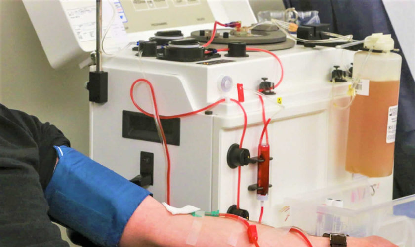
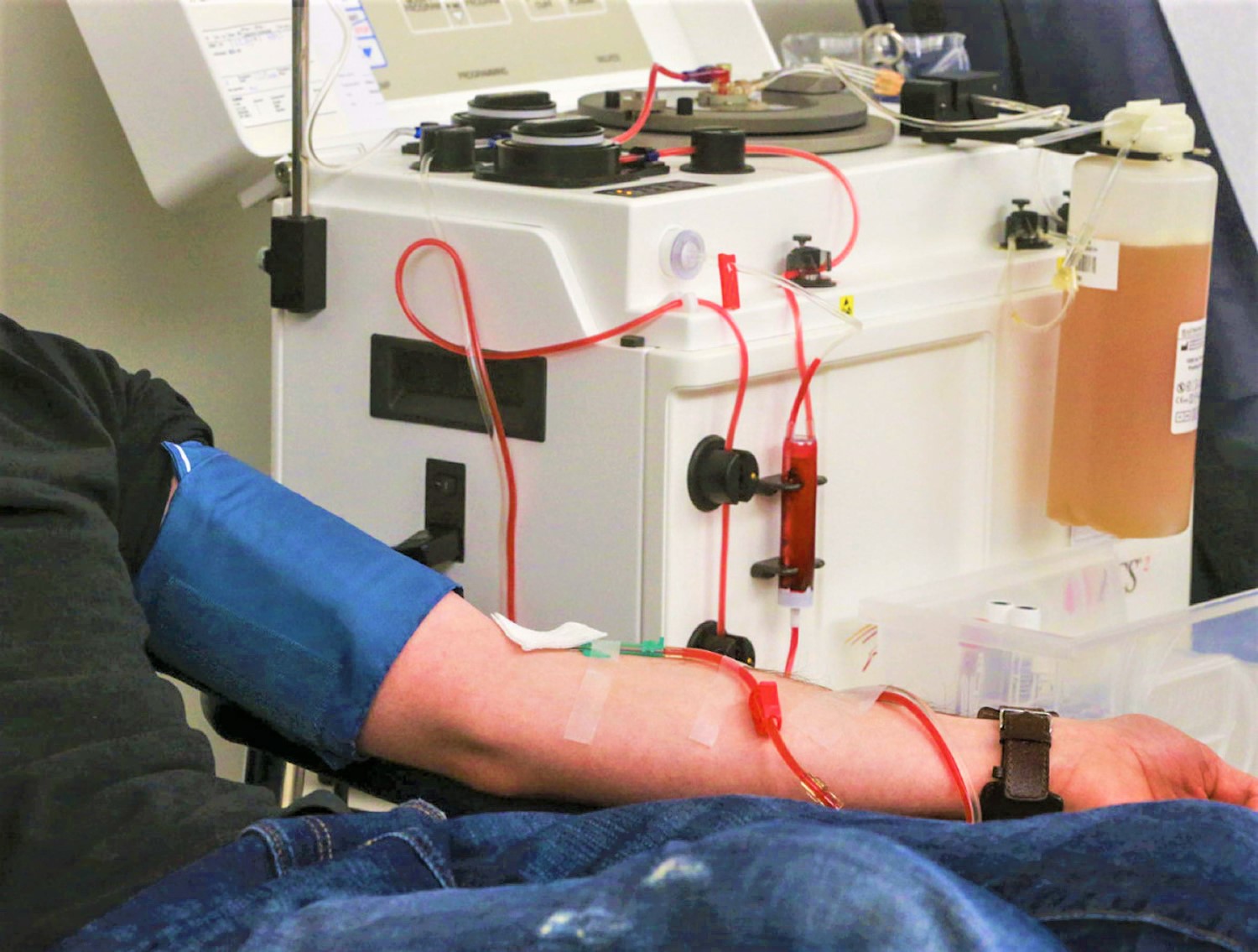
Figure 1. Plasmapheresis machine

Plasmapheresis machine

What is blood plasma

How much blood you have depends mostly on your size and weight. A man who weighs about 70 kg (about 154 pounds) has about 5 to 6 liters of blood in his body. Blood is 55% blood plasma and about 45% different types of blood cells. Blood is composed of solid particles (red blood cells, white blood cells, and cell fragments called platelets) suspended in a fluid extracellular matrix called blood plasma. Over 99% of the solid particles present in blood are cells that are called red blood cells (erythrocytes) due to their red color. The rest are pale or colorless white blood cells (leukocytes) and platelets (thrombocytes).

The blood plasma is the clear, straw-colored, liquid portion of the blood in which the cells (red blood cells, white blood cells) and platelets are suspended. It is
approximately 92% water and less than 8% is dissolved substances, mostly proteins, a complex mixture of organic and inorganic biochemicals. Functions of plasma include transporting gases, vitamins, and other nutrients; helping to regulate fluid and electrolyte balance; and maintaining a favorable pH. Blood plasma also contain antibodies to fight infections (part of the immune system), glucose, amino acids and the proteins that form blood clots (part of the hemostatic system)..

Blood plasma contains electrolytes that are absorbed from the intestine or released as by-products of cellular metabolism. They include sodium, potassium, calcium, magnesium, chloride, bicarbonate, phosphate, and sulfate ions. Sodium and chloride ions are the most abundant. Bicarbonate ions are important in maintaining the pH of plasma. Like other plasma constituents, bicarbonate ions are regulated so that their blood concentrations remain relatively stable.

Blood transports a variety of materials between interior body cells and those that exchange substances with the external environment. In this way, blood helps maintain stable internal environmental conditions.

Hemostasis refers to the process that stops bleeding, which is vitally important when blood vessels are damaged. Following an injury to the blood vessels, several actions may help to limit or prevent blood loss. These include vascular spasm, platelet plug formation, and blood coagulation.

Platelets adhere to any rough surface, particularly to the collagen in connective tissue. When a blood vessel breaks, platelets adhere to the collagen underlying the endothelium lining blood vessels. Platelets also adhere to each other, forming a platelet plug in the vascular break. A plug may control blood loss from a small break, but a larger break may require a blood clot to halt bleeding.

Coagulation, the most effective hemostatic mechanism, forms a blood clot in a series of reactions, each one activating the next. Blood coagulation is complex and utilizes many biochemicals called clotting factors. Some of these factors promote coagulation, and others inhibit it. Whether or not blood coagulates depends on the balance between these two groups of factors. Normally, anticoagulants prevail, and the blood does not clot. However, as a result of injury (trauma), biochemicals that favor coagulation may increase in concentration, and the blood may coagulate. The resulting mass is a blood clot, which may block a vascular break and prevent further blood loss. The clear, yellow liquid that remains after the clot forms is called serum. Serum is plasma minus the clotting factors.

Note: Blood is a complex mixture of formed elements in a liquid extracellular matrix, called blood plasma. Note that water and proteins account for 99% of the blood plasma.

Figure 2. Blood composition

blood composition

blood compositionblood-composition

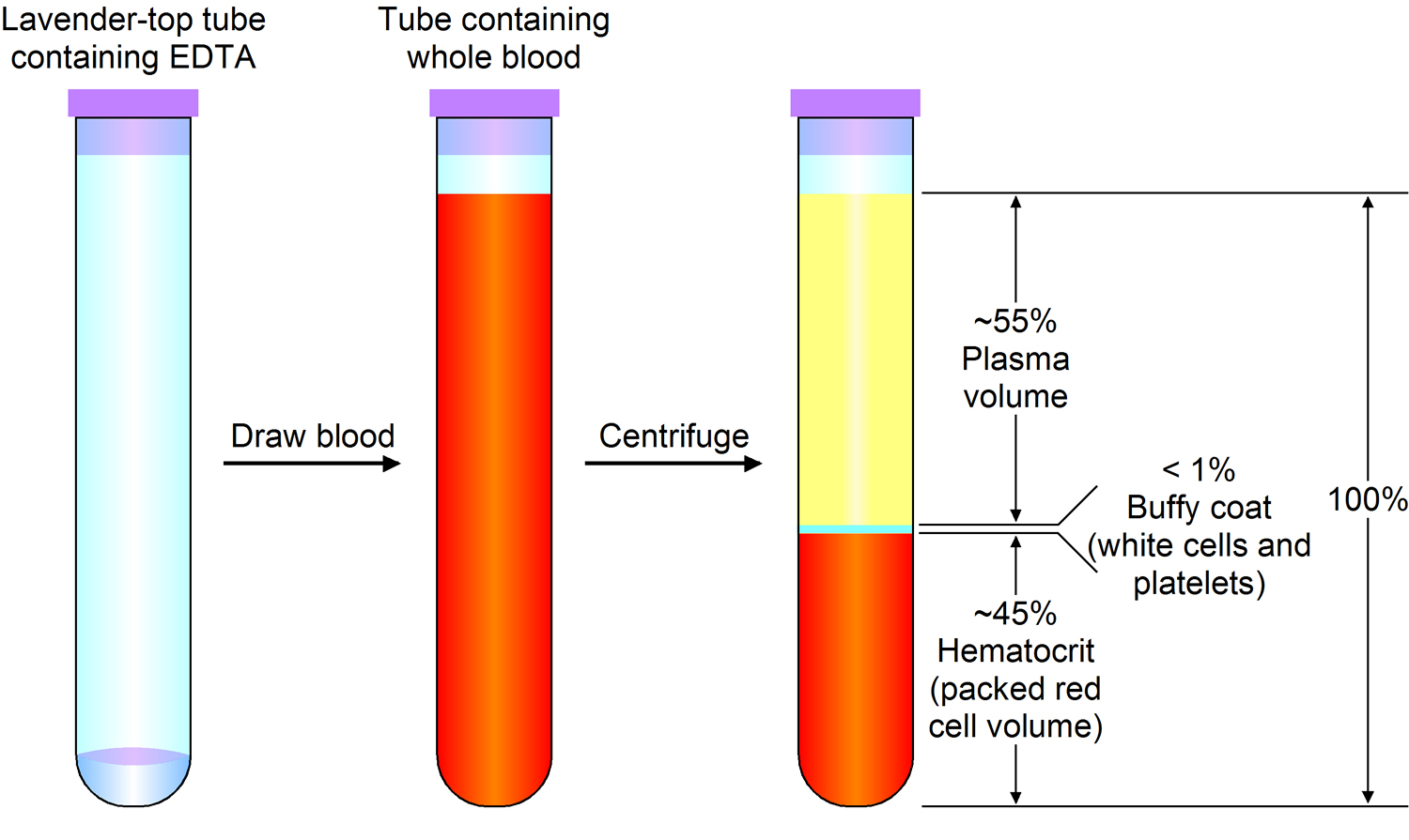
Note: Blood consists of a liquid portion called plasma and a solid portion (the formed elements) that includes red blood cells, white blood cells, and platelets. When blood components are separated by centrifugation, the white blood cells and platelets form a thin layer, called the “buffy coat,” between the plasma and the red blood cells, which accounts for about 1% of the total blood volume. Blood cells and platelets can be seen under a light microscope when a blood sample is smeared onto a glass slide.

Blood Plasma Proteins

Blood plasma proteins are the most abundant of the dissolved substances (solutes) in plasma. These proteins remain in the blood and interstitial fluids, and ordinarily are not used as energy sources. The three main types of blood plasma proteins—albumins, globulins, and fibrinogen—differ in composition and function.

Albumins are the smallest plasma proteins, yet account for about 60% of them by weight. Albumins are synthesized in the liver.

Plasma proteins are too large to pass through the capillary walls, so they are impermeant. They create an osmotic pressure that holds water in the capillaries, despite blood pressure forcing water out of capillaries by filtration. The term colloid osmotic pressure is used to describe this osmotic effect due to the plasma proteins. Because albumins are so plentiful, they are an important determinant of the colloid osmotic pressure of the plasma.

By maintaining the colloid osmotic pressure of plasma, albumins and other plasma proteins help regulate water movement between the blood and the tissues. In doing so, the blood plasma proteins help control blood volume, which, in turn, directly affects blood pressure.

If the concentration of plasma proteins falls, tissues swell. This condition is called edema. As the concentration of plasma proteins drops, so does the colloid osmotic pressure. Water leaves the blood vessels and accumulates in the interstitial spaces, causing swelling. A low plasma protein concentration may result from starvation, a protein-deficient diet, or an impaired liver that cannot synthesize plasma proteins.

Globulins make up about 36% of the plasma proteins. They can be further subdivided into alpha, beta, and gamma globulins. The liver synthesizes alpha and
beta globulins, which have a variety of functions. The globulins transport lipids and fat-soluble vitamins. Lymphatic tissues produce the gamma globulins, which are a type of antibody.

Fibrinogen constitutes about 4% of the plasma proteins, and functions in blood coagulation (clotting). Fibrinogen is synthesized in the liver, fibrinogen is the largest of the plasma proteins.

Plasma products and their uses

Plasma products can be grouped into three main types:

  • clotting or coagulation factors
  • albumin solutions
  • immunoglobulins

Coagulation or clotting factors

Coagulation is the name for the complex process of blood clotting. Clotting factors are proteins that work together with platelets to clot blood.

People need clotting factors for their blood to successfully clot. Missing one or more of these factors leads to blood clotting disorders such as haemophilia and Von Willebrand disease.

In the US, hemophilia is commonly treated with ‘recombinant factors’ that are manufactured in a laboratory and do not come from donated plasma.

Other blood clotting disorders that are treated with coagulation factors made from donated plasma.

Albumin

Albumin is the most common protein in blood plasma. It helps to:

  • carry substances around the body
  • maintain the right amount of fluid circulating in the body

If the circulation is working properly, vital hormones, cells and enzymes are transported to the right parts of the body to do their job.

If it’s not working properly, the circulatory system starts to break down, with serious consequences such as fluids being retained in the cells.

This can be treated by using human albumin solution which makes sure that the right amount of fluid is circulating in the blood stream.

Albumin can also be used to treat people with some types of liver or kidney disease and patients who have suffered burns.

Immunoglobulins

Immunoglobulins are protective antibodies which are produced by the body to fight against invading viruses or bacteria. There are two different types of immunoglobulins, specific and non-specific.

Specific immunoglobulins

Specific immunoglobulins contain high levels of antibody to a particular illness. These are given to people who have been exposed to a specific infection.

Antidotes to tetanus, rabies, chickenpox and hepatitis are all examples of specific immunoglobulins.

For example, a donor who has had chicken pox will have high levels of chicken pox antibodies. So their plasma would be ideal to treat a child with leukaemia who has been exposed to chicken pox.

Non-specific immunoglobulins

Non-specific immunoglobulins contain a wide variety of antibodies. These are given to people:

  • who make faulty antibodies, or can’t make their own antibodies
  • who are having treatments for diseases like cancer, where the treatment harms their ability to make antibodies

People with a faulty immune system need these products to live. Over 1,000 donations of plasma contribute to a single dose of an immunoglobulin product that contains all the necessary antibodies.

Table 1 summarizes the characteristics of the blood plasma proteins.

Table 1. Blood plasma proteins

Protein

Percentage of Total

Origin

Function

Albumins

60%

Liver

Help maintain colloid osmotic pressure

Globulins

36%

Alpha globulins

Liver

Transport lipids and fat-soluble vitamins

Beta globulins

Liver

Transport lipids and fat-soluble vitamins

Gamma globulins

Lymphatic tissues

Constitute the antibodies of immunity

Fibrinogen

4%

Liver

Plays a key role in blood coagulation

Plasmapheresis indications

Plasmapheresis indication guidelines have been defined and revised in 2010 by the American Society for Apheresis and divided into four categories from 1 to 4 on the basis of available literature 9. Category 1 disorders are those for which plasmapheresis is accepted as first-line therapy either as primary standalone treatment or in conjunction with other models of treatment and include disorders such as Guillain–Barre syndrome (GBS), myasthenia gravis, chronic inflammatory demyelinating polyneuropathy, thrombotic thrombocytopenic purpura, Goodpasture’s syndrome, and atypical hemolytic uremic syndrome. The separation of plasma from blood can be achieved by centrifugation devices or with the use of hemodialysis machine and plasma filters. Although an Indian Society for Apheresis was created in 1985, there is a scarcity of data on PE from the Indian subcontinent. This is partly because the facility for PE is available in only large centers located mainly in the cities. With the aim of improving data collection about plasmapheresis procedures in the country, we undertook this retrospective study aiming to look at plasmapheresis procedures conducted in the nephrology department over a fixed time period.

For purposes of Medicare coverage, plasmapheresis (apheresis) is defined as an autologous procedure, i.e., blood is taken from the patient, processed, and returned to the patient as part of a continuous procedure (as distinguished from the procedure in which a patient donates blood preoperatively and is transfused with the donated blood at a later date).

Medicare reimburseable indications for plasmapheresis – this may not be an exhaustive list of all applicable Medicare benefit categories for this item or service 10.

Apheresis is covered for the following indications:

  • Plasma exchange for acquired myasthenia gravis;
  • Leukapheresis in the treatment of leukemia;
  • Plasmapheresis in the treatment of primary macroglobulinemia (Waldenstrom);
  • Treatment of hyperglobulinemias, including (but not limited to) multiple myelomas, cryoglobulinemia and hyperviscosity syndromes;
  • Plasmapheresis or plasma exchange as a last resort treatment of thromobotic thrombocytopenic purpura (TTP);
  • Plasmapheresis or plasma exchange in the last resort treatment of life threatening rheumatoid vasculitis;
  • Plasma perfusion of charcoal filters for treatment of pruritis of cholestatic liver disease;
  • Plasma exchange in the treatment of Goodpasture’s Syndrome;
  • Plasma exchange in the treatment of glomerulonephritis associated with antiglomerular basement membrane antibodies and advancing renal failure or pulmonary hemorrhage;
  • Treatment of chronic relapsing polyneuropathy for patients with severe or life threatening symptoms who have failed to respond to conventional therapy;
  • Treatment of life threatening scleroderma and polymyositis when the patient is unresponsive to conventional therapy;
  • Treatment of Guillain-Barre Syndrome; and
  • Treatment of last resort for life threatening systemic lupus erythematosus (SLE) when conventional therapy has failed to prevent clinical deterioration.

Apheresis is covered only when performed in a hospital setting (either inpatient or outpatient) or in a nonhospital setting, e.g., a physician directed clinic when the following conditions are met:

  • A physician (or a number of physicians) is present to perform medical services and to respond to medical emergencies at all times during patient care hours;
  • Each patient is under the care of a physician; and
  • All nonphysician services are furnished under the direct, personal supervision of a physician.

Plasmapheresis for neurologic disorders

Plasmapheresis is established as effective and should be offered in severe acute inflammatory demyelinating polyneuropathy/Guillain-Barré syndrome and in the short-term management of chronic inflammatory demyelinating polyneuropathy (Class I studies, Level A) 11. Plasmapheresis is established as ineffective and should not be offered for chronic or secondary progressive multiple sclerosis (MS) (Class I studies, Level A) 11. Plasmapheresis is probably effective and should be considered for mild acute inflammatory demyelinating polyneuropathy/Guillain-Barré syndrome, as second-line treatment of steroid-resistant exacerbations in relapsing forms of MS, and for neuropathy associated with immunoglobulin A or immunoglobulin G gammopathy, based on at least one Class I or 2 Class II studies (Level B) 11. Plasmapheresis is probably not effective and should not be considered for neuropathy associated with immunoglobulin M gammopathy, based on one Class I study (Level B). Plasmapheresis is possibly effective and may be considered for acute fulminant demyelinating CNS disease (Level C) 11. There is insufficient evidence to support or refute the use of plasmapheresis for myasthenia gravis, pediatric autoimmune neuropsychiatric disorders associated with streptococcus infection, and Sydenham chorea (Class III evidence, Level U) 11.

Plasmapheresis for myasthenia gravis

Because of the lack of randomized controlled studies with masked outcomes, there is insufficient evidence to support or refute the efficacy of plasmapheresis in the treatment of myasthenic crisis or myasthenia gravis prethymectomy 11.

Plasmapheresis for MS

Plasmapheresis should not be offered for chronic progressive or secondary progressive MS (multiple sclerosis) 11. There is no studies on the efficacy of plasmapheresis compared to other treatment options in MS are available 11. There is good evidence plasmapheresis should be considered for the adjunctive treatment of exacerbations in relapsing forms of MS 11. There is weak evidence plasmapheresis may be considered in the treatment of fulminant CNS demyelinating diseases that fail to respond to high-dose corticosteroid treatment 11.

Plasmapheresis for acute inflammatory demyelinating polyneuropathy or Guillain-Barre Syndrome

There is strong evidence plasmapheresis should be offered in the treatment of acute inflammatory demyelinating polyneuropathy/Guillain-Barre Syndrome severe enough to impair independent walking or to require mechanical ventilation 11. There is good evidence plasmapheresis should be considered in the treatment of milder clinical presentations of acute inflammatory demyelinating polyneuropathy/Guillain-Barre Syndrome 12. IV immunoglobulin (IVIg) is an alternative treatment used in patients with acute inflammatory demyelinating polyneuropathy/Guillain–Barré syndrome. There is insufficient evidence to demonstrate the superiority of plasmapheresis over IV immunoglobulin (IVIg).

Plasmapheresis for chronic inflammatory demyelinating neuropathy

There is strong evidence plasmapheresis should be offered as a short-term treatment for patients with chronic inflammatory demyelinating neuropathy 12. Steroids, IV immunoglobulin (IVIg), and immunosuppressants also have been used in the treatment of chronic inflammatory demyelinating neuropathy 12.

Plasmapheresis for polyneuropathy associated with IgA and IgG monoclonal gammopathy of undetermined significance (MGUS)

There is good evidence plasmapheresis should be considered in polyneuropathy associated with IgA and IgG monoclonal gammopathy of undetermined significance (MGUS) 12. Plasmapheresis should not be considered in the treatment of polyneuropathy associated with IgM MGUS 12.

Plasmapheresis for hematological diseases

Therapeutic plasmapheresis is indicated in the management of various hematological diseases. For most of these diseases, clear pathogenetic mechanisms of the disease are understood, and there are well‐defined criteria with regard to the therapy 13. Most medical management of immunohematological disorders requires the use of plasmapheresis, serological immunomodulation, and classical pharmacological immunosuppression with steroids, cytotoxic agents, and antimetabolites, where overall therapy is individually tailored to the needs of the patient. Controlled trials are difficult if not impossible because of variables such as severity of disease, degree of organ system damage before intervention, age and the existence of co‐morbid conditions. In some rare hematological diseases, it is impossible to recruit a large number of cases to perform a controlled clinical trial. Therefore, for most of these diseases only small series of cases are available for analysis.

Therapeutic plasmapheresis, semi‐selective cascade filtration or IA aimed at the causative antibodies can be used in diseases caused by antibodies or immune complexes. Adjuvant drug therapies are different for different diseases and are typically individualized in type, dose and duration of use. The plasmapheresis method chosen depends on the pathophysiological origin of a given disease. The physician who has chosen the plasmapheresis method must be knowledgeable concerning the half‐life time, the compartmental distribution of pathogenic plasma proteins, and the elimination of other toxic substances and complement components. Table 2 shows a selection of hematological and hemostasiological diseases in which plasmapheresis has been implemented with the categories and the recommendation grade (RG) from the AAC.

Table 2. Therapeutic plasmapheresis in hematological and hemostasiological diseases with immunologic origin

Hematological and hemostasiological diseasesApheresis Applications Committee of the ASFA, 2010, 2013
Therapeutic plasmapheresis modalityCategoryRecommendation gradeTreated volume (TPV)Replacement solutionFrequency
Rhesus incompatibilityTPEII2C1–1.5Human‐albumin‐electrolyte solutionDaily or every other day
Red cell alloimmunization in pregnancyIII2C
Autoimmune hemolytic anemia
Warm autoimmune hemolytic diseaseTPEIII2C1–1.5
Cold agglutinin diseaseII2C
Aplastic anemiaTPEIII2C1–1.5
Pure red cell aplasiaIII1B
ABO incompatible HPC TxTPE, RBC exchange ECPII1B–2B1‐1.5Human‐albumin‐ electrolyteDaily or every other day; series weekly
III2C
Graft‐Versus‐Host Disease
Skin (acute)II1C200–270 mL
Skin (chronic)II1B
Non skin (acute/chronic)III2B
Idiopathic thrombocytopenic purpuraTPE, IA‐Protein‐AIV2C1–1.5Human‐albumin‐electrolyte, solution, plasmaDaily or every other day
III2C
Post‐transfusion purpuraTPEIII2C1–1.5
Coagulator factor inhibitors
AlloantibodyTPE, IA‐Protein‐A TPE, IA‐Protein‐AIV2C1–1.5
III2B
AutoantibodyIII2C1–1.5
III1C

Footnotes:

  • Category I: accepted for therapeutic plasmapheresis as first‐line therapy;
  • Category II: accepted for therapeutic plasmapheresis as second‐line therapy;
  • Category III: not accepted for therapeutic plasmapheresis, decision should be individualized;
  • Category IV: not accepted for therapeutic plasmapheresis, Institutional Review Board (IRB) approval is desirable if therapeutic plasmapheresis is undertaken 14.

Abbreviations: ECP = extracorporeal photopheresis; IA = Protein‐A, Immunoadsorption on protein‐A; HPC = hematopoietic progenitor cell; Tx = transplantation.

Plasmapheresis for autoimmune diseases

The terms “systemic autoimmune disease” and “collagen vascular disease” describe a number of illnesses, the common characteristic of which is immune‐mediated destruction of intracellular structures in connective tissue, resulting in fibrinoid tissue damage 8. Based on an immune pathogenesis, the various organs form antigen components, which provoke formation of autoantibodies on the one hand, and circulating immune complexes causing inflammation in organ tissues on the other.

Antinuclear antibodies are to be found against most nuclear structures. The antibodies are typically directed against both cytoplasmic‐associated and cell membrane‐associated proteins, and also antibodies against cytoplasmic structures and cell membrane components. The different groups of antibodies observed in active and subclinical disease includes those against many extracellular antigens, such as collagen, myelin sheaths, immunoglobulins, basement membrane, intercellular bridges, hormones, and complement components 15.

Table 3. Therapeutic plasmapheresis in systemic lupus erythematosus (SLE), catastrophic antiphospholipid syndrome, and rheumatoid arthritis (RA)

German Working Group of Clinical NephrologyApheresis Applications Committee of the ASFA, 2010, 2013
Therapeutic plasmapheresis modalityEvidence classSeverity gradeTherapeutic plasmapheresis modalityCategoryRecommendation gradeTreated Volume (TPV)Replacement solutionFrequency
Systemic lupus erythematosus (severe)IA, Peptid‐GAM, Tryptophan, Dextran sulfateIIIBTPEII2C1‐1.5Human‐ albumin‐ electrolyte solutionDaily or every other day
Lupus nephritisIV2B
Catastrophic antiphospholipid syndromeIIICTPEIII2C
Rheumatoid arthritisIA, Peptid‐GAMIbAIAII2A

Footnotes:

  • Category I: accepted for therapeutic plasmapheresis as first‐line therapy;
  • Category II: accepted for therapeutic plasmapheresis as second‐line therapy;
  • Category III: not accepted for therapeutic plasmapheresis, decision should be individualized;
  • Category IV: not accepted for therapeutic plasmapheresis, Institutional Review Board (IRB) approval is desirable if therapeutic plasmapheresis is undertaken 14.

Abbreviations: IA‐Protein‐A = Immunoadsorption on protein‐A; Peptid‐GAM = Immunoadsorption on synthetic Peptid‐GAM (Globaffin, Affina Immuntechnik, Germany); Tryptophan = immunoadsorption on tryptophan (Immunosorba, Asahi, Japan); Dextran sulfate, chemical adsorption on dextran sulfate (Liposorber, Kaneka, Japan).

Plasmapheresis for renal disorders

Many primary renal diseases are associated with autoantibodies, rendering them appealing indications for plasmapheresis. Some indications are well established by randomized controlled studies and are considered standard of care (Goodpasture and thrombotic thrombocytopenic purpura [TTP]). Others have less compelling or only anecdotal supporting evidence.

Anti–Glomerular Basement Membrane (anti-GBM) Antibody–Mediated Disease (Goodpasture syndrome)

A randomized controlled trial found plasmapheresis to provide a more rapid decrease in anti-glomerular basement membrane antibodies, lower posttreatment serum creatinine level, and decreased incidence of end-stage renal disease. Given these results and the integral role of the anti-glomerular basement membrane antibody, plasmapheresis as a means of rapidly decreasing anti-glomerular basement membrane titers has become the standard of care.

Treatment strategy:

  1. Early initiation of plasmapheresis is essential to avoid end-stage renal disease
  2. Initial prescription is 14 daily 4-L exchanges
  3. Continued plasmapheresis may be required if antibody titers remain increased
  4. Steroids, cyclophosphamide, or azathioprine are added to decrease production of anti-glomerular basement membrane antibody and minimize the inflammatory response

Crescentic Rapidly Progressive Glomerulonephritis (not associated with anti-GBM antibody)

Several controlled studies have failed to show a generalized benefit of plasmapheresis for all patients with rapidly progressive glomerulonephritis; however, subset analysis of all these studies showed plasmapheresis to be beneficial for patients presenting with severe disease or dialysis dependency. A more recent study (Jayne et al) limited to patients presenting with creatinine levels greater than 5.8 mg/dL (to convert creatinine in mg/dL to μmol/L, multiply by 88.4) appears to support this conclusion.

Patients with Wegener granulomatosis and microscopic polyarteritis who present with pulmonary hemorrhage appear to be more likely to present with IgM antineutrophil cytoplasmic antibodies (ANCAs). These patients may also respond to plasmapheresis.

Renal Failure in Multiple Myeloma

After exclusion of other forms of renal failure associated with multiple myeloma (eg, hypercalcemia, volume depletion, hyperuricemia, infection, and amyloidosis), patients considered to have light-chain–related “cast nephropathy” may benefit from plasmapheresis. plasmapheresis can decrease serum levels of light chains more rapidly than chemotherapy alone. A randomized controlled study found plasmapheresis to provide a more likely return of renal function and better overall survival 16. However, despite a 50% decrease in need for dialysis, a recently reported study did not find a statistically significant benefit for plasmapheresis 17.

Treatment considerations:

  1. Demonstration of free light chains in serum is essential if plasmapheresis is to be considered a rational treatment option (by standard immunofixation or the new free light chain assay)
  2. Successful plasmapheresis prescription is 3 to 4 L of plasma exchanged on 5 consecutive days
  3. Well-established (chronic) renal failure considered to be caused by cast nephropathy may respond less dramatically
  4. Newly available highly permeable hemofilter membranes may allow for light chain removal without significant albumin loss 18

IgA Nephropathy and Henoch-Schönlein Purpura

Case reports and small clinical series suggest a possible beneficial effect of plasmapheresis in the treatment of IgA-associated rapidly progressive glomerulonephritis.

Cryoglobulinemia

Despite a lack of randomized controlled studies, most experts agree plasmapheresis can be a useful adjunct for severe active disease manifested by progressive renal failure, coalescing purpura, or advanced neuropathy. Plasmapheresis can rapidly decrease cryoglobulin levels without the use of immunosuppressive agents, which might be problematic in hepatitis C–associated disease.

Treatment strategy:

  1. A reasonable plasmapheresis prescription is to exchange 1 plasma volume 3 times weekly for 2 to 3 weeks
  2. An average of 13 treatments may be required to induce clinical improvement (range, 4 to 39)
  3. The replacement fluid can be 5% albumin, which must be warmed to prevent precipitation of circulating cryoglobulins

Thrombotic thrombocytopenic purpura

A large randomized controlled study found 78% survival with plasmapheresis and fresh frozen plasma (FFP) replacement compared with 50% survival with fresh frozen plasma infusions alone 19. Plasmapheresis with fresh frozen plasma replacement is the treatment of choice for thrombotic thrombocytopenic purpura and is considered standard of care.

Treatment considerations:

  1. Fresh frozen plasma (FFP) is required as replacement fluid to replace missing metalloprotease (ADAMTS13 [A Disintigrin-like And Metalloprotease with ThromboSpondin type 1 repeats])
  2. Plasma removal with plasmapheresis removes antibody to ADAMTS13
  3. Treatments are performed daily until the platelet count is normalized and hemolysis has largely ceased (normalization of lactate dehydrogenase)
  4. Exchanged volumes should be at least 1 plasma volume. Some experts recommend 1.5 plasma volume exchanges for the first week
  5. Previous recommendations suggest switching to cryoprecipitate-poor plasma in resistant cases because it may contain lower levels of von Willebrand factor. However, a recent review suggests that cryoprecipitate-poor plasma contains less ADAMTS13 and may be less effective than fresh frozen plasma 20

Hemolytic uremic syndrome in adults

Although renal failure tends to dominate the clinical presentation, unless a specific cause can be identified, hemolytic uremic syndrome is often difficult to distinguish from thrombotic thrombocytopenic purpura

Causes:

  • Verotoxin induced by Escherichia coli 0157-H7: prodrome of bloody diarrhea
  • Drugs: cyclosporine, tacrolimus, mitomycin, cisplatinum, quinine, oral contraceptives, antiplatelet agents, and so on
  • Lupus
  • Cancer
  • Bone marrow transplant
  • Post transplantation recurrence

Prognosis in adults is poor:

  • Mortality between 25% and 50%
  • End-stage renal disease in 40%

Although treatment success depends on the cause, hemolytic uremic syndrome in adults is often treated with plasmapheresis as with thrombotic thrombocytopenic purpura.

Hemolytic uremic syndrome in children

Prognosis is usually good in verotoxin-induced disease, with only a small percentage of patients experiencing strokes or sustained renal failure. Controlled trials with plasma infusion have shown only minimal benefit.

Plasmapheresis may be beneficial in children:

  1. Without a diarrheal prodrome
  2. Older than 5 years
  3. With significant central nervous system involvement

Lupus anticoagulant, Anticardiolipin Antibodies, and Antiphospholipid Antibody Syndrome

Lupus anticoagulant and anticardiolipin antibody are antiphospholid antibodies associated with thromboses, recurrent fetal loss, and renal disease. Plasmapheresis has been successful in removing antiphospholipid antibodies to avoid spontaneous abortion, treatment of lupus anticoagulant-associated renal failure, and in the management of catastrophic antiphospholipid syndrome (CAPS).

Scleroderma

Plasmapheresis may be useful in rare coexistence of scleroderma and ANCA-positive or antinuclear antibody (ANA)-positive renal disease.

Focal Segmental Glomerulosclerosis: Recurrence Posttransplantation

Fifteen percent to 55% of patients with end-stage renal disease secondary to focal segmental glomerulosclerosis have rapid recurrence of proteinuria after renal transplantation. Some patients with early recurrence of proteinuria have a circulating 30- to 50,000-d protein capable of increasing glomerular permeability to albumin. Standard plasmapheresis and immunoadsorption have been successful in decreasing the level of proteinuria. The addition of cyclophosphamide to plasmapheresis may lead to more prolonged remission. plasmapheresis may be effective in the treatment of recurrent focal segmental glomerulosclerosis if treatment is initiated promptly after the initiation of proteinuria.

Transplant Candidates With Cytotoxic Antibodies

Plasmapheresis and immunoadsorption have been successful in decreasing high levels of preformed cytotoxic antibodies (panel reactive antibody [PRA]), allowing for successful transplants for up to 34 months.

Often used with concomitant cyclophosphamide and prednisolone.

Renal Allograft Rejection

Plasmapheresis can provide a rapid decrease in anti-human leukocyte antigen (HLA) antibodies. However, 2 controlled trials of plasmapheresis for acute vascular rejection did not find this treatment to be useful.

Plasmapheresis together with cyclophosphamide and methylprednisolone has been reported to result in greater improvement in renal function and improved graft survival.

Renal Transplantation Across Blood Group Type ABO Groups

Plasmapheresis can be used to remove anti-A or anti-B antibodies before transplantation. Five-year graft survival has been as high as 78% when kidneys from donors in blood A2 or B subgroups are transplanted into group O recipients. Donor-specific skin grafting can be used to predict outcome.

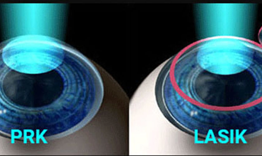
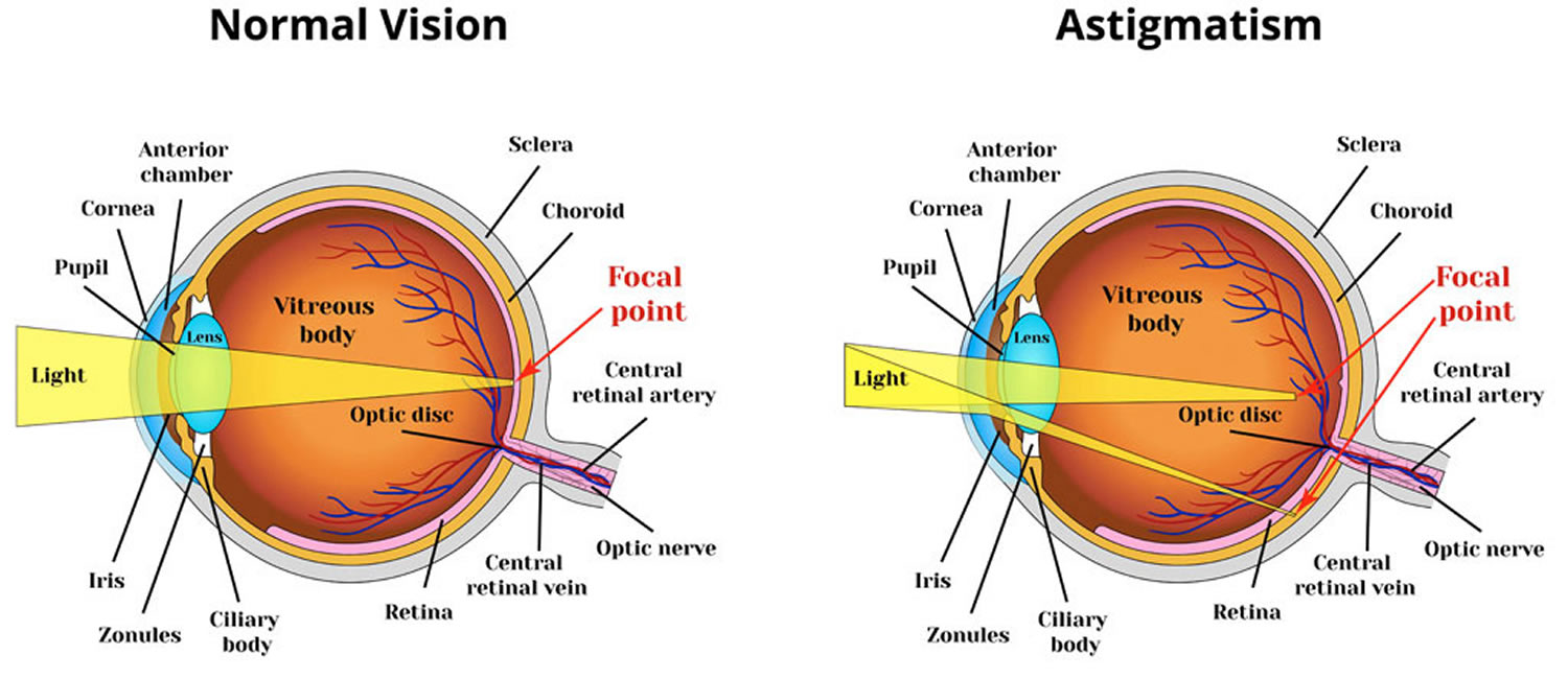
Plasmapheresis for inflammatory eye disease

When conventional therapy with cortisone or immunosuppressive drugs fails or is inadequate in the treatment of immune‐mediated inflammatory eye disease with an auto immunologic pathogenesis, plasmapheresis may be indicated and is increasingly being implemented with success.

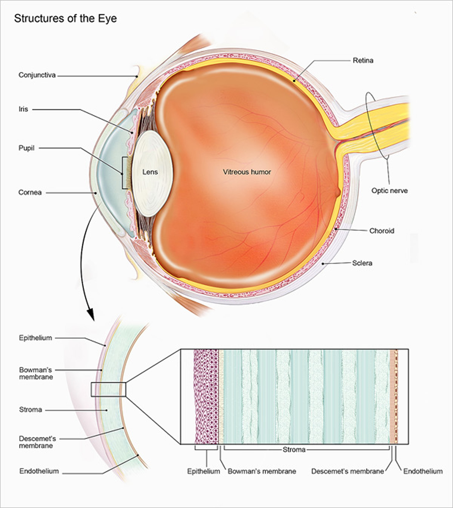
Severe uveitis is potentially associated with visual impairment or blindness in young patients 21. In posterior uveitis, progredient inflammatory processes can lead to morphologic changes in the chorioidea and retina, contributing to functional deterioration. In uveitis intermedia, inflammatory processes in the peripheral retina and in the area of the ciliary body require primary attention and aggressive treatment. In both cases, secondary destructive changes in the vessels can occur, causing reduced perfusion of the retina and chorioidea. Primary inflammatory vascular changes may lead to secondary morphologic chorioretinal changes, which may then further impair function. The inflammatory process and/or the reduced chorioretinal perfusion are important. Therefore, an anti‐inflammatory/immunomodulatory therapy, a hemorheologic therapy, or a combination of both treatments, should bring about improvement of the condition, insofar as no other specific therapy is indicated 6.

Detection of immune complexes or autoantibodies in uveitis is problematic. First, indications for the existence and possible pathomechanism of pathogenic substrates to retinal S antigen were found in patients with uveitis and in animal studies. Both improvement and deterioration in the condition can be regarded as an indication of elimination of a pathogenic substrate.

The improvement in hemorheologic parameters could contribute considerably to the therapeutic success in autoimmune eye diseases accompanied by primary or secondary vascular changes. With improved microcirculation, the damaged tissue can recover. In addition, other mechanisms, such as elimination of a pathogenic substrate or immunomodulatory effects of the exchange medium, probably contribute to the success of this therapy. The immunomodulating mechanism of plasmapheresis, which favors a prompter elimination of inflammation, increases ocular function, and reduces recurrence, has been clarified.

In recent years, the anti‐TNF‐α antibodies, infliximab and adalimumab, and others demonstrated significant efficacy in controlling uveitis associated with seronegative spondyloarthropathies and juvenile idiopathic arthritis 22. The majority of reports of biologic therapies in posterior uveitis have been uncontrolled or retrospective studies in patients with uveitis resistant to immunosuppression.

Biologic therapies have increased the treatment options for sight‐threatening uveitis. Despite an experimental rationale, the lack of evidence from randomized controlled studies limits our understanding of when to commence therapy, which agent to choose, and how long to continue treatment. Additionally, the high cost and potential side‐effects of the biologic agents have limited their current use to uveitis refractory to immunosuppression. Further controlled randomized multicenter studies of TPE and/or immunosuppression versus biologics are necessary to clarify efficacy, side‐effects, and costs.

Plasmapheresis for dermatological diseases

Dermatologic immune mediated diseases represent a heterogenous group of disorders associated with circulating autoantibodies against distinct adhesion molecules of the skin and/or mucosa. According to the level of split formation, the disorders can be divided in the intraepidermal blistering pemphigus, such as pemphigus vulgaris, pemphigus foliaceus, and paraneoplastic pemphigus, and the subepidermal blistering pemphigoid diseases, such as bullous pemphigoid, pemphigoid gestations, and dermatitis herpetiformis 23. The new developed sensitive and specific assays for circulating autoantibodies in these dermatological diseases now enable a serological diagnosis in about 90% of cases.

The incidences of autoimmune blistering skin diseases in Germany has doubled in the last 10 years, to about 25 new cases per million humans per year, because of improved diagnostic techniques as well as the aging of the population 23. There are an estimated 2000 new cases of autoimmune blistering skin diseases per year. The incidence of pemphigus in Europe is one to two cases per million humans per year, and 80% have pemphigus vulgaris. Bullous pemphigoid is the most common type of subepidermal autoimmune blistering skin disease in Europe, with an incidence of about 13 cases per million humans per year. The next common types are mucous membrane pemphigoid and pemphigoid gestationis 24.

The standard of diagnostic testing for autoimmune blistering skin diseases is the direct immunofluorescence (IF) microscopy to demonstrate tissue‐bound autoantibodies and/or C3 in the patients’ skin or mucous membranes. The direct IF microscopy of the patient’s serum can be used as a screening test for circulating antibodies. The diagnostic assessment of autoimmune blistering skin diseases can be expected to improve in the near future as new serological testing systems are developed that employ recombinant forms of the target antigens. But the treatments in use still need to be validated by prospective, controlled trials 66. An example for intradermal blistering pemphigus is pemphigus vulgaris.

Table 4. Therapeutic plasmapheresis in dermatological diseases with immunologic origin

Dermatological diseases treated with therapeutic plasmapheresisApheresis Application Committee of the ASFA, 2010, 2013
Therapeutic plasmapheresis modalityTreated with TATherapeutic plasmapheresismodalityCategoryRecommendation gradeTreated Volume (TPV)Replacement solutionFrequency
Intraepidermal blistering pemphigus1‐1.5human‐albumin‐electrolyte solutiondaily or every other day
Pemphigus vulgarisTPE, IA+TPE, IA, ECPIII2B, 2C
+
Subepidermal blistering pemphigus
Bullous pemphigoidTPE, IA+
+
D‐penicillinamine induced pemphigusTPE, IA+
+
Progressive sclerodermaTPE+TPE, ECPIII, III2C
2B
Pyoderma gangrenosumTPE+
Henoch‐Schönlein purpuraTPE+TPEIII2C

Footnotes:

  • Category I: accepted for therapeutic plasmapheresis as first‐line therapy;
  • Category II: accepted for therapeutic plasmapheresis as second‐line therapy;
  • Category III: not accepted for therapeutic plasmapheresis, decision should be individualized;
  • Category IV: not accepted for therapeutic plasmapheresis, Institutional Review Board (IRB) approval is desirable if therapeutic plasmapheresis is undertaken 14.

Abbreviations: IA‐Protein‐A = Immunoadsorption on protein‐A; ECP = extracorporeal photopheresis

Plasmapheresis procedure

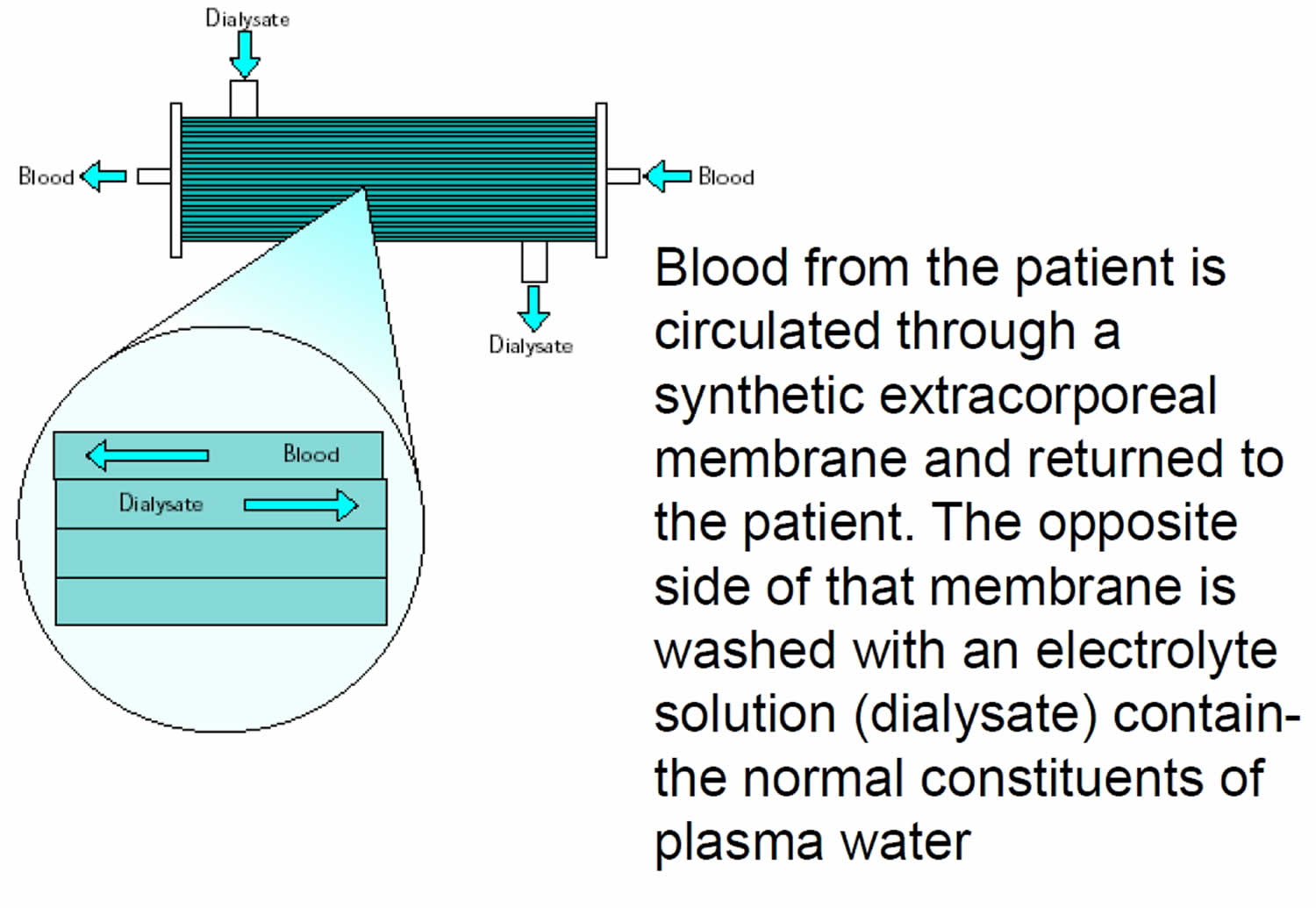
Plasmapheresis (also known as plasma exchange or apheresis) is similar to kidney dialysis (hemodialysis); however, plasmapheresis removes the plasma portion of the blood where the antibodies are located. Plasma is the almost clear part of the blood which carries red cells, white cells, platelets and other substances through your bloodstream. During plasmapheresis, you will need to have a working native fistula, graft or dialysis catheter. If you have a catheter, one line of the catheter is attached to tubing and takes blood to the plasmapheresis machine. A second line of the catheter is used to return the blood. If you have a fistula or graft, needles will be placed as they are for hemodialysis. You may feel some minor discomfort when the needles are placed in position. This is similar to what a blood donor experiences.

Plasmapheresis uses a machine that separates the plasma (the liquid part of the blood) that contains the abnormal antibodies such as monoclonal paraproteins and pathogenic autoantibodies, immune complexes, cryoglobulins, myeloma light chains, endotoxin, and cholesterol-containing lipoproteins from the blood cells. The plasma containing the abnormal protein is discarded, while the blood cells are mixed with salt solution and plasma from a donor and given back to the patient.

Plasmapheresis is done over a few hours while the person lies in a bed or sits in a reclining chair. The blood is removed through an IV line (usually in a vein in the arm), goes through the machine where the plasma is replaced, and then is returned to the body through another IV line. Sometimes, minor surgery is done before the procedure to put a single large catheter in a large vein just below the neck or under the collar bone instead of using IV lines in the arms. This type of catheter, called a central line or central venous catheter (CVC), has both IVs built in.

Plasmapheresis is not painful (aside from the IV lines being put in), but it can be hard to stay sitting or lying down in the same place for 2 or 3 hours. Calcium levels can drop in some people during treatment, causing numbness and tingling (especially in the hands and feet and around the mouth) and muscle spasms, which can sometimes be painful. This can be treated by giving the patient calcium.

Plasmapheresis works quickly to bring down the abnormal antibodies level. However, it does not treat the cause of the abnormal antibodies level, immune complexes, cryoglobulins, myeloma light chains, endotoxin, and cholesterol-containing lipoproteins from the blood cells, so it will go back up again without further treatment.

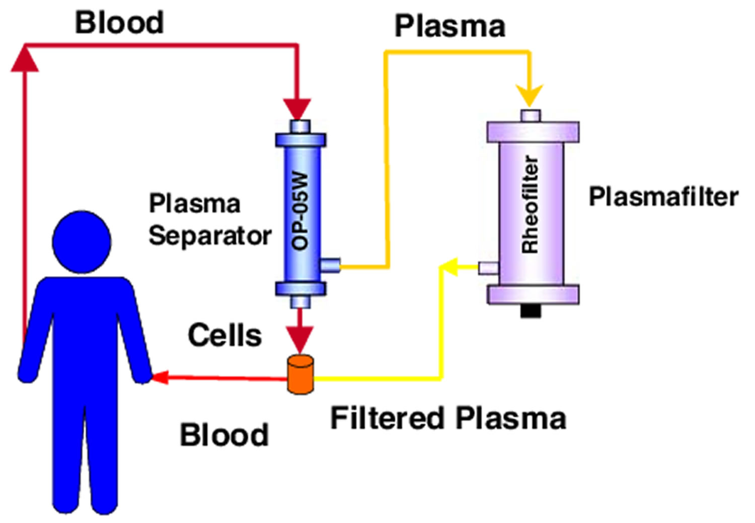
Figure 3. Plasmapheresis procedure

plasmapheresis procedure

plasmapheresis procedure

plasmapheresis machineplasmapheresis-technique

plasmapheresis_technique

Plasmapheresis technique

Traditionally, plasmapheresis was performed with centrifugation devices used in blood-banking procedures. These devices offer the advantage of allowing for selective cell removal (cytapheresis). Plasmapheresis also can be performed using a highly permeable filter and standard dialysis equipment.

1) Centrifugation

Centrifugation separates the plasma by density gradients.Whole-blood constituents are layered into plasma (specific gravity [SG], 1.025 to 1.109), platelets (SG, 1.040), lymph (SG, 1.070), granulocytes (SG, 1.087 to 1.092), and red blood cells (SG, 1.093 to 1.096).

2) Filtration (membrane plasma separation)

Separation of plasma from the blood’s cellular components can also be accomplished by filtration though a highly permeable membrane. Blood is separated into its cellular and noncellular components by subjecting it to sieving through a membrane with pores that allow plasma proteins to pass, but that retain the larger cellular elements within the blood path.

3) Plasmapheresis with dialysis equipment

Plasmapheresis can be performed with a highly permeable filter connected to the blood pump and pressure monitoring system of the dialysis machine. The machine is used in its “isolated” ultrafiltration mode, bypassing the dialysate proportioning system.

4) Anticoagulation

For centrifugal techniques, anticoaglation is often provided by citrate. For membrane plasma separation with dialysis equipment, heparin can be used as during standard dialysis.

5) Replacement Fluids:

  • Albumin
    • Pros: no viral transmission, allergies are rare
    • Cons: depletion coagulopathy, immunoglobulin depletion
    • Electrolyte composition: Sodium, 145 ± 15 mEq/L; potassium, less than 2 mEq/L (sodium and potassium in mEq/L is equivalent to sodium and potassium in mmol/L)
    • Anaphylactic reactions: Rare, antibodies to polymerized albumin
    • “Depletion coagulopathy”: Replacement with albumin will lead to depletion of coagulation factors.
      1. After a single plasma exchange, prothrombin time (PT) increases 30%, partial thromboplastin time (PTT) doubles: these increases often reverse 1 day after treatment
      2. Multiple consecutive treatments result in prolonged increases in PT/PTT
      3. Fresh frozen plasma administered toward the end of the procedure can minimize hemorrhagic risks
    • Immunoglobulin depletion
      1. A single 1-plasma volume exchange reduces serum immunoglobulin levels by 60%
      2. Multiple treatments can decrease immunoglobulin levels for several weeks
      3. A single infusion of immunoglobulin (IVIG) administered after a series of plasmapheresis treatments can reconstitute normal immunoglobulin levels
    • Risk of viral transmission: Albumin is heat treated and considered to be devoid of transmissible virus.
  • Fresh frozen plasma
    • 3 L of fresh frozen plasma is obtained from 10 to 15 donors (15 separate units).
    • Pros: Does not lead to postpheresis coagulopathy or immunoglobulin depletion. Fresh frozen plasma is essential for the treatment of thrombotic thrombocytopenic purpura.
    • Cons: Anaphylactoid reactions, citrate toxicity, small risk of viral transmission.
    • Anaphylactic reactions: Fever, rigors, urticaria, wheezing, hypotension, and laryngeal edema
      • Angiotensin-converting enzyme (ACE) inhibitors should be avoided given their ability to inhibit kinin metabolism
      • Consider pretreatment with diphenhydramine intravenously (IV): 0.3 to 0.5 mL of epinephrine (1:1,000 solution) should be available for subcutaneous administration for severe reactions
    • Citrate toxicity: Fresh frozen plasma contains 14% citrate by volume; can lead to hypocalcemia and metabolic alkalosis
    • Risk of viral transmission: 1/63,000 units for hepatitis B, 1/100,000 units for hepatitis C, 1/680,000 units for human immunodeficiency virus (HIV), and 1/641,000 units for human T-lymphotrophic virus
  • Starch replacement for plasmapheresis
    • Similar attributes with albumin, may be less expensive.

6) Vascular Access

  • Antecubital veins:
    1. Ideal for low-flow treatments
    2. Increasingly difficult to use after multiple punctures
  • Temporary vascular catheters: Catheter removal may be hazardous after an intensive run of plasmapheresis treatments, which can result in depletion coagulopathy and increased PT/PTT.
    1. Femoral vein cannulation
    2. Subclavian and internal jugular catheters
    3. Tunneled jugular venous catheters
  • Permanent arteriovenous access: Preferred if treatments are to be repeated regularly (hyperlipidemia).
    1. Primary arteriovenous fistula
    2. Arteriovenous graft

7) Selective Plasmapheresis Techniques

  • Designed to remove a particular pathogenic substance
  • Decreases need for replacement fluid
  • Minimizes risks of depletion coagulopathy and hypogammaglobulinemia
  • Many systems available in Japan and Europe, few in United States
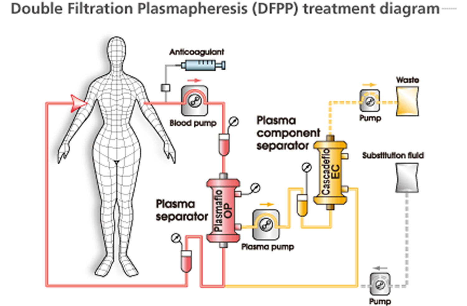
    1. Cascade filtration (“double filtration”)
      • Separated plasma is refiltered through a secondary filter with smaller pore size
      • Larger, unwanted molecules removed by secondary filter
      • Indications: Waldenstrom macroglobulinemia, cryoglobulinemia, familial hypercholesterolemia, and immune complex–mediated disease
    2. Cryofiltration
      • Removed plasma is cooled, causing certain substances to aggregate
      • Increasing size allows for efficient secondary filtration
      • Indications: cyroglobulins and immune complexes
    3. Immunoadsorbant techniques
      • Systems for selective immunoadsorption
      • Indications: nonselective immunoglobulin removal, low- density lipoprotein (LDL) cholesterol.
        • Protein A columns: Protein A 42,000-d protein released from Staphylococcus aureus. Used for the ex vivo adsorption of 3 of the 4 classes of IgG (1, 2, and 4).
          • Prosorba column (Cypress Biosciences Inc, San Diego, CA). Single-use nonregenerating system placed in series with a standard plasma exchange circuit. When the plasma is separated from the blood, it is slowly perfused over the column (at 20 mL/min). This column saturates rapidly with very limited IgG removal. Postulated mode of action is by “immunomodulation” of perfused plasma. Food and Drug Administration approved for idiopathic thrombocytopenic purpura (ITP) and rheumatoid arthritis. Secondary effects are common: fever, chills, musculoskeletal pains, hypotension. Contraindicated in patients using ACE inhibitors.
          • Excorim (Lund, Sweden). Alternating columns repeatedly regenerated to allow for more efficient IgG removal. Renal indications: removal of anti-HLA antibodies in highly sensitized recipients, rapidly progressive glomerulonephritis.
    4. Selective LDL cholesterol removal
      • Limits the loss of plasma proteins and high-density lipoprotein (HDL) cholesterol
      • Indicated in patients with familial hypercholesterolemia who cannot tolerate or whose condition is unresponsive to pharmacological treatment and who have either known cardiovascular disease and a plasma LDL cholesterol level greater than 200 mg/dL or no known cardiovascular disease and a plasma LDL cholesterol level greater than 300 mg/dL
      • Four systems:
        • Imunoadsorbant
        • Dextran sulfate binding to apoprotein B. Contraindication for patients on ACE-inhibitor therapy
        • Heparin-mediated extracorporal LDL precipitation (HELP)
        • Direct adsorption of LDL (DALI). Does not require plasma separation, removes LDL directly from whole blood
    5. Endotoxin adsorption
      1. Fibers impregnated with polymyxin B. Can bind endotoxin fragments
      2. Japanese experience documents improvement in systemic hemodynamics of sepsis
      3. Not currently available in the United States

Plasmapheresis complications

Plasmapheresis side effects include hypotension, allergic reactions, nausea, vomiting paresthesia, and cramps are the most common complications, and these may be seen in 3%–25% of procedures 25. Most of these events are mild and resolve without treatment. The reported incidence of paresthesia and cramps ranges from 1.5% to 9% 25. Reported incidence of hypotension ranges from 2.6% to 8.1% 26. However, the incidence of both of these was quite low in our study. The overall mortality in plasmapheresis is estimated to be 1–3/10,000 procedures 27.

References
  1. Therapeutic Plasma Exchange: Core Curriculum 2008. American Journal of Kidney Diseases , Volume 52 , Issue 6 , 1180 – 1196. https://www.ajkd.org/article/S0272-6386(08)00707-5/fulltext
  2. Therapeutic Apheresis in Hematologic, Autoimmune and Dermatologic Diseases With Immunologic Origin. Bambauer R, Latza R, Burgard D, Schiel R. Ther Apher Dial. 2016 Oct; 20(5):433-452. https://onlinelibrary.wiley.com/doi/full/10.1111/1744-9987.12474
  3. Ahammed Nizar OT, Rai P, Rao SN, Shenoy MP. Plasmapheresis: A Retrospective Audit of Procedures from a Tertiary Care Center in Southern India. Indian Journal of Critical Care Medicine : Peer-reviewed, Official Publication of Indian Society of Critical Care Medicine. 2017;21(12):857-860. doi:10.4103/ijccm.IJCCM_177_17. https://www.ncbi.nlm.nih.gov/pmc/articles/PMC5752796/
  4. Kaplan AA. Therapeutic plasma exchange: Core curriculum 2008. Am J Kidney Dis. 2008;52:1180–96. https://www.ncbi.nlm.nih.gov/pubmed/18562061
  5. Strauss RG, Ciavarella D, Gilcher RO, Kasprisin DO, Kiprov DD, Klein HG, et al. An overview of current management. J Clin Apher. 1993;8:189–94. https://www.ncbi.nlm.nih.gov/pubmed/8113206
  6. Bambauer R, Latza R, Schiel R. Therapeutic Plasma Exchange and Selective Plasma Separation Methods. In: Fundamental Technologies, Pathology and Clinical Results, 4th edn. D‐49525 Lengerich: Pabst Science Publishers, 2013.
  7. Kumilien G, Ullstrom L, Losvall A et al. Clinical experience with a new apheresis filter has specifically depletes ABO blood group antibodies. Transfusion 2006;46:1568–75. https://www.ncbi.nlm.nih.gov/pubmed/16965585
  8. Davidson A, Diamond B. Autoimmune Diseases. N Engl J Med 2001;345:340–50.
  9. Schwartz J, Padmanabhan A, Aqui N, Balogun RA, Connelly-Smith L, Delaney M, et al. Guidelines on the use of therapeutic apheresis in clinical practice-evidence-based approach from the writing committee of the American Society for Apheresis: The Seventh Special Issue. J Clin Apher. 2016;31:149–62. https://www.ncbi.nlm.nih.gov/pubmed/27322218
  10. National Coverage Determination (NCD) for Apheresis (Therapeutic Pheresis). https://www.cms.gov/medicare-coverage-database/details/ncd-details.aspx?ncdid=82
  11. Cortese I, Chaudhry V, So YT, Cantor F, Cornblath DR, Rae-Grant A. Evidence-based guideline update: Plasmapheresis in neurologic disorders: Report of the Therapeutics and Technology Assessment Subcommittee of the American Academy of Neurology. Neurology. 2011;76(3):294-300. doi:10.1212/WNL.0b013e318207b1f6. https://www.ncbi.nlm.nih.gov/pmc/articles/PMC3034395/
  12. Update: Plasmapheresis in Neurologic Disorders. American Academy of Neurology guideline update. Neurology 2011;2011;76:294–300. https://www.aan.com/Guidelines/home/GetGuidelineContent/471
  13. Bambauer R, Latza R, Schiel R. Therapeutic Apheresis in Neurological and Hematological Diseases in Pediatrics. Clin Med Ins: Pediatr in press.
  14. Schwartz J, Winters JL, Padmanabhan A et al. Guidelines on the use of therapeutic apheresis in clinical practice‐evidence‐based approach from the Writing Committee of the American Society for Apheresis: the sixth special issue. J Clin Apher 2013;28:145–284.
  15. Berlit P. Lupus Erythematodes und Nervensystem. Dtsch Ärztebl 1989;86:A3176–82.
  16. Zucchelli P, Pasquali S, Cagnoli L, Ferrari G: Controlled plasma exchange trial in acute renal failure due to multiple myeloma. Kidney Int 33:1175-1189, 1988
  17. Clark WF, Stewart AK, Rock GA, et al: Plasma exchange when myeloma presents as acute renal failure: A randomized, controlled trial. Ann Intern Med 143:777-784, 2005
  18. Hutchison CA, Cockwell P, Reid S, et al: Efficient removal of immunoglobulin free light chains by hemodialysis for multiple myeloma: In vitro and in vivo studies. J Am Soc Nephrol 18:886-895, 2007
  19. Rock GA, Shumak KH, Buskard NA, et al: Comparison of plasma exchange with plasma infusion in the treatment of thrombotic thrombocytopenic purpura. N Engl J Med 325:393-397, 1991
  20. Raife TJ, Friedman KD, Dwyre DM: The pathogenicity of vWF factor in TTP: Reconsideration of treatment with cryopoor plasma. Transfusion 46:74-79, 2006
  21. Bodaghi B, Grendron G, Wechsler B et al. Efficacy of interferon alpha in the treatment of refractory and sight threatening uveitis: a retrospective monocentric study of 45 patients. Br Opthalmol 2007;91:335–9.
  22. Imrie FR, Dick D. Biologics in the treatment of uveitis. Curr Opin Ophthalmol 2007;18:481–6.
  23. Meyersburg D, Schmitt E, Kasperkiewicz M et al. Immunoadsorption in Dermatology. Ther Apher Dial 2012;16(4):311–20.
  24. Langan SM, Smeeth L, Hubbard R et al. Bullous pemphigoid and pemphigus vulgaris‐incidence and mortality in the UK: population based on cohort study. BMJ 2008;337:a180.
  25. Stegmayr B, Ptak J, Wikström B, Berlin G, Axelsson CG, Griskevicius A, et al. World apheresis registry 2003-2007 data. Transfus Apher Sci. 2008;39:247–54. https://www.ncbi.nlm.nih.gov/pubmed/18977177
  26. Shemin D, Briggs D, Greenan M. Complications of therapeutic plasma exchange: A prospective study of 1,727 procedures. J Clin Apher. 2007;22:270–6. https://www.ncbi.nlm.nih.gov/pubmed/17722046
  27. Ward DM. Conventional apheresis therapies: A review. J Clin Apher. 2011;26:230–8. https://www.ncbi.nlm.nih.gov/pubmed/21882233
read more
Procedures

PRK

prk

What is PRK

PRK stands for photorefractive keratectomy, which is a laser vision correction surgery. PRK eye surgery uses a laser to treat vision problems caused by refractive errors. You have a refractive error when your eye does not refract (bend) light properly. PRK is used to treat myopia (nearsightedness), hyperopia (farsightedness) and astigmatism.

The goal of PRK (photorefractive keratectomy) is to correct your refractive error to improve your vision. PRK may reduce your need for eyeglasses or contact lenses. In some cases, PRK may even allow you to do without them completely.

In PRK eye surgery, the top layer of the cornea is removed, then an excimer laser removes tissue from the middle surface of the cornea to reshape the cornea to correct mild to moderate nearsightedness (myopia), to give clearer vision.

Photorefractive keratectomy (PRK) is done with the same kind of excimer laser used for LASIK surgery.

The excimer laser beam reshapes the cornea by removing tiny amounts of tissue from the outer surface. The procedure uses a computer to map the eye’s surface and calculate how much tissue to remove. This surgery generally takes a few minutes. Because the cornea surface is cut, it takes several weeks to heal.

Figure 1. Human eye

human-eye-anatomystructures_of_the_eye

Figure 2. Myopia (nearsightedness) – images are focused in front of the retina

myopia - nearsightedness

Figure 3. Hyperopia (farsighted vision) – images are focused behind the retina

Hyperopia - farsighted vision

Figure 4. In astigmatism, images focus in front of and beyond the retina, causing both close and distant objects to appear blurry.

astigmatism eye

PRK complications and risks

The most common side effects of PRK eye surgery include:

  • Eye pain that may last for several weeks
  • Mild corneal haze right after surgery
  • Glare or halos around lights for months after surgery

Like any surgery, PRK carries risks of problems or complications you should consider. These include:

  • glare and halos around lights, particularly at night
  • scarring of the cornea
  • cloudiness of the cornea (called corneal haze)
  • corneal infection

Also, with PRK, your vision may end up being undercorrected or overcorrected. These problems often can be improved with glasses, contact lenses, or additional laser surgery.

Most complications can be treated without any loss of vision. However, very rare problems may include:

  • having worse vision than before PRK, even with glasses or contacts (called loss of best-corrected vision)
  • blindness

If you are happy wearing contacts or glasses, you may not want to have refractive surgery. Together, you and your ophthalmologist can weigh the risks and rewards of PRK.

Should I get PRK?

If you have dry eyes or thin corneas and want to have refractive surgery, PRK may be a good choice for you. This is because some other types of refractive surgery, such as LASIK, are not recommended if you have these conditions.

Also, if you have a very active lifestyle or job, PRK may be a better option for you than LASIK or similar procedures. This is because PRK does not involve cutting a flap in your cornea like LASIK and similar surgeries do. If you are highly active, you could accidentally dislodge a corneal flap, causing problems.

Some people who have certain lenses put in their eyes during cataract surgery may have PRK to fine-tune their vision.

To have PRK, you need to meet certain requirements:

  • You should be 18 years or older (ideally, over 21 years old, when vision is more likely to have stopped changing).
  • Your eye prescription should not have changed in the last year.
  • Your refractive error must be one that can be treated with PRK.
  • Your corneas need to be healthy, and your overall eye health must be generally good.
  • You need to have realistic expectations about what PRK can and cannot do for you.

Some people are NOT candidates for PRK. They include people with:

  • an unstable (changing) refractive error
  • skin or other disease that can affect healing
  • a history of a lot of scarring
  • cornea abrasions or disease
  • advanced glaucoma
  • a cataract affecting vision
  • uncontrolled diabetes
  • pregnant or nursing women
  • history of certain eye infections

Your ophthalmologist can talk with you about other conditions that may keep you from having PRK.

To determine whether you are a candidate for PRK, your ophthalmologist will examine your eyes. Here’s what will be done:

  • The overall health of your eyes will be checked.
  • Measurements of your cornea will be taken.
  • Your pupil size will be checked.
  • Your refractive error will be measured.

Before PRK Surgery

You and your ophthalmologist will discuss your vision needs based on your lifestyle. For example, if you play sports, you may be seeking clear distance vision from surgery.

Also, you and your ophthalmologist should discuss your expectations for PRK. People who have PRK to achieve perfect vision without glasses or contacts run the risk of being disappointed. PRK allows people to do most of their everyday tasks without corrective lenses. However, you might need to wear glasses for certain activities, such as reading or driving at night.

Your ophthalmologist will thoroughly examine your eyes and make sure you are a candidate for PRK.

Here is what he or she will do:

  • Test your vision. This is to make sure that your vision has not changed. It also shows how high your refractive error is and whether PRK can be used to correct your vision.
  • Check for other eye problems. Your ophthalmologist will make sure that you do not have eye problems. This is because other problems could affect your surgery, or PRK could make those other problems worse.
  • Measure and map the surface of your cornea. Your ophthalmologist will check the thickness of your cornea and make precise measurements of the cornea’s surface. Your eye surgeon uses these measurements to program the computer-based laser used during surgery.
  • Measure your pupil size. He or she will also measure the size of your pupil.

PRK procedure

PRK is usually done in an outpatient surgery center. The procedure usually takes about 15 minutes. Here is what to expect:

  • Your eye will be numbed with eye drops.
  • Your eye surgeon will place an eyelid holder on your eye to keep you from blinking.
  • Then your ophthalmologist will remove the outer layer of cells on your cornea, called the epithelium. To do this, he or she may use a special brush, blade, laser or alcohol solution.
  • You will be asked to stare at a target light so that your eyes will not move. The ophthalmologist then reshapes your cornea using a laser. The laser is a special instrument that has been programmed with measurements for your eye. While your ophthalmologist is using the laser, you will hear a clicking sound.

PRK recovery

Right after surgery, your ophthalmologist will place a “bandage” contact lens over your eye to help it heal.

  • You will need to have someone drive you home after surgery. You should plan to go home and take a nap or just relax after the surgery.
  • Your surgeon may suggest that you take a few days off from work. Also, you should avoid strenuous activity for up to a week after surgery, as this could slow the healing process.
  • For two to three days after PRK, you may have some eye pain. Over-the-counter medicine usually controls the pain. Occasionally, some people may need eye drop pain relievers or other prescription medicine to relieve pain. Be sure to call your ophthalmologist if your pain is not helped by over-the-counter medicines.
  • You will need to use eye drop medicine for up to a month or as prescribed by your ophthalmologist. Be sure to follow your doctor’s instructions for using this medicine to help healing.
  • After PRK, you will need to wear sunglasses outside for as long as your doctor tells you. This is because sun exposure can lead to corneal scarring after surgery, causing vision problems.

PRK recovery timeline

At first, your vision will be blurry after PRK. Over 3–5 days, as you heal, your vision will gradually improve. Keep in mind it may take a month or longer to achieve your best vision.

About 9 out of 10 people (90 percent) who have PRK end up with 20/40 vision or better without glasses or contact lenses.

It is important to know that PRK cannot correct presbyopia. This is the normal, age-related loss of close-up vision. With or without refractive surgery, almost everyone who has excellent distance vision will need reading glasses after around age 40.

To help with presbyopia, some people have PRK to get monovision. This means one eye is left slightly nearsighted and the other eye is adjusted for distance vision. The brain learns to adapt so that the nearsighted eye is used for close work, while the other eye sees distant objects. Monovision is not for everyone. To see if you are able to adapt to this correction, you will probably want to try monovision with contact lenses first.

PRK vs LASIK

LASIK stands for “laser in situ keratomileusis.” LASIK is a type of laser eye surgery to treat nearsightedness (myopia), farsightedness (hyperopia), and astigmatism. LASIK cannot reverse presbyopia, the age-related loss of close-up focusing power, which mainly affects near vision. The LASIK procedure reshapes the cornea with an excimer laser — to improve the way the eye focuses light rays onto the retina at the back of the eye. LASIK has replaced many of the other refractive eye surgery methods.

LASIK is approved by the FDA to treat certain degrees of nearsightedness, farsightedness and certain types and degrees of astigmatism, alone or in combination with near- or farsightedness. In general, severe refractive error reduces the chance of success and increases the chance that retreatment may be needed.

LASIK surgery is done using a computer-controlled excimer cold laser and a tool called a microkeratome, which is like a carpenter’s plane, cuts a thin flap of tissue from the front of the cornea (clear part on the front of the eye). This same procedure can be performed with a femtosecond laser. With these tools, the surgeon cuts a flap in the center of the cornea to remove a thin layer of tissue. The flap is then folded out of the way. Next, an excimer laser removes a very specific amount of corneal tissue from the front surface of the cornea to alter the shape and power. After the laser treatment, the flap is folded back over the cornea. This causes the cornea to flatten. The flap is replaced without stitches and reattaches to the cornea within minutes.

For people who are nearsighted (myopic), LASIK is used to flatten a cornea that is too steep. Farsighted people will have LASIK to achieve a steeper cornea. LASIK can also correct astigmatism by shaping an irregular cornea into a more normal shape.

It is important that anyone considering LASIK have realistic expectations. LASIK (laser in situ keratomileusis) allows people to perform most of their everyday tasks without corrective lenses. However, people looking for perfect vision without glasses or contacts run the risk of being disappointed. More than 90 percent of people who have LASIK achieve somewhere between 20/20 and 20/40 vision without glasses or contact lenses. If sharp, detailed 20/20 vision is essential for your job or leisure activities, consider whether 20/40 vision would be good enough for you.

You should be comfortable with the possibility that you may need a second surgery (called a retreatment or enhancement) or that you might need to wear glasses for certain activities, such as reading or driving at night. Also, you should be aware that LASIK cannot correct presbyopia, the age-related loss of close-up focusing power.

Wavefront-guided LASIK is an advanced method for measuring optical distortions in the eye. The technology can be used to evaluate the eye before surgery. It measures how light is distorted as it passes into the eye and is reflected back. This creates an optical map of the eye and shows problem areas. The wavefront technology lets a LASIK surgeon adjust the laser beam settings for a more precise procedure. This can give sharper vision and reduce nighttime vision problems.

In most cases, recovery from LASIK surgery is fast and involves minimal discomfort. Mild pain medicine and eye drops can help common after-effects of surgery such as:

  • Dry eyes during healing
  • Eye discomfort in the first 24 hours after surgery

How the LASIK procedure works

LASIK is performed while the patient reclines under a surgical device called an excimer laser in an outpatient surgical suite.

First, the eye is numbed with a few drops of topical anesthetic. An eyelid holder is placed between the eyelids to keep them open and prevent the patient from blinking. A suction ring placed on the eye lifts and flattens the cornea and helps keep the eye from moving. The patient may feel pressure from the eyelid holder and suction ring, similar to a finger pressed firmly on the eyelid.

From the time the suction ring is put on the eye until it is removed, vision appears dim or goes black. Once the cornea is flattened, a hinged flap of corneal tissue is created using an automated microsurgical device, either a laser or blade called a microkeratome. This corneal flap is lifted and folded back. Then the excimer laser preprogrammed with the patient’s unique eye measurements is centered above the eye.

The surgeon checks that the laser is positioned correctly. The patient looks at a special pinpoint light, called a fixation or target light, while the excimer laser sculpts the corneal tissue. Then the surgeon places the flap back into position and smoothes the edges. The corneal flap sticks to the underlying corneal tissue within two to five minutes, and stitches are not needed.

The patient should plan to have someone drive him or her home after the procedure and then take a nap or just relax. To help protect the cornea as it heals, the surgeon may place a transparent shield over the eye(s) to protect against accidental bumps and to remind the patient not to rub the eye(s). The patient may need to wear the shield only when sleeping. The surgeon will provide eyedrops to help the eye heal and relieve dryness.

After LASIK surgery, you should avoid rubbing the eye, which may cause the flap to shift out of place. To help protect the cornea as it heals, the surgeon may place a transparent protective shield over your eye. The shield may only be needed at night to prevent you from rubbing the eye during sleep.

Usually your vision will be clear enough to drive to the follow-up visit the next day. The doctor may advise waiting several days before you resume a normal work schedule. The doctor should advise you on how long you should wait before resuming sports, exercise, or strenuous activity.

After LASIK surgery, you will receive eyedrops to help prevent infection and inflammation during the healing process and to alleviate dryness. You must be sure to follow any instructions from your doctor and return for follow-up appointments as directed. Bear in mind that it may take three to six months for vision to stabilize completely.

All LASIK patients should ask their doctors for a record of their pre-LASIK correction prescription. This information is important for you to give to the doctor who may perform a future cataract surgery or other eye disease diagnosis and treatment.

It may take three to six months after LASIK surgery for the improvements in a person’s vision to fully stabilize and any side effects to go away.

LASIK eye surgery possible risks and complications

LASIK, like any surgery, has potential risks and complications that should be carefully considered. Since it was approved by the FDA in 1998, LASIK is has become a popular treatment in the United States and the overall complication rate is low. LASIK has been performed on millions of patients in the United States in the past 10 years, and the overall rate of severe complications is low. Most LASIK complications can be treated without any loss of vision, but vision loss may rarely occur. Infection and inflammation are possibilities, as with any surgical procedure, and usually can be cleared up with medications.

Problems with the corneal flap after surgery sometimes make further treatment necessary. There is a chance, though small, that vision will not be as good after the surgery as before, even with glasses or contacts.

Some people experience side effects after LASIK that usually disappear over time. These side effects may include hazy or blurry vision; difficulty with night vision and/or driving at night; scratchiness, dryness and other symptoms of the condition called “dry eye”; glare, halos or starbursts around lights; light sensitivity; discomfort or pain; or small pink or red patches on the white of the eye. In a small minority of patients, some of these effects may be permanent.

Sometimes a second surgery, called a retreatment or enhancement, may be needed to achieve the desired vision correction. This is more likely for people who were more nearsighted, farsighted, or had higher astigmatism before LASIK — those whose vision originally needed more intensive correction. Approximately 10.5 percent of LASIK patients in the United States require a retreatment.

LASIK eye surgery possible complications include:

  • Overcorrected or undercorrected vision, could mean that the person might still need to wear corrective lenses for some or all activities, or need a retreatment with LASIK or another, similar refractive surgery to achieve the patient’s desired results.
  • Irregular astigmatism
  • Corneal haze or glare
  • Discomfort or pain
  • Sensations of scratchiness or dryness, which are symptoms of “dry eye”
  • Poor night vision and/or difficulty driving at night
  • Glare, halos or starbursts around lights
  • Reduced sharpness of vision called “contrast sensitivity”
  • Small pink or red patches on the white of the eye
  • Sensitivity to light
  • Inability to wear contact lenses
  • Loss of the corneal flap and need for a corneal graft
  • Scarring
  • Infection
  • Blurry vision or vision loss
  • Inflammation and infection are possibilities with any surgical procedure. These can usually be cleared up with medications, but rarely may lead to the need
    for another surgical procedure or to the loss of vision.
  • Problems with the corneal flap sometimes require further treatment, which might include additional surgery.
  • Ectasia, or bulging of the cornea, may require further treatment.
  • There is a chance, though small, that a LASIK patient’s vision will not be as good after the surgery as it was before, even with glasses or contact lenses. The patient may have significantly reduced vision (usually correctable by treatment and/or wearing corrective lenses) or permanent loss of vision (extremely rare).

Your corneal flap will never adhere to the surface of the eye with quite the same strength it did prior to the surgery, so there is a rare but possible risk of the flap becoming displaced with sufficient force.

Risk factors that may increase your risk after LASIK eye procedure

  • Dry eye syndrome. If dry eye is left untreated prior to surgery, patients may be disappointed with their LASIK results. If dry eye is diagnosed and adequately treated before surgery, you will have the same chance of a successful outcome as a patient without pre-existing dry eye. If you have very severe dry eye, however, it might disqualify you as a candidate for the surgery. You are more likely to have dry eye if you are older, especially if you are a woman after menopause. You are also more likely to have dry eye if you have an immune system disorder, or if you are taking hormone replacement therapy or other medications with dry eye as a side effect, such as anti-depressants or certain blood pressure-lowering medications. You should be screened for dry eye before you have LASIK or other refractive surgery.
  • Large pupil size, as evaluated in the pre-LASIK exam, has been thought to be a factor in undesirable side effects such as “glare” and “halos,” but there are conflicting reports about the relationship between pupil size in low light and these disturbing visual symptoms. There is a risk of night vision problems after
    LASIK, irrespective of pupil size.
  • Keratoconus, a degenerative corneal condition, or a family history of this disorder. Your eye specialist should check you for this condition before surgery.
  • Thin corneas. Patients with thin corneas may not be good candidates for LASIK but may be considered for other forms of refractive surgery. Your eye specialist should check the thickness of your cornea before surgery.
  • Degree of refractive error. Very high levels of refractive error (nearsightedness, farsightedness, astigmatism, or certain combinations of these errors) may not be compatible with LASIK. In addition, if your correction prescription has not remained the same for about a year, your vision may not be stable enough to make you a good LASIK candidate.
  • Age. The ideal LASIK patient is over 21 years of age, since the refractive error is more likely to be changing below this age. Some patients over the age of 21 are still experiencing change in refractive error making them unsuitable for LASIK. Your eye specialist should confirm stability of your refractive error before considering LASIK.
  • Pregnancy. If you are pregnant or nursing, you are not a good candidate for LASIK, because your refractive error may fluctuate.
  • Other conditions. A number of other general health conditions and less common eye conditions or injuries may affect whether a person is a good candidate for LASIK. Be certain you and your surgeon review your medical and eye health history, current health status and medications during the pre-LASIK exam.

Should I get LASIK?

LASIK has satisfied millions of patients worldwide, but it is not suited for everyone. As a patient, it is important that you have a clear understanding of the surgery, the procedure’s advantages and risks, and whether or not you would make a good candidate.

LASIK is a type of laser eye surgery that is done as an outpatient surgical procedure to treat nearsightedness (myopia), farsightedness (hyperopia), and astigmatism. However, LASIK cannot reverse presbyopia, the age-related loss of close-up focusing power, which mainly affects near vision.

LASIK was first approved for use by the FDA in 1998 and has been gaining steadily in popularity. Each year, approximately 700,000 Americans have the procedure and the vast majority of patients are happy with their results. As with all surgery, however, there are risks associated with the procedure. As a result, some patients have experienced complications or side effects that have negatively affected their eyes and quality of life.

It is important for anyone considering LASIK to have realistic expectations. LASIK allows many people to perform most of their everyday tasks without wearing corrective lenses. However, those hoping to achieve perfect vision and become completely free of the need to wear eyeglasses or contact lenses run the risk of being disappointed. Everyone develops the need to wear reading glasses in their 40s or 50s due to presbyopia. If your vision is fully corrected for distance with LASIK, you will need reading glasses to correct for presbyopia once it has developed. If you are nearsighted and do not yet need reading glasses, having LASIK may mean you will need reading glasses at an earlier age than had you not had laser eye surgery.

If you are having LASIK over the age of 40 and are interested in correcting your presbyopia (i.e., decreasing your dependence upon reading glasses), you may want to consider a strategy called monovision. This technique corrects your vision to allow for near or intermediate vision in one eye and distance vision in the other eye. This means that each eye is working independently instead of together. For monovision, your dominant eye — the one you would use to look into the viewfinder of a camera — would become the distance eye and the other would be used for near vision. With this technique, the brain learns to adapt to eyes set to focus at different distances. Not everyone is comfortable with this difference in focus, especially those who spend a lot their time playing sports or do a lot of night driving. However, many people find they adapt well to monovision when they try it out first, using contact lenses, before having LASIK. In fact, many preop LASIK patients over 40 are already using monovision with their contact lenses to decrease their dependence upon reading glasses, and are comfortable with it. Contact lenses are actually the best way to demonstrate monovision before surgery, as they most accurately replicate what the patient will see after surgery. Nevertheless, some patients respond so positively to a “monovision demonstration” with trial frames (spectacles) during the preoperative evaluation that a contact lens trial is not necessary.

If 20/20 vision is essential for your job or leisure activities, consider whether 20/40 vision would satisfy you. More than 90 percent of people who have LASIK achieve somewhere between 20/20 and 20/40 vision without eyeglasses or contact lenses. Also, you would need to be comfortable with the possibility that you might need a second surgery (“retreatment”) in order to attain your desired results, or that you might need to wear glasses for certain activities, such as reading or driving at night. The greater your refractive error (that is, the greater your nearsightedness, farsightedness or astigmatism, or combination of these conditions), the more likely you would require retreatment or glasses.

It is important to discuss your lifestyle, including your work and recreational and leisure activities, with your prospective surgeon before deciding to go ahead with LASIK. Some work, sports and other activities are not compatible with LASIK.

Alternatives to LASIK

There are several alternatives to LASIK for correcting your vision. Eyeglasses and contact lenses are the most common methods of correcting refractive errors. They work by refocusing light rays on the retina, compensating for the shape of the eye and cornea. You should discuss your vision status, goals and lifestyle with your eye specialist, who will help you weigh the risks and benefits and decide which of these options would be the best choice for you.

read more
Procedures

Breast biopsy

breast biopsy

What is breast biopsy

A breast biopsy is a procedure to remove a small amount of breast tissue for study under the microscope and laboratory testing. A breast biopsy is a way to evaluate a suspicious area in your breast to determine whether it is breast cancer. However, needing a breast biopsy doesn’t necessarily mean you have breast cancer. Most breast biopsy results are not cancer, but a biopsy is the only way to find out. During a breast biopsy, a doctor will remove cells from the suspicious area so they can be looked at in the lab to see if cancer cells are present.

A breast biopsy provides a sample of tissue that doctors use to identify and diagnose abnormalities in the cells that make up breast lumps, other unusual breast changes, or suspicious or concerning findings on a mammogram or ultrasound. The lab report from the breast biopsy can help determine whether you need additional surgery or other treatment.

There are several types of breast biopsy procedures.

Some use a needle, and some use an incision (cut in the skin). Each has pros and cons. The type you have depends on a number of things, like:

  • How suspicious the breast change looks
  • How big it is
  • Where it is in the breast
  • If there is more than one
  • Other medical problems you might have
  • Your personal preferences

Ask the doctor which type of breast biopsy you will have and what you can expect during and after the procedure. It’s important to ask questions if there’s anything you’re not sure about.

If the doctor thinks you don’t need a breast biopsy, but you still feel there’s something wrong with your breast, follow your instincts. Don’t be afraid to talk to the doctor about this or go to another doctor for a second opinion. A breast biopsy is the only sure way to diagnose breast cancer.

Regardless of what type of breast biopsy you have, the biopsy samples will be sent to a lab where a specialized doctor called a pathologist will look at them. It typically will take at least a few days for you to find out the results.

Figure 1. Normal breast (female)

Breast

Does a breast biopsy hurt?

Some patient has significant discomfort, which can be readily controlled by non-prescription pain medication.

Reasons for why breast biopsy is done

Your doctor may recommend a breast biopsy if:

  • You or your doctor feels a lump or thickening in your breast, and your doctor suspects breast cancer
  • Your mammogram shows a suspicious area in your breast
  • An ultrasound scan reveals a suspicious finding
  • Your breast MRI (magnetic resonance imaging) reveals a suspicious finding
  • You have unusual nipple or areolar changes, including crusting, scaling, dimpling skin or a bloody discharge

Breast biopsy risks

Risks associated with a breast biopsy include:

  • Bruising and swelling of the breast
  • Infection at the biopsy site. The chance of infection requiring antibiotic treatment appears to be less than one in 1,000.
  • There is a risk of bleeding and forming a hematoma, or a collection of blood at the biopsy site. The risk, however, appears to be less than one percent of patients.
  • Depending on the type of biopsy being performed or the design of the biopsy machine, a biopsy of tissue located deep within the breast carries a slight risk that the needle will pass through the chest wall, allowing air around the lung that could cause the lung to collapse. This is an extremely rare occurrence.
  • Altered breast appearance, depending on how much tissue is removed and how your breast heals
  • Additional surgery or other treatment, depending on biopsy results
  • There is a small chance that this procedure will not provide the final answer to explain the imaging abnormality.

Breast biopsy procedures will occasionally miss a lesion or underestimate the extent of disease present. If the diagnosis remains uncertain after a technically successful procedure, surgical biopsy will usually be necessary.

Contact your doctor if you develop a fever, if the biopsy site becomes red or warm, or if you have unusual drainage from the biopsy site. These can be signs of an infection that may require prompt treatment.

How you prepare for breast biopsy

Before the breast biopsy, tell your doctor if you:

  • Have any allergies
  • Have taken aspirin in the last seven days
  • Are taking blood-thinning medications (anticoagulants)
  • Are unable to lie on your stomach for an extended period of time

If your biopsy will be done using magnetic resonance imaging (MRI), tell your doctor if you have a cardiac pacemaker or other electronic device implanted in your body or if you’re pregnant or think you may be pregnant. An MRI generally isn’t recommended under these circumstances.

Wear a bra to your appointment. Your health care team may place a cold pack against the biopsy site after the procedure, and the bra can hold the cold pack in place and provide support for your breast.

Breast biopsy types

Several breast biopsy procedures are used to obtain a tissue sample from your breast. Your doctor may recommend a particular procedure based on the size, location and other characteristics of your breast abnormality. If it’s not clear why you’re having one type of biopsy instead of another, ask your doctor to explain.

For many biopsies, you’ll get an injection to numb the area of the breast to be biopsied.

Types of breast biopsy include:

Fine needle aspiration biopsy breast

Fine needle aspiration biopsy is used to assess breast lump. This is the simplest type of breast biopsy and may be used to evaluate a breast lump that can be felt during a clinical breast exam. In the past, this required a sometimes painful surgical procedure that involved a longer waiting period for the results. With fine needle aspiration, a sample of the breast lump is obtained using a small, thin needle. The test often allows doctors to make a diagnosis within two to three days of the test.

In an needle aspiration biopsy, a very thin, hollow needle attached to a syringe is used to withdraw (aspirate) a small amount of tissue from a suspicious area. The needle used for an needle aspiration biopsy is thinner than the one used for blood tests.

For the fine needle aspiration biopsy procedure, you lie on a table. While steadying the lump with one hand, your doctor uses the other hand to direct a very thin needle into the breast lump.

The needle is attached to a syringe that can collect a sample of cells or fluid from the lump. Fine-needle aspiration is a quick way to distinguish between a fluid-filled cyst and a solid mass and, possibly, to avoid a more invasive biopsy procedure. If, however, the mass is solid, a tissue sample will be obtained.

How is the fine needle aspiration biopsy performed?

A fine needle aspiration biopsy is an outpatient procedure most often done in the doctor’s office. Your doctor may or may not use a numbing medicine (called a local anesthetic). But, the needle used for the biopsy is so thin that getting an anesthetic might hurt more than the biopsy itself.

Your doctor will ask some questions about the breast lump:

  • Where it is?
  • How and when you first became aware of it?
    Have you noticed any changes in it?

Next, the doctor will feel the breast lump. Before the actual biopsy is performed your doctor will give you an opportunity to ask any questions or express any concerns you might have about the procedure. After all your questions and concerns have been addressed, the actual procedure will begin.

Holding the breast lump with one hand, the doctor will precisely sample the lump with a thin needle held in a needle holder, which provides greater control. Usually, two to three samples will be required from the lump to provide an accurate diagnosis. During the procedure, the doctor will usually leave the examination room with one of the slides to check that there is enough tissue to prevent the need for a second office visit.

If the lump can’t be felt easily, the doctor might watch the needle on an ultrasound screen as it moves toward and into the area. This is called an ultrasound-guided biopsy.

If ultrasound is used, you may feel some pressure from the ultrasound wand and as the needle is put in. Once the needle is in the right place, the doctor will use the syringe to pull out a small amount of tissue and/or fluid. This might be repeated a few times. Once the biopsy is done, the area is covered with a sterile dressing or bandage.

Getting each biopsy sample usually takes about 15 seconds. The entire procedure from start to finish generally takes around 20 to 30 minutes if ultrasound is used.

Figure 2. Fine needle aspiration breast biopsy – ultrasound guided

fine needle aspiration breast biopsy

How long does the fine needle aspiration biopsy procedure take?

Each sample takes about 10 to 20 seconds to obtain. The whole procedure from start to finish usually takes no more than 10 to 15 minutes. However, allow an hour for your visit because of registration and possible waiting time in the office.

After a fine needle aspiration breast biopsy

Your doctor or nurse will tell you how to care for the biopsy site and what you can and can’t do while it heals. Biopsies can sometimes cause bleeding and lead to swelling. This can make it seem like the breast lump is larger after the biopsy. Most often, this is nothing to worry about, and the bleeding, bruising, and swelling go away over time.

What does an fine needle aspiration breast biopsy show?

A doctor called a pathologist will look at the biopsy tissue or fluid to find out if there are cancer cells in it.

  • If the fluid is clear, the lump is most likely a cyst, and not cancer.
  • Bloody or cloudy fluid can mean either a cyst that’s not cancer or, very rarely, cancer.
  • If the lump is solid, the doctor will pull out small pieces of tissue.

The main advantages of fine needle aspiration breast biopsy are that the skin doesn’t have to be cut, so no stitches are needed and there is usually no scar. Also, in some cases it’s possible to make the diagnosis the same day.

An fine needle aspiration breast biopsy biopsy is the easiest type of biopsy to have, but it can sometimes miss a cancer if the needle does not go into the cancer cells. Even if an fine needle aspiration breast biopsy does find cancer, there may not be enough cancer cells to do some of the other lab tests that are needed.

If the results of the fine needle aspiration breast biopsy biopsy do not give a clear diagnosis, or your doctor still has concerns, you might need to have a second biopsy or a different type of biopsy.

How reliable is fine needle aspiration breast biopsy test?

In the hands of a skilled fine needle aspiration practitioner, this test is very reliable. In the instance of a clearly benign diagnosis, it may prevent you from undergoing surgery. In the case of a clearly malignant diagnosis, it quickly establishes the need for further treatment. In the less frequent occurrence of a non-definitive diagnosis, either repetition of the fine needle aspiration biopsy or a surgical biopsy is usually recommended.

Some medical center has demonstrated a 2 percent to 3 percent chance that a breast cancer may not be detected. This is why you will be asked to come back for a follow-up visit. Your doctor may also take into account the result of any imaging studies, such as a mammogram or ultrasound scan, and how the breast lump feels to your doctor. By doing this, the chance of missing a breast cancer is reduced to less than 1 percent.

What are fine needle aspiration breast biopsy complications?

When carried out by an experienced practitioner, a fine needle aspiration biopsy is virtually free of significant complications. The most common complication is a slight bruising or tenderness of the area for a few days following the procedure. Discomfort should be relieved by an over-the-counter pain reliever such as Tylenol or the application of an icepack for short periods following your return home.

Core needle biopsy breast

If other tests show you might have breast cancer, your doctor may refer you for a core needle breast biopsy. During a core needle breast biopsy procedure, the doctor uses a wide, hollow needle to take out pieces of breast tissue from the area of concern. This can be done with the doctor feeling the area, or while using an imaging test.

Core needle breast biopsy may also be used to assess a breast lump that’s visible on a mammogram or ultrasound or that your doctor feels (palpates) during a clinical breast exam. A radiologist or surgeon uses a wide, hollow needle to remove tissue samples from the breast mass, most often using ultrasound guidance.

Several samples, each about the size of a grain of rice, are collected and analyzed to identify features indicating the presence of disease. Depending on the location of the mass, other imaging techniques, such as a mammogram, an ultrasound or MRI (magnetic resonance imaging), may be used to guide the positioning of the needle to obtain the tissue sample.

Special types of core needle biopsies

Stereotactic core needle biopsy

For this procedure, a doctor uses mammogram pictures taken from different angles to pinpoint the biopsy site. A computer analyzes the x-rays of the breast and shows exactly where the needle tip needs to go in the abnormal area. This type of core needle breast biopsy is often used to biopsy suspicious microcalcifications (tiny calcium deposits) or small tumors that can’t be seen clearly on an ultrasound.

Vacuum-assisted core biopsy

For a vacuum-assisted biopsy (VAB), a hollow probe is put through a small cut into the abnormal area of breast tissue. The doctor guides the probe into place using an imaging test. A cylinder (core) of tissue is then suctioned into the probe, and a rotating knife inside the probe cuts the tissue sample from the rest of the breast. Several samples can be taken from the same cut. This method usually removes more tissue than a core biopsy done with a regular needle.

Figure 3. Core needle breast biopsy

core needle breast biopsy

During the core needle breast biopsy

A core needle breast biopsy is an outpatient procedure most often done in the doctor’s office with local anesthesia (you’re awake but your breast is numbed). The procedure itself is relatively quick, though it may take more time if imaging tests are needed or if one of the special types of core needle breast biopsy described below is used.

You may be sitting up, lying flat or on your side, or lying face down on a special table with openings for your breasts to fit into. You will have to be still while the biopsy is done.

For any type of core needle breast biopsy, a thin needle will be used to put in medicine to numb your skin. Then a small cut (about ¼ inch) will be made in the breast. The needle or probe is put into the breast tissue through this cut to remove the tissue sample. You might feel pressure as the needle goes in. Again, imaging tests may be used to guide the needle to the right spot.

Typically, a tiny marker (called a clip) is put into the area where the biopsy is done. This marker shows up on mammograms or other imaging tests so the exact area can be located for further treatment or follow up. You can’t feel or see the marker. It can stay in place during MRIs, and it will not set off metal detectors.

Once the tissue is removed, the needle or probe is taken out. No stitches are needed. The area is covered with a sterile dressing. Pressure may be applied for a short time to help limit bleeding.

After the core needle breast biopsy procedure

You may be told to limit strenuous activity for a day or so, but you should be able to go back to your usual activities after that. Your doctor or nurse will give you instructions on this.

A core needle breast biopsy can cause some bruising, but usually it doesn’t leave a scar. Your doctor or nurse will tell you how to care for the biopsy site and what you can and can’t do while it heals. All biopsies can cause bleeding and can lead to swelling. This can make it seem like the breast lump is larger after the biopsy. Most often, this is nothing to worry about, and the bleeding, bruising, and swelling go away over time.

What does a core needle breast biopsy show?

A doctor called a pathologist will look at the biopsy tissue and/or fluid to find out if there are cancer cells in it. A core needle breast biopsy is likely to clearly show if cancer is present, but it can still miss some cancers.

Ask your doctor when you can expect to get the results of your biopsy. If the results of the core needle breast biopsy do not give a clear diagnosis, or your doctor still has concerns, you might need to have a second biopsy or a different type of biopsy.

Ultrasound guided breast biopsy

An ultrasound breast core needle breast biopsy involves ultrasound — an imaging method that uses high-frequency sound waves to produce precise images of structures within your body. An ultrasound breast core needle breast biopsy will require you to lie on your back or side on an ultrasound table with your arm on the biopsy side above your head. Holding the ultrasound device (transducer) against your breast, the radiologist locates the mass within your breast, makes a small incision to insert the needle and takes several core samples of tissue to be sent to a lab for analysis.

During your biopsy you need to lie very still since you will feel pressure and possibly mild pain, and if you feel severe or sharp pain let your doctor know. Your doctor will then use ultrasound to guide the biopsy needle into target area and take approximately five samples. Also, she/he will place a marker to mark the positions of biopsy, as it is important to know the location of biopsy if any further intervention is needed. A post-biopsy mammogram is done to see the location of the clip and changes to the biopsy target. The visit will take more than an hour and the biopsy results will be sent to your doctor who will then contact you.

Stereotactic breast biopsy

The stereotactic breast biopsy uses mammograms to pinpoint the location of suspicious areas within the breast. The stereotactic core biopsy will be performed by a radiologist with help from a radiologic (X-ray) technologist. Before you arrive, the physician will have studied your mammogram to become familiar with the location of the abnormality.

Stereotactic core biopsy was developed as an alternative to surgical biopsy. It is a less invasive way to obtain the tissue samples needed for diagnosis. This procedure requires less recovery time than does a surgical biopsy, and there is no significant scarring to the breast.

Your doctor, the radiologist and you may consider this type of biopsy when there is an abnormality found on a mammogram that cannot be felt. The radiologist can make a judgment about whether the procedure is technically feasible and your doctor may recommend it in your particular situation.

For stereotactic breast biopsy procedure, you generally lie facedown on a padded biopsy table with one of your breasts positioned in a hole in the table, or you may have the procedure in a seated position. You may need to remain in this position for 30 minutes to one hour.

The table is raised several feet, and the equipment used by the radiologist is positioned beneath the table. Your breast is firmly compressed between two plates while mammograms are taken to show the radiologist the exact location of the area for biopsy.

The radiologist makes a small incision — about 1/4-inch long (about 6 millimeters) — into your breast. He or she then inserts either a needle or a vacuum-powered probe and removes several samples of tissue. The samples are sent to a lab for analysis.

What happens during the stereotactic core breast biopsy procedure?

After checking in, you will be asked to change into a hospital gown and escorted to the biopsy room. The technologist will ask you to lie face down on the special examination table, making sure you are as comfortable as possible.

During the stereotactic (mammogram-guided) breast biopsy, you will be lying on your belly with the breast positioned through a special round opening in the table. You will feel pressure and possibly mild pain, but if you feel severe or sharp pain let your doctor know. The table will be elevated so the radiologist will work on your breast beneath the table. Mammography images will identify the target area in the breast. They will insert biopsy needle at the target area, while taking multiple images to ensure the needle is on the right track. They will place a marker at the site as it is important to know the location of biopsy if any further intervention is needed.

Step 1: Finding the Abnormal Tissue

The first part of the procedure will seem much like your mammogram, except that you are lying down instead of standing up. Your breast will be compressed with a compression paddle, just as it was during your mammogram. A confirming X-ray will be taken to ensure that the area of the breast containing the lesion is correctly centered in the paddle window.

When the position is confirmed, two stereo X-rays will be taken. They are called stereo images because they are images of the same area from different angles. With the help of a computer, the exact positioning of the biopsy needle is determined from these stereo images.

Step 2: Biopsy of the Abnormal Tissue

Using this information, the doctor will then position the device which holds the biopsy needle for the correct angle of entry. Next, the doctor will numb the biopsy area by injecting a local anesthetic into your breast. This will be done with a very tiny needle and you may feel a slight sting in your breast at the injection site.

After the local anesthetic has taken effect, the physician will insert the biopsy needle into your breast. Another set of stereo X-rays will then be taken to ensure proper needle placement. Once placement is confirmed, the physician will tell you to hold very still while the tissue samples are acquired.

When the physician has retrieved all the samples, the compression paddle will be released from your breast. The nurse or technologist will then apply pressure to the biopsy site for five to ten minutes to prevent bleeding. Afterwards, a dressing will be applied which you will wear home.

When will I know the results of the breast biopsy?

The pathology results are available in less than one week of your biopsy. Your doctor or nurse will inform you of the results immediately when they are available.

MRI-Guided Breast Biopsy

MRI guided core needle breast biopsy will require you to be lying on your belly and your breast will be compressed on a grid. This type of core needle biopsy is done under guidance of an MRI — an imaging technique that captures multiple cross-sectional images of your breast and combines them, using a computer, to generate detailed 3-D pictures. During the biopsy, you lie facedown on a padded scanning table. Your breasts fit into a hollow depression in the table.

The MRI machine provides images that help determine the exact location for the biopsy. A small incision about 1/4-inch long (about 6 millimeters) is made to allow the core needle to be inserted. A doctor will insert the biopsy needle at the target area, while taking multiple images to ensure the needle is in a good position. With the needle in position they will take the samples, and it takes about one minute to complete. You will feel pressure and possibly mild pain, and if you feel severe or sharp pain let your doctor know.

Also, your doctor will place a marker at the site as it is important to know the location of biopsy if any further intervention is needed. A post-biopsy mammogram will be done to see the location of the clip and to see the changes of the biopsy target.

Several samples of tissue are taken and sent to a lab for analysis.

Surgical breast biopsy

At the time of the breast biopsy procedures noted above, a tiny stainless steel marker or clip may be placed in your breast at the biopsy site. This is done so that if your breast biopsy is shows cancer cells or precancerous cells, your doctor or surgeon can locate the biopsy area to remove more breast tissue surgically (known as the surgical biopsy).

For a surgical (open) biopsy, surgery is used to remove all or part of a lump so it can be checked to see if there are cancer cells in it. During this procedure, a doctor cuts out all or part of the lump so it can be checked for cancer cells.

If other tests show you might have breast cancer, your doctor may refer you for a breast biopsy. Most often this will be a fine needle aspiration (FNA) biopsy or a core needle breast biopsy. But in some situations, such as if the results of a needle biopsy aren’t clear, you might need a surgical (open) biopsy.

There are 2 types of surgical biopsies:

  1. An incisional biopsy removes only part of the abnormal area to make a diagnosis.
  2. An excisional biopsy removes the entire tumor or abnormal area. An edge of normal breast tissue around the tumor may be taken, too, depending on the reason for the biopsy.

During a surgical (open) biopsy, a portion of the breast mass is removed for examination (incisional biopsy) or the entire breast mass may be removed (excisional biopsy, wide local excision or lumpectomy). A surgical biopsy is usually done in an operating room using sedation given through a vein in your hand or arm (intravenously) and a local anesthetic to numb your breast.

Wire localization to guide surgical biopsy

If the change in your breast can’t be felt and/or is hard to find, your radiologist may use a mammogram, ultrasound, or MRI to place a wire in the suspicious area to guide the surgeon the right spot. This is called wire localization or stereotactic wire localization.

During wire localization, the tip of a thin wire is positioned within the breast mass or just through it. This is usually done right before surgery.

During surgery, the surgeon will attempt to remove the entire breast mass along with the wire. To help ensure that the entire mass has been removed, the tissue is sent to the hospital lab to confirm whether breast cancer has been detected and if so, the edges (margins) of the mass are evaluated to determine whether cancer cells are present in the margins (positive margins).

If cancer cells are present at the margins, you will be scheduled for another surgery so more tissue can be removed. If the margins are clear (negative margins), then the cancer has been removed adequately.

Instructions before breast biopsy surgery:

  • Do not eat or drink anything after midnight on the night before your surgery. Any medications that you take routinely should be taken at the usual time with a sip or two of water. People with diabetes, heart disease and other illnesses should contact their primary care doctor for directions. Inform us if you are taking Coumadin or other blood thinning medication.
  • Do not take aspirin or aspirin-containing products for 10 days before your surgery. Tylenol is okay. Also, stop taking vitamin E supplements two weeks before your surgery or as soon as possible, although vitamin E in a multivitamin is okay.
  • Wear comfortable clothing, such as a two-piece, loose outfit with a zipper or buttons in front that is really easy to put on. Some women prefer a loose dress with a zipper or buttons in front. Please bring it with you.
  • Wear a supportive non-wire bra, such as a sports bra or a bra with fasteners in the front. The bra will provide comfort and support after your procedure.
  • Wear a bra for three to four days following surgery, even while you sleep. This minimizes post-operative bleeding and will make you more comfortable.

During a surgical biopsy

Rarely, a surgical biopsy might be done in the doctor’s office. But most often it’s done in a hospital’s outpatient department. You are typically given local anesthesia with intravenous (IV) sedation. (This means you’re awake, but your breast is numbed, and you’re given medicine to make you drowsy.) Another option is to have the biopsy done under general anesthesia (where drugs are used to put you in a deep sleep and not feel pain).

The skin of the breast is cut to allow the doctor to remove the suspicious area. You often need stitches after a surgical biopsy, and pressure may be applied for a short time to help limit bleeding. The area is then covered with a sterile dressing.

Instructions after breast biopsy surgery

Your doctor or nurse will tell you how to care for the biopsy site and what you can and can’t do while it heals. All biopsies can cause bleeding and can lead to swelling. This can make it seem like the breast is larger after the biopsy. Most often, this is nothing to worry about, and the bleeding, bruising, and swelling go away over time.

A surgical biopsy may leave a scar. You might also notice a change in the shape of your breast, depending on how much tissue is removed.

Pain Management

People experience different types and amount of pain or discomfort after breast biopsy surgery. The goal of pain management is to assess your own level of discomfort and to take medication as needed. You will have better results controlling your pain if you take pain medication before your pain is severe.

  • You will be given a prescription for Vicodin for the management of moderate pain. It is recommended to take medication for pain when pain is experienced on a regular schedule. Ibuprofen (Advil or Motrin) or Tylenol can be added to or replace the Vicodin. Everyone is different and if one plan to decrease your pain is not working, it will be changed. Healing and recovery improve with good pain control.
  • Notify your doctor of any drug allergies, reactions or medical problems that would prevent you from taking these drugs. Vicodin is a narcotic and should not be taken with alcoholic drinks. Do not use narcotics while driving.
  • Narcotics also can cause or worsen constipation, so increase your fluid intake, eat high-fiber foods such as prunes and bran, and make sure that you get up and out of bed to take small walks.
  • An ice pack may be helpful to decrease discomfort and swelling, particularly to the armpit after a lymph node dissection. A small pillow positioned in the armpit also may decrease discomfort.
  • Call your doctor or nurse if medication is not helping your pain, if you have problems with medication(s) or you are constipated.

Incision and Dressing Care

Your incision, or scar, has both stitches and steri-strips, which are small white strips of tape, and is covered by a gauze dressing and tape or a plastic dressing.

  • Do not remove the dressing, steri-strips or stitches. Your doctor will remove the dressing in seven to 10 days. Your doctor also will remove the sutures in one to two weeks unless they absorb on their own. If the dressing or steri-strips fall off, do not attempt to replace them.
  • You may shower one day after the drain(s) is out and if you have a plastic dressing.
  • If you have gauze and paper tape, you may remove it two days after surgery and shower after that. Use a towel to dry your incision thoroughly after showering. Be careful not to touch or remove the steri-strips or sutures.
  • Bruising and some swelling are common in women after surgery. If you experience a great deal of swelling, please call the Breast Care Center.
    A low-grade fever that is under 100° Fahrenheit is normal the day after surgery.

Activity

  • Avoid strenuous activity, heavy lifting and vigorous exercise until the stitches are removed.
  • Walking is a normal activity that can be restarted right away.
  • If possible, plan to take the day off or plan a lighter day following the surgery.

Follow-Up Care

The pathology results from your biopsy should be available within one week after your surgery. Your doctor will contact you with the results by telephone or at your next post-operative visit.

What does a surgical biopsy show?

A doctor called a pathologist will look at the biopsy tissue under a microscope to find out if there are cancer cells in it.

Ask your doctor when you can expect to get the results of your biopsy. The next steps will depend on the biopsy results.

If there are no cancer cells in the tissue, your doctor will talk to you about when you need to have your next mammogram and any other follow-up visits.

If cancer is found, the doctor will talk to you about the kinds of tests needed to learn more about the cancer and how to best treat it. You might need to see other doctors, too.

Lymph node biopsy

The doctor may also need to biopsy the lymph nodes under the arm to check them for breast cancer spread. This might be done at the same time as biopsy of the breast tumor, or when the breast tumor is removed at surgery. This is done by needle biopsy, or with a sentinel lymph node biopsy and/or an axillary lymph node dissection.

If breast cancer spreads, it typically goes first to nearby lymph nodes. Knowing whether the cancer has spread to your lymph nodes helps medical providers find the best way to treat your cancer.

If you have been diagnosed with breast cancer, it’s important to find out how far the cancer has spread. To help find out if the cancer has spread beyond the breast, one or more of the lymph nodes under the arm (axillary lymph nodes) are removed and checked under a microscope. This is an important part of staging. When the lymph nodes contain cancer cells, there is a higher chance that cancer cells have also spread to other parts of the body. Treatment decisions will often depend on whether cancer is found in the lymph nodes.

Lymph node removal can be done in different ways, depending on whether any lymph nodes are enlarged, how big the breast tumor is, and other factors.

Breast biopsy recovery

With all types of breast biopsy except a surgical biopsy, you’ll go home with only bandages and an ice pack over the biopsy site. Although you should take it easy for the rest of the day, you’ll be able to resume your normal activities within a day. Bruising is common after core needle biopsy procedures. To ease pain and discomfort after a breast biopsy, you may take a nonaspirin pain reliever containing acetaminophen (Tylenol, others) and apply a cold pack as needed to reduce swelling.

If you have a surgical biopsy, you’ll likely have stitches (sutures) to care for. You will go home the same day of your procedure and you can resume usual activities the next day. Your health care team will tell you how to protect your stitches.

After your breast biopsy

  1. An ice pack will be applied to the biopsy site in the PACU (Peri-Anesthesia Care Unit).
  2. You will need to have a responsible adult with you to drive you home. It is unsafe to permit you to drive home after surgery when you have received any medication that might slow your responses, such as anesthesia, pain medication or medication to relieve anxiety.
  3. After discharge, re-apply the ice pack once, for 30 minutes.
  4. Remove the dressing 24 hours after the breast biopsy.
  5. Keep the biopsy site dry for 24 hours until the dressing is removed. After removing the dressing, you may shower. Wash the incision with soap and water, then pat dry.
  6. If “Steri-Strips” are in place, let them fall off on their own. This usually happens in 7 to 10 days. If you have sutures, contact your surgeon to schedule their removal.
  7. If Dermabond is in place, refer to the printed instruction sheet on this product.
  8. If you have pain, take Tylenol, Motrin, or Advil as prescribed for 3 days. If pain persists for more than 3 days, call the surgeon.
  9. Do no vigorous activity (such as jogging, aerobic exercise, tennis) for 1 week.
  10. If redness, drainage, fever, or chills develop, contact your healthcare provider.

Breast biopsy results

It may be several days before the results of a core needle breast biopsy are available. After breast biopsy procedure, your breast tissue is sent to a lab, where a doctor who specializes in analyzing blood and body tissue (pathologist) examines the sample using a microscope and special procedures.

The pathologist prepares a pathology report that is sent to your doctor, who will share the results with you. The pathology report includes details about the size and consistency of the tissue samples, the location of the biopsy site, and whether cancer, noncancerous (benign) changes or precancerous cells were present.

If your breast biopsy reveals normal results or benign breast changes, your doctor will need to see if the radiologist and pathologist agree on the findings. Sometimes the opinions of these two experts differ. For instance, your radiologist may find that your mammogram results suggest a more-suspicious lesion such as breast cancer or precancerous lesion, but your pathology report reveals normal breast tissue. In this case, you may need more surgery to obtain more tissue to further evaluate the area.

If your pathology report says that breast cancer is present, it will include information about the cancer itself, such as what type of breast cancer you have and additional information, such as whether the cancer is hormone receptor positive or negative. You and your doctor can then develop a treatment plan that best suits your needs.

Generally, your results should be available from your surgeon’s office in two to five working days. The results can be grouped into three categories:

  1. Clearly benign — not cancer
  2. Clearly malignant – cancer
  3. Non-definitive, less clear — most often, this will be followed by a surgical biopsy
read more
Procedures

Abdominoplasty

abdominoplasty

What is abdominoplasty

Abdominoplasty is an abdominal wall surgery, which is a procedure that improves the appearance of flabby, stretched-out abdominal (belly) muscles and skin. Abdominoplasty surgery is also called a tummy tuck. Abdominoplasty surgery can range from a simple mini-abdominoplasty to more extensive surgery. Abdominoplasty is able to eliminate excess skin and stubborn excess deposits of fat in the midsection. Abdominoplasty surgery can also repair abdominal muscle separation, known as diastasis recti. Abdominoplasty surgery can help you achieve a tighter, flatter, more fit appearance of the abdomen and flanks (love handles).

The abdominoplasty is one of the most commonly performed aesthetic surgical procedures across the world. It is estimated that more than 800,000 people undergo this operation each year, making it the sixth most common cosmetic procedure 1. The main objective of an abdominoplasty is to reshape the body contour by means of excising redundant skin and fat tissue to remodel the abdominal wall. Since its initial conception more than a century ago, various surgical alternatives have been proposed 2. However, it was during the 1960s and 1970s that the contributions of Vernon 3, Pitanguy 4 and Grazer 5 established the founding pillars of modern abdominoplasty. Abdominoplasty is also the second most popular body cosmetic surgery nationwide and is performed over 100,000 times per year in the United States.

Abdominoplasty surgery is not the same as liposuction, which is another way to remove fat. But, abdominoplasty is sometimes combined with liposuction. Abdominoplasty in combination with liposuction of the abdomen and flanks can remove stubborn pads of fat that may be resistant to diet and exercise.

An abdominoplasty is not a substitute for weight loss and patients should reach their goal weight prior to undergoing abdominoplasty surgery. However, if you have subcutaneous fat around your abdomen that persists despite your best weight loss efforts, your American Board of Plastic Surgery certified plastic surgeon can include liposuction in your abdominoplasty procedure to better contour the midsection.

The best candidates for an abdominoplasty have loose skin around their abdomen, as well as loose abdominal muscles (diastasis recti) or excess fatty deposits. Most frequently, abdominoplasty patients are women who have been pregnant, but the procedure can also benefit men or women who have lost a significant amount of weight that left them with baggy skin and an unflattering midsection. A consultation with a board certified plastic surgeon (American Board of Plastic Surgery) will help you better understand the results you can expect to see from an abdominoplasty surgery and whether you might be a good candidate

During an abdominoplasty surgery, the surgeon will make a cut in the fold under the belly. This is the “bikini line.” Skin is also cut around the belly button. A large area of fat and skin are then pulled away from the muscle.

Reasons for abdominoplasty

Most of the time, abdominoplasty surgery is an elective or cosmetic procedure because it is an operation you choose to have. Abdominoplasty surgery is not usually needed for health reasons. Cosmetic abdomen repair can help improve appearance, particularly after a lot of weight gain or loss. Abdominoplasty surgery helps flatten the lower abdomen and tighten stretched skin.

A flabby abdomen is caused not only by the accumulation of fat, but also by the poor elasticity of the skin, excess skin, and the stretching of the inner girdle of connective tissue (abdominal fascia) and abdominal muscles that extends from the ribs to the pubic bone. This inner girdle, which holds the internal organs in place, is responsible for the tone and appearance of the abdomen.

Your abdomen is more likely to protrude after your abdominal fascia has been stretched during pregnancy or after significant changes in your weight. A tummy tuck can remove loose, excess skin and fat, and tighten weak fascia. A tummy tuck can also remove stretch marks and excess skin in the lower abdomen below the bellybutton. However, a tummy tuck won’t correct stretch marks outside of this area.

Abdominoplasty surgery may also help relieve skin rashes or infections that develop under large flaps of skin.

Abdominoplasty can be helpful when:

  • Diet and exercise have not helped improve muscle tone, such as in women who have had more than one pregnancy.
  • Skin and muscle cannot regain its normal tone. This can be a problem for very overweight people who lost a lot of weight.

You might consider a abdominoplasty if:

  • You have excess skin that’s accumulated around the area of your bellybutton
  • You have a weak lower abdominal wall
  • Liposuction didn’t adequately improve the appearance of your abdomen
  • You previously had a C-section and have retracted scarring

If you’ve previously had a Caesarean section, your plastic surgeon might be able to incorporate your existing C-section scar into your abdominoplasty scar.

An abdominoplasty can also be done in combination with other body contouring cosmetic procedures, such as breast surgery.

An abdominoplasty isn’t for everyone. Your doctor might caution against an abdominoplasty if you:

  • Plan to lose a significant amount of weight
  • Might consider future pregnancy
  • Have a severe chronic condition, such as heart disease, diabetes or irritable bowel syndrome
  • Have a body mass index (BMI) that’s greater than 30

Abdominoplasty procedure is a major surgery. Be sure you understand the risks and benefits before having it.

Abdominoplasty is not used as an alternative to weight loss.

Abdominoplasty risks

Risks for anesthesia and surgery in general are:

  • Reactions to medicines
  • Breathing problems
  • Bleeding, blood clots, or infection

Risks for abdominoplasty surgery are:

  • Loss of skin
  • Nerve damage that can cause pain or numbness in part of your belly
  • Poor healing
  • Fluid accumulation beneath the skin (seroma). Drainage tubes left in place after surgery can help reduce the risk of seroma. Your doctor might also remove fluid after surgery using a needle and syringe.
  • Poor wound healing. Sometimes areas along the incision line heal poorly or begin to separate. You might be given antibiotics during and after surgery to prevent a resulting infection.
  • Excessive scarring. The incision scar from a abdominoplasty is permanent, but is placed along the easily hidden bikini line. The length and visibility of the scar will vary from person to person.
  • Tissue necrosis. During a abdominoplasty, fatty tissue deep within your skin in the abdominal area might get damaged or die. Smoking increases the risk of tissue necrosis. Depending on the size of the area, tissue might heal on its own within weeks or require a surgical touch-up procedure.
  • Changes in skin sensation. During a abdominoplasty, the repositioning of your abdominal tissues can affect superficial sensory nerves in the abdominal area, and infrequently, in the upper thighs. You’ll likely feel some reduced sensation or numbness. This usually diminishes in the months after the procedure.

Like any other type of major surgery, a abdominoplasty poses a risk of bleeding, infection and an adverse reaction to anesthesia.

According to the published case series, local complications are considerably more common than complications with systemic repercussions 6. Approximately 10% to 20% of patients suffer a local complication following abdominoplasty, while fewer than 1% suffer a systemic complication. Complications including seroma, hematoma, infection, skin necrosis, suture extrusions, hypertrophic scars, neurological symptoms, umbilical anomalies, deep venous thrombosis and pulmonary thromboembolism, respiratory distress, and death 6.

The combination of abdominoplasty with abdominal liposuction remains a controversial subject. This controversy reached its zenith in February of 2004 when, after several patient deaths, the state of Florida placed a temporary moratorium preventing the simultaneous combination of abdominoplasty with liposuction in the office setting, mandating a minimum 14-day interval between the two operations 7. This has led to the evolution of a different operation entirely, sometimes called lipoabdominoplasty 8. Lipoabdominoplasty differs from the combination of abdominoplasty and liposuction in that the liposuction is performed first in lipoabdominoplasty, and there is limited undermining of the abdominal flap in the area above the umbilicus, thus sparing multiple perforator vessels. This new operation slowly evolved out of many surgeons’ efforts. This operation differs from traditional abdominoplasty in several ways. First, the liposuction is performed first and the flap is elevated with hydrodissection and liposuction. Second, there is typically limited undermining of the flap above the umbilicus. Third, there is usually no closure, or only segmental closure of the rectus diastasis 9. The results shown in the article by Brauman and Capocci are impressive, and this is a useful technique for body contouring 9. Avelar’s technique involves even less undermining of the superior abdominal flap, and the umbilicus is not transposed 10. His technique does involve the addition of skin excision in the inframammary area in selected cases.

Despite all the complications mentioned, abdominoplasties and lipoabdominoplasties are reproducible and very gratifying operations for both patients and surgeons.

Seroma

The accumulation of serous fluid underneath the abdominal flap is the most frequent complication following an abdominoplasty. In the series of 1,008 cases published by Neaman et al. 11 in 2013, the reported seroma rate was 15.4%. In this study, the authors identified an association between adjuvant liposuction and a higher risk of suffering a seroma, particularly in male patients.

The possibility of a higher incidence of seroma in lipoabdominoplasties than in abdominoplasties without liposuction remains controversial. Najera et al. 12 published a series of 200 patients, showing that the seroma rates in the abdominoplasty and lipoabdominoplasty groups were 16% and 31.2%, respectively. These percentages are far greater than the 0.04% seroma rate reported by Hurvitz et al. 13. Unfortunately, no consensus exists regarding the definition of a clinically significant seroma or an objective method of assessing this outcome.

Fortunately, most seromas usually resolve after puncture and repeated aspiration. Injection of steroids after draining a seroma to accelerate the process is not backed by supporting evidence. Alkylating agents, such as bleomycin, doxycycline, and talcum powder 14, have been used for recurrent seromas, inspired by their use for persistent pleural effusion. Surgery is a last resort, with the objective of obliterating the space occupied by the seroma by approximating its walls 15.

Infection

Infections are the second most common complication following abdominoplasty, with an estimated incidence between 1% and 3.8% 16, including operative site infections and infected seromas. There is often inflammation of a delimitated area that typically presents erythema, edema, tenderness, and an elevated local temperature. Exudate and systemic symptoms might also be present in more severe infections.

Immunosuppressed states, malnutrition, and diabetes are known risk factors for any kind of infectious process. Particularly, for abdominoplasties, obese and overweight patients seem to have an elevated infection risk 17.

Tobacco consumption also increases the risk of infection, raising the infection rate to 12.7%, compared with 5% in nonsmokers, according to the case series published by Manassa et al. 18. This is explained by the vasoconstriction that follows smoking, which impairs cellular immunity 19. Other complications, such as flap necrosis due to insufficient irrigation and seromas, also increase the risk of infection.

Skin necrosis

Flap compromise due to insufficient perfusion can cause different complications depending on its severity. Epidermolysis is a mild variant, and its natural course is towards spontaneous reepithelization. However, when necrosis occurs in the skin and subdermal tissue, healing can be a tortuous process. Initially, necrosis may manifest with signs of insufficient irrigation, such as delayed capillary fill and diminished local temperature.

The incidence of skin necrosis varies between 3% and 4.4% if a limited dissection technique is used, preserving an adequate number of perforating vessels 20. The rate of reoperations to achieve an acceptable aesthetic result associated with this complication is less than 1%.

The most important risk factor for this complication is tobacco consumption, which triples the risk 21. Cessation of this habit lowers the risk and should be encouraged in every patient before surgery. Performing abdominoplasties along with other aesthetic operations at the same time also increases the risk of skin necrosis.

Hematoma

Hematomas are less frequent than seromas or skin necrosis, with a reported incidence of 2% 20. Neither Samra et al. 22 nor Hensel et al. 23 encountered any differences in the rate of this complication between patients who underwent abdominoplasties and those who underwent lipoabdominoplasties 22.

The clinical presentation of a hematoma depends on its volume. If it is small enough, it can be completely asymptomatic, but if larger it manifests with swelling, localized pain, and ecchymosis, usually during the first 24 hours. Large hematomas with active bleeding can consequently result in hemodynamic instability and hypovolemic shock, which is a reason why they need to be carefully monitored in order to decide promptly whether exploration is indicated 24.

The risk factors for hematoma as a complication of abdominoplasty, as for any other surgical procedure involving the abdominal wall, are hypertension, unsuccessful hemostasis during the operation, and congenital and acquired coagulopathies. Moreover, a higher incidence of hematoma has been demonstrated in patients with a higher body mass index 25.

Other local complications

Suture extrusion is another local complication that, according to the published literature, occurs in at least 5% of cases 26. The consequent inflammation and swelling usually cause great concern, especially when associated with exudate. Fortunately, resolution is fast once the offending suture is removed. The use of slow-reabsorption barbed sutures is associated with higher rates of suture extrusion, while their fast-absorption variants (V-Loc 90, Covidien) are less closely associated with this side effect 27.

Necrosis of the umbilicus due to insufficient irrigation through its pedicle occurs in about 0.2% of cases 26. Special care while performing the plication must be taken to avoid strangulation of the umbilicus.

Approximately 1.9% of patients suffer some degree of neurological symptoms following abdominoplasty, with the lateral femoral cutaneous nerve the most commonly involved, followed by the iliohypogastric nerve 28. A careful dissection around the anterior superior iliac spine allows preservation of the lateral femoral cutaneous nerve. Neurological lesions can cause neuropathic pain, hypoesthesia, paraesthesia, hyperesthesia, or allodynia. Meralgia paresthetica is the most common presentation following a lateral cutaneous nerve injury. Once diagnosed, conservative treatment includes massages and analgesia. The use of anticonvulsants, tricyclic antidepressants, nerve blocks, and steroid injections can also be indicated, depending on the clinical presentation. If after 6 months there is no resolution and symptoms are severe, referral to a pain specialist is recommended. Surgical exploration of the nerve trajectory may be useful to free an entrapment or to excise a neuroma 29.

Rupture of the vertical plication of the rectus abdominis sheath has been reported. It can occur years after the abdominoplasty and present as fast-growing abdominal pseudo-tumors 30. Clinically, these are difficult to differentiate from malignant tumours or hernias, and for this reason, imaging studies are usually required to confirm the diagnosis.

Systemic complications

Systemic complications are the most feared and severe complications and, fortunately, the least frequent ones after an abdominoplasty. The incidence of thromboembolism ranges between 0.3% and 1.1%, depending on the series 20. These reports contain cases diagnosed using Doppler ultrasound imaging, without consideration of subclinical deep venous thromboses that resolve spontaneously without causing symptoms. Apart from the classically described risk factors for this complication, patients undergoing abdominoplasty are at an even higher risk if their body mass index is 30 kg/m2 or more 31. Combining this surgery with other intraabdominal operations at the same time increases the risk of deep venous thrombosis to 2.17%, contrasting with an incidence of 0.76% when it is associated with another aesthetic procedure 31. Reports of fat embolism following an abdominoplasty are scarce 32.

The indication for thromboembolism prophylaxis in abdominoplasties is still a controversial issue. Newall et al. 33 and Hatef et al. 34 have reported that using low-molecular-weight heparin in high-risk patients reduced the rate of deep venous thrombosis. These studies also showed a consequent increase in the incidence of haematomas when chemoprophylaxis was used. Other authors prefer prevention protocols that avoid the use of low-molecular-weight heparin by using intermittent pneumatic compression intraoperatively and until the patient is discharged, in combination with early assisted walking in the first postoperative hours. These precautions, along with ensuring tobacco cessation for at least a month before surgery, allowed Somogyi et al. 35 to report only 1 case of deep venous thrombosis among the 404 patients in their case series.

Death following an abdominoplasty has rarely been reported in the literature 36, with an incidence ranging from 0.04% to 0.16% in series published around 25 years ago 37 and mortality has not been mentioned in more recent series. Most cases of mortality were attributed to massive pulmonary embolism. However, these statistics do not consider abdominoplasties performed by uncertified plastic surgeons working under limited safety conditions.

Before the abdominoplasty procedure

Initially, you’ll talk to a plastic surgeon about a tummy tuck. During your first visit, your plastic surgeon will likely:

  • Review your medical history. Be prepared to answer questions about current and past medical conditions. Talk about any medications you’re taking or you have taken recently, as well as any surgeries you’ve had. Tell your doctor if you are allergic to any medications. If your desire for a tummy tuck is related to weight loss, your doctor will likely ask detailed questions about your weight gain and loss.
  • Do a physical exam. To determine your treatment options, the doctor will examine your abdomen. The doctor might also take pictures of your abdomen for your medical record.
  • Discuss your expectations. Explain why you want a tummy tuck, and what you’re hoping for in terms of appearance after the procedure. Make sure you understand the benefits and risks, including scarring. Keep in mind that previous abdominal surgery might limit your results.

Tell your surgeon or nurse:

  • If you could be pregnant
  • What medicines you are taking, even drugs, supplements, or herbs you bought without a prescription

Before abdominoplasty surgery:

  • Several days before surgery, you may be asked to stop taking blood thinning drugs. These include aspirin, ibuprofen (Advil, Motrin), warfarin (Coumadin), and herbal supplements, which can increase bleeding.
  • Ask your surgeon which drugs you should still take on the day of your surgery.
  • If you smoke, try to stop. Smoking decreases blood flow in the skin and can slow the healing process. In addition, smoking increases the risk of tissue damage. If you smoke, your doctor will recommend that you stop smoking before surgery and during recovery.
  • Maintain a stable weight. Ideally, you’ll maintain a stable weight for at least 12 months before having a abdominoplasty. If you’re severely overweight, your doctor will recommend that you lose weight before the procedure. Significant weight loss after the procedure can diminish your results.
  • Take medication to prevent complications. Shortly before your abdominoplasty, you’ll need to begin taking an anticoagulant to prevent blood clotting.
  • Arrange for help during recovery. Make plans for someone to drive you home after you leave the hospital and stay with you for at least the first night of your recovery at home.

On the day of abdominoplasty surgery:

  • Follow instructions about when to stop eating and drinking.
  • Take the drugs your surgeon told you to take with a small sip of water.
  • Arrive at the hospital on time.

Abdominoplasty procedure

Your abdominoplasty surgery will be done in an operating room in a hospital or an outpatient surgical facility. You will receive general anesthesia. This will keep you asleep and pain-free during the procedure. The abdominoplasty surgery takes 2 to 6 hours. You can expect to stay in the hospital for 1 to 3 days after abdominoplasty surgery.

After you receive anesthesia, your surgeon will make a cut (incision) across your abdomen to open up the area. This cut will be just above your pubic area (see Figure 1). Your plastic surgeon will make incisions to remove most of the skin and fat between your bellybutton and pubic hair in a horizontal oval or elliptical shape. The fascia, which overlies the abdominal muscles, will be tightened with permanent sutures.

Your surgeon will remove fatty tissue and loose skin from the middle and lower sections of your abdomen to make it firmer and flatter. In extended full abdominoplasty surgeries, the surgeon also removes excess fat and skin (love handles) from the sides of the abdomen. Your abdominal muscles may be tightened also.

Your plastic surgeon will then reposition the skin around your bellybutton. Your bellybutton will be brought out through a small incision and sutured in its normal position. The incision from hip to hip above the pubic hair will be stitched together and will leave a scar that falls along the natural crease within the bikini line.

Mini abdominoplasty is performed when there are areas of fat pockets (love handles). It can be done with much smaller cuts.

Your surgeon will close your cut with stitches. Small tubes called drains may be inserted to allow fluid to drain out of your cut. These will be removed later.

A firm elastic dressing (bandage) will be placed over your abdomen.

During the procedure you might be given an antibiotic to prevent infection.

The abdominoplasty procedure typically takes about three hours.

For less complicated abdominoplasty surgery, your surgeon may use a medical device called an endoscope. Endoscopes are tiny cameras that are inserted into the skin through very small cuts. They are connected to a video monitor in the operating room that allows the surgeon to see the area being worked on. Your surgeon will remove excess fat with other small tools that are inserted through other small cuts. This surgery is called endoscopic surgery.

Figure 1. Abdominoplasty surgery

Abdominoplasty surgery

Figure 2. Abdominoplasty surgery – the surgeon tightens the abdominal muscles in the belly

abdominoplasty surgery

Figure 3. Abdominoplasty surgery – the skin is then stretched downward. Extra skin and fat are trimmed away. A new hole will be cut for the belly button. The opening will then be closed with stitches.

abdominoplasty surgery

Figure 4. Abdominoplasty surgery with abdominal liposuction safe areas and unsafe areas

abdominoplasty surgery with abdominal liposuction safe areas and unsafe areas

How long does the abdominoplasty surgery take?

Because an abdominoplasty is such a personalized procedure, the exact duration of your abdominoplasty surgery depends on the approach that your surgeon uses for you. In some patients whose needs are minimal, an abdominoplasty can take less than an hour. For others, the abdominoplasty procedure can last 4 hours or more. The best way to gain a better understanding of how long your particular abdominoplasty will last is to come in for a consultation with a board certified plastic surgeon so that she/he can assess your needs.

Abdominoplasty cost

The cost of an abdominoplasty will vary depending on your location and what cosmetic surgeon you see. If you are after good results and outcome from any surgery, then price shouldn’t be your deciding factor. Just because a doctor offers this procedure does not mean he is qualified or board certified in abdominoplasty surgery.

The average cost of an abdominoplasty is $5,992, according to 2017 statistics from the American Society of Plastic Surgeons.

The average fee referenced above does not include anesthesia, operating room facilities or other related expenses.

A surgeon’s fee will be based on his or her experience, the type of procedure used and the geographic office location.

Most health insurance does not cover an abdominoplasty surgery or its complications, but many plastic surgeons offer patient financing plans, so be sure to ask.

  • Full Abdominoplasty with rectus muscle repair – from $9,995 all inclusive (approx.)
  • Abdominoplasty with liposuction – from $14,995 all inclusive (approx.)

Will I have an obvious abdominoplasty scar?

As with all surgeries, the scarring from an abdominoplasty is permanent. However, your board certified plastic surgeon will take special care to place the scar below your bikini line, meaning your scar shouldn’t be visible even if you’re only wearing a bathing suit. Typically, a full abdominoplasty scar will stretch from one hip to the other, although a mini abdominoplasty scar will be slightly shorter. The abdominoplasty scar will fade with time.

Male patients tend to present less pleasing scars than women following abdominoplasty 6. The inguinal skin in men is thinner and more pigmented than the rest of the skin in the abdominal region. Differences in skin color between both sides of the scar, along with a disparity in skin thickness, result in a suboptimal aesthetic outcome.

Even with these precautions, the reported incidence of keloid and hypertrophic scars ranges between 1% and 3.7% 6. In these cases, compression with silicone strips has proven to be useful 38, leaving intralesional treatments and scar revision as second- and third-line treatments.

The term “dog ear” is frequently used to describe the conic deformity produced by skin excess after a circular or asymmetrical wound is closed. This defect is always iatrogenic (caused by the surgeon) and in the case of a considerably sized dog ear, it is advisable to correct the defect during the operation, mainly because its postoperative improvement is often unpredictable 39. A simple alternative for correcting a dog ear is to extend the skin excision in the same direction. It is preferable to perform a 90° incision at the end of the initial incision and to resect the excess skin or to de-epithelialize the redundant skin, avoiding extending the wound 40.

How long will I have to wait before seeing my final results?

Your abdominoplasty results will be noticeable immediately after your abdominoplasty surgery, although you can expect swelling and bruising to obscure your final outcome. While post-operative swelling should decrease significantly over the first several weeks, your final results may not be apparent until 6 months after surgery.

What is liposuction?

Liposuction is a type of cosmetic surgery. Liposuction removes unwanted excess body fat by suction using special surgical equipment to improve body appearance and to smooth irregular body shapes. The procedure is sometimes called body contouring.

Liposuction may be useful for contouring under the chin, neck, cheeks, upper arms, breasts, abdomen, buttocks, hips, thighs, knees, calves, and ankle areas.

Liposuction is a surgical procedure with risks, and it may involve a painful recovery. Liposuction can have serious or rare fatal complications. So, you should carefully think about your decision to have this surgery.

Mini abdominoplasty

If your skin laxity is primarily focused in the lower abdomen, bellow the belly button, and any abdominal separation (diastasis recti) is minimal, you may be a good candidate for a mini abdominoplasty, which uses a less aggressive approach to address only the lower abdomen.

On the other hand, if you have skin laxity throughout your upper and lower abdomen or more pronounced abdominal separation (diastasis recti), a full abdominoplasty might be the best option for you.

Abdominoplasty before and after

Figure 5. Abdominoplasty before and after

abdominoplasty-before-and-after

Footnotes: This patient presented with protuberant belly with an unsightly vertical Caesarean section scar. Her abdominal area was reconstructed with low-tension abdominoplasty with rectus muscle repair and natural umbilicoplasty.

Figure 6. Abdominoplasty before and after

abdominoplasty before and after

Footnotes: This patient presented with significant abdominal skin laxity, as well as excess fat, and muscle diastasis resulting a protruding abdomen. She had restorative abdominoplasty with rectus muscle repair and umbilicoplasty in order to reconstruct her abdominal area. The patient is seen here with dramatic improvement of her body contour 8 months post-op.

Figure 7. Abdominoplasty before and after

abdominoplasty before and after

Footnotes: A 45-year-old patient with a preoperative body mass index of 28, shown (left) before surgery and (right) 7 months after abdominoplasty combined with concurrent abdomen, flank, and mid-back liposuction.

Figure 8. Abdominoplasty before and after

abdominoplasty before and after

Footnotes: A 29-year-old patient with a preoperative body mass index of 35, shown (left) before surgery and (right) approximately 4 weeks after abdominoplasty with concurrent abdomen, flank, and back liposuction. The purpose of showing this early result is to confirm that the changes present are attributable solely to the surgery and not to patient weight loss after surgery. Note that the incisions are still erythematous and the patient is still slightly swollen after surgery.

Abdominoplasty recovery

After a abdominoplasty, your abdominal incision and your bellybutton will likely be covered with surgical dressing. Small tubes might be placed along the incision site to drain any excess blood or fluid.

Your bed will be positioned to keep your upper body slightly raised and your knees at an angle for the first few days after surgery. Members of your health care team will also help you walk as early as the first day after a abdominoplasty to help prevent the formation of blood clots.

You will have some pain and discomfort for several days after abdominoplasty surgery. Your surgeon will prescribe pain medicine to help you manage your pain or you can manage discomfort with over-the-counter pain medications. It may help to rest with your legs and hips bent during recovery to reduce pressure on your abdomen. Resting with the legs and hips bent will put less stress on your belly.

Your surgeon may also give you a compression garment to wear, which can help speed up the abdominoplasty recovery process by promoting blood flow and reducing swelling. Wearing an elastic support similar to a girdle for 2 to 3 weeks will provide extra support while you heal.

Your scars will become flatter and lighter in color over the next year. DO NOT expose the area to sun, because it can worsen the scar and darken the color. Keep it covered when you are out in the sun.

Most people are happy with the results of abdominoplasty. Many feel a new sense of self-confidence.

Abdominoplasty recovery time

While everyone’s recovery experience is different and your recovery will vary depending on the type of abdominoplasty you get, you can expect to feel some soreness around the site of your incision accompanied by some degree of discomfort in the abdominal area.

After abdominoplasty surgery, there may be some pain and discomfort. This may last for several days.

You should take time away from work and other responsibilities to rest for at least 1 to 2 weeks. You should avoid strenuous activity and anything that makes you strain for 4 to 6 weeks. You will probably be able to return to work in 2 to 4 weeks.

Abdominoplasty results

By removing excess skin and fat and restoring your abdominal wall, a abdominoplasty can give your abdomen a more toned appearance.

Abdominoplasty results are usually long lasting. Keep in mind that maintaining a stable weight is crucial for retaining your results.

References
  1. International Society of Aesthetic Plastic Surgery . International study on aesthetic/cosmetic procedures performed in 2013. Hanover: International Society of Aesthetic Plastic Surgery; 2014.
  2. Kelly HA. Excision of the fat of the abdominal wall lipectomy. Surg Gynecol Obstet. 1910;10:229–31.
  3. Umbilical transplantation upward and abdominal contouring in lipectomy. VERNON S. Am J Surg. 1957 Sep; 94(3):490-2. https://www.ncbi.nlm.nih.gov/pubmed/13458618/
  4. Pitanguy I. Abdominal lipectomy: an approach to it through an analysis of 300 consecutive cases. Plast Reconstr Surg. 1967;40:384–91.
  5. Abdominoplasty. Grazer FM. Plast Reconstr Surg. 1973 Jun; 51(6):617-23. https://www.ncbi.nlm.nih.gov/pubmed/4705318/
  6. Vidal P, Berner JE, Will PA. Managing Complications in Abdominoplasty: A Literature Review. Archives of Plastic Surgery. 2017;44(5):457-468. doi:10.5999/aps.2017.44.5.457. https://www.ncbi.nlm.nih.gov/pmc/articles/PMC5621815/
  7. Florida Board of Medicine. Emergency Rule 64b8er04-2: Moratorium on combined liposuction/abdominoplasty procedures in office surgery settings Florida Administrative Weekly. 2004;30:808.
  8. Samra S, Sawh-Martinez R, Barry O, Persing JA. Complication rates of lipoabdominoplasty versus traditional abdominoplasty in high-risk patients. Plast Reconstr Surg. 2010;125:683–690. https://www.ncbi.nlm.nih.gov/pubmed/20124853
  9. Brauman D, Capocci J. Liposuction abdominoplasty: An advanced body contouring technique. Plast Reconstr Surg. 2009;124:1685–1695. https://www.ncbi.nlm.nih.gov/pubmed/20009857
  10. Avelar JM. Abdominoplasty without panniculus undermining and resection: Analysis and 3-year follow-up of 97 consecutive cases. Aesthet Surg J. 2002;22:16–25. https://www.ncbi.nlm.nih.gov/pubmed/19331948
  11. Neaman KC, Armstrong SD, Baca ME, et al. Outcomes of traditional cosmetic abdominoplasty in a community setting: a retrospective analysis of 1008 patients. Plast Reconstr Surg. 2013;131:403e–410e. https://www.ncbi.nlm.nih.gov/pubmed/23446591
  12. Najera RM, Asheld W, Sayeed SM, et al. Comparison of seroma formation following abdominoplasty with or without liposuction. Plast Reconstr Surg. 2011;127:417–22. https://www.ncbi.nlm.nih.gov/pubmed/21200239
  13. Hurvitz KA, Olaya WA, Nguyen A, et al. Evidence-based medicine: abdominoplasty. Plast Reconstr Surg. 2014;133:1214–21. https://www.ncbi.nlm.nih.gov/pubmed/24776552
  14. Throckmorton AD, Askegard-Giesmann J, Hoskin TL, et al. Sclerotherapy for the treatment of postmastectomy seroma. Am J Surg. 2008;196:541–4. https://www.ncbi.nlm.nih.gov/pubmed/18809059
  15. Nayak N, Narayan D. Buried dermal flap for the treatment of chronic postoperative seroma. BMJ Case Rep. 2010;2010:bcr0120102668 https://www.ncbi.nlm.nih.gov/pmc/articles/PMC3029351/
  16. Matarasso A, Swift RW, Rankin M. Abdominoplasty and abdominal contour surgery: a national plastic surgery survey. Plast Reconstr Surg. 2006;117:1797–808
  17. Rogliani M, Silvi E, Labardi L, et al. Obese and nonobese patients: complications of abdominoplasty. Ann Plast Surg. 2006;57:336–8 https://www.ncbi.nlm.nih.gov/pubmed/16929206
  18. Manassa EH, Hertl CH, Olbrisch RR. Wound healing problems in smokers and nonsmokers after 132 abdominoplasties. Plast Reconstr Surg. 2003;111:2082–7. https://www.ncbi.nlm.nih.gov/pubmed/12711974
  19. Hopf HW, Rollins MD. Wounds: an overview of the role of oxygen. Antioxid Redox Signal. 2007;9:1183–92.
  20. Matarasso A, Swift RW, Rankin M. Abdominoplasty and abdominal contour surgery: a national plastic surgery survey. Plast Reconstr Surg. 2006;117:1797–808.
  21. Manassa EH, Hertl CH, Olbrisch RR. Wound healing problems in smokers and nonsmokers after 132 abdominoplasties. Plast Reconstr Surg. 2003;111:2082–7.
  22. Samra S, Sawh-Martinez R, Barry O, et al. Complication rates of lipoabdominoplasty versus traditional abdominoplasty in high-risk patients. Plast Reconstr Surg. 2010;125:683–90. https://www.ncbi.nlm.nih.gov/pubmed/20124853
  23. Hensel JM, Lehman JA, Jr, Tantri MP, et al. An outcomes analysis and satisfaction survey of 199 consecutive abdominoplasties. Ann Plast Surg. 2001;46:357–63. https://www.ncbi.nlm.nih.gov/pubmed/11324875
  24. David JS, Spann C, Marcotte G, et al. Haemorrhagic shock, therapeutic management. Ann Fr Anesth Reanim. 2013;32:497–503
  25. Rogliani M, Silvi E, Labardi L, et al. Obese and nonobese patients: complications of abdominoplasty. Ann Plast Surg. 2006;57:336–8.
  26. Neaman KC, Armstrong SD, Baca ME, et al. Outcomes of traditional cosmetic abdominoplasty in a community setting: a retrospective analysis of 1008 patients. Plast Reconstr Surg. 2013;131:403e–410e.
  27. Rubin JP, Hunstad JP, Polynice A, et al. A multicenter randomized controlled trial comparing absorbable barbed sutures versus conventional absorbable sutures for dermal closure in open surgical procedures. Aesthet Surg J. 2014;34:272–83. https://www.ncbi.nlm.nih.gov/pubmed/24421408
  28. Ducic I, Zakaria HM, Felder JM 3rd, et al. Abdominoplasty-related nerve injuries: systematic review and treatment options. Aesthet Surg J. 2014;34:284–97. https://www.ncbi.nlm.nih.gov/pubmed/24436448
  29. Madura JA, Madura JA, 2nd, Copper CM, et al. Inguinal neurectomy for inguinal nerve entrapment: an experience with 100 patients. Am J Surg. 2005;189:283–7. https://www.ncbi.nlm.nih.gov/pubmed/15792751
  30. Dragu A, Bach AD, Polykandriotis E, et al. Pseudotumors after primary abdominal lipectomy as a new sequela in patients with abdominal apron. Obes Surg. 2009;19:1599–604
  31. Hatef DA, Kenkel JM, Nguyen MQ, et al. Thromboembolic risk assessment and the efficacy of enoxaparin prophylaxis in excisional body contouring surgery. Plast Reconstr Surg. 2008;122:269–79.
  32. Shaikh N, Hanssens Y, Kettern MA, et al. Cerebral fat embolism as a rare complication of liposuction with abdominoplasty. Rev Neurol. 2008;47:277–8.
  33. Newall G, Ruiz-Razura A, Mentz HA, et al. A retrospective study on the use of a low-molecular-weight heparin for thromboembolism prophylaxis in large-volume liposuction and body contouring procedures. Aesthetic Plast Surg. 2006;30:86–95.
  34. Hatef DA, Trussler AP, Kenkel JM. Procedural risk for venous thromboembolism in abdominal contouring surgery: a systematic review of the literature. Plast Reconstr Surg. 2010;125:352–62.
  35. Somogyi RB, Ahmad J, Shih JG, et al. Venous thromboembolism in abdominoplasty: a comprehensive approach to lower procedural risk. Aesthet Surg J. 2012;32:322–9.
  36. Christman KD. Death following suction lipectomy and abdominoplasty. Plast Reconstr Surg. 1986;78:428.
  37. Teimourian B, Rogers WB., 3rd A national survey of complications associated with suction lipectomy: a comparative study. Plast Reconstr Surg. 1989;84:628–31
  38. O’Brien L, Jones DJ. Silicone gel sheeting for preventing and treating hypertrophic and keloid scars. Cochrane Database of Systematic Reviews 2013, Issue 9. Art. No.: CD003826. DOI: 10.1002/14651858.CD003826.pub3. http://cochranelibrary-wiley.com/doi/10.1002/14651858.CD003826.pub3/full
  39. Jaibaji M, Morton JD, Green AR. Dog ear: an overview of causes and treatment. Ann R Coll Surg Engl. 2001;83:136–8 https://www.ncbi.nlm.nih.gov/pmc/articles/PMC2503349/pdf/annrcse01630-0070.pdf
  40. Grassetti L, Lazzeri D, Torresetti M, et al. Aesthetic refinement of the dog ear correction: the 90 degrees incision technique and review of the literature. Arch Plast Surg. 2013;40:268–9. https://www.ncbi.nlm.nih.gov/pmc/articles/PMC3665876/
read more
Procedures

Nuclear stress test

nuclear stress test

What is a nuclear stress test

A nuclear stress test provides images of the blood flow to the heart muscle both under resting conditions and during stress (this can be accomplished with either exercise or medications). The nuclear heart stress test usually involves injecting radioactive dye (such as thallium or sestamibi), then taking two sets of images of your heart — one while you’re at rest and another after exertion. A nuclear stress test can be used to detect abnormal blood flow to the heart muscle from blockages or narrowing in the coronary arteries and the presence of prior heart attacks. The nuclear stress test can show the size of your heart’s chambers, how well your heart is pumping blood, and whether your heart has any damaged or dead muscle. Nuclear stress tests can also give doctors information about your arteries and whether they might be narrowed or blocked because of coronary artery disease. However, nuclear stress test will not identify the presence of plaques that do not limit blood flow in your coronary arteries 1.

A nuclear stress test is almost the same as the exercise stress test, except doctors will give you a small amount of a radioactive substance just before the end of the exercise part of the test. A nuclear stress test uses radioactive dye and an imaging machine to create pictures showing the blood flow to your heart. The test measures blood flow while you are at rest and are exerting yourself, showing areas with poor blood flow or damage in your heart. The radioactive substance is not harmful to your body or your organs.

Certain factors may interfere with or affect the results of nuclear stress test. These include:

  • Use of medicines containing theophylline
  • Caffeine intake within 48 hours of the procedure
  • Smoking or using any form of tobacco within 48 hours of the procedure
  • Certain heart medicines, such as those that slow the heart rate

There are 2 types of nuclear stress tests. The most commonly available test is single-photon emission computed tomography (SPECT), although some centers may use positron emission tomography (PET). Positron emission tomography (PET) has limited availability but may provide improved images, particularly in obese patients.

Nuclear stress test techniques:

  1. Thallium-201 SPECT
    • excessive radiation dose (c.f. 99mTc-MIBI)
    • redistribution may occur
    • single injection for stress and resting phase
  2. 99mTc-Sestamibi SPECT
    • less radiation dose
    • no redistribution
    • separate injections for stress and resting phase
  3. FDG-PET (for viability)
    • based on the fact that myocardium (heart muscle) utilizes glucose for metabolism when under effect of ischemia (hence the ischemic myocardium will show greater uptake than normal cells)
    • under normal circumstances, it utilizes fatty acids for energy
    • non-viable myocardium will not show any uptake

Your doctor may recommend a nuclear stress test to:

  • Diagnose coronary artery disease. Your coronary arteries are the major blood vessels that supply your heart with blood, oxygen and nutrients. Coronary artery disease develops when these arteries become damaged or diseased — usually due to a buildup of deposits containing cholesterol and other substances (plaques). If you have symptoms such as chest pain or shortness of breath, a nuclear stress test can help determine if you have coronary artery disease and how severe the condition is.
  • Guide treatment of heart disorders. If you’ve been diagnosed with coronary artery disease, a nuclear stress test can help your doctor find out how well treatment is working. It may also be used to help establish the right treatment plan for you by determining how much exercise your heart can handle.

Nuclear heart scans are used for three main purposes:

  1. To check how blood is flowing to the heart muscle. If part of the heart muscle isn’t getting blood, it may be a sign of coronary heart disease (coronary artery disease). Coronary artery disease can lead to chest pain called angina, a heart attack, and other heart problems. When a nuclear stress test is done for this purpose, it’s called myocardial perfusion scanning.
  2. To look for damaged heart muscle. Damage might be the result of a previous heart attack, injury, infection, or medicine. When a nuclear stress test is done for this purpose, it’s called myocardial viability testing.
  3. To see how well your heart pumps blood to your body. When a nuclear stress test is done for this purpose, it’s called ventricular function scanning.

Usually, two sets of pictures are taken during a nuclear cardiac stress test. The first set is taken right after a stress test, while your heart is beating fast.

  • During a nuclear cardiac stress test, you exercise to make your heart work hard and beat fast. If you can’t exercise, you might be given medicine to increase your heart rate. This is called a pharmacological stress test.
  • The second set of pictures is taken later, while your heart is at rest and beating at a normal rate..

The results of the nuclear stress test can show doctors if the heart is not working properly while you are resting, exercising, or both. If the nuclear stress test shows that blood flow is normal while you are resting but not normal while you are exercising, then doctors know that your blood flow to your heart is not adequate during times of stress. The heart normally pumps more blood during times of physical exertion. If the nuclear stress test results are not normal during both parts of the test (rest and exercise), part of your heart is permanently deprived of blood or is scarred. If doctors cannot see the radioactive substance in one part of your heart, it probably means that section of heart muscle has died, either because of a previous heart attack or because the coronary arteries supplying blood to that area of the heart are blocked.

Just like the exercise stress test, you will have small metal disks called electrodes placed on your chest and back. The electrodes are attached to wires called leads, which are attached to an electrocardiogram machine. Doctors will then have you walk on a treadmill.

After your doctors have the information they need from the exercise part of the test, you will step off of the treadmill and go into another room. You will be given an injection of a radioactive substance (either thallium or sestamibi), and you will be asked to lie on an examination table, which has a gamma-ray camera above it. The camera is used to take pictures of your heart. The camera can pick up traces of the radioactive substance in your body and then send a picture to a television monitor.

After this part of the test is over, you can leave the testing area for 3 or 4 hours. Doctors will ask you not to exercise or drink or eat anything with caffeine, such as coffee, tea, sodas, or chocolate. When you return, doctors will give you another injection of the radioactive substance. You will be asked to lie down on the examination table, and the gamma-ray camera will take pictures of your heart while you are resting. This will give your doctor an idea of how your heart works during both exercise and rest.

After the test is over, you may eat, drink, and go back to your normal activities right away.

nuclear stress test

Does a nuclear stress test show blocked arteries?

Yes. Because the radioactive tracer highlights areas of blood flow, nuclear stress test SPECT (single-photon emission computed tomography) can check for:

  • Clogged coronary arteries. If the arteries that feed the heart muscle become narrowed or clogged, the portions of the heart muscle served by these arteries can become damaged or even die.
  • Reduced pumping efficiency. Nuclear stress test SPECT can show how completely your heart chambers empty during contractions.

In some cases, other organs and structures can cause false positive results. However, special steps can be taken to avoid this problem.

You may need additional tests, such as cardiac catheterization, depending on your test results.

Is there anyone that a nuclear heart stress test wouldn’t be suitable for?

Nuclear stress test is NOT recommended for:

  • Patients with severe obesity because the test may have limited images
  • If you are pregnant or breastfeeding due to risk of radiation from the radioactive materials. If you are breastfeeding, you should notify your health care provider due to the risk of contaminating breast milk with the tracer.

Women should always inform their physician or technologist if there is any possibility that they are pregnant or if they are breastfeeding.

If your doctor know that you have a severe cardiac problem then he/she wouldn’t always consider a stress test, but it is suitable for most people.

How long does a nuclear stress test take?

A nuclear stress test can take two or more hours, depending on the radioactive material and imaging tests used.

Reasons for performing a nuclear stress test

The nuclear stress test is done to see if your heart muscle is getting enough blood flow and oxygen when it is working hard (under stress).

You may need a nuclear stress test if a routine stress test didn’t pinpoint the cause of symptoms such as chest pain or shortness of breath. A nuclear stress test may also be used to guide your treatment if you’ve been diagnosed with a heart condition.

Your provider may order nuclear stress test to find out:

  • How well a treatment (medicines, angioplasty, or heart surgery) is working
  • If you are at high risk for heart disease or complications
  • If you are planning to start an exercise program or have surgery
  • The cause of new chest pain or worsening angina
  • What you can expect after you have had a heart attack
  • Unexplained chest pain.
  • Chest pain brought on by exercise (called angina).
  • Shortness of breath with exertion.
  • Abnormal electrocardiogram.

Cardiac nuclear medicine imaging is also performed:

  • to visualize blood flow patterns to the heart walls, called a myocardial perfusion scan.
  • to evaluate the presence and extent of suspected or known coronary artery disease.
  • to determine the extent of injury to the heart following a heart attack, or myocardial infarction.
  • to evaluate the results of bypass surgery or other revascularization procedures designed to restore blood supply to the heart.
  • in conjunction with an electrocardiogram (ECG), to evaluate heart-wall movement and overall heart function with a technique called cardiac gating.

The results of a nuclear stress test can help:

  • Determine how well your heart is pumping
  • Determine the proper treatment for coronary heart disease
  • Diagnose coronary artery disease
  • See whether your heart is too large

A normal nuclear stress test most often means that you were able to exercise as long as or longer than most people of your age and gender. You also did not have symptoms or changes in blood pressure, your ECG or the images of your heart that caused concern.

A normal result means blood flow through the coronary arteries is probably normal.

The meaning of your test results depends on the reason for the test, your age, and your history of heart and other medical problems.

Nuclear stress test prep

Your doctor will give you specific instructions on how to prepare for your nuclear stress test.

Women should always inform their physician or technologist if there is any possibility that they are pregnant or if they are breastfeeding.

You should inform your physician and the technologist performing your exam of any medications you are taking, including vitamins and herbal supplements. You should also inform them if you have any allergies and about recent illnesses or other medical conditions.

You should inform your physician if you are pregnant or breastfeeding and/or if you have:

  • had a recent heart attack or myocardial infarction
  • heart failure
  • asthma
  • chronic lung disease
  • conduction abnormalities within the heart (such as AV block), aortic stenosis or other abnormalities with the valves of your heart
  • any abnormality with the heart and lungs

Also, if you have problems with your knees, hips or keeping your balance, tell your doctor as this may limit your ability to perform the exercise needed for this procedure. You should wear comfortable clothing and walking shoes. Do not apply oil, lotion, or cream to your skin the day of the exam. If you use an inhaler for asthma or other breathing problems, bring it to the test and make sure the health care team monitoring your stress test knows that you use an inhaler.

Jewelry and other metallic accessories should be left at home if possible, or removed prior to the exam because they may interfere with the procedure.

You should wear comfortable clothes and shoes with non-skid soles. You may be asked not to eat or drink after midnight. You will be allowed to have a few sips of water if you need to take medicines.

Food and medications

You may be asked not to eat, drink or smoke for a period of time before a nuclear stress test. You may need to avoid caffeine the day before and the day of the test.

Ask your doctor if it’s safe for you to continue taking all of your prescription and over-the-counter medications before the test, because they might interfere with certain stress tests.

If you use an inhaler for asthma or other breathing problems, bring it to the test. Make sure your doctor and the health care team member monitoring your stress test know that you use an inhaler.

You will need to AVOID caffeine for 24 hours before the nuclear stress test. This includes:

  • Tea and coffee
  • All sodas, even ones that are labeled caffeine-free
  • Chocolates, and certain pain relievers that contain caffeine

Many medicines can interfere with blood test results.

  • Your provider will tell you if you need to stop taking any medicines before you have this test.
  • DO NOT stop or change your medicines without talking to your doctor first.

Nuclear stress test procedure

Nuclear stress test is done at a medical center or health care provider’s office. Nuclear stress test is done in stages:

Before a nuclear stress test

First, your doctor will ask you some questions about your medical history and how often and strenuously you exercise. This helps determine the amount of exercise that’s appropriate for you during the test. Your doctor will also listen to your heart and lungs for any abnormalities that might affect your test results.

You will have an intravenous (IV) line started.

  • Before you start the test, a technician inserts an intravenous (IV) line into your arm and injects a radioactive dye (radiopharmaceutical or radiotracer), such as thallium or sestamibi, will be injected into one of your veins.
  • The radiotracer may feel cold when it’s first injected into your arm. It takes about 20 to 40 minutes for your heart cells to absorb the radiotracer. Then, you’ll lie still on a table and have your first set of images taken while your heart is at rest.
  • You will lie down and wait for between 15 and 45 minutes.
  • A special camera will scan your heart and create pictures to show how the substance has traveled through your blood and into your heart.

A nuclear stress test may be performed in combination with an exercise stress test, in which you walk on a treadmill. If you aren’t able to exercise, you’ll receive a drug through an IV that mimics exercise by increasing blood flow to your heart.

Most people will then walk on a treadmill (or pedal on an exercise machine).

  • A nurse or technician will place sticky patches (electrodes) on your chest, legs and arms. Some areas may need to be shaved to help them stick. The electrodes have wires connected to an electrocardiogram machine, which records the electrical signals that trigger your heartbeats. A cuff on your arm checks your blood pressure during the test. You may be asked to breathe into a tube during the test to show how well you’re able to breathe during exercise.
  • After the treadmill starts moving slowly, you will be asked to walk (or pedal ride a stationary bike) faster and on an incline.
  • You’ll start slowly, and the exercise gets more difficult as the test progresses. You can use the railing on the treadmill for balance. Don’t hang on tightly, as this may skew the results.
  • You’ll continue exercising until either your heart rate has reached a set target, you develop symptoms that don’t allow you to continue or you develop:
    • Moderate to severe chest pain
    • Severe shortness of breath
    • Abnormally high or low blood pressure
    • An abnormal heart rhythm
    • Dizziness
    • Certain changes in your electrocardiogram
  • You and your doctor will discuss your safe limits for exercise. You can stop the test anytime you’re too uncomfortable to continue.
  • If you are not able to exercise, you may be given a medicine called a vasodilator. This drug widens (dilates) your heart arteries. Possible side effects may be similar to those caused by exercise, such as flushing or shortness of breath. You might get a headache.
  • In other cases, you may get a medicine (dobutamine) that will make your heart beat faster and harder, similar to when you exercise.

Your blood pressure and heart rhythm (ECG) will be watched throughout the test.

When your heart is working as hard as it can, a radioactive substance is again injected into one of your veins.

  • You will wait for 15 to 45 minutes.
  • Again, the special camera will scan your heart and create pictures. You’ll lie still on a table and have a second set of images made of your heart muscle. The dye shows any areas of your heart receiving inadequate blood flow.
  • You may be allowed to get up from the table or chair and have a snack or drink.

Your provider will compare the 1st (at rest) and 2nd set (during exercise or under stress) of pictures using a computer. This can help detect if you have heart disease or if your heart disease is becoming worse.

During the nuclear stress test, some people feel:

  • Chest pain
  • Fatigue
  • Muscle cramps in the legs or feet
  • Shortness of breath

If you are given the vasodilator drug, you may feel a sting as the medicine is injected. This is followed by a feeling of warmth. Some people also have a headache, nausea, and a feeling that their heart is racing.

If you are given medicine to make your heart beat stronger and faster (dobutamine), you may have a headache, nausea, or your heart may pound faster and more strongly.

Rarely, during the nuclear stress test people experience:

  • Chest discomfort
  • Dizziness
  • Palpitations
  • Shortness of breath

If any of these symptoms occur during your nuclear stress test, tell the person performing the test right away.

Precautions after nuclear stress test

After you stop exercising, you might be asked to stand still for several seconds and then lie down for a period of time with the monitors in place. Your doctor can watch for any abnormalities as your heart rate and breathing return to normal.

When the test is complete, you may return to normal activities unless your doctor tells you otherwise.

Through the natural process of radioactive decay, the small amount of radiotracer in your body will lose its radioactivity over time. It may also pass out of your body through your urine or stool during the first few hours or days following the test. You should also drink plenty of water for 24 to 48 hours to help flush the radioactive material out of your body as instructed by the nuclear medicine personnel.

Your healthcare provider may give your other instructions after the procedure, depending on your particular situation.

Abnormal nuclear stress test results

Your doctor will discuss your nuclear stress test results with you. Your results could show:

  • Normal blood flow during exercise and rest. You may not need further tests.
  • Normal blood flow during rest, but not during exercise. Part of your heart isn’t receiving enough blood when you’re exerting yourself. This might mean that you have one or more blocked arteries (coronary artery disease).
  • Low blood flow during rest and exercise. Part of your heart isn’t getting enough blood at all times, which could be due to severe coronary artery disease or a previous heart attack.
  • Lack of radioactive dye in parts of your heart. Areas of your heart that don’t show the radioactive dye have tissue damage from a heart attack.

Abnormal nuclear stress test results may be due to:

  • Reduced blood flow to a part of the heart. The most likely cause is a narrowing or blockage of one or more of the arteries that supply your heart muscle.
  • Scarring of the heart muscle due to a previous heart attack.

In some cases, other organs and structures can cause false positive results. However, special steps can be taken to avoid this problem.

You may need additional tests, such as cardiac catheterization, depending on your test results.

After the nuclear stress test you may need:

  • Angioplasty and stent placement
  • Changes in your heart medicines
  • Coronary angiography
  • Heart bypass surgery

If you don’t have enough blood flow through your heart, you may need to undergo coronary angiography. This test looks directly at the blood vessels supplying your heart. If you have severe blockages, you may need a coronary intervention (angioplasty and stent placement) or open-heart surgery (coronary artery bypass).

Nuclear stress test side effects

A nuclear stress test is generally safe, and complications are rare. Your symptoms will be carefully monitored and the person doing the nuclear stress test will know that they need to stop if any symptoms occur. There is no significant risk from the nuclear stress test, as it will be performed under controlled and monitored conditions. The nuclear stress test would be stopped well before you get to a level of exercise that would cause the heart to struggle.

It’s much safer to do a controlled nuclear stress test like this than to run 10km knowing that there might be something wrong with the heart.

Women should always inform their physician or radiology technologist if there is any possibility that they are pregnant or if they are breastfeeding.

Complications of nuclear stress test are rare, but may include:

  • Allergic reaction. Though rare, you could be allergic to the radioactive dye that’s injected during a nuclear stress test.
  • Abnormal heart rhythms (arrhythmias). Arrhythmias brought on during a stress test usually go away shortly after you stop exercising or the medication wears off. Life-threatening arrhythmias are rare.
  • Increased angina pain during the test
  • Breathing problems or asthma-like reactions
  • Extreme swings in blood pressure
  • Skin rashes
  • Heart attack (myocardial infarction). Although extremely rare, it’s possible that a nuclear stress test could cause a heart attack.
  • Dizziness or chest pain. These symptoms can occur during a stress test. Other possible signs and symptoms include nausea, shakiness, headache, flushing, shortness of breath and anxiety. These signs and symptoms are usually mild and brief, but tell your doctor if they occur.
  • Low blood pressure. Blood pressure may drop during or immediately after exercise, possibly causing you to feel dizzy or faint. The problem should go away after you stop exercising.

Your provider will explain the risks before the nuclear stress test.

Nuclear stress test risks

The majority of cardiac imaging tests are extremely safe. During tests that use exercise or medications that simulate the effects of exercise, the chance of having a heart attack or dying as a result of the test is less than 1 in 10,000 1.

Table 1. Typical Radiation Exposure Associated With Cardiac Imaging Tests Relative to Naturally Occurring Background Radiation Exposure

Test TypeExposure (Relative to Row 1)
Naturally occurring annual background radiation exposure for a person living in the United States (≈3 mSv)1
Coronary artery calcium score0.5
Cardiac computed tomography angiography1–4
Nuclear stress test (single-photon emission computed tomography)3–4
Exercise treadmill testing (with no imaging)0
Cardiac magnetic resonance imaging/echocardiogram0

Footnote: The above estimates are based on typical estimates using the most commonly performed techniques; actual exposure may vary between individuals and among different centers.

[Sources 2, 1] References
  1. Introduction to Noninvasive Cardiac Imaging. Circulation. January 24, 2012, Volume 125, Issue 3. e267-e271 https://doi.org/10.1161/CIRCULATIONAHA.110.017665 http://circ.ahajournals.org/content/125/3/e267
  2. Fazel R, Dilsizian V, Einstein AJ, Ficaro EP, Henzlova M, Shaw LJ. Strategies for defining an optimal risk-benefit ratio for stress myocardial perfusion spect. J Nucl Cardiol. 2011;18:385–392.
read more
Procedures

Kidney function test

kidney function test

What is a kidney function test

You need to have kidney function test to check for your kidney’s health because you can’t feel kidney disease. Kidney tests are very important for people who have diabetes, high blood pressure, or heart disease. These conditions can hurt your kidneys. The routine blood and urine tests listed below may provide the first indication of a kidney problem or may be ordered if chronic kidney disease (CKD) is suspected due to a person’s signs and symptoms. These tests reflect how well the kidneys are removing excess fluids and wastes and some are included in the basic and comprehensive metabolic panels.

A blood pressure measurement is also important since high blood pressure (hypertension) can lead to chronic kidney disease (CKD). When a structural problem is suspected, a variety of imaging tests can be used to evaluate your kidneys. A sample of kidney tissue, a biopsy, is sometimes helpful in diagnosing the specific cause of a problem.

The National Kidney Foundation (https://www.kidney.org) and the National Kidney Disease Education Program (https://www.niddk.nih.gov/health-information/communication-programs/nkdep) recommend that people who are at high risk be screened for kidney disease to detect it in its earliest stages. Risk factors include diabetes, high blood pressure, heart disease, or a family history of these or kidney disease. The National Kidney Foundation (https://www.kidney.org) recommends that everyone with diabetes between the ages of 12 and 70 be screened for kidney disease at least once a year. At this time, there is no consensus on screening people who have no risk factors or symptoms. The National Kidney Foundation and National Kidney Disease Education Program recommend two tests, in addition to blood pressure measurement, to screen for kidney disease:

Two tests are used routinely to check for kidney disease:

  1. A blood test checks your GFR (glomerular filtration rate), which tells how well your kidneys are filtering.
  2. A urine test checks for albumin in your urine, a sign of kidney damage.

GFR (glomerular filtration rate) is the best test to measure your level of kidney function and determine your stage of kidney disease. Your doctor can calculate it from the results of your blood creatinine test, your age, body size and gender. Your GFR tells your doctor your stage of kidney disease and helps the doctor plan your treatment. If your GFR number is low, your kidneys are not working as well as they should. The earlier kidney disease is detected, the better the chance of slowing or stopping its progression. The normal level of glomerular filtration rate (GFR) varies according to age, sex, and body size. Normal glomerular filtration rate (GFR) in young adults is approximately 120 to 130 mL/min per 1.73 m² and declines with age 1.

Blood tests for kidney disease

The best measure of kidney function is the glomerular filtration rate (GFR), which can be estimated from a blood test that checks the blood for creatinine (a waste product made by muscle tissue). A normal result is higher than 90 mL/min/1.73 m². If the result is persistently less than 60 mL/min/1.73 m² for at least three months, this confirms that the person has chronic kidney disease.

Blood tests can reveal other abnormalities of kidney function, such as:

  • high levels of acids (acidosis)
  • anemia (insufficient red blood cells or hemoglobin, the protein in red blood cells that transports oxygen)
  • high levels of potassium (hyperkalemia)
  • low levels of salt (hyponatremia)
  • changes to the levels of calcium and phosphate.

Urine tests for kidney disease

Albumin is a major protein normally present in your blood. A healthy kidney does not let albumin pass into the urine. A damaged kidney lets some albumin pass into the urine. The less albumin in your urine, the better. Normal urine protein elimination is less than 150 mg/day and less than 30 mg of albumin/day. A small amount of albumin in the urine is sometimes referred to as urine microalbumin or microalbuminuria. “Microalbuminuria” is slowly being replaced with the term “albuminuria,” which refers to any elevation of albumin in the urine. Elevated urine microalbumin levels may be seen temporarily with conditions such as infections, stress, pregnancy, diet, cold exposure, or heavy exercise. Persistent protein in the urine (microalbuminuria) suggests possible kidney damage or some other condition that requires additional testing to determine the cause.

Damaged or inflamed kidneys ‘leak’ substances such as blood or protein into the urine. The preferred test for detecting protein in the urine is a urine albumin-to-creatinine ratio (urine ACR) test, which shows the amount of albumin (a type of protein) in the urine.

A urine albumin-to-creatinine ratio (urine ACR) test should be done at least once a year if the person has diabetes or high blood pressure, and every two years if the person has any of the other identified risk factors for developing chronic kidney disease.

A urine albumin-to-creatinine ratio (urine ACR) test is performed by sending a sample of your urine to a laboratory for analysis.

Studies have shown that elevated levels of urinary albumin in people with diabetes or hypertension are associated with increased risk of developing cardiovascular disease. More recently, research has been focused on trying to determine if increased levels of albumin in the urine are also indicative of cardiovascular disease risk in those who do not have diabetes or high blood pressure.

How is the urine sample collected for testing?

A random sample of urine, a timed urine sample (such as 4 hours or overnight), or a complete 24-hour urine sample is collected in a clean container. The health practitioner or laboratory will provide a container and instructions for properly collecting the sample that is needed.

Kidney function tests

  • Urine protein—a few different tests may be used to screen for protein in the urine:
  • Urine total protein or urine protein to creatinine ratio (UP/CR)—detects not just albumin, but all types of proteins that may be present in the urine.
  • Urinalysis—this is a routine test that can detect protein in the urine as well as red blood cells and white blood cells. These are not normally found in the urine and, if present, may indicate kidney disease.
  • Urine albumin—this test may be done on a 24-hour urine sample, or both urine albumin and creatinine can be measured in a random urine sample and the albumin/creatinine ratio (ACR) can be calculated. The American Diabetes Association recommends albumin/creatinine ratio (ACR) as the preferred test for screening for albumin in the urine (microalbuminuria).

While urinalysis and urine total protein are not as sensitive as urine albumin for detecting kidney damage, these tests give fewer false signals of kidney damage.

  • Estimated glomerular filtration rate (eGFR)—a blood creatinine test or possibly a cystatin C test is performed in order to calculate the eGFR. The glomerular filtration rate refers to the amount of blood that is filtered by the glomeruli per minute. As a person’s kidney function declines due to damage or disease, the filtration rate decreases and waste products begin to accumulate in the blood.

Some additional tests that may be ordered to evaluate for kidney disease include:

  • Urea (urea nitrogen or BUN)—the level of this waste product in the blood increases as kidney filtration declines. Increased BUN levels suggest impaired kidney function, although they can also be elevated due to a condition that results in decreased blood flow to the kidneys, such as congestive hear failure, heart attack, or shock.
  • Creatinine clearance—this test measures creatinine levels in both a sample of blood and a sample of urine from a 24-hour urine collection. The results are used to calculate the amount of creatinine that has been cleared from the blood and passed into the urine. This calculation allows for a general evaluation of the amount of blood that is being filtered by the kidneys in a 24-hour time period.

Tests to monitor kidney function

If a person has been diagnosed with a kidney disease, several laboratory tests may be ordered to help monitor kidney function. Some of these include:

  • Blood levels of urea nitrogen (BUN) and creatinine are measured from time to time to see if the kidney disease is getting worse.
  • The amount of calcium and phosphorus in the blood, blood gases (ABGs), and the balance of serum and urine electrolytes can also be measured as these are often affected by kidney disease.
  • Hemoglobin in the blood, measured as part of a complete blood count (CBC), may also be evaluated as the kidneys make a hormone, erythropoietin, that controls red blood cell production and this may be affected by kidney damage.
  • Erythropoietin may be measured directly, although this is not a routine test.
  • Parathyroid hormone (PTH), which controls calcium levels, is often increased in kidney disease and may be checked to help determine if enough calcium and vitamin D are being taken to prevent bone damage.
  • Cystatin C is another test that may sometimes be used as an alternative to creatinine to screen for and monitor kidney dysfunction in those with known or suspected kidney diseases.
  • Both blood and urine beta 2 microglobulin (B2M) tests may be ordered along with other kidney function tests to evaluate kidney damage and disease and to distinguish between disorders that affect the glomeruli and the renal tubules. Normally, only small amounts of B2M are present in the urine, but when the renal tubules become damaged or diseased, B2M concentrations increase due to the decreased ability to reabsorb this protein. When the glomeruli in the kidneys are damaged, they are unable to filter out B2M, so the level in the blood rises. B2M tests may sometimes be ordered to monitor people who have had a kidney transplant, to detect early signs of rejection, and ordered to monitor people who are exposed to high levels of cadmium and mercury, such as with occupational exposure.

Tests to help determine the cause and/or guide treatment

Other tests may be ordered to help determine the cause and/or guide treatment, depending on several factors including a person’s signs and symptoms, physical examination, and medical history. Some examples of these tests include:

  • Urinalysis with a urine culture may be done when someone has symptoms suggesting infection to confirm the presence of a bacterial infection.
  • Hepatitis B or C testing—to detect a hepatitis viral infection associated with some types of kidney disease
  • Antinuclear antibody (ANA)—to help identify an autoimmune condition such as lupus that may be affecting the kidneys.
  • Kidney stone risk panel—this test evaluates a person’s risk of developing a kidney stone, to help guide and monitor treatment and prevention
  • Kidney stone analysis—this test determines the composition of a kidney stone passed or removed from the urinary tract and may be done to help determine the cause of its formation, to guide treatment, and prevent recurrence
  • Complement tests, most commonly C3 and C4—may be tested and monitored with glomerulonephritis
  • Urine protein electrophoresis—may be done to determine the source of a high level of protein in the urine
  • Myoglobin—in people who have had extensive damage to their skeletal muscles (rhabdomyolysis), a urine myoglobin test may be ordered to determine the risk of kidney damage. With severe muscle injury, blood and urine levels of myoglobin can rise very quickly.

Imaging techniques

If a structural problem or blockage is suspected, imaging of the kidneys can be helpful. Imaging techniques such as an ultrasound, CT scan (computed tomography), isotope scan, or intravenous pyelogram (IVP) may be used.

Kidney biopsy

A biopsy is sometimes used to help determine the nature and extent of structural damage to a kidney. Analyzing a small piece of kidney tissue, obtained using a biopsy needle and diagnostic imaging equipment, can sometimes be useful when disease of the glomeruli (or sometimes the tubules) is suspected.

Tests for biomarkers of acute kidney injury

Several biomarkers are gaining attention as early indicators of acute kidney injury (AKI). Studies suggest that blood or urine tests for these biomarkers can detect acute kidney damage earlier than currently used kidney function tests, such as serum creatinine. Early detection of acute kidney injury is critical because injury occurs rapidly over a period of hours to days. Acute kidney injury (AKI) biomarkers are still being studied and may become more widely available in the future.

Although acute kidney injury is a serious condition and costs the U.S. healthcare system millions of dollars each year, the measurement of acute kidney injury biomarkers does not directly help in treating people with acute kidney injury because there are no FDA-approved therapies currently available. When acute kidney injury is diagnosed, imaging scans of the kidneys are frequently performed to rule out the presence of an obstruction in the urinary tract. General supportive treatment may be given, such as IV fluids or transfusion of blood components. Drugs used to improve blood pressure and heart function may be used if a person is in shock. If a person does not recover from acute kidney injury spontaneously, some type of dialysis is required.

Examples of promising acute kidney injury biomarkers include:

  • Urinary insulin-like growth factor-binding protein 7 (IGFBP7) and urinary tissue inhibitor of metalloproteinases-2 (TIMP-2)—these two markers have been combined into a point-of-care test. It is the first test approved by the U.S. Food and Drug Administration to help assess critically ill patients for risk of developing acute kidney injury within the next 12 hours.
  • Neutrophil gelatinase-associated lipocalin (NGAL)—this is a protein that is found in many tissues in the body, including kidney cells. Part of the reason NGAL is a good indicator of acute kidney injury is that its level rises rapidly in response to kidney injury, typically within 2-4 hours.

What happens if you have kidney disease

Kidney disease can be treated. The sooner you know you have kidney disease, the sooner you can get treatment to help delay or prevent kidney failure. Treating kidney disease may also help prevent heart disease.

Treatment goals are to:

  • Keep your GFR from going down
  • Lower your urine albumin

No matter what your results are:

  • Keep your blood pressure, blood glucose and blood cholesterol in your target range.
  • Choose foods that are healthy for your heart and cut back on salt.
  • Be more physically active.
  • If you smoke, take steps to quit.
  • Take medicines the way your provider tells you to.

Kidney

The paired kidneys are reddish, kidney bean–shaped organs located just above the waist between the peritoneum and the posterior wall of the abdomen. Because their position is posterior to the peritoneum of the abdominal cavity, the organs are said to be retroperitoneal (Figure 1). The kidneys are located between the levels of the last thoracic vertebrae T12 and third lumbar (L3) vertebrae, a position where they are partially protected by ribs 11 and 12. If these lower ribs are fractured, they can puncture the kidneys and cause significant, even life-threatening damage. The right kidney is slightly lower than the left (see Figure 1) because the liver occupies considerable space on the right side superior to the kidney.

A typical adult kidney is 10–12 cm (4–5 in.) long, 5–7 cm (2–3 in.) wide, and 3 cm (1 in.) thick—about the size of a bar of bath soap—and weighs about 135–150 g (4.5–5 oz). The concave medial border of each kidney faces the vertebral column (see Figure 2). Near the center of the concave border is an indentation called the renal hilum, through which the ureter emerges from the kidney along with blood vessels, lymphatic vessels, and nerves.

Three layers of tissue surround each kidney. The deep layer, the renal capsule, is a smooth, transparent sheet of dense irregular connective tissue that is continuous with the outer coat of the ureter. It serves as a barrier against trauma and helps maintain the shape of the kidney. The middle layer, the adipose capsule, is a mass of fatty tissue surrounding the renal capsule. It also protects the kidney from trauma and holds it firmly in place within the abdominal cavity. The superficial layer, the renal fascia, is another thin layer of dense irregular connective tissue that anchors the kidney to the surrounding structures and to the abdominal wall. On the anterior surface of the kidneys, the renal fascia is deep to the peritoneum.

Figure 1. Kidney location

kidney location

Figure 2. Kidney location (transverse section)

kidney location cross section

Kidney Anatomy

A frontal section through the kidney reveals two distinct regions: a superficial, light red region called the renal cortex and a deep, darker reddish-brown inner region called the renal medulla (medulla = inner portion) (Figure 3). The renal medulla consists of several cone-shaped renal pyramids. The base (wider end) of each pyramid faces the renal cortex, and its apex (narrower end), called a renal papilla, points toward the renal hilum. The renal cortex is the smooth-textured area extending from the renal capsule to the bases of the renal pyramids and into the spaces between them. It is divided into an outer cortical zone and an inner juxtamedullary zone. Those portions of the renal cortex that extend between renal pyramids are called renal columns.

Together, the renal cortex and renal pyramids of the renal medulla constitute the parenchyma or functional portion of the kidney. Within the parenchyma are the functional units of the kidney—about 1 million microscopic structures called nephrons. Filtrate (filtered fluid) formed by the nephrons drains into large papillary ducts, which extend through the renal papillae of the pyramids. The papillary ducts drain into cuplike structures called minor and major calyces. Each kidney has 8 to 18 minor calyces and 2 or 3 major calyces. A minor calyx receives filtrate from the papillary ducts of one renal papilla and delivers it to a major calyx. Once the filtrate enters the calyces it becomes urine because no further reabsorption can occur. The reason for this is that the simple epithelium of the nephron and ducts becomes transitional epithelium in the calyces. From the major calyces, urine drains into a single large cavity called the renal pelvis and then out through the ureter to the urinary bladder.

The hilum expands into a cavity within the kidney called the renal sinus, which contains part of the renal pelvis, the calyces, and branches of the renal blood vessels and nerves. Adipose tissue helps stabilize the position of these structures in the renal sinus.

Figure 3. Kidney anatomy

kidney anatomy

Figure 4. Kidney structure

kidney structure

Figure 5. Microcirculation of the kidney

kidney microcirculation

Note: DCT = distal convoluted tubule; PCT = proximal convoluted tubule

Kidney Function

The primary function of the kidneys is to help maintain homeostasis by regulating the composition (including pH) and the volume of the extracellular fluid. The kidneys accomplish this by removing metabolic wastes from the blood and combining the wastes with excess water and electrolytes to form urine, which they then excrete.

Kidneys maintain homeostasis

  1. Regulatory function
    + Control composition and volume of blood
    + Maintain stable concentrations of inorganic anions such as sodium (Na), potassium (K), and calcium (Ca)
    + Maintain acid-base balance
  2. Excretory function
    + Produce urine
    + Remove metabolic wastes
    + Including nitrogenous waste

Kidneys blood filtration and urine production

  • Filtration: Glomeruli generate ultrafiltrate of the plasma.
  • Reabsorption: Tubules selectively reabsorb substances from the ultrafiltrate.
  • Secretion: Tubules secrete substances into the urine.

Examples:

  • Potassium is reabsorbed from and secreted into the urine by the tubules.
  • Sodium is generally reabsorbed by the tubules.
  • Organic acids are secreted into the urine.
  • Albumin is generally reabsorbed within the tubules.

Damaged kidneys allow albumin to cross the filtration barrier into the urine

  • Increased glomerular permeability allows albumin (and other proteins) to cross the glomerulus into the urine.
  • Higher levels of protein within the tubule may exacerbate kidney damage by exceeding tubules’ ability to reabsorb the proteins.
  • An elevated urine albumin-to-creatinine ratio (UACR) is used to identify damaged kidneys. Urine albumin (UACR) results are used for screening, diagnosing, and treating chronic kidney disease. Forty percent of people are identified with chronic kidney disease on the basis of urine albumin alone.

The kidneys have several other important functions:

  • Produce Erythropoietin which stimulates marrow production of red blood cells.
  • Playing a role in the activation of vitamin D [activate 25(OH)D to 1,25 (OH)2D (active vitamin D)].
  • Helping to maintain blood volume and blood pressure by secreting the enzyme Renin.
  • Metabolize drugs and endogenous substances (e.g., insulin).

In patients with kidney failure:

  • Kidneys cannot maintain homeostasis.
  • Kidney failure is associated with fluid, electrolyte, and hormonal imbalances and metabolic abnormalities.
  • End stage kidney failure means the patient is on dialysis or has a kidney transplant.

Ultrafiltration of plasma is the main function of the glomeruli

Filtration is based on size and charge.

  • Small solutes cross readily.
  • Larger substances are generally restricted.
  • Negatively charged molecules are restricted.

Volume of ultrafiltrate = 135–180 liters(L)/day

  • 99% water reabsorbed 1–1.5 L urine excreted
  • Glomerular filtration rate (GFR) provides an estimate of how much blood is filtered by the kidneys each minute. In normal kidneys are GFR > 90 ml/minute. A GFR of less than 60 ml/minute/1.73m² may mean you have kidney disease
  • The formula used to estimate GFR uses serum creatinine, age, gender, and race.
  • eGFR (mL/min/1.73 m2) = 175 x (serum creatinine)–1.154 x (Age)–0.203 x (0.742 if female) x (1.212 if African American)
  • Kidney failure is an eGFR < 15 ml/minute. Most people below this level need dialysis or a kidney transplant. Talk with your health care provider about your treatment options.
  • eGFR is not reliable for patients with rapidly changing creatinine levels, extremes in muscle mass and body size, or altered diet patterns.
  • For a free Glomerular Filtration Rate (GFR) Calculators please go here: https://www.niddk.nih.gov/health-information/communication-programs/nkdep/laboratory-evaluation/glomerular-filtration-rate-calculators

Table 1. Reference Table for Population Mean eGFR from NHANES III

Age (years)Mean eGFR (mL/min/1.73 m²)
20–29116
30–39107
40–4999
50–5993
60–6985
7075
[Source 2 ]

How to test kidney function

Urine Albumin and Albumin/Creatinine Ratio

Urine Albumin and Albumin/Creatinine Ratio test is also known as microalbumin, ACR or UACR.

Albumin is a major protein normally present in your blood. A healthy kidney does not let albumin pass into the urine. A damaged kidney lets some albumin pass into the urine. The less albumin in your urine, the better. Normal urine protein elimination is less than 150 mg/day and less than 30 mg of albumin/day. Elevated levels may be seen temporarily with conditions such as infections, stress, pregnancy, diet, cold exposure, or heavy exercise. Persistent protein in the urine suggests possible kidney damage or some other condition that requires additional testing to determine the cause.

The urine albumin test detects and measures the amount of albumin in the urine. The presence of a small amount of albumin in the urine may be an early indicator of kidney disease. A small amount of albumin in the urine is sometimes referred to as urine microalbumin or microalbuminuria. “Microalbuminuria” is slowly being replaced with the term “albuminuria,” which refers to any elevation of albumin in the urine.

Plasma, the liquid portion of blood, contains many different proteins, including albumin. One of the many functions of the kidneys is to conserve plasma proteins so that they are not released along with waste products when urine is produced. There are two mechanisms that normally prevent protein from passing into urine: (1) the glomeruli provide a barrier that keeps most large plasma proteins inside the blood vessels and (2) the smaller proteins that do get through are almost entirely reabsorbed by the tubules.

Protein in the urine (proteinuria) most often occurs when either the glomeruli or tubules in the kidney are damaged. Inflammation and/or scarring of the glomeruli can allow increasing amounts of protein to leak into the urine. Damage to the tubules can prevent protein from being reabsorbed.

Albumin is a plasma protein that is present in high concentrations in the blood, and when the kidneys are functioning properly, virtually no albumin is present in the urine. If a person’s kidneys become damaged or diseased, however, they begin to lose their ability to conserve albumin and other proteins. This is frequently seen in chronic diseases, such as diabetes and hypertension, with increasing amounts of protein in the urine reflecting increasing kidney dysfunction.

Albumin is one of the first proteins to be detected in the urine with kidney damage. People who have consistently detectable small amounts of albumin in their urine (albuminuria) have an increased risk of developing progressive kidney failure and cardiovascular disease in the future.

A urine albumin test is used to screen for kidney disease in people with chronic conditions such as diabetes and high blood pressure. It can detect small amounts of albumin that escape from the blood through the kidneys into the urine several years before significant kidney damage becomes apparent. Most of the time, tests for albumin and creatinine are done on a urine sample collected randomly (not timed) and an albumin-to-creatinine ratio is calculated. This is done to provide a more accurate indication of the how much albumin is being released into the urine.

How is the urine sample collected for testing?

A random sample of urine, a timed urine sample (such as 4 hours or overnight), or a complete 24-hour urine sample is collected in a clean container. The health practitioner or laboratory will provide a container and instructions for properly collecting the sample that is needed.

How is urine albumin/creatinine ratio test used?

The urine albumin test or albumin/creatinine ratio (ACR) is used to screen people with chronic conditions, such as diabetes and high blood pressure (hypertension) that put them at an increased risk of developing kidney disease. Studies have shown that identifying individuals in the very early stages of kidney disease helps people and healthcare providers adjust treatment. Controlling diabetes and hypertension by maintaining tight glycemic control and reducing blood pressure delay or prevent the progression of kidney disease.

Albumin is a protein that is present in high concentrations in the blood. Virtually no albumin is present in the urine when the kidneys are functioning properly. However, albumin may be detected in the urine even in the early stages of kidney disease.

If albumin is detected in a urine sample collected at random, over 4 hours, or overnight, the test may be repeated and/or confirmed with urine that is collected over a 24-hour period (24-hour urine).

Most of the time, both albumin and creatinine are measured in a random urine sample and an albumin/creatinine ratio (ACR) is calculated. This may be done to more accurately determine how much albumin is escaping from the kidneys into the urine. The concentration (or dilution) of urine varies throughout the day with more or less liquid being released in addition to the body’s waste products. Thus, the concentration of albumin in the urine may also vary.

Creatinine, a byproduct of muscle metabolism, is normally released into the urine at a constant rate and its level in the urine is an indication of the urine concentration. This property of creatinine allows its measurement to be used to correct for urine concentration in a random urine sample. The American Diabetes Association has stated a preference for the albumin/creatinine ratio (ACR) for screening for albuminuria indicating early kidney disease. Since the amount of albumin in the urine can vary considerably, an elevated albumin/creatinine ratio (ACR) should be repeated twice within 3 to 6 months to confirm the diagnosis.

Studies have shown that elevated levels of urinary albumin in people with diabetes or hypertension are associated with increased risk of developing cardiovascular disease. More recently, research has been focused on trying to determine if increased levels of albumin in the urine are also indicative of cardiovascular disease risk in those who do not have diabetes or high blood pressure.

When is urine albumin/creatinine ratio ordered?

According to the American Diabetes Association and National Kidney Foundation, everyone with type 1 diabetes should get tested annually, starting 5 years after onset of the disease, and all those with type 2 diabetes should get tested annually, starting at the time of diagnosis. If albumin in the urine (albuminuria) is detected, it should be confirmed by retesting twice within a 3 to 6 month period. People with hypertension may be tested at regular intervals, with the frequency determined by their healthcare provider.

What does abnormal urine albumin/creatinine ratio test result mean?

Moderately increased albumin levels found in both initial and repeat urine tests indicate that a person is likely to have early kidney disease. Very high levels are an indication that kidney disease is present in a more severe form. Undetectable levels are an indication that kidney function is normal.

The presence of blood in the urine, a urinary tract infection, vigorous exercise, and other acute illnesses may cause a positive test result that is not related to kidney disease. Testing should be repeated after these conditions have resolved.

What is the difference between serum/plasma albumin, prealbumin, and urine albumin tests?

Although the names are similar, albumin and prealbumin are completely different molecules. They are both proteins made by the liver, however, and both have been used historically to evaluate nutritional status. Serum/plasma (or blood) albumin is now more often used to screen for and help diagnose liver or kidney disease and is tested on a blood sample. The urine albumin test (also called a microalbumin test) detects and measures albumin in the urine as an early indicator of kidney damage.

Is there anything I can do to prevent microalbuminuria?

Yes, if you are diabetic, follow your healthcare provider’s instructions for maintaining tight control over your blood glucose level. Keeping high blood pressure under control is also effective in preventing kidney damage that leads to microalbuminuria. Some studies have shown that those who have albuminuria can prevent it from worsening or may reverse it with good glycemic and blood pressure control, or by quitting smoking.

Are there other reasons for having increased urine albumin levels?

Yes, albuminuria is not specific for diabetes. It may also be associated with hypertension (high blood pressure), some lipid abnormalities, and several immune disorders. Elevated results may also be caused by vigorous exercise, blood in the urine, urinary tract infection, dehydration, and some drugs.

What blood test for kidney function

Blood Creatinine

Creatinine is a waste product produced by muscles from the breakdown of a compound called creatine. Creatinine is removed from the body by the kidneys, which filter almost all of it from the blood and release it into the urine, so blood levels are usually a good indicator of how well the kidneys are working. Blood creatinine test measures the amount of creatinine in the blood and/or urine.

Creatine is part of the cycle that produces energy needed to contract muscles. Both creatine and creatinine are produced by the body at a relatively constant rate. Since almost all creatinine is filtered from the blood by the kidneys and released into the urine, blood levels are usually a good indicator of how well the kidneys are working. The quantity produced depends on the size of the person and their muscle mass. For this reason, creatinine concentrations will be slightly higher in men than in women and children.

A normal blood creatinine result is 0.7 to 1.3 mg/dL (61.9 to 114.9 µmol/L) for men and 0.6 to 1.1 mg/dL (53 to 97.2 µmol/L) for women.

Women usually have a lower creatinine level than men. This is because women usually have less muscle mass than men. Creatinine level varies based on a person’s size and muscle mass.

The examples above are common measurements for results of creatinine tests. Normal value ranges may vary slightly among different laboratories. Some labs use different measurements or test different samples. Talk to your doctor about the meaning of your specific test results.

Results from a blood creatinine test may be used in combination with results from other tests, such as a 24-hour urine creatinine test, to perform calculations that are used to evaluate kidney function.

A blood sample is drawn from a vein in the arm. A 24-hour urine sample may also be collected, in which all urine produced during 24 hours is saved. Sometimes a single, random urine sample may be collected and tested.

When is creatinine blood test ordered?

Creatinine may be ordered routinely as part of a comprehensive or basic metabolic panel during a health examination. It may be ordered when someone is acutely ill and/or when a health practitioner suspects that a person’s kidneys are not working properly. Some signs and symptoms of kidney dysfunction include:

  • Fatigue, lack of concentration, poor appetite, or trouble sleeping
  • Swelling or puffiness, particularly around the eyes or in the face, wrists, abdomen, thighs or ankles
  • Urine that is foamy, bloody, or coffee-colored
  • A decrease in the amount of urine
  • Problems urinating, such as a burning feeling or abnormal discharge during urination, or a change in the frequency of urination, especially at night
  • Mid-back pain (flank), below the ribs, near where the kidneys are located
  • High blood pressure

The creatinine blood test may be ordered, along with a BUN (blood urea nitrogen) test and urine albumin, at regular intervals when someone has a known kidney disorder or has a disease that may affect kidney function. Both blood urea nitrogen (BUN) and creatinine may be ordered when a CT scan is planned, prior to and during certain drug therapies, and before and after dialysis to monitor the effectiveness of treatments.

How is creatinine blood test used?

The creatinine blood test is used to assess kidney function. It is frequently ordered along with a BUN (blood urea nitrogen) test or as part of a basic or comprehensive metabolic panel, groups of tests that are performed to evaluate the function of the body’s major organs. Basic or comprehensive metabolic panel tests are used to screen healthy people during routine physical exams and to help evaluate acutely or chronically ill people in the emergency room and/or hospital. Sometimes, creatinine may be performed as part of a renal panel to evaluate kidney function.

If the creatinine and blood urea nitrogen (BUN) tests are found to be abnormal or if someone has an underlying disease that is known to affect the kidneys, such as diabetes or high blood pressure, then creatinine and blood urea nitrogen (BUN) tests may be used to monitor for kidney dysfunction and the effectiveness of treatment. Blood creatinine and blood urea nitrogen (BUN) tests may also be ordered to evaluate kidney function prior to some procedures, such as a CT (computed tomography) scan, that may require the use of drugs that can damage the kidneys.

Results from creatinine tests may be used in calculations that help assess kidney function:

Urine creatinine may also be used with a variety of other urine tests as a correction factor. The concentration (or dilution) of urine varies throughout the day, with more or less liquid being released in addition to the body’s waste products. Since creatinine is produced and removed at a relatively constant rate, the amount of urine creatinine can be compared to the amount of another substance being measured. This stable excretion rate is useful when evaluating both 24-hour urine samples and random urine samples. Examples include:

  • Urine albumin/creatinine ratio (ACR). This more accurately determines how much albumin is escaping from the kidneys into the urine. It is used to screen people with chronic conditions, such as diabetes and high blood pressure (hypertension) that put them at an increased risk of developing kidney disease.
  • Urine protein/creatinine ratio (UP/CR). This may be used to monitor a person with known kidney disease or damage or to screen people on a regular basis when they are taking a medication that may affect their kidney function.

Is any test preparation needed to ensure the quality of the sample?

You may be instructed to fast overnight or refrain from eating cooked meat; some studies have shown that eating cooked meat prior to testing can temporarily increase the level of creatinine. If a 24-hour urine sample is being collected, it is important to save all of the urine produced during that time period.

The health care provider may also tell you to temporarily stop taking certain medicines that can affect the test. These medicines include:

  • Cimetidine, famotidine, and ranitidine
  • Certain antibiotics, such as trimethoprim

Tell your provider about all the medicines you take.

What does a high creatinine blood test mean?

Increased creatinine levels in the blood suggest kidney disease or other conditions that affect kidney function. These can include:

  • Damage to or swelling of blood vessels in the kidneys (glomerulonephritis) caused by, for example, infection or autoimmune diseases
  • Bacterial infection of the kidneys (pyelonephritis)
  • Death of cells in the kidneys’ small tubes (acute tubular necrosis) caused by, for example, drugs or toxins
  • Prostate disease, kidney stone, or other causes of urinary tract obstruction
  • Reduced blood flow to the kidney due to shock, dehydration, congestive heart failure, atherosclerosis, or complications of diabetes

Creatinine blood levels can also increase temporarily as a result of muscle injury and are generally slightly lower during pregnancy.

Some drugs may cause increased creatinine levels. Inform your healthcare provider of any drugs you are taking.

Note: Blood creatinine levels are evaluated with 24-hour urine creatinine levels as part of a creatinine clearance test.

Single, random urine creatinine levels have no standard reference ranges. They are usually used with other tests to reference levels of other substances measured in the urine. Some examples include the urine albumin test and urine albumin/creatinine ratio and the urine protein test.

What does a low creatinine blood test mean?

Low blood levels of creatinine are not common, but they are also not usually a cause for concern. They can be seen with conditions that result in decreased muscle mass.

What can I do to make my creatinine level normal?

Creatinine is a reflection of processes that are going on in your body and of kidney function. It is not generally responsive to lifestyle changes. If you have an elevated creatinine that is due to a temporary condition, such as a kidney infection, then it should normalize as the infection resolves. If it is elevated due to an underlying chronic condition that can affect kidney function, such as diabetes, then it will reflect changes in kidney function and is likely to stabilize if/when the condition is under control.

How does diet affect creatinine levels?

Some studies have shown that eating cooked meat prior to testing can temporarily increase the level of creatinine. Creatinine levels may be 10%-30% higher in people who eat a diet that is very high in meat.

What is creatine? If I take creatine, will my creatinine levels go up?

Creatine is a compound that is made primarily in the liver and then transported to your muscles, where it is used as an energy source for muscle activity. Once in the muscle, some of the creatine is spontaneously converted to creatinine. The amount of both creatine and creatinine depend on muscle mass, so men usually have higher levels than women. Creatine is now available as a dietary supplement. If you take creatine, your creatinine levels may be higher than when you do not take the supplement. You should tell your healthcare provider about all of the dietary supplements you are taking to help in the evaluation of your lab results.

Will exercise affect my creatinine levels?

In general, moderate exercise will not affect your creatinine levels. As you continue to exercise and build muscle mass, your creatinine levels may increase slightly but not to abnormal levels.

Do creatinine levels change with age?

Creatinine levels relate to both muscle mass and to kidney function. As you age, your muscle mass decreases but your kidneys tend to function less effectively. The net result is not much change in creatinine levels in the blood as you get older.

What is a BUN/Creatinine ratio?

Occasionally, a health practitioner will look at the ratio between a person’s blood urea nitrogen (BUN) and blood creatinine to help determine what is causing these concentrations to be higher than normal. The ratio of blood urea nitrogen (BUN) to creatinine is usually between 10:1 and 20:1. An increased ratio may be due to a condition that causes a decrease in the flow of blood to the kidneys, such as congestive heart failure or dehydration. It may also be seen with increased protein, from gastrointestinal bleeding, or increased protein in the diet. The ratio may be decreased with liver disease (due to decrease in the formation of urea) and malnutrition.

Besides measuring blood creatinine, are there other ways of estimating GFR?

Yes, a set of new Chronic Kidney Disease Epidemiology Collaboration (CKD-EPI) calculators was published in 2012. One uses the measure of a substance called cystatin C instead of creatinine to calculate eGFR (estimated GFR) while the other uses measures of both blood creatinine and cystatin C to estimate GFR. Cystatin C is a relatively small protein that is produced throughout the body by all cells that contain a nucleus and is found in a variety of body fluids, including the blood. It is produced, filtered from the blood by the kidneys, and broken down at a constant rate. Unlike creatinine, cystatin C is not significantly affected by muscle mass.

According to the National Kidney Foundation, the newer Chronic Kidney Disease Epidemiology Collaboration (CKD-EPI) equations may be useful for assessing kidney function in people who have differences in diet, such as vegans, very high or very low muscle mass (e.g., body builders or those with muscle-wasting diseases), or for those who have changing muscle mass. They also may be useful for identifying people diagnosed with chronic kidney disease who have the highest risk of complications.

Urine creatinine test

The creatinine urine test measures the amount of creatinine in urine. This test is done to see how well your kidneys are working.

This creatinine urine test is done to see how well your kidneys work. Creatinine is removed by the body entirely by the kidneys. If kidney function is not normal, creatinine level in your urine decreases.

Normal urine creatinine (24-hour sample) values can range from 500 to 2000 mg/day (4420 to 17680 mmol/day). Results depend on your age and amount of lean body mass.

Another way of expressing the normal range for test results is:

  • 14 to 26 mg per kg of body mass per day for men ( 123.8 to 229.8 µmol/kg/day)
  • 11 to 20 mg per kg of body mass per day for women (97.2 to 176.8 µmol/kg/day)

Normal value ranges may vary slightly among different laboratories. Some labs use different measurements or test different samples. Talk to your doctor about the meaning of your specific test results.

Creatinine urine test can be used for the following:

  • To evaluate how well the kidneys are working
  • As part of the creatinine clearance test
  • To provide information on other chemicals in the urine, such as albumin or protein

How the urine creatinine test is performed

After you provide a urine sample, it is tested in the lab. If needed, your doctor may ask you to collect your urine at home over 24 hours. Your health care provider will tell you how to do this. Follow instructions exactly so that the results are accurate.

How to prepare for the urine creatinine test

Your health care provider may tell you to temporarily stop taking certain medicines that may affect test results. Be sure to tell your provider about all the medicines you take. These include:

  • Antibiotics such as cefoxitin or trimethoprim
  • Cimetidine

DO NOT stop taking any medicine before talking to your provider.

How the creatinine urine test will feel

The test involves only normal urination. There is no discomfort.

Creatinine clearance test

The creatinine clearance test helps provide information about how well the kidneys are working. The test compares the creatinine level in urine with the creatinine level in blood.

By comparing the creatinine level in urine with the creatinine level in blood, this test estimates the glomerular filtration rate (GFR). GFR (glomerular filtration rate) is a measure of how well the kidneys are working, especially the kidneys’ filtering units. These filtering units are called glomeruli.

Creatinine is removed, or cleared, from the body entirely by the kidneys. If kidney function is abnormal, creatinine level increases in the blood because less creatinine is released through the urine.

How the creatinine clearance test is performed

Creatinine clearance test test requires both a urine sample and blood sample. You will collect your urine for 24 hours and then have blood taken. Follow instructions exactly. This ensures accurate results.

How to prepare for the creatinine clearance test

Your health care provider may ask you to temporarily stop any medicines that may affect the test results. These include some antibiotics and stomach acid medicines. Be sure to tell your provider about all the medicines you take.

DO NOT stop taking any medicine before talking to your provider.

How the creatinine clearance test will feel

The urine test involves only normal urination. There is no discomfort.

When the needle is inserted to draw blood, some people feel moderate pain. Others feel only a prick or stinging. Afterward, there may be some throbbing or a slight bruise. This soon goes away.

Glomerular filtration rate

Glomerular filtration rate (GFR) is a measure of the function of the kidneys. This test measures the level of creatinine in the blood and uses the result in a formula to calculate a number that reflects how well the kidneys are functioning, called the estimated GFR or eGFR.

Glomeruli are tiny filters in the kidneys that allow waste products to be removed from the blood, while preventing the loss of important constituents, including proteins and blood cells. Every day, healthy kidneys filter about 200 quarts of blood and produce about 2 quarts of urine. The GFR refers to the amount of blood that is filtered by the glomeruli per minute. As a person’s kidney function declines due to damage or disease, the filtration rate decreases and waste products begin to accumulate in the blood.

Chronic kidney disease (CKD) is associated with a decrease in kidney function that is often progressive. Chronic kidney disease (CKD) can be seen with a variety of conditions, including diabetes and high blood pressure. Early detection of kidney dysfunction can help to minimize the damage. This is important as symptoms of kidney disease may not be noticeable until as much as 30-40% of kidney function is lost.

Measuring glomerular filtration rate directly is considered the most accurate way to detect changes in kidney status, but measuring the GFR directly is complicated, requires experienced personnel, and is typically performed only in research settings and transplant centers. Because of this, the estimated GFR (eGFR) is usually used.

The eGFR (estimated GFR) is a calculation based on a serum creatinine test. Creatinine is a muscle waste product that is filtered from the blood by the kidneys and released into the urine at a relatively steady rate. When kidney function decreases, less creatinine is eliminated and concentrations increase in the blood. With the creatinine test, a reasonable estimate of the actual GFR can be determined.

Different equations may be used to calculate eGFR. The following two are most common and require a person’s blood creatinine result, age, and assigned values based upon sex and race.

  • Chronic Kidney Disease Epidemiology Collaboration (CKD-EPI) creatinine equation (2009)—recommended by the National Kidney Foundation for calculating eGFR in adults
  • Modification of Diet in Renal Disease Study (MDRD) equation—some laboratories continue to use this equation

The results reported using one equation versus the other will not be identical but should give a healthcare practitioner similar information.

A different set of Chronic Kidney Disease Epidemiology Collaboration (CKD-EPI) calculators was published in 2012. These equations use the result of a cystatin C test. There is also a modified equation for people 18 and under that takes the blood urea nitrogen (BUN) level into consideration along with the factors listed above.

How is estimated glomerular filtration rate used?

The estimated glomerular filtration rate (eGFR) is used to screen for and detect early kidney damage, to help diagnose chronic kidney disease (CKD), and to monitor kidney status. It is a calculation based on the results of a blood creatinine test along with other variables such as age, sex, and race (e.g., African-American, non-African American), depending on the equation used.

The National Kidney Disease Education Program, American Society of Nephrology, and the National Kidney Foundation all recommend that an eGFR be calculated every time a creatinine blood test is done. The creatinine test is ordered frequently as part of a routine comprehensive metabolic panel (CMP) or basic metabolic panel (BMP), or along with a blood urea nitrogen (BUN) test to evaluate the status of a person’s kidneys.

Creatinine, along with estimated glomerular filtration rate (eGFR), is often used to monitor people with known chronic kidney disease and those with conditions such as diabetes and high blood pressure (hypertension) that may lead to kidney damage.

Other tests that may be done at the same time to help detect kidney damage and/or evaluate kidney function are:

  • Urine albumin (microalbumin) and albumin/creatinine ratio (ACR)—used to screen people with chronic conditions, such as diabetes and hypertension, that put them at an increased risk of developing kidney disease; increased levels of albumin in the urine may indicate kidney damage.
  • Urinalysis—may be used to help detect signs of kidney damage, such as the presence of blood or casts in the urine

When is estimated glomerular filtration rate ordered?

A creatinine test and estimated glomerular filtration rate (eGFR) may be ordered when a healthcare practitioner wants to evaluate a person’s kidney function as part of a health checkup or if kidney disease is suspected. Signs and symptoms of kidney disease may include:

  • Swelling or puffiness, particularly around the eyes or in the face, wrists, abdomen, thighs, or ankles
  • Urine that is foamy, bloody, or coffee-colored
  • A decrease in the amount of urine
  • Problems urinating, such as a burning feeling or abnormal discharge during urination, or a change in the frequency of urination, especially at night
  • Mid-back pain (flank), below the ribs, near where the kidneys are located
  • High blood pressure (hypertension)

As kidney disease worsens, symptoms may include:

  • Urinating more or less often
  • Feeling itchy
  • Tiredness, loss of concentration
  • Loss of appetite, nausea and/or vomiting
  • Swelling and/or numbness in hands and feet
  • Darkened skin
  • Muscle cramps

An estimated glomerular filtration rate (eGFR) may be repeated if the initial result is abnormal to see if it persists.

The test is usually ordered periodically when a person has a chronic kidney disease (CKD) or a condition such as diabetes or hypertension that is associated with an increased risk of kidney damage.

How can actual glomerular filtration rate be determined?

The best method for directly determining the GFR is a procedure called an “inulin clearance.” It involves introducing a fluid containing the marker molecule inulin (NOT insulin) into your veins (IV – intravenous infusion) and then collecting timed urines over a period of hours. The urine volumes are noted and the inulin in each sample is measured to allow determination of the GFR. This test and other methods of determining GFR, such as those that use radioactive markers, are not routinely ordered and are primarily performed in research settings.

Could I calculate my own eGFR?

If you have had a recent creatinine or cystatin C measurement, you can calculate the eGFR by using one of the calculators for people 19 years of age or older on the National Kidney Foundation (https://www.kidney.org/professionals/KDOQI/gfr) web site. If you have questions about the interpretation of your results, it is best to consult with your healthcare provider. For children and teens younger than 19, see the pediatric eGFR calculator on the National Kidney Foundation website here (https://www.kidney.org/professionals/KDOQI/gfr_calculatorPed).

What does abnormal glomerular filtration rate test result mean?

Estimated GFR results are reported as milliliters/minute/1.73m² (mL/min/1.73m²). Because some laboratories do not collect information on a patient’s race when the sample is collected for testing, they may report calculated results for both African Americans and non-African Americans. The healthcare practitioner uses the result that applies to the particular patient in order to interpret the results correctly.

A normal eGFR for adults is greater than 90 mL/min/1.73m², according to the National Kidney Foundation. (Because the calculation works best for estimating reduced kidney function, actual numbers are only reported once values are less than 60 mL/min/1.73m²).

An eGFR below 60 mL/min/1.73m² suggests that some kidney damage has occurred. The test may be repeated to see if the abnormal result persists. Chronic kidney disease is diagnosed when a person has an eGFR less than 60 mL/min/1.73m² for more than three months.

A person may have some kidney damage even with an eGFR greater than 90 mL/min/1.73m². Other evidence, such as increased urine albumin, may indicate some degree of kidney damage. Thus, a person’s eGFR should be interpreted in relation to the person’s clinical history and presenting conditions.

GFR is a measure of how well your kidneys filter blood.

  • A GFR of 60 ml/min/1.73 m2 or higher is in the normal range.
  • A GFR below 60 ml/min/1.73 m2 may mean kidney disease.
  • A GFR of 15 ml/min/1.73 m2 or lower may mean kidney failure.

Figure 6. Kidney function test results

Kidney function test results

Another method of evaluating kidney function and potentially estimating GFR involves the measurement of the blood level of cystatin C. There is increasing interest in the use of this test for these purposes and several studies have been performed comparing calculations of eGFR using creatinine, cystatin C, or both. According to the National Kidney Foundation (NKF), two meta-analyses concluded that cystatin C is superior to creatinine as a marker of kidney function. The NKF also states that a formula for calculating eGFR that includes both blood creatinine and cystatin C values may improve that estimate (see below).

The creatinine clearance test also provides an estimate of kidney function and of the actual GFR. However, in addition to the serum creatinine, this test requires a timed urine collection (24 hours) for urine creatinine measurement in order to compare blood and urine creatinine concentrations and to calculate the clearance.

The actual amount of creatinine that a person produces and excretes is affected by their muscle mass and by the amount of protein in their diet. Men tend to have higher creatinine levels than women and children.

A person’s GFR decreases with age and some illnesses and usually increases during pregnancy.

A slightly different equation should be used to calculate the eGFR for those under the age of 18 (see below). The eGFR equations are not valid for those who are 70 years of age or older because muscle mass normally decreases with age.

An eGFR may not be as useful for those who differ from normal creatinine concentrations. This may include people who have significantly more muscle (such as a body builder) or less muscle (such as a muscle-wasting disease) than the norm, those who are extremely obese, malnourished, follow a strict vegetarian diet, ingest little protein, or who take creatine dietary supplements.

The eGFR may also be affected by a variety of drugs, such as gentamicin, cisplatin, and cefoxitin that increase creatinine levels, and by any condition that decreases blood flow to the kidneys.

The calculation for eGFR is intended to be used when kidney function and creatinine production are stable. If a creatinine level is measured when the kidney function is changing rapidly, such as with acute kidney failure, then it will not give a useful estimate of the filtration rate.

Why might my healthcare provider repeat my eGFR test?

Besides for periodic monitoring, the eGFR might be repeated if your healthcare provider feels that a temporary condition may be affecting your results.

What other findings might suggest kidney dysfunction?

Diabetics and others at risk for developing kidney disease may be monitored for small amounts of albumin in their urine by performing a urine albumin test. The presence of albumin and other plasma proteins as well as blood in the urine can all be signs of potential kidney damage.

Why are factors such as age, sex, and race used in eGFR calculations?

This is because these factors are known to affect the level of creatinine in the blood. Creatinine is a waste product produced by muscles from the breakdown of a compound called creatine. According to the National Kidney Foundation, men tend to have more muscle mass than women so their creatinine levels tend to be higher. African Americans have a higher average muscle mass and generate more creatinine. As we age, we lose muscle mass and thus have lower blood creatinine levels for the same kidney function the older we get.

Blood Urea Nitrogen (BUN)

Urea is a waste product formed in the liver when protein is metabolized into its component parts (amino acids) . This process produces ammonia, which is then converted into the less toxic waste product urea. Blood urea nitrogen (BUN) test measures the amount of urea nitrogen in your blood.

Nitrogen is a component of both ammonia and urea. Urea and urea nitrogen are referred to somewhat interchangeably because urea contains nitrogen and because urea/urea nitrogen is the “transport method” used by the body to rid itself of excess nitrogen. Urea is released by the liver into the blood and is carried to the kidneys, where it is filtered out of the blood and released into the urine. Since this is an ongoing process, there is usually a small but stable amount of urea nitrogen in the blood. However, when the kidneys cannot filter wastes out of the blood due to disease or damage, then the level of urea in the blood will rise.

Most diseases or conditions that affect the kidneys or liver have the potential to affect the amount of urea present in the blood. If increased amounts of urea are produced by the liver or if the kidneys are not working properly and have difficulty filtering wastes out of the blood, then urea concentrations will rise in the blood. If significant liver damage or disease inhibits the production of urea, then blood urea nitrogen (BUN) concentrations may fall.

The blood urea nitrogen (BUN) test is primarily used, along with the creatinine test, to evaluate kidney function in a wide range of circumstances, to help diagnose kidney disease, and to monitor people with acute or chronic kidney dysfunction or failure. It also may be used to evaluate a person’s general health status when ordered as part of a renal panel, basic metabolic panel or comprehensive metabolic panel.

If the creatinine and BUN tests are found to be abnormal or if someone has an underlying disease that is known to affect the kidneys, such as diabetes or high blood pressure, then creatinine and blood urea nitrogen (BUN) tests may be used to monitor for kidney dysfunction and the effectiveness of treatment. Blood creatinine and BUN tests may also be ordered to evaluate kidney function prior to some procedures, such as a CT (computed tomography) scan, that may require the use of drugs that can damage the kidneys.

Blood urea nitrogen (BUN) levels increase with age. Blood urea nitrogen levels in very young babies are about 2/3 of the levels found in healthy young adults, while levels in adults over 60 years of age are slightly higher than younger adults.

When is blood urea nitrogen (BUN) ordered?

Blood urea nitrogen (BUN) is part of both the basic metabolic panel and comprehensive metabolic panel, groups of tests that are widely used:

  • As part of a routine health checkup
  • To check how the kidneys are functioning before starting to take certain drug therapies
  • When an acutely ill person comes to the emergency room and/or is admitted to the hospital
  • During a hospital stay

Although early kidney disease usually does not have any signs or symptoms, certain factors can put you at a higher risk. These include:

  • Family history of kidney problems
  • Diabetes
  • High blood pressure
  • Heart disease

Blood urea nitrogen (BUN) is often ordered with creatinine or renal panel when kidney problems are suspected. Some signs and symptoms of kidney dysfunction include:

  • Fatigue, lack of concentration, poor appetite, or trouble sleeping
  • Swelling or puffiness (edema), particularly around the eyes or in the face, wrists, abdomen, thighs, or ankles
  • Urine that is foamy, bloody, or coffee-colored
  • A decrease in the amount of urine
  • Problems urinating, such as a burning feeling or abnormal discharge during urination, or a change in the frequency of urination, especially at night
  • Mid-back pain (flank), below the ribs, near where the kidneys are located
  • High blood pressure
  • Needing to go the bathroom (urinate) frequently or infrequently
  • Itching
  • Recurring fatigue
  • Muscle cramps
  • Trouble sleeping

Blood urea nitrogen (BUN) also may be ordered:

  • At regular intervals to monitor kidney function in those with chronic diseases or conditions such as diabetes, congestive heart failure, and myocardial infarction (heart attack)
  • At regular intervals to monitor kidney function and treatment in people with known kidney disease
  • Prior to and during certain drug treatments to monitor kidney function
  • Along with a creatinine when a CT scan is planned
  • At regular intervals to monitor the effectiveness of dialysis

What happens during a blood urea nitrogen (BUN) test?

A health care professional will take a blood sample from a vein in your arm, using a small needle. After the needle is inserted, a small amount of blood will be collected into a test tube or vial. You may feel a little sting when the needle goes in or out. This usually takes less than five minutes.

Will I need to do anything to prepare for the blood urea nitrogen (BUN) test?

You don’t need any special preparations for a BUN test. If your health care provider has also ordered other blood tests, you may need to fast (not eat or drink) for several hours before the test. Your health care provider will let you know if there are any special instructions to follow.

What do the blood urea nitrogen (BUN) results mean?

Normal blood urea nitrogen (BUN) levels can vary, but generally a high level of blood urea nitrogen is a sign that your kidneys are not working correctly. However, abnormal results don’t always indicate that you have a medical condition needing treatment. Higher than normal blood urea nitrogen (BUN) levels can also be caused by dehydration, burns, certain medications, a high protein diet, or other factors. To learn what your results mean, talk to your health care provider.

Blood Urea Nitrogen (BUN) levels can increase with the amount of protein in the diet. High-protein diets may cause abnormally high blood urea nitrogen (BUN) levels while very low-protein diets can cause an abnormally low blood urea nitrogen (BUN).

A wide variety of drugs can cause an increase in blood urea nitrogen (BUN). Drugs that can decrease blood urea nitrogen (BUN) include chloramphenicol and streptomycin. Inform your healthcare provider of any medications you are taking.

Both decreased and increased blood urea nitrogen (BUN) concentrations may be seen during a normal pregnancy.

What is a BUN/Creatinine ratio?

Occasionally, a health practitioner will look at the ratio between a person’s blood urea nitrogen (BUN) and blood creatinine to help determine what is causing these concentrations to be higher than normal. The ratio of BUN to creatinine is usually between 10:1 and 20:1. An increased ratio may be due to a condition that causes a decrease in the flow of blood to the kidneys, such as congestive heart failure or dehydration. It may also be seen with increased protein, from gastrointestinal bleeding, or increased protein in the diet. The ratio may be decreased with liver disease (due to decrease in the formation of urea) and malnutrition.

What other tests are used with the blood urea nitrogen test to check how my kidneys are functioning?

BUN and creatinine are the primary tests used to check how well the kidneys are able to filter waste products from your blood. Your healthcare provider may also order a renal panel or electrolyte tests, such as sodium and potassium, or calcium to help understand how your kidneys are functioning.

Cystatin C

Cystatin C is a relatively small protein that is produced throughout the body by all cells that contain a nucleus and is found in a variety of body fluids, including the blood. It is produced, filtered from the blood by the kidneys, and broken down at a constant rate. Cystatin C blood test measures the amount of cystatin C in blood to help evaluate kidney function.

Cystatin C is filtered out of the blood by the glomeruli, clusters of tiny blood vessels in the kidneys that allow water, dissolved substances, and wastes to pass through their walls while retaining blood cells and larger proteins. What passes through the walls of the glomeruli forms a filtrate fluid. From this fluid, the kidneys reabsorb cystatin C, glucose, and other substances. The remaining fluid and wastes are carried to the bladder and excreted as urine. The reabsorbed cystatin C is then broken down and is not returned to the blood.

The rate at which the fluid is filtered is called the glomerular filtration rate (GFR). A decline in kidney function leads to decreases in the GFR and to increases in cystatin C and other measures of kidney function, such as creatinine and urea in the blood. The increases in these levels occur because the kidneys are not able to properly filter the blood at a normal rate, causing their accumulation in the blood. On the other hand, improvement in kidney function is expected to lead to increases in GFR, which would cause cystatin C, creatinine, and urea to decline as a result of the kidneys being able to effectively clear them from the blood.

When the kidneys are functioning normally, concentrations of cystatin C in the blood are stable. However, as kidney function deteriorates, the concentrations begin to rise. This increase in cystatin C occurs as the GFR falls and is often detectable before there is a measurable decrease in kidney function (GFR).

Because cystatin C levels fluctuate with changes in GFR, there has been interest in the cystatin C test as one method of evaluating kidney function. Tests currently used include creatinine, a byproduct of muscle metabolism that is measured in the blood and urine, blood urea nitrogen (BUN), and eGFR (an estimate of the GFR usually calculated from the blood creatinine level). Unlike creatinine, cystatin C is not significantly affected by muscle mass (hence, sex or age), race, or diet, which has led to the idea that it could be a more reliable marker of kidney function and potentially used to generate a more precise estimate of GFR.

While there are growing data and literature supporting the use of cystatin C, there is still a degree of uncertainty about when and how it should be used. However, testing is becoming increasingly more available and steps are being taken toward standardizing the calibration of cystatin C results.

How is cystatin C blood test used?

A cystatin C test may be used as an alternative to creatinine and creatinine clearance to screen for and monitor kidney dysfunction in those with known or suspected kidney disease. Cystatin C test is most useful in special cases where creatinine measurement could be misleading.

For example, in those who have liver cirrhosis, are very obese, are malnourished, practice a vegetarian diet, have amputated limbs, or have reduced muscle mass (elderly and children), creatinine measurements may not be reliable. Since creatinine depends on muscle mass, assessment of kidney function may therefore not be accurate in these individuals with abnormally high or low body mass. Cystatin C is not affected by body mass or diet, and hence is a more reliable marker of kidney function than creatinine.

Measuring cystatin C may also be useful in the early detection of kidney disease when other test results (eGFR, creatinine or urine albumin) may still be normal or borderline and an affected person may have few, if any, symptoms. In this case, the healthcare practitioner may want to confirm if chronic kidney disease is present by measuring cystatin C.

Although cystatin C is less variable and less affected by age, body mass, and diet than creatinine in some individuals, it is not a perfect test and can be affected by a number of drugs and other medical conditions.

Some studies have reported increased cystatin C levels associated with higher levels of C-reactive protein (CRP) or body mass index (BMI), hyperthyroidism, steroid use, malignant diseases, HIV/AIDS, rheumatic diseases, and certain metabolic conditions such as hyperhomocysteinemia (increased homocysteine). In addition, other studies suggest that cystatin C can be cleared by non-kidney pathways, such as in the gut, and that its levels tend to fluctuate among patients with kidney transplants.

Researchers are exploring other uses of cystatin C, such as using it alone or in combination with blood creatinine for estimating the glomerular filtration rate (GFR). A recent study found that an equation for eGFR that includes both creatinine and cystatin C was more accurate than one that uses either of these alone and could be used to confirm chronic kidney disease (CKD) in people with an eGFR near 60, the threshold for CKD. In addition to kidney dysfunction, it has been associated with an increased risk of mortality, cardiovascular disease and heart failure in older adults. These equations are currently being validated in different patient populations prior to it being fully implemented into clinical practice.

Lastly, there is some research suggesting that cystatin C returns to a normal level more quickly than creatinine and could be used to assess kidney function and severity of illness when GFR is rapidly changing in critically ill hospitalized patients.

When is cystatin C ordered?

Cystatin C is gaining acceptance as studies confirm and define its usefulness, especially as an early, sensitive marker for chronic kidney disease (CKD). It may be ordered when a person has a known or suspected disease that affects or potentially affects kidney function and reduces the rate at which the kidneys filter impurities from the blood, the glomerular filtration rate (GFR).

It may be ordered when a healthcare practitioner is not satisfied with the results of other tests, such as a creatinine or creatinine clearance, or wants to check for early kidney dysfunction, particularly in the elderly or in sick babies, and/or wants to monitor known impairment over time.

Research is ongoing to learn more about cystatin C as an indicator of risk of end stage renal disease, heart failure, and death. Studies have also found that, in diverse populations, cystatin C may improve the estimate of GFR when combined in an equation with blood creatinine.

Can cystatin C be measured in my urine?

No. Unlike creatinine, cystatin C is reabsorbed from the glomerular filtrate and then metabolized in the kidneys. Under normal conditions, cystatin C is not found at detectable levels in the urine.

Kidney function test results

Glomerular filtration rate (GFR)

The following table summarizes estimated GFR (eGFR) and the stages of kidney damage

Table 2. Estimated GFR (eGFR) and the stages of kidney damage

Kidney damage stageDescriptionEstimated GFR (ml/min/1.73M2)Other findings
1Normal or minimal kidney damage with normal GFR90+Protein or albumin in urine may be high, cells or casts rarely seen in urine
2Mild decrease in GFR60-89Protein or albumin in urine may be high, cells or casts rarely seen in urine
3Moderate decrease in GFR30-59
4Severe decrease in GFR15-29
5Kidney failure<15

What does abnormal blood creatinine results mean

A normal blood creatinine result is 0.7 to 1.3 mg/dL (61.9 to 114.9 µmol/L) for men and 0.6 to 1.1 mg/dL (53 to 97.2 µmol/L) for women.

Women usually have a lower creatinine level than men. This is because women usually have less muscle mass than men. Creatinine level varies based on a person’s size and muscle mass.

A higher than normal blood creatinine level may be due to:

  • Blocked urinary tract
  • Kidney problems, such as kidney damage or failure, infection, or reduced blood flow
  • Loss of body fluid (dehydration)
  • Muscle problems, such as breakdown of muscle fibers (rhabdomyolysis)
  • Problems during pregnancy, such as seizures caused by eclampsia or high blood pressure caused by preeclampsia

A lower than normal blood creatinine level may be due to:

  • Conditions involving the muscles and nerves that lead to decreased muscle mass
  • Malnutrition

There are many other conditions for which the test may be ordered, such as high blood pressure, diabetes, or medicine overdose. Your provider will tell you more, if needed.

What does abnormal urine creatinine results mean

Normal urine creatinine (24-hour sample) values can range from 500 to 2000 mg/day (4420 to 17680 mmol/day). Results depend on your age and amount of lean body mass.

Another way of expressing the normal range for test results is:

  • 14 to 26 mg per kg of body mass per day for men ( 123.8 to 229.8 µmol/kg/day)
  • 11 to 20 mg per kg of body mass per day for women (97.2 to 176.8 µmol/kg/day)

Normal value ranges may vary slightly among different laboratories. Some labs use different measurements or test different samples. Talk to your doctor about the meaning of your specific test results.

Abnormal results of urine creatinine may be due to any of the following:

  • High meat diet
  • Kidney problems, such as damage to the tubule cells
  • Kidney failure
  • Too little blood flow to the kidneys, damage to filtering units
  • Kidney infection (pyelonephritis)
  • Muscle breakdown (rhabdomyolysis), or loss of muscle tissue (myasthenia gravis)
  • Urinary tract obstruction

Creatinine clearance test

Normal creatinine clearance test results

Clearance is often measured as milliliters per minute (mL/min) or milliliters per second (mL/s). Normal values are:

  • Male: 97 to 137 mL/min (1.65 to 2.33 mL/s).
  • Female: 88 to 128 mL/min (14.96 to 2.18 mL/s).

Normal value ranges may vary slightly among different laboratories. Some labs use different measurements or test different samples. Talk to your doctor to know about the meaning of your specific test results.

What abnormal creatinine clearance test results mean

Abnormal results (lower than normal creatinine clearance) may indicate:

  • Kidney problems, such as damage to the tubule cells
  • Kidney failure
  • Too little blood flow to the kidneys
  • Damage to the filtering units of the kidneys
  • Loss of body fluids (dehydration)
  • Bladder outlet obstruction
  • Heart failure

What does abnormal blood urea nitrogen (BUN) test result mean?

Increased blood urea nitrogen (BUN) levels suggest impaired kidney function. This may be due to acute or chronic kidney disease, damage, or failure. It may also be due to a condition that results in decreased blood flow to the kidneys, such as congestive heart failure, shock, stress, recent heart attack, or severe burns, to conditions that cause obstruction of urine flow, or to dehydration.

Blood urea nitrogen (BUN) concentrations may be elevated when there is excessive protein breakdown (catabolism), significantly increased protein in the diet, or gastrointestinal bleeding (because of the proteins present in the blood).

Low blood urea nitrogen (BUN) levels are not common and are not usually a cause for concern. They may be seen in severe liver disease, malnutrition, and sometimes when a person is overhydrated (too much fluid volume), but the BUN test is not usually used to diagnose or monitor these conditions.

If one kidney is fully functional, blood urea nitrogen (BUN) concentrations may be normal even when significant dysfunction is present in the other kidney.

What does abnormal cystatin C test result mean?

A high level of cystatin C in the blood corresponds to a decreased glomerular filtration rate (GFR) and hence to kidney dysfunction.

Since cystatin C is produced throughout the body at a constant rate and removed and broken down by the kidneys, it should remain at a steady level in the blood if the kidneys are working efficiently and the GFR is normal.

Recent studies suggest that increased levels of cystatin C may also indicate an increased risk of heart disease, heart failure, and mortality.

What causes kidney failure

Kidneys can become damaged from a physical injury or a disease like diabetes, high blood pressure, or other disorders.

Diabetes is the most common cause of kidney failure. High blood pressure is the second most common cause of kidney failure.

Other problems that can cause kidney failure include 3:

  • Autoimmune diseases, such as lupus and IgA nephropathy
  • Genetic diseases (diseases you are born with), such as polycystic kidney disease
  • Nephrotic syndrome
  • Urinary tract problems

Sometimes the kidneys can stop working very suddenly (within two days). This type of kidney failure is called acute kidney injury or acute renal failure. Common causes of acute renal failure include:

  • Heart attack
  • Illegal drug use and drug abuse
  • Not enough blood flowing to the kidneys
  • Urinary tract problems

This type of kidney failure is not always permanent. Your kidneys may go back to normal or almost normal with treatment and if you do not have other serious health problems.

Having one of the health problems that can lead to kidney failure does not mean that you will definitely have kidney failure.

Kidney failure does not happen overnight. It is the end result of a gradual loss of kidney function. In fact, some people do not even know they have kidney disease until their kidneys fail. Why not? Because people with early kidney disease may not have any symptoms. Symptoms usually show up late in the progression of the disease.

Types of Kidney Disease

Kidney disease occurs when the kidneys are damaged and cannot function properly. Numerous conditions and diseases can result in damage to the kidneys, thus affecting their ability to filter waste from the blood while reabsorbing important substances. Generally, kidney disease may present or develop in a few different ways:

  • Acute kidney injury (AKI) is the rapid loss of kidney function. It may be recognized when a person suddenly produces urine much less frequently and/or has a dramatic increase in the level of waste products in the blood that the kidneys normally filter out. Acute kidney injury (AKI) is often the result of trauma, illness, or a medication that damages the kidneys. It is most common in people who are already hospitalized, such as those who are critically ill and in the intensive care unit. If the damage caused by acute kidney injury persists, it can eventually progress to chronic kidney disease.
  • Chronic kidney disease (CKD) occurs over time and is usually defined as lasting over 3 months. The most common causes are diabetes and high blood pressure (hypertension). According to the National Kidney Foundation, 26 million American adults have chronic kidney disease (CKD) and many more are at risk. However, in some cases, it is preventable or, if detected early enough, treatable to prevent or delay progression to kidney failure.
  • Nephrotic syndrome is characterized by the loss of too much protein in the urine. It is caused by damage to the glomeruli and can be a primary disorder of the kidney or secondary to an illness or other condition, such as cancer or lupus. Along with a high amount of protein in the urine, signs and symptoms of nephrotic syndrome include a low amount of albumin in the blood, higher than normal lipid levels in the blood, and swelling (edema) in the legs, feet, and ankles. The condition may be acute or chronic, and the outcome can vary.
  • Kidney failure, also called end-stage renal disease or ESRD, is the total or near total loss of kidney function and is permanent. Treatment with hemodialysis or kidney transplant is the only option at this stage of kidney disease to sustain life.

Various factors can cause different patterns of injury to the kidneys and can affect kidney function. Some factors affect the blood-filtering units, the nephrons, or parts of the nephrons, such as the glomeruli or the tubules. Some factors affect the passage of urine from the kidney while others cause damage to the kidney(s) as a whole.

The most common causes of and main risk factors for kidney disease are:

  • Diabetes: a sustained high level of blood glucose from uncontrolled diabetes can over time damage the nephrons in the kidneys. This can be avoided by maintaining good glucose control.
  • High blood pressure (hypertension): can damage blood vessels within the kidneys, preventing them from filtering wastes from the blood as they should. Hypertension can therefore cause chronic kidney disease (CKD), but having chronic kidney disease (CKD) can cause high blood pressure as well.
  • Family history of kidney disease: for example, polycystic kidney disease (PKD) is an inherited disorder in which cysts grow in the kidneys, reducing kidney function over time and eventually leading to kidney failure.

Some other examples of factors affecting the kidneys or patterns of kidney disease include:

  • Glomerulonephritis (also called chronic nephritis or nephritic syndrome): a group of diseases that cause inflammation and damage to the blood filtering units of the kidneys (glomeruli) and the third most common type of kidney disease. As blood filtering becomes impaired, urine output decreases, water and waste products accumulate in the blood, and blood appears in the urine. Because the blood cells break down, urine often becomes brown instead of red. Certain body tissues swell with the excess water (a condition called edema). Outcomes can vary: the condition may go away in a few weeks, permanently reduce kidney function, or progress to end-stage renal disease.
  • Obstruction: the urinary tract can become blocked, or obstructed, from such things as a kidney stone or tumor. The blockage can lead to infection and injury of the kidney.
  • Autoimmune disease: sometimes an autoimmune disorder such as systemic lupus erythematosus or Goodpasture syndrome can lead to glomerular disease and affect the kidneys. In autoimmune diseases, the body’s immune system mistakenly attacks and damages its own tissue and organs, including the kidneys.
  • Infections: certain bacteria and viruses can infect the kidneys and cause damage. Repeated urinary tract infections (UTIs) that spread to the kidneys is an example.
  • Immune response: infections in other parts of the body can stimulate an immune response that has an adverse effect on the kidneys. Examples include strep infection of the throat or skin, the skin infection impetigo, an infection inside the heart (endocarditis), or a viral infection such as HIV, hepatitis B, or hepatitis C.
  • Congenital defects: defects present at birth, such as those that impede the normal flow of urine.
  • Injury: trauma to the kidneys can cause AKI that can lead to chronic kidney disease.
  • Toxins: some contrast dyes used for imaging procedures and certain medications can have toxic effects on the kidneys.
  • Drugs: use and/or overuse of non-steroidal anti-inflammatory drugs (NSAIDS), such as over-the-counter ibuprofen, and various prescription drugs can damage the kidneys. Use of analgesics (pain killers) has been associated with two different forms of kidney damage: acute renal failure and a type of chronic kidney disease called analgesic nephropathy. Certain antibiotics can be directly toxic to the kidneys if their levels are too high. Other drugs may trigger an immune response by the body that subsequently causes kidney damage.
  • Pre-renal azotemia: any situation in which there is severe blood loss or reduced blood flow may prevent the kidneys from working properly, such as a blood clot, severe burn, severe dehydration, or septic shock.
  • Interstitial nephritis: a kidney disorder in which the spaces between the kidney tubules become inflamed and swollen. It may be acute or chronic. Causes include side effects of certain medications, certain autoimmune disorders, and having a low blood potassium level or a high blood level of calcium or uric acid. It is associated with decreased urine output, blood in the urine, and edema. Usually, this is a short-term condition.
  • Acute tubular necrosis (ATN): a kidney disorder involving damage to the tubules in the kidneys. It is one of the most common causes of kidney failure in hospitalized patients. It is caused by a lack of oxygen to the kidney tissues or from damage to the kidneys by toxic substances such as contrast dyes used for x-ray studies and certain medications. In most cases, acute tubular necrosis is reversible.

According to the National Kidney Foundation 4:

  • 26 million American adults are estimated to have chronic kidney disease (CKD), although most don’t know it because early signs are often missed
  • 2,941,360 Medicare patients are diagnosed with chronic kidney disease (CKD), but not kidney failure
  • 548,000 Americans have irreversible kidney failure, or end-stage renal disease (ESRD), and require dialysis or a kidney transplant to survive
  • 382,343 End-stage renal disease (ESRD) patients receive dialysis at least 3 times per week to replace kidney function
  • 158,739 Americans live with a functioning kidney transplant
  • 87,812 People die annually due to end-stage renal disease (ESRD), the ninth leading cause of death
  • 15,430 Americans received a kidney transplant in 2010
  • 83,621 Americans were on the kidney transplant waiting list on March 3, 2010
  • 72% Of new dialysis patients have diabetes and/or hypertension as cause of end-stage renal disease (ESRD)
  • 76.3% Of new end-stage renal disease (ESRD) patients apply for Medicare for their primary health insurance

Kidney failure signs and symptoms

Healthy kidneys remove wastes and extra fluid from your blood. But when your kidneys fail, wastes and extra fluid can build up in your blood and make you feel sick.

Chronic kidney disease (CKD) can progress silently over many years, with no signs or symptoms or with ones that are too general for a person to suspect as related to kidney function. For that reason, routine blood and urine tests are especially important. They detect blood or protein in the urine and abnormal levels of certain waste products in the blood, such as creatinine and urea (blood urea nitrogen or BUN), which are early signs of kidney dysfunction. The following problems may, however, be warning signs of kidney disease and should not be ignored.

Prompt medical attention is required when any of these are present:

  • Swelling or puffiness, particularly around the eyes or in the face, wrists, abdomen, thighs or ankles
  • Urine that is foamy, bloody, or coffee-colored
  • A decrease in the amount of urine
  • Problems urinating, such as a burning feeling or abnormal discharge during urination, or a change in the frequency of urination, especially at night
  • Mid-back pain (flank), below the ribs, near where the kidneys are located
  • High blood pressure (hypertension)

As kidney disease worsens, symptoms may include:

  • Urinating more or less often
  • Feeling itchy
  • Tiredness, loss of concentration
  • Loss of appetite, nausea and/or vomiting
  • Swelling and/or numbness in hands and feet
  • Darkened skin
  • Muscle cramps

Acute kidney injury (AKI) is a sudden loss of kidney function and can be fatal. It requires prompt treatment. Symptoms may include:

  • Urinating less frequently
  • Fluid retention, causing swelling in the legs, ankles or feet
  • Drowsiness, fatigue
  • Shortness of breath
  • Nausea
  • Confusion
  • Seizures or coma
  • Chest pain

When these conditions occur, you need treatment to replace the work your damaged kidneys have stopped doing. Left untreated, kidney failure will lead to coma, seizures, and death.

Once you begin treatment for kidney failure, your symptoms will improve and you will begin to feel much better.

Stages of kidney failure

Chronic kidney disease progresses in stages.

Each stage of chronic kidney disease is related to the level of kidney function and kidney damage.

  • Stage 1 – a normal eGFR greater than or equal to 90 ml per minute per 1.73 m2, and albuminuria, hematuria, a pathological abnormality or a structural abnormality.
  • Stage 2 – a slightly decreased eGFR between 60 and 89 ml per minute per 1.73 m2, and albuminuria, haematuria, a pathological abnormality or a structural abnormality.

Note: If your kidney function is at stage 1 or 2, you only have chronic kidney disease if you have albuminuria, hematuria, a pathological abnormality or a structural abnormality.

  • Stage 3a – a mild to moderate decrease in eGFR between 45 and 59 millilitres per minute per 1.73 m2.
  • Stage 3b – a moderate to severe decrease in eGFR between 30 and 44 millilitres per minute per 1.73 m2.
  • Stage 4 – a severe decrease in eGFR between 15 and 29 millilitres per minute per 1.73 m2.
  • Stage 5 – end stage kidney disease, as eGFR decreases to less than 15 millilitres per minute per 1.73 m2 or dialysis is started.

Figure 7. Stages of Chronic Kidney Disease

Stages-of-Chronic-Kidney-Disease

What is chronic kidney disease?

Chronic kidney disease (CKD) is defined as abnormalities of kidney structure or function, present for >3 months, with implications for health.

Chronic kidney disease (CKD) can be slowed and progression halted, but it generally cannot be reversed.

Chronic kidney disease (CKD) means that there has been irreversible loss of kidney function and glomeruli. This is not reversible with present existing medical technology. With treatment, however, you may see an improvement in the glomerular filtration rate (eGFR) and stabilization of kidney function. This is the goal of medical treatment of CKD.

Over 30 million American adults have chronic kidney disease (CKD). It is the ninth leading cause of death in the U.S., yet most people are not aware they have this illness or are at risk.

Table 3. Criteria for chronic kidney disease (CKD)

Either of the following present for > 3 months
Markers of kidney damage
(one or more)
  • Albuminuria (ACR ≥ 30 mg/g)
  • Urine sediment abnormalities
  • Electrolyte and other abnormalities due to tubular disorders
  • Abnormalities detected by histology
  • Structural abnormalities detected by imaging
  • History of kidney transplantation
Decreased GFRGFR <60 ml/min/1.73 m2
Abbreviations:  GFR, glomerular filtration rate.

How will kidney failure affect your life ?

Kidney failure will affect your life in many ways. You may find you cannot do all the things you used to do at home or at work. You may have less energy and may feel depressed. Physical problems may include:

  • ankle or belly swelling
  • stomach sickness
  • throwing up
  • loss of appetite
  • feeling tired
  • weakness
  • confusion
  • headaches

Having kidney failure does not have to take over your life. Having kidney failure does not have to mean giving up hobbies, work, social activities, or time with family.

Kidney disorder and failure treatment

Appropriate treatment will vary, depending on the cause of the kidney disorder. In general, the earlier kidney disease is recognized, the more likely it is to be treatable, and sometimes – as may occur with acute kidney disease – the damage may be reversible. Goals of treatment are to treat underlying conditions, minimize kidney dysfunction, control symptoms, and prevent the progression of kidney disease to the extent possible.

In the case of diabetics, monitoring and controlling blood glucose levels are paramount. For people with hypertension, lowering blood pressure, sometimes through the use of medications, can help protect the kidneys from damage.

Other medications may be used to relieve some of the symptoms of kidney disease, such as anemia and edema, or to lower cholesterol in order to reduce the risk of heart disease. Dietary changes may also be recommended.

Some kidney conditions, such as infections and some acute kidney injuries, can be resolved without causing permanent kidney damage. In many cases, however, the damage cannot be reversed. If the damage is severe and end-stage renal disease has been reached, treatment involves dialysis – a machine used several times a week to take over kidney filtration – or kidney transplant surgery.

You have three treatment options to choose from to filter your blood if you have kidney failure or end-stage renal disease. A fourth option offers care without replacing the work of the kidneys. None of these treatments helps the kidneys get better. However, they all can help you feel better.

  1. Hemodialysis uses a machine to move your blood through a filter outside your body, removing wastes.
  2. Peritoneal dialysis uses the lining of your belly to filter your blood inside your body, removing wastes.
  3. Kidney transplantation is surgery to place a healthy kidney from a person who has just died or a living person, usually a family member, into your body to take over the job of filtering your blood.
  4. Conservative management is the choice not to treat kidney failure with dialysis or a transplant. Instead, the focus is on using medicines to keep you comfortable, preserving kidney function through diet, and treating the problems of kidney failure, such as anemia—a shortage of red blood cells that can make you tired—and weak bones.
References
  1. Lindeman RD, TobinJ, ShockNW. Longitudinal studies on the rate of decline in renal function with age, J Am Geriatr Soc, 1985, vol. 33, pg. 278-85
  2. http://nkdep.nih.gov/professionals/gfr_calculators/gfr_faq.htm
  3. Kidney failure. American Kidney Fund. http://www.kidneyfund.org/kidney-disease/kidney-failure/
  4. Kidney Disease by Numbers. National Kidney Foundation. https://www.kidney.org/sites/default/files/docs/kidney_disease_by_the_numbers.pdf
read more
Procedures

Pneumococcal vaccine

pneumococcal vaccine

What is pneumococcal vaccine

Pneumococcal vaccines are very good at preventing severe pneumococcal disease, which is any type of infection caused by Streptococcus pneumoniae bacteria, needing treatment in the hospital and death. However, pneumococcal vaccination is not guaranteed to prevent infection and symptoms in all people. Streptococcus pneumoniae or pneumococcus, is a type of bacterium that causes pneumococcal disease. Pneumococcal infections can range from ear and sinus infections to pneumonia and bloodstream infections. Each year in the United States, pneumococcal disease causes thousands of infections, such as meningitis, bloodstream infections, pneumonia, and ear infections. Children younger than 2 years old and adults 65 years or older are among those most at risk for disease, but older adults are at greatest risk of serious illness and death.

The Food and Drug Administration (FDA) licensed 2 pneumococcal vaccines for use in the United States. The two kinds of pneumococcal vaccines available in the United States that help prevent pneumococcal disease in children and adults are:

  • Pneumococcal conjugate vaccine: the 13-valent pneumococcal conjugate vaccine (PCV13 [Prevnar 13]) 
  • Pneumococcal polysaccharide vaccine: the 23-valent pneumococcal polysaccharide vaccine (PPSV23 [Pneumovax 23])

The Centers for Disease Control and Prevention (CDC) recommends pneumococcal conjugate vaccine (PCV13 [Prevnar 13]) for all children younger than 2 years old, all adults 65 years or older, and people 2 through 64 years old with certain medical conditions. CDC recommends pneumococcal polysaccharide vaccine (PPSV23 [Pneumovax 23]) for all adults 65 years or older, people 2 through 64 years old with certain medical conditions, and adults 19 through 64 years old who smoke cigarettes.

One dose of PCV13 [Prevnar 13] is recommended for adults:

  • 65 years or older who have not previously received PCV13 [Prevnar 13].‚‚
  • 19 years or older with certain medical conditions and who have not previously received PCV13 [Prevnar 13]. See Table 1 below for specific guidance.

One dose of PPSV23 [Pneumovax 23] is recommended for adults:

  • 65 years or older, regardless of previous history of vaccination with pneumococcal vaccines.
    • Once a dose of PPSV23 [Pneumovax 23] is given at age 65 years or older, no additional doses of PPSV23 should be administered.‚
  • 19 through 64 years with certain medical conditions.
    • A second PPSV23 [Pneumovax 23] dose may be indicated depending on the medical condition. See Table 1 for specific guidance.

Where can I find pneumococcal vaccines?

Your healthcare professional’s office is usually the best place to receive recommended vaccines for you or your child.

Pneumococcal conjugate vaccine is part of the routine childhood immunization schedule. Therefore, it is regularly available for children at:

  • Pediatric offices
  • Family practice offices
  • Community health clinics
  • Public health departments

If your healthcare professional does not have pneumococcal vaccines for adults, ask for a referral.

Pneumococcal vaccines may also be available for adults at:

  • Pharmacies
  • Workplaces
  • Community health clinics
  • Health departments
  • Other community locations such as schools and religious centers

Federally funded health centers can also provide services if you don’t have a regular source of health care. Locate one near you here (https://www.vaccines.gov/getting/where/index.html). You can also contact your state health department here (https://www.cdc.gov/vaccines/imz-managers/awardee-imz-websites.html) to learn more about where to get pneumococcal vaccines in your community.

When receiving any vaccine, ask the provider to record the vaccine in the state or local registry, if available. This helps healthcare professionals know what vaccines you or your child has already received.

How do I pay for these pneumococcal vaccines?

There are several ways to cover the cost of pneumococcal vaccines:

Medicare

Medicare Part B covers 100% of the cost for both pneumococcal vaccines (when administered at least 12 months apart).

Private Health Insurance

Most private health insurance plans cover pneumococcal vaccines. Check with your insurance provider for details on whether there is any cost to you and for a list of in-network vaccine providers.

Vaccines for Children Program

The Vaccines for Children (VFC) Program provides vaccines to children whose parents or guardians may not be able to afford them. A child is eligible if they are younger than 19 years old and meets one of the following requirements:

  • Medicaid-eligible
  • Uninsured
  • American Indian or Alaska Native
  • Underinsured (have health insurance that does not cover vaccines or does not cover certain vaccines)

If your child is VFC-eligible, ask if your healthcare professional is a VFC provider. For help in finding a VFC provider near you, contact your state or local health department’s VFC Program Coordinator here: https://www.cdc.gov/vaccines/imz-managers/awardee-imz-websites.html

How well do these pneumococcal vaccines work?

Some pneumococcal infections are “invasive.” Invasive disease means that germs invade parts of the body that are normally free from germs. Invasive disease is usually very serious and can sometimes result in death.

Vaccines that help protect against pneumococcal disease work well, but cannot prevent all cases.

Studies 1, 2, 3show that at least 1 dose of pneumococcal conjugate vaccine protects:

  • At least 8 in 10 babies from serious infections called invasive pneumococcal disease
  • 75 in 100 adults 65 years or older against invasive pneumococcal disease
  • 45 in 100 adults 65 years or older against pneumococcal pneumonia

Studies* show that 1 dose of pneumococcal polysaccharide vaccine protects

Between 50 to 85 in 100 healthy adults against invasive pneumococcal disease

* Studies looked at protection against infections caused by the serotypes covered by the specific vaccine used

Composition of Pneumococcal Vaccines

Pneumococcal Conjugate Vaccine

Pneumococcal conjugate vaccine (PCV13 or Prevnar13®) includes purified capsular polysaccharide of 13 serotypes of Streptococcus pneumoniae (1, 3, 4, 5, 6A, 6B, 7F, 9V, 14, 19A, 19F, 18C, and 23F) conjugated to a nontoxic variant of diphtheria toxin known as CRM197. A 0.5-milliliter (mL) PCV13 dose contains approximately 2.2 micrograms (µg) of polysaccharide from each of 12 serotypes and approximately 4.4 µg of polysaccharide from serotype 6B; the total concentration of CRM197 is approximately 34 μg. The vaccine contains 0.02% polysorbate 80, 0.125 milligrams of aluminum as aluminum phosphate adjuvant, and 5 mL of succinate buffer. The vaccine does not contain thimerosal preservative.

Pneumococcal Polysaccharide Vaccine

Pneumococcal polysaccharide vaccine (PPSV23 or Pneumovax23®) includes purified preparations of pneumococcal capsular polysaccharide. PPSV23 contains polysaccharide antigen from 23 types of pneumococcal bacteria. It contains 25 µg of each antigen per dose and contains 0.25% phenol as a preservative.

Pneumococcal vaccine contraindications and precautions

You should NOT receive PCV13 [Prevnar 13]:

  • If you ever had a severe allergic reaction (e.g., anaphylaxis) after a previous dose of PCV7 or PCV13 or to any vaccine containing diphtheria toxoid
  • If you have a severe allergy to any component of this vaccine
  • Anyone who has had a life-threatening allergic reaction to any of the following should not get PCV13:
    • A dose of this vaccine
    • An earlier pneumococcal conjugate vaccine called PCV7 (or Prevnar®)
    • Any vaccine containing diphtheria toxoid (for example, DTaP)
  • Anyone with a severe allergy to any component of PCV13 should not get the vaccine. Your or your child’s healthcare professional can tell you about the vaccine’s components.

You should NOT receive PPSV23 [Pneumovax 23]:

  • Children younger than 2 years old should not get this vaccine.
  • If you ever had a severe allergic reaction (e.g., anaphylaxis) after a previous dose
  • If you have a severe allergy to any component of this vaccine
  • You or your child have had a life-threatening allergic reaction or have a severe allergy.
    • Anyone who has had a life-threatening allergic reaction to PPSV23 should not get another dose.
    • Anyone who has a severe allergy to any component of PPSV23 should not get it. Your or your child’s healthcare professional can tell you about the vaccine’s components.

You or your child are not feeling well.

  • People who have a mild illness, such as a cold, can probably get the vaccine. People who have a more serious illness should probably wait until they recover. Your or your child’s healthcare professional can advise you.

You are pregnant.

  • There is no evidence that PPSV23 is harmful either to a pregnant woman or to her baby. However, as a precaution, women who need the vaccine should get it before becoming pregnant, if possible.

You may have pneumococcal vaccines, if your healthcare provider and parent or you deem the benefits of vaccination to outweigh the risks.

  • People who have a mild illness, such as a cold, can probably get the vaccine. People who have a more serious illness should probably wait until they recover. Your or your child’s healthcare professional can advise you.

Pneumococcal vaccine schedule

The Advisory Committee on Immunization Practices 4 currently recommends that a dose of the 13-valent pneumococcal conjugate vaccine (PCV13 [Prevnar 13]) be followed by a dose of the 23-valent pneumococcal polysaccharide vaccine (PPSV23 [Pneumovax 23]) in all adults aged ≥65 years who have not previously received pneumococcal vaccine and in persons aged ≥2 years who are at high risk for pneumococcal disease because of underlying medical conditions (see Table 1). The recommended intervals between PCV13 [Prevnar 13] and PPSV23 [Pneumovax 23] given in series differ by age and risk group and the order in which the two vaccines are given 5, 6.

Recommended intervals between PCV13 [Prevnar 13] and PPSV23 [Pneumovax 23] for persons aged ≥2 years with medical indications to receive both vaccines remain unchanged. PPSV23 [Pneumovax 23] is recommended to be given ≥8 weeks after PCV13 [Prevnar 13] for children and adults aged ≥19 years with certain underlying medical conditions (including adults aged ≥65 years with immunocompromising conditions, functional or anatomic asplenia, CSF leaks, or cochlear implants). Studies among HIV-positive adults evaluating the immune response to PPSV23 [Pneumovax 23] administered 4 or 8 weeks after PCV7 showed statistically significant increases in antibody levels compared with response to PPSV23 [Pneumovax 23] alone 7, 8. The currently recommended 8-week interval minimizes the risk window for invasive pneumococcal disease caused by serotypes unique to PPSV23 [Pneumovax 23] in these highly vulnerable groups.

For immunocompetent adults aged ≥65 years who have not previously received pneumococcal vaccine, the Advisory Committee on Immunization Practices makes the following recommendation for intervals between PCV13 followed by PPSV23: A dose of PPSV23 [Pneumovax 23] should be given ≥1 year following a dose of PCV13 [Prevnar 13]. The two vaccines should not be co-administered. If a dose of PPSV23 [Pneumovax 23] is inadvertently given earlier than the recommended interval, the dose need not be repeated.

Table 1. Summary of pneumococcal vaccine recommended intervals

Risk group/Underlying medical condition

Intervals for PCV13–PPSV23
sequence, by age group

Intervals for PPSV23–PCV13
sequence, by age group

24–71 months

6–18 years

19–64 years

≥65 years

24–71 months

6–18 years

19–64 years

≥65 years

No underlying chronic conditions

NA

NA

NA

≥1 year

NA

NA

NA

≥1 year

Immunocompetent persons

  • Chronic heart disease
  • Chronic lung disease
  • Diabetes mellitus
  • Alcoholism*
  • Chronic liver disease, cirrhosis*
  • Cigarette smoking*

≥8 weeks

NA

NA

≥1 year

≥8 weeks

NA

NA

≥1 year

Immunocompetent persons

  • Cerebrospinal fluid leak
  • Cochlear implant

≥8 weeks

≥8 weeks

≥8 weeks

≥8 weeks

≥8 weeks

≥8 weeks

≥1 year

≥1 year

Persons with functional or anatomic asplenia

  • Sickle cell disease/other hemaglobinopathy
  • Congenital or acquired asplenia

≥8 weeks

≥8 weeks

≥8 weeks

≥8 weeks

≥8 weeks

≥8 weeks

≥1 year

≥1 year

Immunocompromised persons

  • Congenital or acquired immunodeficiency
  • Human immunodeficiency virus (HIV) infection
  • Chronic renal failure
  • Nephrotic syndrome
  • Leukemia
  • Lymphoma
  • Hodgkin disease
  • Generalized malignancy
  • Iatrogenic immunosuppression
  • Solid organ transplant
  • Multiple myeloma*

≥8 weeks

≥8 weeks

≥8 weeks

≥8 weeks

≥8 weeks

≥8 weeks

≥1 year

≥1 year

Abbreviation: NA = not applicable, sequential use of PCV13 [Prevnar 13] and PPSV23 [Pneumovax 23] is not recommended for these age and risk groups.

* Underlying medical conditions that are not included in the recommendations for children aged <6 years.

[Source 9]

Figure 1. Recommended intervals for sequential use of PCV13 [Prevnar 13] and PPSV23 [Pneumovax 23] for immunocompetent adults aged ≥65 years

Recommended intervals for sequential use of pneumococcal vaccines

Footnotes: The above figure outlines the Advisory Committee on Immunization Practices recommended intervals for sequential use of 13-valent pneumococcal conjugate vaccine (PCV13 [Prevnar 13]) and 23-valent pneumococcal polysaccharide vaccine (PPSV23 [Pneumovax 23]) for immunocompetent adults aged ≥65 years in the United States.

For adults aged ≥65 years with immunocompromising conditions, functional or anatomic asplenia, cerebrospinal fluid leaks, or cochlear implants, the recommended interval between PCV13 [Prevnar 13] followed by PPSV23 [Pneumovax 23] is ≥8 weeks. For those for who previously received PPSV23 [Pneumovax 23] when aged <65 years and for whom an additional dose of PPSV23 is indicated when aged ≥65 years, this subsequent PPSV23 [Pneumovax 23] dose should be given ≥1 year after PCV13 [Prevnar 13] and ≥5 years after the most recent dose of PPSV23 [Pneumovax 23].

[Source 9]

Pneumococcal vaccine guidelines

The Advisory Committee on Immunization Practices currently recommends that both 13-valent pneumococcal conjugate vaccine (PCV13 [Prevnar 13]) and 23-valent pneumococcal polysaccharide vaccine (PPSV23 [Pneumovax 23]) be given in series to adults aged ≥65 years. A dose of PCV13 [Prevnar 13] should be given first followed by a dose of PPSV23 [Pneumovax 23] at least 1 year later to immunocompetent adults aged ≥65 years. The two vaccines should not be co-administered. If a dose of PPSV23 [Pneumovax 23] is inadvertently given earlier than the recommended interval, the dose need not be repeated. The Advisory Committee on Immunization Practices also recommends that adults aged ≥65 years who already received a dose of PPSV23 [Pneumovax 23], should also receive a dose of PCV13 [Prevnar 13] ≥1 year after the dose of PPSV23 [Pneumovax 23]. Among persons aged ≥2 years with medical indications to receive both PCV13 [Prevnar 13] and PPSV23 [Pneumovax 23] in a series, including adults aged ≥65 years with immunocompromising conditions, functional or anatomic asplenia, cochlear implants, or cerebrospinal fluid leaks, a dose of PPSV23 [Pneumovax 23] should be given ≥8 weeks after a dose of PCV13 [Prevnar 13].

CDC recommends pneumococcal conjugate – the 13-valent pneumococcal conjugate vaccine (PCV13 [Prevnar 13]) vaccination for:

  • All babies and children younger than 2 years old
  • All adults 65 years or older
  • People 2 through 64 years old with certain medical conditions

CDC recommends pneumococcal polysaccharide – the 23-valent pneumococcal polysaccharide vaccine (PPSV23 [Pneumovax 23]) vaccination for:

  • All adults 65 years or older
  • People 2 through 64 years old with certain medical conditions
  • Adults 19 through 64 years old who smoke cigarettes

Pneumococcal vaccine by age

Children Younger than 2 Years Old

CDC recommends routine administration of pneumococcal conjugate vaccine (PCV13 or Prevnar13®) for all children younger than 2 years of age:

  • Give PCV13 [Prevnar 13] to infants as a series of 4 doses, one dose at each of these ages:
    • 2 months,
    • 4 months,
    • 6 months, and
    • 12 through 15 months.
  • Children who miss their shots or start the series later should still get the vaccine. The number of doses recommended and the intervals between doses will depend on the child’s age when vaccination begins (see pneumococcal vaccine catch-up guidance below).

Children 2 through 4 Years Old without Certain Medical Conditions

CDC recommends PCV13 [Prevnar 13]vaccination for children 2 through 4 years old who are unvaccinated or received an incomplete PCV13 series.

  • Have 1 dose of PCV13 [Prevnar 13].

Children 2 through 5 Years Old with Certain Medical Conditions

CDC recommends pneumococcal vaccination for children 2 through 5 years old who have certain medical conditions.

For a child with any of these conditions:

  • Chronic heart disease
  • Chronic lung disease
  • Diabetes mellitus
  • Cerebrospinal fluid leaks
  • Cochlear implant(s)

CDC recommends you:

  • Have 2 doses of PCV13 [Prevnar 13] if they are unvaccinated or received an incomplete PCV13 series with <3 doses. Have the second dose at least 8 weeks after the first.
  • Have 1 dose of PCV13 [Prevnar 13] if they received 3 doses of PCV13 but none were given after 12 months of age.
  • Have 1 dose of PPSV23 [Pneumovax 23] at least 8 weeks after the PCV13 series is complete.

For a child with any of these conditions:

  • Sickle cell disease or other hemoglobinopathies
  • Congenital or acquired asplenia, or splenic dysfunction
  • HIV infection
  • Chronic renal failure or nephrotic syndrome
  • Diseases associated with treatment with immunosuppressive drugs or radiation therapy, including malignant neoplasm, leukemia, lymphomas, and
  • Hodgkin’s disease, or solid organ transplantation
  • Congenital immunodeficiency

CDC recommends you:

  • Have 2 doses of PCV13 [Prevnar 13] if they are unvaccinated or received an incomplete PCV13 series with <3 doses. Have the second dose at least 8 weeks after the first.
  • Have 1 dose of PCV13 if they received 3 doses of PCV13 but none were given after 12 months of age.
  • Have 2 doses of PPSV23 [Pneumovax 23] after the PCV13 series is complete. Have the first dose at least 8 weeks after any prior PCV13 dose, then give the second dose of PPSV23 at least 5 years after the first.

Children 6 through 18 Years Old with Certain Medical Conditions

CDC recommends pneumococcal vaccination for children 6 through 18 years old who have certain medical conditions.

For a child with any of these conditions:

  • Cerebrospinal fluid leaks
  • Cochlear implant(s)

CDC recommends you:

  • Have 1 dose of PCV13 [Prevnar 13] if they have not received any doses of PCV13. Administer PCV13 before giving any recommended doses of PPSV23.
  • Have 1 dose of PPSV23 [Pneumovax 23] (if not already given earlier in childhood) at least 8 weeks after PCV13.

For a child with any of these conditions:

  • Sickle cell disease or other hemoglobinopathies
  • Congenital or acquired asplenia, or splenic dysfunction
  • HIV infection
  • Chronic renal failure or nephrotic syndrome
  • Diseases associated with treatment with immunosuppressive drugs or radiation therapy, including malignant neoplasm, leukemia, lymphomas, and
  • Hodgkin’s disease, or solid organ transplantation
  • Congenital immunodeficiency

CDC recommends you:

  • Have 1 dose of PCV13 [Prevnar 13] if they have not received any doses of PCV13. Administer PCV13 before giving any recommended doses of PPSV23.
  • Ensure the child receives 2 doses of PPSV23 [Pneumovax 23]. The first dose of PPSV23 should be given at least 8 weeks after any prior PCV13 dose, then the second dose of PPSV23 should be given at least 5 years after the first.

For a child with any of these conditions:

  • Chronic heart disease
  • Chronic lung disease
  • Diabetes mellitus
  • Alcoholism
  • Chronic liver disease
  • Cigarette smoking

CDC recommends you:

  • Have 1 dose of PPSV23 [Pneumovax 23] (if not already given earlier in childhood).

Adults 19 through 64 Years

CDC recommends pneumococcal vaccination for adults 19 through 64 years old who have certain medical conditions or who smoke.

For anyone with any of the conditions listed below who has not previously received the recommended pneumococcal vaccines:

  • Cerebrospinal fluid leaks
  • Cochlear implant(s)

CDC recommends you:

  • Have 1 dose of PCV13 [Prevnar 13] and 1 dose of PPSV23 [Pneumovax 23]. Administer PCV13 first, then give the PPSV23 dose at least 8 weeks later.

For anyone with any of the conditions listed below who has not previously received the recommended pneumococcal vaccines:

  • Sickle cell disease or other hemoglobinopathies
  • Congenital or acquired asplenia
  • Congenital or acquired immunodeficiency
  • HIV infection
  • Chronic renal failure or nephrotic syndrome
  • Leukemia or lymphoma
  • Hodgkin’s disease
  • Generalized malignancy
  • Iatrogenic immunosuppression (diseases requiring treatment with immunosuppressive drugs, including long-term systemic corticosteroids and radiation therapy)
  • Solid organ transplantation
  • Multiple myeloma

CDC recommends you:

  • Have 1 dose of PCV13 [Prevnar 13] and 2 doses of PPSV23 [Pneumovax 23]. Administer PCV13 first, then give the first PPSV23 dose at least 8 weeks later. Have the second dose of PPSV23 at least 5 years after the first.

For anyone who smokes and has not previously received the recommended pneumococcal vaccine

CDC recommends you:

  • Have 1 dose of PPSV23 [Pneumovax 23].

For anyone with any of the conditions listed below who has not previously received the recommended pneumococcal vaccine:

  • Alcoholism
  • Chronic heart disease
  • Chronic liver disease
  • Chronic lung disease
  • Diabetes mellitus

CDC recommends you:

  • Have 1 dose of PPSV23 [Pneumovax 23].

Adults 65 Years or Older

CDC recommends pneumococcal vaccination for all adults 65 years or older. Have a dose of PCV13 [Prevnar 13] to adults 65 years or older who have not previously received a dose. Then administer a dose of PPSV23 [Pneumovax 23] at least 1 year later.

  • Have 1 dose of PCV13 [Prevnar 13] to all adults 65 years or older who have not previously received a dose.
  • Have 1 dose of PPSV23 [Pneumovax 23] to all adults 65 years or older at least 1 year after any prior PCV13 dose and at least 5 years after any prior PPSV23 dose.
    • Adults who received one or two doses of PPSV23 before age 65 should receive one final dose of the vaccine at age 65 or older.

Figure 2. Pneumococcal vaccine timing for adults 65 years or older

Pneumococcal vaccine timing for adults 65 years or older

Pneumococcal vaccine Catch-Up Guidance

Pneumococcal vaccine Catch-Up Guidance for Healthy Children 4 Months to 4 Years of Age

Figure 3. Pneumococcal vaccine Catch-Up Guidance for Healthy Children 4 Months to 4 Years of Age

Pneumococcal vaccine Catch-Up Guidance for Healthy Children 4 Months to 4 Years of AgePneumococcal vaccine Catch-Up Guidance for Healthy Children 4 Months to 4 Years of Age Pneumococcal vaccine Catch-Up Guidance for Healthy Children 4 Months to 4 Years of Age

Pneumococcal Vaccine Effectiveness

Pneumococcal Conjugate Vaccine

FDA licensed the first pneumococcal conjugate vaccine (PCV7) in 2000. A large clinical trial showed PCV7 reduced invasive disease caused by vaccine serotypes by 97%. Compared to unvaccinated children, children who received PCV7 10:

  • Had 20% fewer episodes of chest X-ray confirmed pneumonia
  • Had 7% fewer episodes of acute otitis media
  • Underwent 20% fewer tympanostomy tube placements

PCV7 also reduced nasopharyngeal carriage, among children, of pneumococcal serotypes in the vaccine.

FDA licensed PCV13 [Prevnar 13] in 2010 based on studies comparing the serologic response of children who received PCV13 to those who received PCV7. These studies showed PCV13 induced antibody levels comparable to those induced by PCV7 and shown to be protective against invasive disease.

In another study, children aged 7 through 71 months received up to 3 PCV13 doses according to age-appropriate immunization schedules. None of the children had previously received pneumococcal conjugate vaccine. The antibody responses were comparable to those achieved after the 3-dose infant PCV13 series in the U.S. immunogenicity trial with the exception of serotype 1. The IgG geometric mean concentration was lower for serotype 1 among children aged 24 through 71 months.

Researchers conducted a randomized placebo-controlled trial (CAPiTA trial) in the Netherlands among approximately 85,000 adults 65 years or older during 2008–2013. This trial evaluated the clinical benefit of PCV13 in the prevention of pneumococcal pneumonia. The results of the CAPiTA trial demonstrated 10:

  • 45.6% efficacy of PCV13 against vaccine-type pneumococcal pneumonia
  • 45.0% efficacy against vaccine-type non-bacteremic pneumococcal pneumonia
  • 75.0% efficacy of PCV13 against vaccine-type invasive pneumococcal disease (IPD)

Substantial evidence demonstrates routine infant PCV7 and PCV13 vaccination reduced carriage and transmission of vaccine serotypes. This resulted in lower invasive pneumococcal disease incidence among unvaccinated persons of all ages, including infants too young to receive the vaccine.

Pneumococcal Polysaccharide Vaccine

More than 80% of healthy adults who receive PPSV23 develop antibodies against the serotypes contained in the vaccine 10. This immune response usually occurs within 2 to 3 weeks after vaccination. Older adults and persons with some chronic illnesses or immunodeficiency may not respond as well Elevated antibody levels persist for at least 5 years in healthy adults but decline more quickly in persons with certain underlying illnesses. Children younger than 2 years of age generally have a poor antibody response to PPSV23.

PPSV23 vaccine efficacy studies have resulted in various estimates of clinical effectiveness. Overall, the vaccine is 60% to 70% effective in preventing invasive disease caused by serotypes in the vaccine. PPSV23 shows reduced effectiveness among immunocompromised persons; however, CDC recommends PPSV23 for these groups because of their increased risk of invasive pneumococcal disease. There is no consensus regarding the ability of PPSV23 to prevent non-bacteremic pneumococcal pneumonia.

Studies comparing patterns of asymptomatic pneumococcal carriage before and after PPSV23 vaccination have not shown decreases in carrier rates among those vaccinated.

Pneumococcal vaccine side effects

Most people who get a pneumococcal vaccine do not have any serious problems with it. With any medicine, including vaccines, there is a chance of side effects. These are usually mild and go away on their own within a few days, but serious reactions are possible.

Mild Problems

Pneumococcal Conjugate Vaccine PCV13 [Prevnar 13]

Mild problems following pneumococcal conjugate vaccination can include:

  • Reactions where the shot was given
  • Redness
  • Swelling
  • Pain or tenderness
  • Fever
  • Loss of appetite
  • Fussiness (irritability)
  • Feeling tired
  • Headache
  • Chills

Young children who get pneumococcal conjugate vaccine at the same time as inactivated flu vaccine may be at increased risk for seizures caused by fever. Ask your doctor for more information.

Pneumococcal Polysaccharide Vaccine PPSV23 [Pneumovax 23]

Mild problems following pneumococcal polysaccharide vaccination can include:

  • Reactions where the shot was given
  • Redness
  • Pain
  • Fever
  • Muscle aches

If these problems occur, they usually go away within about two days.

Problems that Could Happen After Getting Any Injected Vaccine

People sometimes faint after a medical procedure, including vaccination. Sitting or lying down for about 15 minutes can help prevent fainting and injuries caused by a fall. Tell your healthcare professional if you or your child:

  • Feels dizzy
  • Has vision changes
  • Has ringing in the ears

Some people get severe pain in the shoulder and have difficulty moving the arm where the doctor gave the shot. This happens very rarely.

Any medicine can cause a severe allergic reaction. Such reactions from a vaccine are very rare, estimated at about 1 in a million doses. These types of reactions would happen within a few minutes to a few hours after the vaccination.

As with any medicine, there is a very remote chance of a vaccine causing a serious injury or death.

References
  1. Bonten MJ, Huijts SM, Bolkenbaas M, et al. Polysaccharide conjugate vaccine against pneumococcal pneumonia in adults. N Engl J Med. 2015;372(12):1114–25. https://www.nejm.org/doi/10.1056/NEJMoa1408544
  2. Moore MR, Link-Gelles R, Schaffner W, et al. Effectiveness of 13-valent pneumococcal conjugate vaccine for prevention of invasive pneumococcal disease in children in the USA: A matched case-control study. Lancet Respir Med. 2016;4(5):399–406. https://www.thelancet.com/journals/lanres/article/PIIS2213-2600(16)00052-7/fulltext
  3. Pilishvili T, Bennett NM. Pneumococcal disease prevention among adults: Strategies for the use of pneumococcal vaccines. Vaccine. 2015;33(4):D60–5. https://www.ncbi.nlm.nih.gov/pubmed/26116257
  4. Intervals Between PCV13 and PPSV23 Vaccines: Recommendations of the Advisory Committee on Immunization Practices (ACIP). https://www.cdc.gov/mmwr/preview/mmwrhtml/mm6434a4.htm
  5. Use of 13-Valent Pneumococcal Conjugate Vaccine and 23-Valent Pneumococcal Polysaccharide Vaccine Among Adults Aged ≥65 Years: Recommendations of the Advisory Committee on Immunization Practices (ACIP) https://www.cdc.gov/mmwr/preview/mmwrhtml/mm6337a4.htm
  6. Use of 13-Valent Pneumococcal Conjugate Vaccine and 23-Valent Pneumococcal Polysaccharide Vaccine Among Children Aged 6–18 Years with Immunocompromising Conditions: Recommendations of the Advisory Committee on Immunization Practices (ACIP) https://www.cdc.gov/mmwr/preview/mmwrhtml/mm6225a3.htm
  7. Feikin DR, Elie CM, Goetz MB, et al. Randomized trial of the quantitative and functional antibody responses to a 7-valent pneumococcal conjugate vaccine and/or 23-valent polysaccharide vaccine among HIV-infected adults. Vaccine 2001;20:545–53.
  8. Lesprit P, Pédrono G, Molina JM, et al. Immunological efficacy of a prime-boost pneumococcal vaccination in HIV-infected adults. AIDS 2007;21:2425–34.
  9. Intervals Between PCV13 and PPSV23 Vaccines: Recommendations of the Advisory Committee on Immunization Practices (ACIP) https://www.cdc.gov/mmwr/preview/mmwrhtml/mm6434a4.htm
  10. About Pneumococcal Vaccines. https://www.cdc.gov/vaccines/vpd/pneumo/hcp/about-vaccine.html
read more
Procedures

Photodynamic therapy reviews

photodynamic therapy

What is photodynamic therapy

Photodynamic therapy is a non-invasive treatment that uses special drugs, called photosensitizing agents, along with high-intensity light energy, such as from lasers, to kill cancer cells and treat inflammatory and infectious skin diseases. Photodynamic therapy is also sometimes used off-label for facial rejuvenation and to treat mild to moderate acne. The drugs (photosensitizing agents) only work after they have been activated or “turned on” by certain kinds of light. Photodynamic therapy may also be called photoradiation therapy, phototherapy, or photochemotherapy.

Depending on the part of the body being treated, the photosensitizing agent is either put into the bloodstream through a vein or put on your skin. Over a certain amount of time the drug is absorbed by the cancer cells. Then light is applied to the area to be treated. The light causes the drug to react with oxygen, which forms a chemical that kills the cells. Photodynamic therapy might also help by destroying the blood vessels that feed the cancer cells and by alerting the immune system to attack the cancer.

The period of time between when the drug is given and when the light is applied is called the drug-to-light interval. It can be anywhere from a couple of hours to a couple of days, depending on the drug used.

Photodynamic therapy can be done by topically applying the photosensitizer prodrug 5-aminolaevulinic acid (ALA) or its methylated ester of ALA (MAL), which are converted by the heme biosynthetic pathway predominantly to protoporphyrin IX 1. Subsequent activation by light of an appropriate wavelength produces reactive oxygen species (ROS), especially singlet oxygen, triggering both apoptosis and necrosis of target cells. Photodynamic therapy is a well-established treatment for actinic keratosis, in situ squamous cell carcinoma (Bowen’s disease), superficial and nodular basal cell carcinoma (BCC) 2. Experimental and clinical studies also demonstrated various anti-inflammatory effects and immunological activities of photodynamic therapy 3. The goal of Non Melanoma Skin Cancer treatment with photodynamic therapy is to achieve complete eradication of the tumor while preserving aesthetically and functionally important structures. Photodynamic therapy displays several major strengths: it is a non-invasive, easily repeatable, outpatient treatment that can be applied to wide areas of affected skin with an overall good profile of safety. Photodynamic therapy can be used in elderly patients in whom surgery is contraindicated, in immuno-depressed subjects, or to treat large or multiple lesions localized in poorly healing areas, i.e., lower legs. Moreover, photodynamic therapy shows superior cosmetic outcome compared with surgery and cryotherapy, with no scarring and pigmentary changes 4. Faced with the above listed advantages, photodynamic therapy is not without complications, with side effects that can be classified, according to the time of onset, in early (immediately or within days after treatment) and late (after weeks or months) onset side effects.

photodynamic therapy

Photodynamic therapy drugs approved in the US to treat cancer

Several photosensitizing agents are currently approved by the US Food and Drug Administration (FDA) to treat certain cancers or pre-cancers.

Porfimer sodium (Photofrin®)

Porfimer sodium is the most widely used and studied photosensitizer. It’s activated by red light from a laser. It’s approved by the FDA to treat patients with:

  • Cancer of the esophagus (the swallowing tube) to relieve symptoms when a tumor totally blocks the esophagus or partly blocks the esophagus and can’t be treated with laser therapy alone
  • Barrett’s esophagus with dysplasia, a pre-cancerous condition that may lead to esophageal cancer in people who don’t have surgery
  • A type of non-small cell lung cancer that affects the lining of the large breathing tubes (bronchi) called endobronchial cancer. Photodynamic therapy can help to shrink tumors that are blocking the bronchi. It’s used if there is very little spread of the cancer cells (the cancer is micro-invasive). Photodynamic therapy can help those who can’t have other types of treatment, such as surgery or radiation therapy.

Aminolevulinic acid (ALA or Levulan®)

Aminolevulinic acid (ALA) is a drug that’s put right on the skin. It’s used to treat actinic keratosis, a skin condition that can become cancer, and is used only on the face or scalp. A special blue light, rather than laser light, is used to activate this drug.

Methyl ester of ALA (Metvixia® cream)

Methyl ester of ALA (MAL) is one of several other forms of ALA that have been developed. A disadvantage of the older forms of aminolevulinic acid (ALA) is that they do not get into the cancer cells very easily. Newer ester forms, like this one, do. It is approved for treatment of some types of actinic keratoses of the face and scalp. Again, these are skin conditions that can become cancer. Methyl ester of ALA (MAL) is activated with a red light.

Newer photodynamic therapy drugs

Researchers are looking for new photodynamic therapy drugs, and many are being studied. Photodynamic therapy is also being tested for use against several other types of cancer. An example of one of these new drugs, Photochlor®, is now being used in clinical trials. Photochlor, or HPPH, is a second-generation photosensitizer. It’s being studied in the treatment of esophageal, lung, skin, and mouth and throat cancers. So far, studies have shown that photosensitivity lasts a much shorter time, and the drug is removed from the body much faster than porfimer sodium (Photofrin).

Studies are also being done to try to make photodynamic therapy work better and have fewer side effects. Scientists are looking at things like using ointments containing ferrous or cobalt ions and using hydrogen peroxide on the treated area to improve photodynamic therapy outcomes, but these studies are in their early stages and more research is needed.

What is photodynamic therapy used for?

Photodynamic therapy can be used in people with certain types of cancer to help them live longer and improve their quality of life. Although photodynamic therapy works and causes few long-term problems, it’s not widely used to treat cancer today. Still, it is offered in some treatment centers, and is being studied in many clinical trials. It’s becoming more widely recognized as a valuable treatment option for localized cancers (cancers that have not spread far from where they started).

Photodynamic therapy is also used to treat pre-cancers of the skin, and is being tested against pre-cancers in the mouth and other places.

Other light sources

Researchers are also looking at different types of lasers and other light sources. Some newer agents may respond to small doses of radiation as well as to light. This could allow doctors to use smaller amounts of radiation than the doses used in standard radiation therapy, which could lead to fewer side effects.

Combining treatments

Another exciting area of research is looking at the use of photodynamic therapy along with other therapies to make it more effective.

One example of this is to use photodynamic therapy during surgery to help keep cancer from coming back on large surface areas inside the body, such as the pleura (lining of the lung) and the peritoneum (lining of the belly or abdomen). The light treatment can be given while these areas are already exposed during the surgery. Cancers that start in the pleura or peritoneum are called mesotheliomas. These are also common sites of spread for some other types of cancer.

Interstitial treatments

Someday photodynamic therapy may be used to help treat larger solid tumors, too. A technique known as interstitial therapy involves using imaging tests (such as CT scans) to guide fiber optics directly into tumors using needles. This may be especially useful in areas that would require major surgery. Early results of studies of interstitial therapy in head and neck, prostate, and liver tumors have been promising.

Pros and cons of photodynamic therapy

Studies have shown that photodynamic therapy can work as well as surgery or radiation therapy in treating certain kinds of cancers and pre-cancers. It has some advantages, such as:

  • It has no long-term side effects when used properly.
  • It’s less invasive than surgery.
  • It usually takes only a short time and is most often done as an outpatient.
  • It can be targeted very precisely.
  • Unlike radiation, photodynamic therapy can be repeated many times at the same site if needed.
  • There’s usually little or no scarring after the site heals.
  • It often costs less than other cancer treatments.

But photodynamic therapy has limits, too:

  • photodynamic therapy can only treat areas where light can reach. This means it’s mainly used to treat problems on or just under the skin, or in the lining of organs that can be reached with a light source. Because light can’t travel very far through body tissues, photodynamic therapy can’t be used to treat large cancers or cancers that have grown deeply into the skin or other organs.
  • photodynamic therapy can’t be used to treat cancers that have spread to many places.
  • the drugs used for photodynamic therapy leave people very sensitive to light for some time, so special precautions must be taken after the drugs are put in or on the body.
  • photodynamic therapy can’t be used in people who have certain blood diseases, such as any of the porphyrias (a rare group of diseases that affect the skin or nervous system) or people who are allergic to porphyrins. This allergy is rare, but it may happen in those who have gotten porphyrins in the past.

Photodynamic therapy for psoriasis

Psoriasis is a chronic, recurrent, and immune-mediated inflammatory disease that affects 2–3% of the world population. It is associated with genetic predisposition, autoimmune disorders, psychiatry and psychological health, environmental factors (e.g., infection, stress, trauma), and so on. The pathogenesis is closely related to abnormal interactions among innate immunity, T cells, keratinocytes, etc. Immune cells in the patients release excess proinflammatory factors, leading to uncontrollable activation of congenital and acquired immune system, such as nuclear factor-κB (NF-kB) signaling pathway and differentiation of T helper (Th) cells toward Th1 and/or Th17 cells 5. The complex pathogenesis results in tissue and organ damage over time, manifested by hyperproliferation, inflammation, and other clinical syndromes at the lesion sites. Therapeutic options for psoriasis can be divided into two aspects: systemic and topical treatments. The former involves immune inhibitors, like methotrexate, cyclosporine; retinoids (acitretin); immune modulators, such as glycyrrhizin, leflunomide 6. Additionally, newly developed biological agents have been employed to treat moderate to severe psoriasis with body surface area (BSA) greater than 10% or psoriasis area and severity index (PASI) higher than 10 7, including tumor necrosis factor α antagonists (etanercept, infliximab, etc.), alefacept, efalizumab, and ustekinumab 8. As for topical treatment that is mainly for mild or moderate psoriasis, it includes ointments (e.g., calcipotriol, calcineurin inhibitors, tretinoin, glucocorticoid), medicated bath with diastase or herbal extracts, and phototherapy. Phototherapy is an effective, safe, and accessible treatment without incurring any systemic side effects, in contrast to biologic agents or other drugs, especially for stable plaque psoriasis. Moreover, phototherapy can be combined with biologic agents for the treatment of severe psoriasis 9.

Although phototherapy is convenient to use without severe adverse events, inadequate choice of laser/light types or parameters or unnecessary laser exposure could cause erythema, skin burning, photoaging, etc. It is therefore critical for clinicians to properly choose a right light source for a special type of psoriasis.

Low-level light is also called “cold laser,” which involves ultraviolet, visible, and near infrared with much lower energy densities than those lasers used for ablation, cutting, and thermally coagulating tissues. Light sources of low-level light/laser therapy include LED, helium-neon (He-Ne, 632.8 nm), and gallium arsenide (GaAs) laser. LED is the complex semiconductor that converts electrical current into incoherent narrow spectrum light. LED or lasers at a wavelength ranging from 600 to 1070 nm have been widely applied to low-level light/laser therapy. Although longer wavelengths penetrate deeper, lasers at 700-770 nm have been found to limit biochemical activity 10. Blue light (400-480 nm) can reduce the proliferative activity of keratinocytes, modulate T cell immune responses, and safely improve plaque psoriasis 11. It is thus propitious to treat chronic hyperplastic and inflammatory dermatosis, such as psoriasis and atopic dermatitis. A prospective randomized study by comparing the efficacy of blue light (420 and 453 nm, LED) in treatment of psoriasis vulgaris once daily for 4 weeks showed significant improvement in either wavelength 12. Red light (620–770 nm) is able to deeply penetrate skin to about 6 mm 13, stimulate mitochondrial activity, and modulate cytokine release from macrophages to reduce topical inflammation 14. When patients with plaque psoriasis were treated sequentially with LED delivering continuous 830 and 633 nm in two 20-min sessions for 4 or 5 weeks, 60–100% of clearance rates were achieved without any significant side effects 15.

Low-level light is characterized by its ability to induce photobiological processes in cells. For instance, laser at 810 nm was shown to activate NF-kB in primary murine embryonic fibroblasts cells 16. In contrast, a combination of curcumin (anti-proliferation) with LED blue light, along with red light radiation in psoriasis, inhibited NF-κB activity, activated caspase-8/9, and downregulated the phosphorylation level of Akt and ERK 17. A growing number of investigations have demonstrated that low-level light can improve mitochondrial function under stress by increasing ATP synthesis, reactive oxygen species (ROS) generation, and cell redox activity 18. The beneficial functions of low-level light/laser therapy rely on its ability to activate cytochrome c oxidase at the mitochondrial respiratory chain. Cytochrome c oxidase has strong absorption of light at wavelengths ranging from 670 to 830 nm 19. Recent studies also showed that low-level light/laser therapy increased mitochondrial biogenesis in megakaryocytes, facilitating platelet biogenesis both in vivo and in vitro in preclinical studies 20. Finally, laser irradiation at green, red, or infrared wavelength with special parameters can change gene expression and release of various mediators in human and animal cells 21. low-level light/laser therapy has been also commonly used in a variety of conditions for acceleration of healing and relief of pain and inflammation, etc. 22. Its advantages of non-invasion, few side effects, and measurable benefits merit to be explored in the treatment of psoriasis.

Topical ALA-photodynamic therapy was inadequate for chronic plaque psoriasis because of variability in clinical responses and severe pain 23. Single intense pulsed light has been applied to vascular skin disease, such as facial telangiectasias and port-wine stains. At present, intense pulsed light has been widely used to photorejuvenation as non-invasive therapy either alone or in combination with the photosensitizer 24. A fewer literatures reported that intense pulsed light with 550-nm filter was effective to plaque psoriasis 25. In this regard, sunlight contains mainly UV and visible and infrared light, and UVB from the sun works the same way as UVB in phototherapy. Thus, multiple and short exposures to sunlight are recommended in psoriasis patients if they are tolerant to sunlight in the basis of clinical studies of phototherapy. Yet, natural sunbath is so unbound, rendering patients to the risks of photosensitivity and sunburn increase, and thus it should be carefully monitored. The patients should apply broad-spectrum sunscreen to all areas of unaffected skin and wear sunglasses during sunbath.

The outcome of phototherapy depends on a delicate balance between beneficial and detrimental effects of a specific laser. In comparison with other laser modalities, PUVA and UVB have the advantages of large radiation sizes, low price, and efficacy and safety that have been intensively demonstrated. In addition, PUVA has better effects than UVB on refractory psoriasis plaque and palmoplantar pustular psoriasis, but its side effects limit its broad application. Pulsed dye laser provides optimal outcomes on nail psoriasis compared with other lasers. The trails of low-level light/laser therapy are still limited, but the near infrared and visible red light with low energy show prospects for treating psoriasis due to its strong penetration and encouraging photomodulation. Intense pulsed light is rarely reported for the treatment of psoriasis, but photodynamic therapy-intense pulsed light has been found to offer a moderate effect on nail psoriasis.

Photodynamic therapy for acne

Acne vulgaris is a common adolescence disorder, affecting almost 80% of people mostly in their teens, with the most severity in females aged 14–17 and males aged 16–19 26. The prevalence is almost same for both sexes with a higher severity in males. Major factors in acne are hyperactivity of sebaceous glands and the involvement of acne proprium bacterium. Acne entails clinical manifestations and leaves scars in untreated cases and this makes it important mainly due to adverse effects on the patient’s self-confidence, social communication, and psychological functions that result in psychosocial and clinical disorders and even suicide 26. Even though various single and combinational treatments have been introduced, the best method is still controversial and this necessitates the search for less invasive, fast, more tolerable and efficient, and long-lasting options 27. Topical antibacterial agents are preferred to systemic treatments and benzoyl peroxide has distinct advantages among these topical options. Benzoyl peroxide is a nonantibiotic antibacterial agent and its keratolytic property reduces the sebaceous glands activity. Benzoyl peroxide is more effective than topical antibacterials, especially in inflamed lesions 28.

In recent decades, photodynamic therapy with the prodrug 5-aminolevulinic acid (ALA) or its ester derivative (eg, methyl ester of ALA or MAL) as a porphyrin precursor have been gaining increasing popularity and it is regarded as the most evidential treatment of all laser and light devices for acne by the U.S guidelines 29. Previous study reported that ALA is able to be absorbed and accumulated in sebaceous glands and then intracellularly converted to photosensitizer protoporphyrin IX (PpIX).4,5 When irradiated with special wavelength light, PpIX can produce reactive oxygen species (ROS) such as singlet oxygen and leads to a series of reactions. What’s more, photodynamic photorejuvenation has also been reported for this treating method 30.

A well conducted 2016 Cochrane Systematic Study 31 found high-quality evidence on the use of light therapies for people with acne is lacking. There is low certainty of the usefulness of MAL-photodynamic therapy (red light) or ALA-photodynamic therapy (blue light) as standard therapies for people with moderate to severe acne. The review authors 31 were unable to draw firm conclusions from the results of their review, as it was not clear whether the light therapies (including photodynamic therapy) assessed in their studies were more effective than the other comparators tested such as placebo, no treatment, or treatments rubbed on the skin, nor how long the possible benefits lasted. Most studies reported side-effects, but not adequately. Scarring was reported as absent, and blistering was reported in studies on intense pulsed light, infrared light and on photodynamic therapy. Three studies, with a total of 360 participants with moderate to severe acne, showed that photodynamic therapy with methyl aminolevulinate (MAL), activated by red light, had a similar effect on changes in numbers of blackheads, whiteheads and inflamed spots when compared with placebo cream with red light. The review authors judged the quality of this evidence moderate. Future well planned studies comparing the effectiveness of common acne treatments with light therapies are needed to assess the true clinical effects and side-effects of light therapies for acne.

There is a variety of light sources for photodynamic therapy of acne. Red LED arrays (∼ 635 nm) have the advantage of accuracy, intensity, robustness and it has been sufficiently the wide-area sources for facial acne photodynamic therapy 28. A study 32 previously reported that 89.61% (69/77) patients showed excellent improvement (>90% clearance of acne lesions) after three or fewer treatment sessions of ALA-photodynamic therapy conducted by red light (633 ± 6 nm). However, under the circumstance of low doses, low fluency and short exposure time, lots of cases of adverse reactions such as pain, hyperpigmentation and acute inflammatory reactions were still reported. Therefore, a method which can balance the efficacy and adverse events is urgently needed.

Intense pulsed lights (intense-pulsed light) are filtered, noncoherent, nonlaser broadband light and are reported effective and well-tolerated for photodynamic therapy of acne. Studies on ALA-intense-pulsed light-photodynamic therapy for the treatment of acne reported a reduction of lesion counts of 71.8%–87.7% after 12 weeks and side effects were mild and reversible 33.

Usefulness of intense-pulsed light in acne is controversial, especially as a single treatment 34. Intense-pulsed light has shown no superiority over benzoyl peroxide in some previous reports 35. However, intense-pulsed light could have merits over topical options in some conditions 36. Despite the single or combinational therapies with intense-pulsed light in previous studies, no study has addressed the efficacy of intense-pulsed light and benzoyl peroxide in the acne treatment. This combinational choice could reduce the treatment period and increase patient compliance. As results suggest, all severity indices, patient satisfaction, and complications patterns are clearly steeper for treatment group and the effect of combined therapy becomes much distinct by passing the time. The difference 1 month after the last therapeutic visit is remarkable and indicates more long-term benefit could be expected from intense-pulsed light.

In intense-pulsed light therapy, patients need to refer to clinic several times and this may be a disadvantage. intense-pulsed light could result in postinflammatory hyperpigmentation in dark skin patients. It also costs more than conventional treatments. Free visits in our study and lower amounts of prescribed drugs may be an explanation for patient adherence and satisfaction and this could be different in public practice.

Generally, complications following laser and light-based therapies are more frequent than topical treatments 37.

Topical treatments with benzoyl peroxide component have been shown more effective than benzoyl peroxide alone. Erythromycin 3%/benzoyl peroxide 5% combination has been shown to be effective in treating mild-to-moderate inflammatory acne by affecting the antioxidant defense enzymes 38. This combination gives more reduction in levels of superoxide dismutase, glutathione peroxidase, and catalase in leukocytes than benzoyl peroxide alone 39. It also has in vivo anti-propionibacterial activity greater than erythromycin 3% alone 38. Although benzoyl peroxide has a greater and more rapid suppressive effect on follicular population of Propionibacterium acnes than clindamycin, their combinational gel has proven clinical efficacy through both antibacterial and anti-inflammatory superior to single treatments 38. Using these combinational alternatives along with intense-pulsed light could be promising areas of the future work. Efficacy and safety of single intense-pulsed light therapy in acne treatments could be assessed in a trial framework. Increasing the number of visits and/or reducing the visit interval could give an optimal therapy policy.

Intense-pulsed light affects other normal structures of the skin and may result in local hair loss and depigmentation in treated areas. Each filter has its own features. Even though pain is common during intense-pulsed light session, patients tolerated it well because of its positive effects. One patient had postinflammatory hyperpigmentation after first intense-pulsed light session that recovered before the next intense-pulsed light session.

Intense-pulsed light could help improve results from topical agents such as benzoyl peroxide in treating mild-to-moderate acne vulgaris. Higher frequencies of complication are common in laser and light-based therapies. Future research is warranted to assess the effect of intense-pulsed light and combinational topical agents such as erythromycin/benzoyl peroxide and clindamycin/benzoyl peroxide.

Photodynamic therapy for skin cancer

Photodynamic therapy is treatment used mainly for superficial types of skin cancer. Photodynamic therapy is effective in treating actinic keratoses and superficial basal cell carcinomas.

Photodynamic therapy is currently being used or investigated as a treatment for the following skin conditions:

  • Actinic keratoses on the face and scalp
  • Basal cell carcinomas
  • Intraepidermal squamous carcinoma (squamous cell carcinoma in situ, Bowen disease)
  • Squamous cell carcinoma
  • Mycosis fungoides (cutaneous T-cell lymphoma)
  • Kaposi sarcoma
  • Psoriasis
  • Viral warts.

How does photodynamic therapy work?

Photodynamic therapy utilises photosensitizing agents, oxygen and light, to create a photochemical reaction that selectively destroys cancer cells. Photosensitising agents are drugs that are administered into the body through topical, oral or intravenous methods. In the body, they concentrate in cancer cells and only become active when light of a certain wavelength is directed onto the area where the cancer is. The photodynamic reaction between the photosensitising agent, light and oxygen kills the cancer cells.

Photosensitizing agents

Methyl aminolevulinic acid cream

  • Registered for use in the treatment of actinic keratoses and superficial basal cell carcinoma
  • Used with red light or daylight
  • Cutaneous photosensitvity resolves within 24 hours after application

Aminolevulinic acid hydrochloride topical solution

  • Registered in the treatment of actinic keratoses
  • Used with blue light

BF-200 ALA gel

  • Registered for the treatment of actinic keratoses
  • Nanoemulsion formulation containing 10% aminolaevulinic acid hydrochloride
  • Used with red light, BF-RhodoLED

Porfimer sodium

  • Administered intravenously
  • Causes generalised cutaneous photosenstivity that can last for months

Benzoporphyrin derivative monacid ring A

  • Second-generation photosensitisers undergoing evaluation

Tin ethyl etiopurpurin and Lutetium texaphyrin

Light sources

Light sources used in photodynamic therapy include laser or nonlaser light.

Laser light has the advantages of being:

  • Monochromatic (exactly one colour/wavelength that corresponds with the peak absorption of the photosensitising agent)
  • Coherent (able to focus lightwaves to specific site)
  • Intense (high irradiance allowing for shorter treatment times).

Laser light is suitable for small skin lesions whilst nonlaser light is better for the treatment of large skin lesions as the field of illumination is larger. Nonlaser light that emits polychromatic light is also suitable when using different photosensitisers with different absorption maxima.

Natural daylight is used successfully as a light source for the treatment of actinic keratoses.

How is photodynamic therapy given for skin cancer?

Stage 1

Photosensitizing drug is applied to the lesion. The skin may be gently scraped (curettage) or needled beforehand to increase the amount of the drug absorbed.
Waiting for a period of time (usually between 3 and 6 hours) allows the drug to concentrate in the cancer cells.

Stage 2

  • Laser light or nonlaser light is shone directly on to the treated area.
  • Treatment usually last between 5 and 45 minutes.
  • The treated area is covered with a dressing.
  • Depending on the type of lesion being treated and the photosensitizing chemical used, a 2nd cycle of treatment may be given 7–10 days later.

Stage 3

A sunburn reaction occurs, which usually heals within 4 to 8 weeks.

What are the possible side effects of photodynamic therapy for skin cancers?

Side effects from photodynamic therapy are due to the treated area being sensitive to light. The photosensitivity usually lasts about 24 hours (depending on the specific agent). Side effects may include:

  • Burning/stinging sensation
  • Swelling and redness
  • Crusting
  • Itchiness
  • Peeling and blisters
  • Skin infections.

The treated area should be protected from light exposure using a dressing. A local anaesthetic such as lignocaine (lidocaine) spray may be applied to the treatment area before or during Stage 2 of the procedure to help relieve pain.

The treated skin lesion may blister and ulcerate as the cancer cells die off. This may take several weeks to heal. Scarring is generally minimal (but can be moderate). Loss of pigmentation may occur sometimes and can be permanent.

Although photosensitizing drugs concentrate in cancer cells, they can also make healthy cells more sensitive to light. This is not a problem when photosensitizing creams are used as they are localised to the treatment site. It is more of a problem when photosensitizing drugs are given by mouth or injected intravenously. These patients may find all parts of their body sensitive to light and should take precautions to protect themselves from light for the necessary period of time (may be days or weeks depending on the photosensitizing drug used).

Photodynamic therapy for actinic keratosis

Photodynamic therapy is a frequently utilized treatment for actinic keratoses and, in some instances, considered the first-line treatment 40. Several studies of efficacy have been conducted for both aminolevulinic acid (ALA)/photodynamic therapy and methyl ester of ALA (MALA)/photodynamic therapy in treatment of actinic keratoses. A phase III clinical trial of ALA/photodynamic therapy for the treatment of multiple actinic keratoses of the face and scalp found 89% of patients had 75% or more of their actinic keratoses treated by week 12 41. In a phase IV clinical trial to assess longer term results, ALA/photodynamic therapy resulted in an overall lesional recurrence rate of 24% judged by clinical exam. Of the 162 lesions clinically diagnosed as recurrent actinic keratoses, 139 lesions were biopsied. The other lesions were either lost to follow-up (16) or cleared (7). 91% of biopsied lesions were confirmed histologically as actinic keratosis, 7% were found to be squamous cell carcinoma, and 0.7% basal cell carcinoma. Recurrent and non-responding lesions did not show an anatomic predilection, as they were found widely distributed on the face and scalp 42. These studies were conducted with the FDA-approved incubation regimen of illumination occurring with blue light for 14–18 hours after ALA application. Studies have suggested efficacy rates approaching those in the larger clinical trials for shortened incubation times, as short as 1 hour, which is off-label use 42. The newer gel formulation of ALA showed patient complete clearance rates of 78.2%, significantly higher than MAL cream, at the 3 month follow-up point.

MAL/photodynamic therapy has also demonstrated success in treatment of actinic keratoses. Use with red light showed an 89% complete lesion response rate compared to a 38% placebo rate at 3 months. Excellent or good cosmetic results were seen in over 90% of patients treated with MAL/photodynamic therapy 43. Tarstedt et al. 44 analyzed MAL/photodynamic therapy for treatment of thin versus thick actinic keratoses and found that a single treatment was effective for thin actinic keratoses, whereas a repeat treatment 1 week after the initial treatment was more effective for thick lesions.

Photodynamic therapy for basal cell carcinoma

A number of studies have assessed the efficacy, cosmetic results, and recurrence rates of basal cell carcinoma treated with photodynamic therapy 45. MAL/photodynamic therapy is approved in the EU for treatment of basal cell carcinoma, but remains off-label in the United States. Photodynamic therapy has shown to generally be more effective for superficial basal cell carcinoma as compared to nodular basal cell carcinoma, and also for smaller lesions <2 cm 46. However, when considering use of photodynamic therapy for treatment of larger and nodular basal cell carcinoma, MAL/photodynamic therapy specifically has shown more promise as compared to ALA/photodynamic therapy. Whereas ALA/photodynamic therapy was found to have a 30.7% recurrence rate for treatment of nodular basal cell carcinoma in one study 47, MAL/photodynamic therapy showed a 14% recurrence rate for treatment of nodular basal cell carcinoma in another study 48. Christensen et al. 49 conducted the longest follow-up of any study to date, which spanned 10 years. The overall complete response rate was 75% for all subtypes of basal cell carcinoma treated with ALA/photodynamic therapy, with a 60% complete response after one treatment and 87% response after two treatments. Further long term studies are warranted to better assess the effectiveness of photodynamic therapy on basal cell carcinoma.

Compared to surgical excision, photodynamic therapy appears to result in higher basal cell carcinoma recurrence rates [42,47,51,52,54]. Rhodes et al. 48 found the recurrence rate for primary nodular basal cell carcinoma to be 14% with photodynamic therapy and 4% with surgical excision at the 5-year follow-up point. A meta-analysis of photodynamic therapy versus surgical excision by Zou et al. 50 concluded that photodynamic therapy is comparably effective to excision for treatment of basal cell carcinoma, but with increased risk of recurrence. Multiple studies have noted that photodynamic therapy compared to excision results in better cosmetic outcomes 51.

Vinciullo et al. 52 specifically examined MAL/photodynamic therapy for “difficult-to-treat” basal cell carcinoma, defined as large lesions, lesions in the H-zone, or lesions in patients with a high risk of surgical complications. Failure rate of treatment was 18% at 12 months and 24% at 24 months, with a cosmetic outcome of excellent or good in 84% of patients at 24 months. The authors concluded that MAL/photodynamic therapy is an attractive treatment option for the subset of “difficult-to-treat” basal cell carcinomas given that surgical treatment would have been extensive and resulted in a worse cosmetic outcome.

A couple of recent small studies comparing red light LED-photodynamic therapy with Pulsed Dye Laser-photodynamic therapy for treatment of basal cell carcinoma have demonstrated slightly better clearance and recurrence rates with red light LED-photodynamic therapy. A small pilot study with 6 patients, each with 1 large superficial basal cell carcinoma (average diameter of 3.5 cm), was conducted using a split lesion design. One half of each lesion was treated with 630 nm LED-photodynamic therapy, and the other half was treated with 595 nm Pulsed Dye Laser-photodynamic therapy, both using MAL as the photosensitizer. 5 patients achieved complete response with 630 nm LED–photodynamic therapy, but an incomplete response with Pulsed Dye Laser-photodynamic therapy. 1 patient did not respond to either treatment 53. Another study with 15 patients, using an intra-individual split design, with 630 nm LED-photodynamic therapy and 585 nm Pulsed Dye Laser-photodynamic therapy on similarly sized basal cell carcinomas (nodular or superficial), showed similar clearance rates with both treatments, but higher recurrence rates with PDL-photodynamic therapy 54.

For small and superficial basal cell carcinoma, photodynamic therapy is a reasonable option for treatment, although it is not considered first line. Larger and nodular basal cell carcinoma may also be treated with MAL/photodynamic therapy, however the risk of recurrence must be weighed against the gains in cosmetic outcomes when compared to surgical excision.

Photodynamic therapy for squamous cell carcinoma of the skin

Use of MAL/photodynamic therapy for treatment of Bowen’s disease, or squamous cell carcinoma in situ, is approved in several European countries. The dosing regimen for MAL/photodynamic therapy in Europe consists of two treatments 7 days apart, repeated at 3 months, as needed 55.

Historically, there has been controversy regarding use of photodynamic therapy for Bowen’s disease, given recurrence rates and the potential for squamous cell carcinoma to metastasize. Fink-Puches et al. 56 studied ALA/photodynamic therapy for superficial squamous cell carcinoma, defined as squamous cell carcinoma confined to the papillary dermis, and projected a disease-free rate of just 8% at 36 months after treatment. These lesions were not in situ, and subsequent studies have shown better response rates. A retrospective study of 31 Bowen’s lesions (in situ squamous cell carcinoma) treated with MAL/photodynamic therapy in Brazil found 14 of 31 lesions recurrent, for a rate of 53.8%, with a mean follow-up of 43.5 months 57. Comparing MAL/photodynamic therapy to cryotherapy and 5-fluorouracil showed similar response rates with all treatments used against squamous cell carcinoma in situ at the 12 month follow-up point 58. A study comparing ALA/photodynamic therapy to MAL/photodynamic therapy with 9 and 18 Bowen’s disease lesions, respectively, showed an 89% and 78% response rate, respectively, at approximately 6 months after treatment 59.

One study using 585 nm Pulsed Dye Laser-photodynamic therapy for treatment of Bowen’s disease (in situ squamous cell carcinoma), with ALA as the photosensitizer, demonstrated a complete clinical response rate of 82% at 1-year follow-up, which is in-line with LED-photodynamic therapy response rates in other studies. Morbidity after the procedure, however, was high, as 1 patient (out of the 13 patients in the study) developed cellulitis at the site of treatment, 8 patients had prolonged crusting lasting 8 weeks, and 4 patients had prolonged discomfort lasting 6 weeks after treatment.

Photodynamic therapy shows promise for treatment of Bowen’s disease, but larger studies with longer follow-up are needed to better assess response rates. Caution should be used with this treatment, given the potential for squamous cell carcinoma to metastasize. Similar to treatment outcomes of basal cell carcinoma, cosmetic outcomes have overall been good in most patients 58.

Photodynamic therapy for Organ Transplant Recipients

Organ transplant recipients on long-term immunosuppressive therapy are at an increased risk of non-melanoma skin cancer, particularly squamous cell carcinoma. Photodynamic therapy has proven useful in reducing the incidence of actinic keratoses and squamous cell carcinomas in this special population. Willey et al. 60 carried out cyclic ALA/photodynamic therapy, defined as treatments at 4 to 8 week intervals over a 2-year period, on twelve patients who were solid organ transplant recipients. There was a 95% mean reduction in squamous cell carcinoma lesion count at 24 months post-treatment, compared to 1 month pre-treatment. Wennberg et al. 61 found that repeat MAL/photodynamic therapy treatments 1 week apart at months 0, 3, 9, and 15 reduced the occurrence of new actinic keratoses in this special population.

Photodynamic therapy for cancer

Photodynamic therapy using porfimer sodium

Porfimer sodium (Photofrin) is given through a vein (IV). It travels through the bloodstream and is absorbed by both normal and cancer cells all over the body. The normal cells get rid of most of the porfimer sodium over a couple of days. But a lot of the drug stays in the cancer cells, with less in normal cells.

Porfimer sodium alone does not destroy cancer cells. It must be activated or “turned on” with light. This is done about 2 or 3 days after the drug is given. (This gives normal cells a chance to get rid of the drug.) The doctor directs a laser light at the area of cancer cells using a very thin fiber-optic glass strand.

To treat esophageal cancer or Barrett’s esophagus, the fiber-optic strand is passed down the throat through a thin, flexible tube called an endoscope. For lung cancer treatment, the strand is passed through a bronchoscope, which is an endoscope designed to go into the lungs.

The laser used is a low-power light so it does not burn. It causes little or no pain. The light is applied for 5 to 40 minutes, depending on the size of the tumor. Any dead tissue left in the treated area is removed about 4 or 5 days later during endoscopy or bronchoscopy. The treatment can be repeated if needed.

Who should not get treated with porfimer sodium?

Porfimer sodium is NOT recommended for people with:

  • A fistula (abnormal opening) between the esophagus and the windpipe (trachea) or one of the lower large breathing tubes (a bronchus)
  • A tumor that’s spreading into a major blood vessel
  • Enlarged veins in the stomach or esophagus, or ulcers in the esophagus
  • Porphyria, or an allergy to porphyrins

Possible side effects

The major possible side effects from porfimer sodium are photosensitivity reactions (reactions triggered by light) and swelling in the treated area. Swelling may cause pain or trouble swallowing or breathing. Other minor side effects are possible, too.

Photosensitivity reactions: As soon as porfimer sodium is put into the bloodstream, it starts to collect in the cells of the body. Some of it will stay in the cells for several weeks. The skin and eyes become very sensitive to light during this time. If exposed to sunlight or other forms of bright light, the skin can quickly become swollen, sunburned, and blistered. It takes only a few minutes for this to happen, so it’s very important to protect the eyes and skin during this time.

After you get porfimer sodium, you should take precautions (see below) for at least 30 days to prevent reactions. Sensitivity to light can last as long as 3 months, but the length of time is different with each person. If you have a reaction, call your doctor right away.

You should try to avoid bright lights and direct sunlight, but you don’t have stay in dark rooms. Some indoor light is important because it will help to slowly break down the drug in your skin. As this happens, your skin becomes less sensitive to light over time. Ask your doctor when and how you should test your skin for photosensitivity. This is usually done no sooner than 30 days after you get the drug.

You can help prevent a photosensitivity reaction if you prepare before treatment and use precautions after it. Here are some ways to do this:

  • Before going to your doctor’s office or hospital for treatment, close the shades and curtains on the windows in your home. Be sure windows and skylights are fully covered.
  • Bring dark sunglasses, gloves, a wide brimmed hat, long pants, socks, shoes, and a long-sleeved shirt to wear after your appointment. Clothing should be light in color and the fabric should be tightly woven.
  • Do not count on sunscreen to protect you. Most sunscreens only protect against ultraviolet (UV) light, and they will not prevent a photosensitivity reaction.
  • For at least 30 days after you get the drug, limit your time outdoors, especially when the sun’s rays are strongest (between about 10 am and 4 pm). Cover your skin when you do go outside, even on cloudy days and when you are in the car.
  • Try to do your daily errands after sundown.
  • Do not expose your skin to reading lamps or exam lamps (like those used in a dentist’s office).
  • Don’t use helmet-type hair dryers (such as those found in beauty salons). High heat can activate any drug left in your scalp and cause redness or burning. If you use a hand-held hair dryer, use a low heat setting.

Swelling: Swelling in the treated area can lead to pain in the chest or back. If the esophagus is treated, it may lead to narrowing (stricture) of the esophagus, which could cause problems swallowing. Treatment of the breathing tubes or lungs could lead to trouble breathing. If you notice any of these problems, let your doctor know right away.

Other possible side effects: Side effects depend on the part of the body being treated. If the esophagus is treated, possible side effects include nausea, vomiting, fever, dehydration, headache, scarring and narrowing of the esophagus, hiccups, trouble swallowing, and fluid collecting around the lungs. In people treated for lung cancer, possible side effects include shortness of breath, coughing up blood, fever, pneumonia, and bronchitis.

If you are treated with porfimer sodium, ask your doctor which side effects you might expect and which you need to report right away. Get the phone number to call if you have problems after regular office hours.

Photodynamic therapy using aminolevulinic acid (ALA)

Aminolevulinic acid (Levulan Kerastick) is a solution that’s put right on the spots (called lesions) of actinic keratosis. Unlike porfimer sodium, it does not reach other parts of the body. This means the lesions are sensitive to the light but the rest of the body is not.

The drug is left on the affected skin for about 14 to 18 hours, usually until the next day. At that time your doctor will expose the area being treated to a blue light for about 15 minutes. During the light therapy you and the doctor will wear protective eyewear. You may feel stinging or burning once the area is exposed to the blue light, but it should go away within a day or so. The treated area may get red and scale and crust for up to 4 weeks before healing. If a lesion does not completely go away after treatment, it can be treated again 8 weeks later.

Who should NOT get treated with aminolevulinic acid?

Aminolevulinic acid is NOT recommended for people with skin sensitivity to blue light, people with porphyria, or anyone with an allergy to porphyrins.

Possible side effects

Photosensitivity reactions: Reactions caused by light can show up on the skin where the drug is applied. They usually involve redness and a tingling or burning sensation. For about 2 days after the drug is used, you should take care to not expose treated areas of your face and scalp to light.

  • Stay out of strong, direct light.
  • Stay indoors as much as possible.
  • Wear protective clothing and wide-brimmed hats to avoid sunlight when outdoors.
  • Avoid beaches, snow, light colored concrete, or other surfaces where strong light may be reflected.

Sunscreens will not protect the skin from photosensitivity reactions.

Skin changes: The treated skin will likely turn red and may swell after treatment. This usually peaks about a day after treatment and gets better within a week. It should be gone about 4 weeks after treatment. The skin may also be itchy or change color after treatment.

Talk to your doctor about what you should expect your treated skin to look and feel like. Also ask about which side effects you should report right away and what phone number to call if you have problems after regular office hours.

Photodynamic therapy using methyl ester of ALA (MAL)

Methyl ester of ALA (Metvixia cream) is used very much like aminolevulinic acid. It’s a cream that’s put on the skin of the face or scalp to treat actinic keratosis lesions. The doctor will likely first scrape the area with a small, sharp blade. The lesions where the cream is applied will become sensitive to light, but the rest of the body will not. (This drug does not reach other parts of the body.) The cream should not be left on the skin for more than 4 hours.

The cream is applied and covered with a bandage. About 3 hours later the doctor will take off the bandage, wash off the cream, and expose the area to a red light source for 5 to 20 minutes. During the light therapy you and the doctor will wear protective goggles. You may feel stinging or burning when light reaches the area. Two treatment sessions are usually done 7 days apart. The treated area may turn red, blister, scale, and crust for up to 10 days before healing. The doctor will look at the lesion about 3 months after the last treatment to see whether it worked.

Who should NOT get treated with methyl ester of ALA?

Methyl ester of ALA is NOT recommended for those with:

  • Skin sensitivity to light
  • Allergies to peanuts or almonds (these oils are used to make the cream)
  • Immunosuppression (a weakened immune system)
  • Porphyria, or an allergy to porphyrins

Methyl ester of ALA cream has not been studied for more than 2 treatment sessions. Information regarding more treatments done after 3 months for remaining or new actinic keratosis lesions is not available.

Possible side effects

Photosensitivity reactions: These are reactions triggered by light. They can happen at the area where the drug was applied, and usually involve redness and stinging or burning. You should stay out of the sun, away from bright indoor lights, and avoid extreme cold after the cream is applied and before the light treatment is done. For about 2 days after the light treatment, you should take care to keep the treated area away from any light.

  • Keep the treated area covered.
  • Stay out of strong, direct, bright indoor light.
  • Stay indoors as much as possible.
  • When outdoors, wearing protective clothing and wide-brimmed hats to avoid sunlight.
  • Avoid beaches, snow, light-colored concrete, or other surfaces where strong light may be reflected.

Sunscreens will not protect the skin from photosensitivity.

Skin changes: The skin being treated will likely turn red and may blister and swell after treatment. Burning and stinging are common. The skin may also be itchy, scaly, or change color after treatment. These side effects should get better with time and should be gone by 3 weeks after treatment. If they get worse or are not gone in 3 weeks, call your doctor. Ask what other side effects should be reported to the doctor and what phone number you should use if you have problems after regular office hours.

Allergic reactions: Repeated exposure to methyl ester of ALA cream can cause sensitization, or development of allergy to the cream. Rashes such as eczema and hives (raised itchy bumps) can appear at the area of contact within a few hours after exposure to the cream. Very rarely, more serious allergic reactions can happen.

Photodynamic therapy side effects

Early onset side effects

Pain and local skin reactions, including erythema, edema, desquamation, or pustulae, often in association with each other, are commonly observed in course of exposure to the light source and in the hours/days immediately after photodynamic therapy 62. More rarely, urticaria, contact dermatitis, or erosive pustular dermatosis of the scalp occur. Photodynamic therapy has also a significant effect on the immune system, with acute onset, but with a potential long-term effect on treatment-related carcinogenesis.

Pain

Pain is an issue of general concern, as it represents the most frequent and limiting side effect of conventional photodynamic therapy, with up to 58% of patients reporting severe pain 63. Painful burning sensation usually starts immediately or very early during light exposure, becoming rapidly very intense, with a peak in the first minutes of treatment. Thereafter, pain usually tends to decrease or even subside towards the end of the treatment 64. In some cases, pain can be so severe to induce the premature stop of light exposure, which results in insufficient protoporphyrin IX (PpIX) formation and inadequate therapeutic result. Pain can also induce systemic symptoms. Borroni et al. 65 reported the occurrence of post-methylated ester-photodynamic therapy acute postoperative hypertension in 8 out of 36 patients (22%); 11% of patients developed hypertensive crisis, requiring immediate treatment. Interestingly, the majority of these patients had a positive history for hypertension, which may represent an important risk factor and identify high-risk subjects 65. Photodynamic therapy has a significant temporary impact on patients’ quality of life, with a marked increase of the dermatology life quality index scores from 1.6 ± 1.7 prior to photodynamic therapy to 7.3 ± 4.9 immediately post photodynamic therapy 66. Moreover, most patients experiencing severe pain are dissatisfied about effectiveness, convenience and overall experience. Pain can negatively influence patients’ adherence, leading to refusal of further treatment 67.

The exact mechanism of photodynamic therapy-related pain is yet unknown. Reactive oxygen species (ROS) are the main mediators of pain experience during photodynamic therapy, and contribute to stimulation of sensory neurons that conduct pain to sensory cortex of the brain. Intensity of pain can be determined by the depth of singlet oxygen production in the skin, which in turn depends on the nature of the photosensitizer and on the wavelength of the stimulating light. Local hypoxia secondary to oxygen-consuming reactions, like PpIX photobleaching or tumour destruction, can cause a decrease of the pH in the tissue, and trigger pain signals due to the low oxygen level around the mitochondria-rich nerve endings 68. Pain is the result of the interplay between many intrinsic and extrinsic factors. No correlation was found with age or sex, and no studies investigated racial influence 69. Skin phototype seems to not influence pain experience, although some studies reported higher intensity of pain in fair-skinned patients. However, these patients are constitutionally more prone to develop sun-induced tumors in larger areas than dark phototypes 70.

About photosensitizer, many studies compared the pain intensity experienced using 5-aminolaevulinic acid (ALA) or methylated ester (MAL). Unfortunately, these comparisons are difficult to interpret, as clinicians use the drugs differently in clinical practice; moreover, some authors compared branded versus compounded drugs. Kasche et al. 71 evaluated 69 patients affected by multiple actinic keratoses on the scalp, and reported that ALA-photodynamic therapy caused a higher level of pain than MAL-photodynamic therapy. Similar results were obtained some years later by Steinbauer et al. 72. Gaal et al. 73 compared the pain caused by ALA-photodynamic therapy and MAL-photodynamic therapy in different body areas (head, trunk, extremities), and found that ALA-photodynamic therapy was more painful than MAL-photodynamic therapy in all cases, but the difference was statistically significant only for head lesions.

On the other hand, Ibbotson et al., in a cohort of patients affected by Bowen’s disease and basal cell carcinoma, found no significant differences in visual analogue scale (VAS) scores between ALA-photodynamic therapy and MAL-photodynamic therapy 74. Such results were confirmed by Yazdanyar et al. 75, who reported no significant difference in an intra-individual split-forehead and scalp study, where MAL-photodynamic therapy and ALA-photodynamic therapy were given to each patient in two similar areas. Some studies used measurements of fluorescence intensity to evaluate PpIX generation with both photosensitizers. Pretreatment fluorescence directly correlates with pain intensity and is a good predictor of erythema and lesion clearance 76. The redness of the actinic lesions was found to be related to photodynamic therapy-induced pain, the reduction of actinic area, and the cure rate. The redder the actinic area, the better the treatment outcome and the more pain experienced 77. It is not clear, however, whether different incubation times may influence PpIX concentration and clinical outcome.

A few studies investigated the correlation between clearance rate and different incubation times with both ALA and MAL, showing no significant differences between 1 hour vs. 3 hours regimens 78. Lerche et al. 79 recently introduced the concept of pulse–photodynamic therapy, in which MAL is applied for 30 min under occlusion before it is removed. After removal, the skin is covered with a light-impermeable dressing for 2.5 hours, followed by red light illumination. The short-time incubation should promote selective PpIX accumulation in the mitochondria and the endoplasmic reticulum (which are considered the main targets for achieving apoptosis), preventing excessive PpIX production in the surrounding tissue. This would limit death to diseased cells and decrease the severity of adverse events such as pain and erythema, with no influence on treatment efficacy 79. Fluence shows a strong positive correlation with pain, lower fluences being less painful 80. Light dose also plays an important role in modulating pain sensation. Radakovic-Fijan et al. 81 found no significant correlation between high light dose and pain intensity when total light dosage was higher than 70 J/cm². Consistent with these observations, Wang et al. 82 proposed a threshold theory for photodynamic therapy-induced pain, postulating a positive correlation with both fluence rate and dose below a certain threshold (rate of 60 mW/cm², dose of 50 J/cm²). Exceeding this threshold, no significant increase in pain is experienced 82. The link may be ROS generation. Increasing light dose and fluence rate causes a progressive increase in ROS production; when the threshold is reached, desensitization of nociceptors and/or saturation of cell capacity to produce ROS may determine a plateau of pain perception. The constant and slow dynamics of ROS production is probably the mechanism through which Daylight photodynamic therapy (DL-photodynamic therapy) is quite less painful respect to conventional photodynamic therapy, with increased patient tolerance and satisfaction. Compared to conventional photodynamic therapy, pain intensity during Daylight photodynamic therapy is significantly lower, probably due to continuous production and photoactivation of small amounts of PpIX, with decreased local concentration of ROS and, consequently, reduced stimulation of nerve endings 83. Other important factors influencing pain are lesion type, location and treatment area size. Many studies identified actinic keratosis as the most painful lesion to treat, with head and neck location having the greatest impact on pain perception, because of the high nerve density; lesions located on the limbs cause a greater degree of pain than those on the trunk 84. Nevertheless, other researchers found nodular basal cell carcinoma and squamous cell carcinoma to be the most painful lesions to treat, suggesting a role for lesion thickness 85. The treatment area size positively correlates with severe pain, with larger areas being more painful 86. The first treatment is frequently less painful than the second, as demonstrated by Lindeburg et al. 87, a patient with low pain experience during the first photodynamic therapy has a greater risk of more pain during the second photodynamic therapy, while a patient with high pain experience during the first photodynamic therapy is more likely to feel a reduction of pain during the following light exposure 87.

Pain management is a major challenge. Different strategies, including cold air analgesia, topical anesthesia, infiltration anesthesia, nerve block, hypnosis, have been studied, none of them being completely effective 88. When indicated, Daylight photodynamic therapy is the real painless alternative to conventional photodynamic therapy.

Local Skin Reactions

Erythema and edema are the main phototoxic effects of photodynamic therapy and develop in the treated area during and after light exposure 89. The often severe erythema can be followed by crusting and generally resolves in 1–2 weeks. In one large study of patients (n = 2031) receiving topical photodynamic therapy over a 5-year period, erythema and oedema occurred in 89% of subjects and 80% reported scaling and itch. Crusting (9%), pustules (6%), erosions (1.2%) and infections (0.4%) were other reported adverse effects 90. Especially during photodynamic therapy treatment of large areas on the face and scalp, patients are discomforted by the inflamed appearance that may prevent them from going to work for days 91.

Brooke and colleagues 92 studied the effects of ALA-photodynamic therapy on human skin, demonstrating that the acute inflammatory response comprises immediate stinging, followed by a more prolonged erythema, and that histamine, at least in part, mediates the acute reaction to photodynamic therapy. Post-treatment dermal histamine levels peak at 30 min after light exposure, remain stably elevated for 4 hours, and gradually return to baseline by 24 hours posttreatment. However, a recent clinical trial which evaluated the impact of oral H1 antihistamine therapy in the reduction of Local Skin Reaction showed no effects both on inflammatory response and ALA-photodynamic therapy efficacy 93. If histamine is a key mediator of the immediate urticarial response, the delayed erythema is more closely attributable to other proinflammatory mediators such as prostaglandin E2 and nitric oxide, owing to their vasodilatory properties and their involvement in apoptosis and tumorigenesis in experimental models 94. However, these data refer to human healthy skin, and may not reflect the changes induced by photodynamic therapy in damaged skin.

Urticaria

Urticarial reactions to photodynamic therapy with ALA and MAL were described in the literature 95. A 2008 study reported a 0.9% prevalence (12/1353 patients) for severe itching and wheals within the first minute of illumination 96. The patients most predisposed to reaction were those who had received more than 7 courses of treatment (3.8% prevalence). The proposed mechanism was histamine release from mast cells in the dermis. This pathogenesis is consistent with the recurrent nature of the reactions in subsequent treatments, the satisfactory control of these reactions through administration of an antihistamine, such as cetirizine, before treatment, and the immediate appearance of urticaria in areas not previously treated with photodynamic therapy. Only two pediatric cases of urticaria during photodynamic therapy were reported until now 97.

Contact Dermatitis

Allergic contact dermatitis was reported with MAL and, more rarely, with ALA 98. Despite their structural similarity, no evidence of cross-reactivity between the two agents was highlighted until now. In the twenty cases reviewed by Pastor-Nieto et al. 99, patch tests with the licensed 16% MAL preparation were positive in all patients while patch tests with the vehicle were negative, confirming the causative role of the active ingredient. A single case of systemic allergic contact dermatitis caused by MAL was recently described in a patient with keratosis–ichthyosis–deafness syndrome 100. It is likely that the incidence of sensitization to MAL is underestimated, as a number of intense inflammatory post-photodynamic therapy reactions probably reflects genuine contact dermatitis. Conversely, the use of many different ALA compounded drugs could explain the lower reported occurrence of contact dermatitis caused by this molecule.

Immunosuppression

photodynamic therapy has a significant effect on the immune system, by either stimulating or, in some circumstances, repressing innate and adaptive immune response 101. photodynamic therapy causes release or expression of various pro-inflammatory and acute phase response mediators from the treated site, with local recruitment of neutrophils and other inflammatory cells in large numbers and activation of the complement system, targeting the tumor microenvironment 102. In addition to stimulating local inflammation, photodynamic therapy can induce potent, systemic, antigen specific anti-tumor immunity 103. The other side of the coin is the ability of photodynamic therapy to cause local and systemic immunosuppression, with reduction of delayed-type hypersensitivity responses to recall antigens. By measuring Mantoux erythema and diameter, Matthews and Damian found that, at the light doses and irradiance rates in current clinical use, both ALA-photodynamic therapy and MAL-photodynamic therapy were locally immune suppressive even after one treatment session 104. Moreover, it was shown that photodynamic therapy reduces the number of Langerhans cells (epidermis-resident antigen-presenting cells) both in healthy skin and in biopsy samples from basal cell carcinoma. Such reduction can induce antigenic tolerance and can be responsible for suppression of contact hypersensitivity both at the site of irradiation (local immunosuppression) as well as at distant, non-irradiated sites (systemic immunosuppression), with potential negative impact on antitumor response 105. In this respect, Thanos et al. showed that both oral and topical nicotinamide (vitamin B3) reduce the immune suppressive effects of photodynamic therapy on delayed-type hypersensitivity responses in humans, and proposed administration of nicotinamide as a simple method to increase the effectiveness of photodynamic therapy 106. Moreover, the same authors demonstrated a synergic effect of administration of nicotinamide and low irradiance rate photodynamic therapy, with no negative impact on tumour clearance rate 107. On the basis of these considerations, we could consider the association of low light intensity daylight-photodynamic therapy and oral nicotinamide as the best strategy to achieve good therapeutic efficacy with high safety profile. The exact mechanism by which nicotinamide exerts this effect is still not fully understood. Probably is involved its ability, as an NAD precursor, to replenish cellular ATP levels decreased by photodynamic therapy, thus favoring the highly energy-dependent processes of DNA repair. Furthermore, nicotinamide does not exert antioxidant effects in vitro in human keratinocytes, and, consequently, does not decrease ROS generation, which is required for the antitumoral action of photodynamic therapy 106.

Miscellaneous

It is known that photodynamic therapy exerts a wide spectrum of antimicrobial effects, so it is not unexpected that occurrence of infections at the site of treatment is a rare complication, with only four cases of bacterial cellulitis reported over 700 treatments 108. To date, only one case of erosive pustular dermatosis of the scalp was reported, with extensive sterile pustular lesions, non-healing erosions, and crusting of the scalp 109. Interestingly, three cases of erosive pustular dermatosis of the scalp were recently reported after ingenol mebutate application, suggesting that post-treatment inflammation could act as trigger factor in highly photodamaged areas 110. The good response to both topical and systemic steroids could lead us to hypothesize a common pathogenic mechanism linked to neutrophilic infiltration, which is a key component of the inflammatory response in both treatments 111. Gemigniani et al. reported a case of complete left peripheral facial palsy occurred 1 week after topical photodynamic therapy for left hemifacial actinic keratosis 112. The authors considered it as a possible, although uncommon, complication of photodynamic therapy, on the basis of the close relation among the treated zone, the superficial localization of facial nerve branches, and the short elapsed time.

Late onset side effects

Pigmentary changes and scarring

photodynamic therapy can rarely induce hyperpigmentation and scarring. In the large experience of the Scottish photodynamic therapy Centre, pigmentary change and mild/moderate scarring accounted only for 1% and 0.8% of lesions treated, respectively, on a patient population predominantly of skin phototypes I–III [86]. Hyperpigmentation is generally transient, with slow resolution within the months following photodynamic therapy. Hypopigmentation, presumably due to phototoxic damage to melanocytes, can also occur, although this is not well documented in the literature [47].

Bullous Pemphigoid

Two cases of post-photodynamic therapy bullous pemphigoid (BP) were described, one strictly confined to the areas treated with photodynamic therapy for Bowen’s disease, the other involving other sites too [87,88]. In both cases, BP lesions were detected after 3–4 months, at follow-up visit. The pathogenetic mechanism remains unknown; Wolf’s isotopic response was suggested as possible explanation.

Carcinogenicity

In the spectrum of the possible side effects caused by photodynamic therapy, the most worrisome is certainly its potential to induce or stimulate skin carcinogenesis. Several reports showed onset of basal cell carcinoma, keratoacanthoma and invasive squamous cell carcinoma after treatment with photodynamic therapy 113. In 1997, a case of melanoma of the scalp, at a site repeatedly exposed to topical ALA-photodynamic therapy for solar keratoses and superficial squamous cell carcinomas, was reported by Wolf et al. 114. In 2009, Schreml and colleagues described another case of melanoma developed after photodynamic therapy treatment of Bowen’s disease on the right cheek 115. These reports highlight the dilemma of whether photodynamic therapy may promote tumor development and growth, and should be considered with caution. Indeed, these patients often have a great predisposition to skin cancer (immunosuppression, prior history of Non Melanoma Skin Cancer, heavily photodamaged skin, multiple treatment fields) and photodynamic therapy could have a coincidental, rather than causal, role in promoting carcinogenesis. The carcinogenic risk may be the consequence of different pathogenic mechanisms, including the previously discussed immunosuppression, mutagenesis and isotopic response. The mutagenic effect of photodynamic therapy is controversial. Some authors affirm that photodynamic therapy is not directly mutagenic on DNA, others demonstrated that ROS generated after photodynamic therapy photosensitization can cause DNA damage and oncogene activation 116. Kick et al. 117 described the photodynamic therapy-related induction of some proto-oncogenes (c-jun and c-fos) involved in the carcinogenesis of human epithelial cells. Giri et al. 118 reported a particular effect of the photosensitizer protoporphyrin in mouse skin (double dose-dependent effect), resulting in the in situ generation of ROS, and able to induce DNA damage in normal epithelial cells. They demonstrated that mice treated by photodynamic therapy face an anti-tumoral effect (destruction of tumor cells) with a high dose of haematoporphyrin (5 mg/kg) and a pro-tumoral result (DNA damage) with a lower dose (2.5 mg/kg) 118. Miyazu and colleagues evaluated telomerase protein expression in noncancerous bronchial epithelium of patients with lung cancer, and concluded that photodynamic therapy is useful to treat lung cancer, but does not destroy normal cells that express telomerase and are, for this reason, predisposed to squamous cell carcinoma development 119. This aspect turns out to be relevant when the important role of telomerase is considered in skin carcinogenesis [104]. photodynamic therapy could also modify the course of tumor. Gilaberte and colleagues 120 studied recurrences and aggressiveness of skin tumors non respondent to photodynamic therapy, reporting an increased Epidermal Growth Factor Receptor (EGFR) expression after MAL-photodynamic therapy. Some authors found high expression of Epidermal Growth Factor Receptor in tumors characterized by aggressiveness, poor prognosis, short survival of patients and development of resistence to cytotoxic agents 121. This correlation was demonstrated also in squamous cell carcinoma 122. Gilaberte and colleagues also focused on the role of mitogen-activated protein kinase (MAPK), mediated in most human cancer by fosforilation of ERK1/2. They hypothesized that photodynamic therapy may promote the selection of more aggressive tumor cells, and the MAPK/ERK signal pathway may be involved in the resistance to photodynamic therapy 120. Moreover, the activation of Epidermal Growth Factor Receptor induces stimulation of ERK, with consequent overexpression of cyclin D1, which is frequently involved in keratinocyte carcinogenesis 123. In this regard, Moreno Romero et al. reported two cases of rapidly growing squamous cell carcinoma after treatment of ingenol mebutate for actinic keratoses on the forehead (time to onset: 4 weeks) and the neck (time to onset: 5 weeks), and proposed that, in some cases, the inflammatory process induced by ingenol mebutate could accelerate the transformation of actinic keratoses into squamous cell carcinomas 124. It is interesting to highlight that the MAPK/ERK signal pathway is involved also in the mechanism of action of ingenol mebutate and could explain, at least in part, this paradoxical response, which consists of reduction of cell viability and proliferation, and, on the other hand, promotion of tumor cell growth 125.

Lastly, the role of photodynamic therapy as promoter of skin malignancies remains not completely understood and its influence on tumor development in humans requires further study. However, taking into account the cases of skin cancers after photodynamic therapy that were reported in literature, it appears crucial to perform continued and careful follow-up after this useful treatment, especially in patients with multiple risk factors for skin cancers, and to make biopsies when, in a treated area, a new suspicious lesion appears or invasion is likely.

References
  1. Borgia F, Giuffrida R, Caradonna E, Vaccaro M, Guarneri F, Cannavò SP. Early and Late Onset Side Effects of Photodynamic Therapy. Biomedicines. 2018;6(1):12. doi:10.3390/biomedicines6010012. https://www.ncbi.nlm.nih.gov/pmc/articles/PMC5874669/
  2. Morton C., Szeimies R.M., Sidoroff A., Wennberg A.M., Basset-Seguin N., Calzavara-Pinton P., Gilaberte Y., Hofbauer G., Hunger R., Karrer S., et al. European Dermatology Forum. European Dermatology Forum Guidelines on topical photodynamic therapy. Eur. J. Dermatol. 2015;25:296–311. doi: 10.1684/ejd.2015.2570
  3. Borgia F., Saitta C., Vaccaro M., Franzè M.S., Lentini M., Cannavò S.P. Nodular-cystic eruption in course of sorafenib administration for hepatocarcinoma: An unconventional skin reaction requiring unconventional treatment. Int. J. Immunopathol. Pharmacol. 2017;30:327–331. doi: 10.1177/0394632017727618 https://www.ncbi.nlm.nih.gov/pmc/articles/PMC5815258/
  4. Tierney E.P., Eide M.J., Jacobsen G., Ozog D. Photodynamic therapy for actinic keratoses: Survey of patient perceptions of treatment satisfaction and outcomes. J. Cosmet. Laser Ther. 2008;10:81–86. doi: 10.1080/14764170802056117 https://www.ncbi.nlm.nih.gov/pubmed/18569260
  5. Greb JE, Goldminz AM, Elder JT, Lebwohl MG, Gladman DD, JJ W, Mehta NN, Finlay AY, Gottlieb AB. Psoriasis. Nat Rev Dis Primers. 2016;2:16082. doi: 10.1038/nrdp.2016.82
  6. Goldenberg G, Lanoue J, Dong J. New oral therapies for psoriasis: a comprehensive review. J Clin Aesthet Dermatol. 2016;9(8):25–28 https://www.ncbi.nlm.nih.gov/pmc/articles/PMC5022993/
  7. Menter A, Korman NJ, Elmets CA, Feldman SR, Gelfand JM, Gordon KB, Gottlieb AB, Koo JY, Lebwohl M, Lim HW, Van Voorhees AS, Beutner KR, Bhushan R. Guidelines of care for the management of psoriasis and psoriatic arthritis: section 4. Guidelines of care for the management and treatment of psoriasis with traditional systemic agents. J Am AcadDermatol. 2009;61(3):451–485. doi: 10.1016/j.jaad.2009.03.027
  8. Laws PM, Warren RB. Ustekinumab for the treatment of psoriasis. Expert Rev Clin Immunol. 2011;7(2):155–164. doi: 10.1586/eci.11.4
  9. Calzavara-Pinton PG, Sala R, Arisi M, Rossi MT, Venturini M, Ortel B. Synergism between narrowband ultraviolet B phototherapy and etanercept for the treatment of plaque-type psoriasis. Br J Dermatol. 2013;169(1):130–136. doi: 10.1111/bjd.12277
  10. Avci P, Gupta A, Sadasivam M, Vecchio D, Pam Z, Pam N, Hamblin MR. Low-level laser (light) therapy (LLLT)in skin: stimulating, healing, restoring. SeminCutan Med Surg. 2013;32(1):41–52.
  11. Pfaff S, Liebmann J, Born M, Merk HF, von Felbert V. Prospective randomized long-term study on the efficacy and safety of UV-free blue light for treating mild psoriasis vulgaris. Dermatology. 2015;231(1):24–34. doi: 10.1159/000430495
  12. Weinstabl A, Hoff-Lesch S, Merk HF, von Felbert V. Prospective randomized study on the efficacy of blue light in the treatment of psoriasis vulgaris. Dermatol. 2011;223:251–259. doi: 10.1159/000333364
  13. Niu T, Tian Y, Ren Q, Wei L, Li X, Cai Q. Red light interferes in UVA-induced photoaging of human skin fibroblast cells. Photochem Photobiol. 2014;90(6):1349–1358. doi: 10.1111/php.12316
  14. Kwon HH, Lee JB, Yoon JY, Park SY, Ryu HH, Park BM, Kim YJ, Suh DH. The clinical and histological effect of home-use, combination blue-red LED phototherapy for mild-to-moderate acne vulgaris in Korean patients: a double-blind, randomized controlled trial. Br J Dermatol. 2013;168:1088–1094. doi: 10.1111/bjd.12186
  15. Ablon G. Combination 830-nm and 633-nm light-emitting diode phototherapy shows promise in the treatment of recalcitrant psoriasis: preliminary findings. Photomed Laser Surg. 2010;28(1):141–146. doi: 10.1089/pho.2009.2484
  16. Chen AC, Arany PR, Huang YY, Tomkinson EM, Sharma SK, Kharkwal GB, Saleem T, Mooney D, Yull FE, Blackwell TS, Hamblin MR. Low level laser therapy activates NF-jB via generation of reactive oxygen species in mouse embryonic fibroblasts. PLoS One. 2011;6(7):e22453. doi: 10.1371/journal.pone.0022453
  17. Niu T, Tian Y, Cai Q, Ren Q, Wei L. Red light combined with blue light irradiation regulates proliferation and apoptosis in skin keratinocytes in combination with low concentrations of curcumin. PLoS One. 2015;10(9):e0138754. doi: 10.1371/journal.pone.0138754
  18. Chung H, Dai T, Sharma SK, Huang YY, Carroll JD, Hamblin MR. The nuts and bolts of low-level laser (light) therapy. Ann Biomed Eng. 2012;40(2):516–533. doi: 10.1007/s10439-011-0454-7
  19. Wong-Riley MT, Liang HL, Eells JT, Chance B, Henry MM, Buchmann E, Kane M, Whelan HT. Photobiomodulation directly benefits primary neurons functionally inactivated by toxins: role of cytochrome c oxidase. J Biol Chem. 2005;280(6):4761–4771. doi: 10.1074/jbc.M409650200
  20. Zhang Q, Dong T, Li P, Wu MX. Noninvasive low-level laser therapy for thrombocytopenia. Sci Transl Med. 2016;8(349):349 ra101. doi: 10.1126/scitranslmed.aaf4964
  21. Peplow PV, Chung TY, Ryan B, Baxter GD. Laser photobiomodulation of gene expression and release of growth factors and cytokines from cells in culture: a review of human and animal studies. Photomed Laser Surg. 2011;29(5):285–304. doi: 10.1089/pho.2010.2846
  22. Dong T, Zhang Q, Hamblin MR, MX W. Low-level light in combination with metabolic modulators for effective therapy of injured brain. J Cereb Blood Flow Metab. 2015;35(9):1435–1444. doi: 10.1038/jcbfm.2015.87
  23. Zhang P, Wu MX. A clinical review of phototherapy for psoriasis. Lasers in Medical Science. 2018;33(1):173-180. doi:10.1007/s10103-017-2360-1. https://www.ncbi.nlm.nih.gov/pmc/articles/PMC5756569/
  24. Le Pillouer-Prost A, Cartier H. Photodynamic photorejuvenation: a review. Dermatol Surg. 2016;42(1):21–30. doi: 10.1097/DSS.0000000000000528
  25. Tawfik AA. Novel treatment of nail psoriasis using the intense pulsed light: a one-year follow-up study. Dermatol Surg. 2014;40(7):763–768
  26. Williams HC, Dellavalle RP, Garner S. Acne vulgaris. The Lancet. 2012;379(9813):361–72. doi: 10.1016/S0140-6736(11)60321-8
  27. Zhang HY, Ji J, Tan YM, Zhang LL, Wang XJ, Wang PR, Yang DG, Shi L, Huang Z, Chen SY, Wang XL. Evaluation of 5-aminolevulinic acid-mediated photorejuvenation of neck skin. Photodiagnosis Photodyn Ther. 2014;11(4):498–509. doi: 10.1016/j.pdpdt.2014.10.003. doi:10.1016/j.pdpdt.2014.10.003
  28. Sakamoto FH, Torezan L, Anderson RR. Photodynamic therapy for acne vulgaris: a critical review from basics to clinical practice: part II. Understanding parameters for acne treatment with photodynamic therapy. J Am Acad Dermatol. 2010;63(2):195–211; quiz -2. doi:10.1016/j.jaad.2009.09.057 10.1016/j.jaad.2009.09.056
  29. Zaenglein AL, Pathy AL, Schlosser BJ, Alikhan A, Baldwin HE, Berson DS, Bowe WP, Graber EM, Harper JC, Kang S, et al. Guidelines of care for the management of acne vulgaris.J Am Acad Dermatol. 2016;74(5):945–73.e33. doi: 10.1016/j.jaad.2015.12.037 https://www.ncbi.nlm.nih.gov/pubmed/26897386
  30. Ruiz-Rodriguez R, Sanz-Sanchez T, Cordoba S. Photodynamic photorejuvenation. Dermatol Surg. 2002;28(8):742–4; discussion 744. doi:10.1097/00042728-200208000-00019 10.1046/j.1524-4725.2002.02018.x
  31. Barbaric J, Abbott R, Posadzki P, Car M, Gunn LH, Layton AM, Majeed A, Car J. Light therapies for acne. Cochrane Database of Systematic Reviews 2016, Issue 9. Art. No.: CD007917. DOI: 10.1002/14651858.CD007917.pub2. http://cochranelibrary-wiley.com/doi/10.1002/14651858.CD007917.pub2/full
  32. Wang XL, Wang HW, Zhang LL, Guo MX, Huang Z. Topical ALA PDT for the treatment of severe acne vulgaris. Photodiagnosis Photodyn Ther. 2010;7(1):33–8. doi:10.1016/j.pdpdt.2010.01.003
  33. Mei X, Shi W, Piao Y. Effectiveness of photodynamic therapy with topical 5-aminolevulinic acid and intense pulsed light in Chinese acne vulgaris patients. Photodermatol Photoimmunol Photomed. 2013;29(2):90–6. doi:10.1111/phpp.12031
  34. Mokhtari F, Gholami M, Siadat AH, et al. Efficacy of Intense-pulsed Light Therapy with Topical Benzoyl Peroxide 5% versus Benzoyl Peroxide 5% Alone in Mild-to-moderate Acne Vulgaris: A Randomized Controlled Trial. Journal of Research in Pharmacy Practice. 2017;6(4):199-205. doi:10.4103/jrpp.JRPP_17_29. https://www.ncbi.nlm.nih.gov/pmc/articles/PMC5787904/
  35. El-Latif AA, Hassan FA, Elshahed AR, Mohamed AG, Elsaie ML. Intense pulsed light versus benzoyl peroxide 5% gel in treatment of acne vulgaris. Lasers Med Sci. 2014;29:1009–15 https://www.ncbi.nlm.nih.gov/pubmed/24114421
  36. Faghihi G, Isfahani AK, Hosseini SM, Radan MR. Efficacy of intense pulsed light combined with topical erythromycin solution 2% versus topical erythromycin solution 2% alone in the treatment of persistent facial erythematous acne macules. Adv Biomed Res. 2012;1:70
  37. Mariwalla K, Rohrer TE. Use of lasers and light-based therapies for treatment of acne vulgaris. Lasers Surg Med. 2005;37:333–42
  38. Tan HH. Topical antibacterial treatments for acne vulgaris: Comparative review and guide to selection. Am J Clin Dermatol. 2004;5:79–84 https://www.ncbi.nlm.nih.gov/pubmed/15109272
  39. Basak PY, Gultekin F, Kilinc I, Delibas N. The effect of benzoyl peroxide and benzoyl peroxide/erythromycin combination on the antioxidative defence system in papulopustular acne. Eur J Dermatol. 2002;12:53–7
  40. Cohen DK, Lee PK. Photodynamic Therapy for Non-Melanoma Skin Cancers. Hamblin MR, ed. Cancers. 2016;8(10):90. doi:10.3390/cancers8100090. https://www.ncbi.nlm.nih.gov/pmc/articles/PMC5082380/
  41. Piacquadio D.J., Chen D.M., Farber H.F., Fowler J.F., Glazer S.D., Goodman J.J., Hruza L.L., Jeffes E.W.B., Ling M.R., Phillips T.J., et al. Photodynamic therapy with aminolevulinic acid topical solution and visible blue light in the treatment of multiple actinic keratoses of the face and scalp. Arch. Dermatol. 2004;140:41–46. doi: 10.1001/archderm.140.1.41
  42. Tschen E.H., Wong D.S., Pariser D.M., Dunlap F.E., Houlihan A., Ferdon M.B., Bruce S., Jarratt M.T., Loss R.W., Weiss J., et al. Photodynamic therapy using aminolaevulinic acid for patients with nonhyperkeratotic actinic keratoses of the face and scalp: Phase IV multicentre clinical trial with 12-month follow up. Br. J. Dermatol. 2006;155:1262–1269. doi: 10.1111/j.1365-2133.2006.07520.x
  43. Pariser D.M., Lowe N.J., Stewart D.M., Jarratt M.T., Lucky A.W., Pariser R.J., Yamauchi P.S. Photodynamic therapy with topical methyl aminolevulinate for actinic keratosis: Results of a prospective randomized multicenter trial. J. Am. Acad. Dermatol. 2003;48:227–232. doi: 10.1067/mjd.2003.49
  44. Tarstedt M., Rosdahl I., Berne B., Svanberg K., Wennberg A.M. A randomized multicenter study to compare two treatment regimens of topical methyl aminolevulinate (Metvix®)-PDT in actinic keratosis of the face and scalp. Acta Derm. Venereol. 2005;85:424–428. doi: 10.1080/00015550510032887
  45. Roozeboom M.H., Arits A.H.M.M., Mosterd K., Sommer A., Essers B.A.B., de Rooij M.J.M., Quaedvlieg P.J.F., Steijlen P.M., Nelemans P.J., Kelleners-Smeets N.W.J. Three-year follow-up results of photodynamic therapy vs. imiquimod vs. fluorouracil for treatment of superficial basal cell carcinoma: A single-blind, noninferiority, randomized controlled trial. J. Investig. Dermatol. 2016;136:1568–1574. doi: 10.1016/j.jid.2016.03.043
  46. Kessels J., Hendriks J., Nelemans P., Mosterd K., Kelleners-Smeets N. Two-fold illumination in topical 5-aminolevulinic acid (ALA)-mediated photodynamic therapy (PDT) for superficial basal cell carcinoma (sBCC): A retrospective case series and cohort study. J. Am. Acad. Dermatol. 2016;74:899–906. doi: 10.1016/j.jaad.2015.12.009
  47. Roozeboom M.H., Aardoom M.A., Nelemans P.J., Thissen M.R.T.M., Kelleners-Smeets N.W.J., Kuijpers D.I.M., Mosterd K. Fractionated 5-aminolevulinic acid photodynamic therapy after partial debulking versus surgical excision for nodular basal cell carcinoma: A randomized controlled trial with at least 5-year follow-up. J. Am. Acad. Dermatol. 2013;69:280–287. doi: 10.1016/j.jaad.2013.02.014
  48. Rhodes L.E., de Rie M., Enstrom Y., Groves R., Morken T., Goulden V., Wong G.A., Grob J.J., Varma S., Wolf P. Photodynamic therapy using topical methyl aminolevulinate vs. surgery for nodular basal cell carcinoma: Results of a multicenter randomized prospective trial. Arch. Dermatol. 2007;143:1131–1136. doi: 10.1001/archderm.143.9.1131
  49. Christensen E., Mørk C., Skogvoll E. High and sustained efficacy after two sessions of topical 5-aminolaevulinic acid photodynamic therapy for basal cell carcinoma: A prospective, clinical and histological 10-year follow-up study. Br. J. Dermatol. 2012;166:1342–1348. doi: 10.1111/j.1365-2133.2012.10878.x
  50. Zou Y., Zhao Y., Yu J., Luo X., Han J., Ye Z., Li J., Lin H. Photodynamic therapy versus surgical excision to basal cell carcinoma: Meta-analysis. J. Cosmet. Dermatol. 2016 doi: 10.1111/jocd.12236
  51. Szeimies R.M., Ibbotson S., Murrell D.F., Rubel D., Frambach Y., De Berker D., Dummer R., Kerrouche N., Villemagne H. A clinical study comparing methyl aminolevulinate photodynamic therapy and surgery in small superficial basal cell carcinoma (8–20 mm), with a 12-month follow-up. J. Eur. Acad. Dermatol. Venereol. 2008;22:1302–1311. doi: 10.1111/j.1468-3083.2008.02803.x
  52. Vinciullo C., Elliott T., Francis D., Gebauer K., Spelman L., Nguyen R., Weightman W., Sheridan A., Reid C., Czarnecki D., et al. Photodynamic therapy with topical methyl aminolaevulinate for “difficult-to-treat” basal cell carcinoma. Br. J. Dermatol. 2005;152:765–772. doi: 10.1111/j.1365-2133.2005.06484.x
  53. Fernandez-Guarino M., Harto A., Jaen P. Pulsed dye laser does not seem as effective as red light in basal cell carcinoma mal-pdt: A small pilot study. J. Skin Cancer. 2012 doi: 10.1155/2012/396481
  54. Carija A., Puizina-Iviv N., Vukovic D., Kovacevic L.M., Capkun V. Single treatment of low-risk basal cell carcinomas with pulsed dye laser-mediated photodynamic therapy (PDL-PDT) compared with photodynamic therapy (PDT): A controlled, investigator-blinded, intra-individual prospective study. Photodiagn. Photodyn. Ther. 2016;16:60–65. doi: 10.1016/j.pdpdt.2016.08.003
  55. Morton C.A. Methyl Aminolevulinate: Actinic Keratoses and Bowen’s Disease. Dermatol. Clin. 2007;25:81–87. doi: 10.1016/j.det.2006.09.009
  56. Fink-Puches R., Soyer H.P., Hofer A., Kerl H., Wolf P. Long-term follow-up and histological changes of superficial nonmelanoma skin cancers treated with topical δ-aminolevulinic acid photodynamic therapy. Arch. Dermatol. 1998;134:821–826. doi: 10.1001/archderm.134.7.821
  57. Cabete J., Rafael M., Cravo M., Moura C., Sachse F., Pecegueiro M. Long-term recurrence of nonmelanoma skin cancer after topical methylaminolevulinate photodynamic therapy in a dermato-oncology department. An. Bras. Dermatol. 2015;90:846–850. doi: 10.1590/abd1806-4841.20154080
  58. Morton C., Horn M., Leman J., Tack B., Bedane C., Tjioe M., Ibbotson S., Khemis A., Wolf P. Comparison of topical methyl aminolevulinate photodynamic therapy with cryotherapy or fluorouracil for treatment of squamous cell carcinoma in situ—Results of a multicenter randomized trial. Arch. Dermatol. 2006;142:729–735. doi: 10.1001/archderm.142.6.729
  59. Tarstedt M., Gillstedt M., Wennberg Larkö A.-M., Paoli J. Aminolevulinic acid and methyl aminolevulinate equally effective in topical photodynamic therapy for non-melanoma skin cancers. J. Eur. Acad. Dermatol. Venereol. 2016;30:420–423. doi: 10.1111/jdv.13558
  60. Willey A., Mehta S., Lee P.K. Reduction in the incidence of squamous cell carcinoma in solid organ transplant recipients treated with cyclic photodynamic therapy. Dermatologic Surg. 2010;36:652–658. doi: 10.1111/j.1524-4725.2009.01384.x
  61. Wennberg A.M., Stenquist B., Stockfleth E., Keohane S., Lear J.T., Jemec G., Mork C., Christensen E., Kapp A., Solvsten H., et al. Photodynamic therapy with methyl aminolevulinate for prevention of new skin lesions in transplant recipients: A randomized study. Transplantation. 2008;86:423–429. doi: 10.1097/TP.0b013e318180731e
  62. Borgia F, Giuffrida R, Caradonna E, Vaccaro M, Guarneri F, Cannavò SP. Early and Late Onset Side Effects of Photodynamic Therapy. Biomedicines. 2018;6(1):12. doi:10.3390/biomedicines6010012 https://www.ncbi.nlm.nih.gov/pmc/articles/PMC5874669/
  63. Tran D.T., Salmon R. Field treatment of facial and scalp actinic keratoses with photodynamic therapy: Survey of patient perceptions of treatment satisfaction and outcomes. Australas. J. Dermatol. 2011;52:195–201. doi: 10.1111/j.1440-0960.2011.00785.x https://www.ncbi.nlm.nih.gov/pubmed/21834815
  64. Valentine R., Ibbotson S.H., Brown T.A., Wood K., Moseley H. A quantitative comparison of 5-aminolaevulinic acid and methyl aminolevulinate-induced fluorescence, photobleaching and pain during photodynamic therapy. Photochem. Photobiol. 2011;87:242–249. doi: 10.1111/j.1751-1097.2010.00829.x https://www.ncbi.nlm.nih.gov/pubmed/21077899
  65. Borroni R.G., Carugno A., Rivetti N., Arbustini E., Brazzelli V. Risk of acute postoperative hypertension after topical photodynamic therapy for non-melanoma skin cancer. Photodermatol. Photoimmunol. Photomed. 2013;29:73–77. doi: 10.1111/phpp.12019 https://www.ncbi.nlm.nih.gov/pubmed/23458390
  66. Gholam P., Kroehl V., Enk A.H. Dermatology life quality index and side effects after topical photodynamic therapy of actinic keratosis. Dermatology. 2013;226:253–259. doi: 10.1159/000349992
  67. Middelburg T.A., Nijsten T., Neumann M.H., de Haas E.R., Robinson D.J. Red light ALA-PDT for large areas of actinic keratosis is limited by severe pain and patient dissatisfaction. Photodermatol. Photoimmunol. Photomed. 2013;29:276–278. doi: 10.1111/phpp.12055 https://www.ncbi.nlm.nih.gov/pubmed/24001386
  68. Mahmound B.H., Hexsel C.L., Hamzavi I., Lim H.W. Effects of visible light on the skin. Photochem. Photobiol. Sci. 2008;84:450–462. doi: 10.1111/j.1751-1097.2007.00286.x https://www.ncbi.nlm.nih.gov/pubmed/18248499
  69. Hambly R.A., Mansoor N., Quinlan C., Shah Z., Lenane P., Ralph N., Moloney F.J. Factors predicting pain and effect of oral analgesia in topical photodynamic therapy. Photodermatol. Photoimmunol. Photomed. 2017;33:176–179. doi: 10.1111/phpp.12301 https://www.ncbi.nlm.nih.gov/pubmed/28196277
  70. Halldin C.B., Gillstedt M., Paoli J., Wennberg A.M., Gonzalez H. Predictors of pain associated with photodynamic therapy: A retrospective study of 658 treatments. Acta Derm. Venereol. 2011;91:545–551. doi: 10.2340/00015555-1101 https://www.ncbi.nlm.nih.gov/pubmed/21611684
  71. Kasche A., Luderschmidt S., Ring J., Hein R. Photodynamic therapy induces less pain in patients treated with methyl aminolevulinate compared to aminolevulinic acid. J. Drugs Dermatol. 2006;5:353–356. https://www.ncbi.nlm.nih.gov/pubmed/16673803
  72. Steinbauer J.M., Schreml S., Babilas P., Zeman F., Karrer S., Landthaler M., Szeimies R.M. Topical photodynamic therapy with porphyrin precursors-assessment of treatment—Associated pain in a retrospective study. Photochem. Photobiol. Sci. 2009;8:1111–1116. doi: 10.1039/b823378k
  73. Gaál M., Otrosinka S., Baltás E., Ocsai H., Oláh J., Kemény L., Gyulai R. Photodynamic therapy of non-melanoma skin cancer with methyl aminolaevulinate is associated with less pain than with aminolaevulinic acid. Acta Derm. Venereol. 2012;92:173–175. doi: 10.2340/00015555-1223
  74. Ibbotson S.H., Valentine R., Hearn R. Is the pain of topical photodynamic therapy with methyl aminolevulinate any different from thatA with 5-aminolaevulinic acid? Photodermatol. Photoimmunol. Photomed. 2012;28:272–273. doi: 10.1111/j.1600-0781.2012.00684.x
  75. Yazdanyar S., Zarchi K., Jemec G.B.E. Pain during topical photodynamic therapy—Comparing methyl aminolevulinate (Metvix®) to aminolaevulinic acid (Ameluz®); an intra-individual clinical study. Photodiagn. Photodyn. Ther. 2017;20:6–9. doi: 10.1016/j.pdpdt.2017.07.010 https://www.ncbi.nlm.nih.gov/pubmed/28780136
  76. Kanick S.C., Davis S.C., Zhao Y., Sheehan K.L., Hasan T., Maytin E.V., Pogue B.W., Chapman M.S. Pre-treatment protoporphyrin IX concentration in actinic keratosis lesions may be a predictive biomarker of response to aminolevulinic-acid based photodynamic therapy. Photodiagn. Photodyn. Ther. 2015;12:561–566. doi: 10.1016/j.pdpdt.2015.10.006 https://www.ncbi.nlm.nih.gov/pmc/articles/PMC4684466/
  77. Sandberg C., Stenquist B., Rosdahl I., Ros A.M., Synnerstad I., Karlsson M., Gudmundson F., Ericson M.B., Larkö O., Wennberg A.M. Important factors for pain during photodynamic therapy for actinic keratosis. Acta Derm. Venereol. 2006;86:404–408. doi: 10.2340/00015555-0098 https://www.ncbi.nlm.nih.gov/pubmed/16955183
  78. Braathen L.R., Paredes B.E., Saksela O., Fritsch C., Gardlo K., Morken T., Frølich K.W., Warloe T., Solér A.M., Ros A.M. Short incubation with methyl aminolevulinate for photodynamic therapy of actinic keratoses. J. Eur. Acad. Dermatol. Venereol. 2009;23:550–555. doi: 10.1111/j.1468-3083.2008.03029.x https://www.ncbi.nlm.nih.gov/pubmed/19415804
  79. Lerche C.M., Fabricius S., Philipsen P.A., Wulf H.C. Correlation between treatment time, photobleaching, inflammation and pain after photodynamic therapy with methyl aminolevulinate on tape-stripped skin in healthy volunteers. Photochem. Photobiol. Sci. 2015;14:875–882. doi: 10.1039/C5PP00069F https://www.ncbi.nlm.nih.gov/pubmed/25812618
  80. Cottrell W.J., Paquette A.D., Keymel K.R., Foster T.H., Oseroff A.R. Irradiance-dependent photobleaching and pain in delta-aminolevulinic acid-photodynamic therapy of superficial basal cell carcinomas. Clin. Cancer Res. 2008;14:4475–4483. doi: 10.1158/1078-0432.CCR-07-5199
  81. Radakovic-Fijan S., Blecha-Thalhammer U., Kittler H., Hönigsmann H., Tanew A. Efficacy of 3 different light doses in the treatment of actinic keratosis with 5-aminolevulinic acid photodynamic therapy: A randomized, observer-blinded, intrapatient, comparison study. J. Am. Acad. Dermatol. 2005;53:823–827. doi: 10.1016/j.jaad.2005.06.010 https://www.ncbi.nlm.nih.gov/pubmed/16243131
  82. Wang B., Shi L., Zhang Y.F., Zhou Q., Zheng J., Szeimies R.M., Wang X.L. Gain with no pain? Pain management in dermatological photodynamic therapy. Br. J. Dermatol. 2017;177:656–665. doi: 10.1111/bjd.15344 https://www.ncbi.nlm.nih.gov/pubmed/28122416
  83. Fargnoli M.C., Ibbotson S.H., Hunger R.E., Rostain G., Gaastra M.-T.W., Eibenschutz L., Cantisani C., Venema A.W., Medina S., Kerrouche N., et al. Patient and physician satisfaction in an observational study with methyl aminolevulinate daylight-photodynamic therapy in the treatment of multiple actinic keratoses of the face and scalp in 6 European countries. J. Eur. Acad. Dermatol. Venereol. 2017 doi: 10.1111/jdv.14691 https://www.ncbi.nlm.nih.gov/pubmed/29136306
  84. Gholam P., Kroehl V., Enk A.H. Dermatology life quality index and side effects after topical photodynamic therapy of actinic keratosis. Dermatology. 2013;226:253–259. doi: 10.1159/000349992 https://www.ncbi.nlm.nih.gov/pubmed/23796769
  85. Hambly R.A., Mansoor N., Quinlan C., Shah Z., Lenane P., Ralph N., Moloney F.J. Factors predicting pain and effect of oral analgesia in topical photodynamic therapy. Photodermatol. Photoimmunol. Photomed. 2017;33:176–179. doi: 10.1111/phpp.12301
  86. Middelburg T.A., Nijsten T., Neumann M.H., de Haas E.R., Robinson D.J. Red light ALA-PDT for large areas of actinic keratosis is limited by severe pain and patient dissatisfaction. Photodermatol. Photoimmunol. Photomed. 2013;29:276–278. doi: 10.1111/phpp.12055
  87. Lindeburg K.E., Brogaard H.M., Jemec G.B. Pain and photodynamic therapy. Dermatology. 2007;215:206–208. doi: 10.1159/000106577 https://www.ncbi.nlm.nih.gov/pubmed/17823516
  88. Fink C., Enk A., Gholam P. Photodynamic therapy—Aspects of pain management. J. Dtsch. Dermatol. Ges. 2015;13:15–22. doi: 10.1111/ddg.12546 https://www.ncbi.nlm.nih.gov/pubmed/25640485
  89. Ibbotson S.H. Adverse effects of topical photodynamic therapy. Photodermatol. Photoimmunol. Photomed. 2011;27:116–130. doi: 10.1111/j.1600-0781.2010.00560.x
  90. Lehmann P. Side effects of topical photodynamic therapy. Hautarzt. 2007;58:597–603. doi: 10.1007/s00105-007-1363-4
  91. Esmann S., Jemec G.B. Patients’ perceptions of topical treatments of actinic keratosis. J. Dermatol. Treat. 2014;25:375–379. doi: 10.3109/09546634.2012.757285
  92. Brooke R.C., Sinha A., Sidhu M.K., Watson R.E., Church M.K., Friedmann P.S., Clough G.F., Rhodes L.E. Histamine is released following aminolevulinic acid-photodynamic therapy of human skin and mediates an aminolevulinic acid dose-related immediate inflammatory response. J. Investig. Dermatol. 2006;126:2296–2301. doi: 10.1038/sj.jid.5700449
  93. Vanaman Wilson M.J., Jones I.T., Wu D.C., Goldman M.P. A randomized, double-blind, placebo-controlled clinical trial evaluating the role of systemic antihistamine therapy for the reduction of adverse effects associated with topical 5-aminolevulinic acid photodynamic therapy. Lasers Surg. Med. 2017;49:738–742. doi: 10.1002/lsm.22682
  94. Brooke R.C., Sidhu M., Sinha A., Watson R.E., Friedmann P.S., Clough G.F., Rhodes L.E. Prostaglandin E2 and nitric oxide mediate the acute inflammatory (erythemal) response to topical 5-aminolaevulinic acid therapy in human skin. Br. J. Dermatol. 2013;169:645–652. doi: 10.1111/bjd.12562
  95. Kerr A.C., Ferguson J., Ibbotson S.H. Acute phototoxicity with urticarial features during topical 5-aminolaevulinic acid photodynamic therapy. Clin. Exp. Dermatol. 2007;32:201–202. doi: 10.1111/j.1365-2230.2006.02327.x.
  96. Kaae J., Philipsen P.A., Haedersdal M., Wulf H.C. Immediate whealing urticaria in red light exposed areas during photodynamic therapy. Acta Derm. Venereol. 2008;88:480–483. doi: 10.2340/00015555-0474
  97. Miguélez A., Martín-Santiago A., Bauzá A., Gilaberte Y. Urticaria-like reaction secondary to photodynamic therapy in 2 pediatric patients. Actas Dermosifiliogr. 2013;104:727–729. doi: 10.1016/j.ad.2012.12.002
  98. Wulf H.C., Philipsen P. Allergic contact dermatitis to 5-aminolaevulinic acid methylester but not to 5-aminolaevulinic acid after photodynamic therapy. Br. J. Dermatol. 2004;150:143–145. doi: 10.1111/j.1365-2133.2004.05723.x
  99. Pastor-Nieto M.A., Jiménez-Blázquez E., Sánchez-Herreros C., Belmar-Flores P. Allergic contact dermatitis caused by methyl aminolevulinate. Actas Dermosifiliogr. 2013;104:168–170. doi: 10.1016/j.ad.2012.02.016
  100. Al Malki A., Marguery M.C., Giordano-Labadie F., Konstantinou M.P., Mokeddem L., Lamant L., Paul C., Maza A., Mazereeuw-Hautier J. Systemic allergic contact dermatitis caused by methyl aminolaevulinate in a patient with keratosis-ichthyosis-deafness syndrome. Contact Dermat. 2017;76:190–192. doi: 10.1111/cod.12703
  101. Mroz P., Hamblin M.R. The immunosuppressive side of PDT. Photochem. Photobiol. Sci. 2011;10:751–758. doi: 10.1039/c0pp00345j
  102. Korbelik M. PDT-associated host response and its role in the therapy outcome. Lasers Surg. Med. 2006;38:500–508. doi: 10.1002/lsm.20337
  103. Mroz P., Szokalska A., Wu M.X., Hamblin M.R. Photodynamic Therapy of Tumors Can Lead to Development of Systemic Antigen-Specific Immune Response. PLoS ONE. 2010;5:e15194 doi: 10.1371/journal.pone.0015194
  104. Matthews Y.J., Damian D.L. Topical photodynamic therapy is immunosuppressive in humans. Br. J. Dermatol. 2010;162:637–641. doi: 10.1111/j.1365-2133.2009.09562.x
  105. Evangelou G., Farrar M.D., Cotterell L., Andrew S., Tosca A.D., Watson R.B.E., Rhodes L.E. Topical photodynamic therapy significantly reduces epidermal Langerhans cells during clinical treatment of basal cell carcinoma. Br. J. Dermatol. 2012;166:1112–1115. doi: 10.1111/j.1365-2133.2012.10823.x
  106. Thanos S.M., Halliday G.M., Damian D.L. Nicotinamide reduces photodynamic therapy-induced immunosuppression in humans. Br. J. Dermatol. 2012;167:631–636. doi: 10.1111/j.1365-2133.2012.11109.x
  107. Frost G.A., Halliday G.M., Damian D.L. Photodynamic therapy-induced immunosuppression in humans is prevented by reducing the rate of light delivery. J. Investig. Dermatol. 2011;131:962–968. doi: 10.1038/jid.2010.429
  108. Wolfe C.M., Hatfield K., Cognetta A.B. Cellulitis as a postprocedural complication of topical 5-aminolevulinic acid photodynamic therapy in the treatment of actinic keratosis. J. Drugs Dermatol. 2007;6:544–548
  109. Guarneri C., Vaccaro M. Erosive pustular dermatosis of the scalp following topical methylaminolaevulinate photodynamic therapy. J. Am. Acad. Dermatol. 2009;60:521–522. doi: 10.1016/j.jaad.2008.09.006
  110. Rongioletti F., Chinazzo C., Javor S. Erosive pustular dermatosis of the scalp induced by ingenol mebutate. J. Eur. Acad. Dermatol. Venereol. 2016;30:e110–e111. doi: 10.1111/jdv.13350
  111. Le T.T., Skak K., Schroder K., Schroder W.A., Boyle G.M., Pierce C.J., Suhrbier A. IL-1 Contributes to the Anti-Cancer Efficacy of Ingenol Mebutate. PLoS ONE. 2016;21:e0153975 doi: 10.1371/journal.pone.0153975
  112. Gemigniani F., Bodet D., González-Llavona B., García-Patos V. Peripheral facial palsy after topical photodynamic therapy for facial actinic keratoses. J. Am. Acad. Dermatol. 2014;71:e90–e92. doi: 10.1016/j.jaad.2014.03.008
  113. Ratour-Bigot C., Chemidling M., Montlahuc C., Abirached G., Madjlessi N., Bullier C., Battistella M., Bagot M., Lebbe C., Basset-Seguin N. Squamous Cell Carcinoma Following Photodynamic Therapy for Cutaneous Bowen’s Disease in a Series of 105 Patients. Acta Derm. Venereol. 2016;96:658–663. doi: 10.2340/00015555-2330 https://www.ncbi.nlm.nih.gov/pubmed/26714426
  114. Wolf P., Fink-Puches R., Reimann-Weber A., Kerl H. Development of malignant melanoma after repeated topical photodynamic therapy with 5-aminolevulinic acid at the exposed site. Dermatology. 1997;194:53–54. doi: 10.1159/000246057 https://www.karger.com/Article/Abstract/246057
  115. Schreml S., Gantner S., Steinbauer J., Babilas P., Landthaler M., Szeimies R.M. Melanoma promotion after photodynamic therapy of a suspected Bowen’s disease (in situ squamous cell carcinoma) lesion. Dermatology. 2009;219:279–281. doi: 10.1159/000236026
  116. Wolf P., Kerl H. Photodynamic therapy in patient with xeroderma pigmentosum. Lancet. 1991;337:1613–1614. doi: 10.1016/0140-6736(91)93315-Z
  117. Kick G., Messer G., Plewig G., Kind P., Goetz A.E. Strong and prolonged induction of c-jun and c-fos proto-oncogenes by photodynamic therapy. Br. J. Cancer. 1996;74:30–36. doi: 10.1038/bjc.1996.311
  118. Giri U., Iqbal M., Athar M. Porphyrin-mediated photosensitization has a weak tumor promoting activity in mouse skin: Possible role of in situ-generated reactive oxygen species. Carcinogenesis. 1996;17:2023–2028. doi: 10.1093/carcin/17.9.2023
  119. Miyazu Y.M., Miyazawa T., Hiyama K., Kurimoto N., Iwamoto Y., Matsuura H., Kanoh K., Kohno N., Nishiyama M., Hiyama E. Telomerase expression in noncancerous bronchial epithelia is a possible marker of early development of lung cancer. Cancer Res. 2005;65:9623–9627. doi: 10.1158/0008-5472.CAN-05-0976.
  120. Gilaberte Y., Milla L., Salazar N., Vera-Alvarez J., Kourani O., Damian A., Rivarola V., Roca M.J., Espada J., González S., Juarranz A. Cellular intrinsic factors involved in the resistance of squamous cell carcinoma to photodynamic therapy. J. Investig. Dermatol. 2014;134:2428–2437. doi: 10.1038/jid.2014.178
  121. Brabender J., Danenberg K.D., Metzger R., Schneider P.M., Park J., Salonga D., Hölscher A.H., Danenberg P.V. Epidermal growth factor receptor and HER2-neu mRNA expression in non-small cell lung cancer Is correlated with survival. Clin. Cancer Res. 2001;7:1850–1855
  122. Shimizu T., Izumi H., Oga A., Furumoto H., Murakami T., Ofuji R., Muto M., Sasaki K. Epidermal growth factor receptor overexpression and genetic aberrations in metastatic squamous-cell carcinoma of the skin. Dermatology. 2001;202:203–206. doi: 10.1159/000051637
  123. Alao J.P. The regulation of cyclin D1 degradation: Roles in cancer development and the potential for therapeutic invention. Mol. Cancer. 2007;6:24. doi: 10.1186/1476-4598-6-24
  124. Moreno Romero J.A., Campoy A., Perez N., Garcia F., Grimalt R. Rapidly-growing squamous cell carcinoma shortly after treatment with ingenol mebutate for actinic keratoses: Report of two cases. Br. J. Dermatol. 2015;173:1514–1517. doi: 10.1111/bjd.14054
  125. Freiberger S.N., Cheng P.F., Iotzova-Weiss G., Neu J., Liu Q., Dziunycz P., Zibert J.R., Dummer R., Skak K., Levesque M.P., et al. Ingenol Mebutate Signals via PKC/MEK/ERK in Keratinocytes and Induces Interleukin Decoy Receptors IL1R2 and IL13RA2. Mol. Cancer Ther. 2015;14:2132–2142. doi: 10.1158/1535-7163.MCT-15-0023-T
read more
Procedures

TENS unit

TENS_unit

What is a TENS unit

TENS is the abbreviation for transcutaneous electrical nerve stimulation and TENS is also called electrotherapy. Transcutaneous means across the skin. A TENS unit passes electricity across your skin to stimulate your nerves and relieve your pain 1. TENS unit consists of a battery operated stimulator (about the size of your palm), lead wires and 2 or more electrodes (pads) which stick to your skin. By adjusting control knobs on the stimulator you are able to start or stop the electrical impulses and you can vary the type and intensity of each electrical impulse. The electrical current, which produces a mild tingling sensation, travels from the TENS unit through the lead wires to the electrodes which are placed near the painful areas. The exact electrode placement may be anywhere along this path, but often 1 pair of electrodes is located either at the pain site or near the spine where the nerve pathway connects to the spinal cord. A physiotherapist can show you how to correctly place the electrodes and then trial it to see if it works for you.

TENS therapy is a method of pain relief that is used to treat localized or regional pain. During TENS (transcutaneous electrical nerve stimulation) therapy, a TENS machine delivers a small electrical current to nearby nerve pathways through electrodes attached to your skin — which can help control or relieve some types of pain. TENS is often used to treat osteoarthritis, arthritis, migraine headaches, back and neck injuries, pulled muscles, and postoperative pain. TENS machine is also used for people with chronic pain or women in labor. TENS has been used in childbirth since the 1970s 2.

A TENS unit runs on batteries. You put small electrodes on your skin, and the electrodes are connected to the TENS machine. The machine sends pulses of gentle electric current to the electrodes. The current stimulates the nerves near your pain.

Scientists aren’t clear how TENS works. It’s possible that TENS blocks pain signals by stimulating different nerves in your spinal cord. TENS might also cause the release of endorphins – the body’s natural pain relievers. Some people find TENS gives some pain relief. TENS therapy uses no medicines, no needles and no injections.

TENS interventions tend to be described according to technical characteristics as either high frequency, low intensity (conventional TENS) or low frequency, high intensity (acupuncture-like TENS, AL-TENS). The physiological intention when administering conventional TENS is to activate selectively non-noxious low threshold afferent nerve fibers in the skin (Aβ-fibres) which are claimed to inhibit transmission of nociceptive information at the level of the spinal cord (i.e. segmental modulation) 3. In practice, Aβ nerve fiber activity is recognized by the user reporting strong electrical paresthesia (pins and needles) beneath the electrodes. The physiological intention of acupuncture-like TENS (AL-TENS) is to generate a muscle twitch which is believed to increase activity in small diameter afferent nerve fibers in muscles (Aδ) leading to activation of descending pain inhibitory pathways. In practice, acupuncture-like TENS (AL-TENS) is achieved by administering low frequency and high intensity, but non-painful, currents over muscles 4. Interestingly, experimental evidence to establish the roles of different afferent fibers in TENS outcome is inconclusive 5.

Healthcare professionals have reported that TENS seems to help some people, although how well TENS works depends on the individual and the condition being treated.

TENS isn’t a cure for pain and often only provides short-term relief while the TENS machine is being used.

However, the treatment is generally very safe and you may feel it’s worth trying instead of, or in addition to, the usual medical treatments.

If you’re thinking about trying TENS, it’s a good idea to speak to your doctor about a referral to a physiotherapist or pain clinic.

A physiotherapist or pain specialist may be able to loan you a TENS machine for a short period if they think it could help.

You can choose to buy your own TENS machine without getting medical advice, but it’s generally better to have a proper assessment first, so you can find out whether a TENS machine is appropriate for you and be taught how to use it properly.

To get the most benefit from TENS, it’s important that the settings are adjusted correctly for you and your individual condition.

If you find TENS effective, you can buy a TENS machine from a pharmacy. They range in price from about $15 to $200. More expensive machines aren’t necessarily any better than lower-priced ones, so it’s best to do some research before you buy.

Figure 1. TENS unit

TENS unit

TENS unit benefits

TENS first received serious consideration from the medical community in the 1960’s. At that time surgeons started implanting electrodes in back pain sufferers. The doctors soon discovered that electrodes taped to the skin produced similar pain relief. A few years later the first commercial TENS unit was introduced.
TENS has not become widely used in the medical community. In fact many doctors have never tried TENS. However the method is widely used by health care professionals whose primary job is to treat patients with pain.

TENS has been used to control acute and chronic pain in a wide variety of cases. TENS can give pain relief in labor. TENS unit is also used for chronic pain in people who have conditions such as cancer or arthritis. Physiotherapists sometime use it to treat muscle pain.

What TENS is used for

TENS may be able to help reduce pain and muscle spasms caused by a wide range of conditions including:

  • arthritis
  • period pain
  • knee pain
  • neck pain
  • back pain
  • sports injuries

It’s also sometimes used as a method of pain relief during labor.

The Spinal cord

The spinal cord consists of thirty-one segments, each of which gives rise to a pair of spinal nerves. Although the spinal cord is not visibly segmented, the part supplied by each pair of nerves is called a segment. The cord exhibits longitudinal grooves on its anterior and posterior sides—the anterior median fissure and posterior median sulcus, respectively. These nerves (part of the peripheral nervous system) branch to various body parts and connect them with the central nervous system.

The spinal cord is divided into cervical, thoracic, lumbar, and sacral regions. It may seem odd that it has a sacral region when the cord itself ends well above the sacrum. These regions, however, are named for the level of the vertebral column from which the spinal nerves emerge, not for the vertebrae that contain the cord itself.

In two areas, the spinal cord is a little thicker than elsewhere. In the inferior cervical region, a cervical enlargement gives rise to nerves of the upper limbs. In the lumbosacral region, there is a similar lumbar enlargement that issues nerves to the pelvic region and lower limbs. Inferior to the lumbar enlargement, the cord tapers to a point called the medullary cone. Arising from the lumbar enlargement and medullary cone is a bundle of nerve roots that occupy the vertebral canal from L2 (lumbar vertbra L2) to S5 (sacral vertbra S5). This bundle, named the cauda equina for its resemblance to a horse’s tail, innervates the pelvic organs and lower limbs.

Figure 2. Spinal cord segments

spinal cord nerves and segments

Spinal Nerves

A nerve is a cordlike organ composed of numerous nerve fibers (axons) bound together by connective tissue. If you compare a nerve fiber to a wire carrying an electrical current in one direction, a nerve would be comparable to an electrical cable composed of thousands of wires carrying currents in opposite directions. A nerve contains anywhere from a few nerve fibers to (in the optic nerve) a million. Nerves usually have a pearly white color and resemble frayed string as they divide into smaller and smaller branches. As we move away from the spinal nerves proper, the smaller branches are called peripheral nerves, and their disorders are collectively called peripheral neuropathy.

There are 31 pairs of spinal nerves: 8 cervical (C1–C8), 12 thoracic (T1–T12), 5 lumbar (L1–L5), 5 sacral (S1–S5), and 1 coccygeal (Co1). The first cervical nerve emerges between the skull and atlas, and the others emerge through intervertebral foramina, including the anterior and posterior foramina of the sacrum and the sacral hiatus. Thus, spinal nerves C1 through C7 emerge superior to the correspondingly numbered vertebrae (nerve C5 above vertebra C5, for example); nerve C8 emerges inferior to vertebra C7; and below this, all the remaining nerves emerge inferior to the correspondingly numbered vertebrae (nerve L3 inferior to vertebra L3, for example).

Proximal Branches

Each spinal nerve arises from two points of attachment to the spinal cord. In each segment of the cord, six to eight nerve rootlets emerge from the anterior surface and converge to form the anterior (ventral) root of the spinal nerve. Another six to eight rootlets emerge from the posterior surface and converge to form the posterior (dorsal) root. A short distance away from the spinal cord, the posterior root swells into a posterior (dorsal) root ganglion, which contains the somas (neuron bodies) of sensory neurons. There is no corresponding ganglion on the anterior root.

Slightly distal to the ganglion, the anterior and posterior roots merge, leave the dural sheath, and form the spinal nerve proper. The nerve then exits the vertebral canal through the intervertebral foramen. The spinal nerve is a mixed nerve, carrying sensory signals to the spinal cord by way of the posterior root and ganglion, and motor signals out to more distant parts of the body by way of the anterior root.

The anterior and posterior roots are shortest in the cervical region and become longer inferiorly. The roots that arise from segments L2 to Co1 of the cord form the cauda equina. Some viruses can invade the CNS by way of the spinal nerve roots (e.g varicella-zoster virus of shingles and herpes simplex virus of core sores or genital herpes).

Figure 3. Spinal nerve

spinal nerve

Distal Branches

Distal to the vertebrae, the branches of a spinal nerve are more complex. Immediately after emerging from the intervertebral foramen, the nerve divides into an anterior ramus, posterior ramus, and a small meningeal branch. Thus, each spinal nerve branches on both ends—into anterior and posterior roots approaching the spinal cord, and anterior and posterior rami leading away from the vertebral column.

The meningeal branch reenters the vertebral canal and innervates the meninges, vertebrae, and spinal ligaments with sensory and motor fibers. The posterior ramus innervates the muscles and joints in that region of the spine and the skin of the back. The larger anterior ramus innervates the anterior and lateral skin and muscles of the trunk, and gives rise to nerves of the limbs.

The anterior ramus differs from one region of the trunk to another. In the thoracic region, it forms an intercostal nerve, which travels along the inferior margin of a rib and innervates the skin and intercostal muscles (thus contributing to breathing). Sensory fibers of the intercostal nerve branches to the skin are the most common routes of viral migration in the painful disease known as shingles. Motor fibers of the intercostal nerves innervate the internal oblique, external oblique, and transverse abdominal muscles. All other anterior rami form the nerve plexuses.

The anterior ramus also gives off a pair of communicating rami, which connect with a string of sympathetic chain ganglia alongside the vertebral column. These are seen only in spinal nerves T1 through L2. They are components of the sympathetic nervous system.

Figure 4. Rami of the spinal nerve

rami of the spinal nerve

Figure 5. Spinal nerve fiber anatomy

nerve fiber anatomy

If a nerve resembles a thread, a ganglion resembles a knot in the thread. A ganglion is a cluster of neurosomas outside the central nervous system. It is enveloped in an epineurium continuous with that of the nerve. Among the neurosomas are bundles of nerve fibers leading into and out of the ganglion. Figure 9 shows a type of ganglion associated with the spinal nerves.

Figure 6. Spinal nerve ganglion

spinal nerve ganglionFootnote: The posterior root ganglion contains the somas of unipolar sensory neurons conducting signals from peripheral sense organs toward the spinal cord. Below this is the anterior root of the spinal nerve, which conducts motor signals away from the spinal cord, toward peripheral effectors. Note that the anterior root is not part of the ganglion.

Cutaneous Innervation and Dermatomes

Each spinal nerve except C1 receives sensory input from a specific area of skin called a dermatome. A dermatome map is a diagram of the cutaneous regions innervated by each spinal nerve. Such a map is oversimplified, however, because the dermatomes overlap at their edges by as much as 50%. Therefore, severance of one sensory nerve root does not entirely deaden sensation from a dermatome. It is necessary to sever or anesthetize three sequential spinal nerves to produce a total loss of sensation from one dermatome. Spinal nerve damage is assessed by testing the dermatomes with pinpricks and noting areas in which the patient has no sensation.

Figure 7. Dermatome (spinal nerves sensory innvervation)

dermatomes

Footnote: Each zone of the skin is innervated by sensory branches of the spinal nerves indicated by the labels. Nerve C1 does not innervate the skin.

Spinal Cord Tracts

Ascending tracts carry sensory information up the cord, and descending tracts conduct motor impulses down. All nerve fibers in a given tract have a similar origin, destination, and function. Many of these fibers have their origin or destination in a region called the brainstem. Described more fully in the human brain article.

Several of these tracts undergo decussation as they pass up or down the brainstem and spinal cord—meaning that they cross over from the left side of the body to the right, or vice versa. As a result, the left side of the brain receives sensory information from the right side of the body and sends motor commands to that side, while the right side of the brain senses and controls the left side of the body. Therefore, a stroke that damages motor centers of the right side of the brain can cause paralysis of the left limbs and vice versa.

When the origin and destination of a tract are on opposite sides of the body, anatomists say they are contralateral to each other. When a tract does not decussate, its origin and destination are on the same side of the body and anatomists say they are ipsilateral. Bear in mind that each tract is repeated on the right and left sides of the spinal cord.

Figure 8. Spinal cord tracts

spinal cord tracts

Figure 9. Processing of sensory input and motor output by the spinal cord

sensory input and motor output of the spinal cord

Note: Sensory input is conveyed from sensory receptors to the posterior gray horns of the spinal cord, and motor output is conveyed from the anterior and lateral gray horns of the spinal cord to effectors (muscles and glands).

Ascending Tracts

Ascending tracts carry sensory signals up the spinal cord. Sensory signals typically travel across three neurons from their origin in the receptors to their destination in the brain: a first-order neuron that detects a stimulus and transmits a signal to the spinal cord or brainstem; a second-order neuron that continues as far as a “gateway” called the thalamus at the upper end of the brainstem; and a third-order neuron that carries the signal the rest of the way to the cerebral cortex. The axons of these neurons are called the first- through third-order nerve fibers.

Figure 10. Spinal cord ascending tracts to the brain

spinal cord ascending pathways to the brain

The major ascending tracts are as follows. The names of most of them consist of the prefix spino- followed by a root denoting the destination of its fibers in the brain, although this naming system does not apply to the first two.

Gracile fasciculus

The gracile fasciculus carries signals from the midthoracic and lower parts of the body. Below vertebra T6, it composes the entire posterior column. At T6, it is joined by the cuneate fasciculus, discussed next. It consists of first-order nerve fibers that travel up the ipsilateral side of the spinal cord and terminate at the gracile nucleus in the medulla oblongata of the brainstem. These fibers carry signals for vibration, visceral pain, deep and discriminative touch (touch whose location one can precisely identify), and especially proprioception from the lower limbs and lower trunk. Proprioception is the nonvisual sense of the position and movements of the body.

Cuneate fasciculus

The cuneate fasciculus joins the gracile fasciculus at the T6 level. It occupies the lateral portion of the posterior column and forces the gracile fasciculus medially. It carries the same type of sensory signals, originating from T6 and up (from the upper limbs and chest). Its fibers end in the cuneate nucleus on the ipsilateral side of the medulla oblongata. In the medulla, second-order fibers of the gracile and cuneate systems decussate and form the medial lemniscus, a tract of nerve fibers that leads the rest of the way up the brainstem to the thalamus. Third-order fibers go from the thalamus to the cerebral cortex. Because of decussation, the signals carried by the gracile and cuneate fasciculi ultimately go to the contralateral cerebral hemisphere.

Spinothalamic tract

The spinothalamic tract and some smaller tracts form the anterolateral system, which passes up the anterior and lateral columns of the spinal cord. The spinothalamic tract carries signals for pain, temperature, pressure, tickle, itch, and light or crude touch. Light touch is the sensation produced by stroking hairless skin with a feather or cotton wisp, without indenting the skin; crude touch is touch whose location one can only vaguely identify.

In this pathway, first-order neurons end in the posterior horn of the spinal cord near the point of entry. Here they synapse with second-order neurons, which decussate and form the contralateral ascending spinothalamic tract. These fibers lead all the way to the thalamus. Third-order neurons continue from there to the cerebral cortex. Because of decussation, sensory signals in this tract arrive in the cerebral hemisphere contralateral to their point of origin.

Spinoreticular tract

The spinoreticular tract also travels up the anterolateral system. It carries pain signals resulting from tissue injury. The first-order sensory neurons enter the posterior horn and immediately synapse with second-order neurons. These decussate to the opposite anterolateral system, ascend the cord, and end in a loosely organized core of gray matter called the reticular formation in the medulla and pons. Third-order neurons continue from the pons to the thalamus, and fourth-order neurons complete the path from there to the cerebral cortex.

Posterior and anterior spinocerebellar tracts

The posterior and anterior spinocerebellar tracts travel through the lateral column and carry proprioceptive signals from the limbs and trunk to the cerebellum at the rear of the brain. Their first-order neurons originate in muscles and tendons and end in the posterior horn of the spinal cord. Second-order neurons send  their fibers up the spinocerebellar tracts and end in the cerebellum.

Fibers of the posterior tract travel up the ipsilateral side of the spinal cord. Those of the anterior tract cross over and travel up the contralateral side but then cross back in the brainstem to enter the ipsilateral side of the cerebellum. Both tracts provide the cerebellum with feedback needed to coordinate muscle action.

What is pain?

Pain is the body’s warning system when you are sick or injured. Pain leads people to take action, a good thing, and has been important in humans’ ability to evolve and survive. This type of pain is acute pain (nociceptive pain) and is a reaction to a noxious stimulus. Acute pain is generally simple to treat and tends to fade away as you begin to feel better.

Chronic pain is pain that persists after the body should have healed, usually about three months. This pain may not be warning you of damage occurring in the body so there is no longer a direct link between pain and harm being caused by the (preceding) injury or disease. The terms chronic pain and persistent pain are often used interchangeably. Pain is said to be chronic if it persists beyond the normal healing time of about three months. ‘Chronic’ simply means ongoing and doesn’t tell us much about the severity or quality of the pain. Chronic pain is a complex phenomenon and may not be easy to treat, especially with analgesia alone.

Sometimes, it is not possible for doctors to pin point the cause of the pain and it can be frustrating not to have a diagnosis. Chronic pain is complex because it involves the nerves and nervous systems, including the central nervous system made up of the brain and spinal cord.

Chronic pain occurs because of changes to the nerves or nervous system which keeps the nerves firing and signaling pain. However, there are likely to be other precipitating factors with chronic pain including genetics, gender and previous episodes of acute pain. Chronic pain can be intense and unrelenting, and lead to various degrees of disability if it is not managed well.

Chronic pain is a condition in its own right because of the changes in the nervous system unrelated to the original diagnosis or injury, if there was one. Medical scientists are able to map pain centers in the brain using brain imaging, bringing hope to the many Americans who have not had their pain properly believed, assessed or treated in the past.

How does a TENS unit work?

The exact mechanism for TENS is not known, Some scientists believe that the electrical impulses override the pain messages traveling along the nerve pathway to the brain. Others theorize that the current triggers the brain to release its own pain killing chemicals. Recent studies have shown that both these theories are probably involved, plus several others.

TENS induced analgesia is thought to be multifactorial and encompasses likely peripheral, spinal and supraspinal mechanisms 6. In one animal study, the increased mechanical sensitivity caused by peripheral injection of serotonin (a substance naturally produced following injury/inflammation) was decreased by application of TENS 7. Importantly, it was demonstrated that this analgesia was partly mediated by peripheral mechanisms as preinjection of a peripheral opioid receptor blocker decreased the analgesia produced, implying the TENS effect was mediated via activation of these peripheral receptors 7. A spinal effect for electrical stimulation was initially demonstrated by Wall 1967, and was suggested to work via the ‘pain-gate’ mechanism proposed in 1965 8. The pain gate theory proposes that large diameter (Aβ) afferent fibers (carrying sensations such as vibration, touch, etc.) inhibit nociceptive activity in the dorsal horn of the spinal cord, with a resultant decrease in pain perception 8. TENS application and its stimulation of peripheral neural structures is a source of considerable large diameter afferent activity and this is therefore a plausible means of TENS induced analgesia. TENS is also thought to have additional spinal segmental effects; decreased inflammation-induced dorsal horn neuron sensitization 9, altered levels of neurotransmitters such as gamma-aminobutyric acid (GABA) and glycine, which are thought to be involved in inhibition of nociceptive traffic 10, and modulation of the activity of the cells that provide support/surround neurons (glial cells) in the spinal cord 11, have all been suggested as means by which TENS may produce analgesia at a spinal segmental level.

Further, it appears that TENS may have an effect on endogenous analgesia. Descending activity relayed via the midbrain periaqueductal grey and the rostral ventral medulla in the brainstem may have inhibitory effects at the segmental level 12. This periaqueductal grey-rostral ventral medulla relayed segmental inhibition is mediated in part via opioidergic pathways 13. TENS induced analgesia has been shown to be reversible with preinjection of opioid receptor blockers in both the periaqueductal grey and rostral ventral medulla in rats with experimentally induced peripheral inflammation implying that this may be an operational pathway by which TENS contributes to analgesia 14. This descending mechanism may also exist in humans with pain. An enhanced conditioned pain modulation (descending modulation) response has been observed in people with fibromyalgia during active TENS application compared to no TENS or placebo TENS 15. The descending modulation of pain is apparently not related to frequency of TENS stimulation employed 14, rather it is the intensity of stimulation that appears to be critical in TENS analgesia 16.

Low frequency and high frequency TENS effects have been shown to be mediated via µ- and δ-opioid receptor classes, respectively and as such low frequency TENS effects may be limited in people using opioids for pain relief as they primarily act via µ-opioid receptor pathways 17.

These descending inhibitory mechanisms have also been implicated in placebo analgesia (the phenomena of improvements in pain that follow the delivery of an inert treatment) 18; therefore, it is possible that the suggested mechanisms of TENS induced analgesia described above may not necessarily represent specific effects of electrical stimulation but could possibly result purely from the therapeutic ritual of providing a TENS unit.

What does a TENS unit do

TENS devices create pulsed currents with asymmetrical biphasic rectangular or symmetrical biphasic rectangular waveforms. TENS devices are designed so that users can adjust the electrical characteristics of the currents including: pulse frequency (usually less than 200 Hz), pulse amplitude (usually less than 70 mA), pulse duration (usually 50 μseconds to 250 μseconds), and pulse pattern (sometimes termed ‘mode’ and including continuous, burst, and modulated). Modulated pulse patterns may help to reduce tolerance to TENS caused by repeated use and include modulated frequency, modulated amplitude, and modulated duration 19.

The International Association for the Study of Pain defined two TENS techniques which are commonly used in the literature 20: conventional TENS administered using high-frequency, low-intensity currents to produce a strong non-painful TENS sensation; and acupuncture-like TENS (AL-TENS) using low-frequency, high-intensity currents to produce strong non-painful pulsate sensations, phasic muscle contractions (twitching), or both 21. Low-frequency TENS is consistently defined as the delivery of pulsed current of 10 Hz or less or low-frequency trains (bursts) of high-frequency pulsed current (i.e. burst mode TENS). High-frequency TENS is often described as pulsed current between about 50 Hz and 100 Hz, although this neglects frequencies between 11 Hz and 49 Hz and frequencies above 100 Hz. The term medium-frequency TENS is rarely used in the literature so high-frequency TENS should be used to describe frequencies greater than 10 Hz to the maximum setting on the TENS device, which is usually 150 Hz to 200 Hz 22. High-frequency TENS is not always applied at a low intensity and low-frequency TENS is not always applied at a high intensity. Low-frequency TENS applied 10% below motor threshold generates analgesia in humans and reduces primary and secondary joint inflammation in animal models of nociception 19. The critical factor for response to TENS is the perceptual experience of the intensity of currents during stimulation regardless of frequency. Evidence suggests that optimal hypoalgesia is achieved using pulse amplitudes (mA) that generate a strong, non-painful TENS sensation and therefore pulse amplitude should be titrated during treatment to maintain this intensity level 23.

Response to TENS is also influenced by site of stimulation according to the placement of electrodes. Best practice guidelines suggest that electrodes should be placed on healthy sensate skin so that the TENS sensation covers (permeates) the painful area. This is achieved by placing electrodes directly over or ‘bracketing’ the painful site. This may not always be possible because, for example, skin sensation is altered, there is a skin lesion, or a body part is absent. In these circumstances, electrodes are placed over the main nerves proximal to the site of pain, close to vertebrae of spinal segments, over contralateral dermatomes, over acupuncture points (acu-TENS), or over myofascial trigger points. Research findings on the effect of the site of stimulation on treatment outcome are ambiguous 22. Consideration also needs to be given to the duration and regularity of treatment and the timing of outcome measurements. In particular, evidence suggests that the effects of TENS are maximal during stimulation or immediately after stimulation 19 and that some studies have failed to measure outcome during stimulation 24.

How to use a TENS unit

The information below is a general guide on how to use a TENS unit. You should always follow the manufacturer’s specific instructions.

Technological advances have produced a variety of TENS devices with a wide range of stimulation parameters for clinicians and patients to choose from (e.g. pulse frequency, pulse amplitude, pulse duration and electrode placement site).

TENS units are small and lightweight, so you can use them while you’re working or on the move. You can put it in your pocket, clip it to your belt or hold it in your hand.

You can use TENS throughout the day for as long as you like, although it shouldn’t be used while you’re driving, operating machinery, or in the bath or shower.

TENS units typically use adhesive electrodes applied to the skin surface to apply pulsed electrical stimulation that can be modified in terms of frequency (stimulation rate), intensity and duration 25.

Several types of TENS applications, differing in frequency, amplitude, pulse width and waveform, are used in clinical practice. The two most common application modes include:

  1. High frequency or conventional TENS (frequencyranging up to 50 Hz or 100 Hz and above, pulse width less than 150 μsec, low intensity sufficient to produce a comfortable tingling sensation) 7 and
  2. Low frequency or so called acupuncture-like TENS (frequency of 10 Hz or less, pulse width greater than 150 μsec, high intensity sufficient to elicit muscle twitching) 16.

Low frequency TENS is often used at higher intensities eliciting motor contraction, while high frequency TENS has traditionally been used at lower intensities 26. Modulated TENS applies stimulation across a range of frequencies and may help ameliorate development of tolerance to TENS 19.

Acupuncture-like TENS is associated with a slower onset and longer duration of analgesia compared to conventional TENS 27. However, whether there is a significant difference in clinical effectiveness between high frequency and low frequency modes is unclear and not well defined 27. Indeed, patient preference for, and response to, different stimulation settings may be highly individualized 28.

Three other standard modes of TENS include:

  1. Brief-Intense TENS (frequency greater than 80Hz, pulse width greater than 150 μsec, brief duration of stimulation, very high intensity sufficient to activate nociceptive fibres in addition to motor fibres and primary sensory afferents),
  2. Burst TENS (bursts of high frequency pulses delivered at low frequency (less than 10 Hz) and at a high enough intensity sufficient to activate both motor fibres and primary sensory afferents) and
  3. Modulation TENS (one or more parameters are randomly modulated during therapy).

Intensity appears to be a critical factor in optimizing TENS efficacy and increasingly it is thought that regardless of frequency of application, the intensity needs to produce a strong, non-painful sensation that ideally is titrated during treatment to maintain the intensity level 19. Placement of electrodes may influence response, although this issue is somewhat ambiguous with local, related spinal segment and contralateral electrode placement demonstrating an effect in both animal and human studies 15. Theory predicts that the TENS analgesia induced should peak during or immediately after use 19.

Positioning the TENS pads

Make sure the TENS unit is switched off before you attach the pads to your skin. Position the pads either side of the painful area, at least 2.5cm (1 inch) apart.

NEVER place the TENS pads over:

  • the front or sides of your neck
  • your temples
  • your mouth or eyes
  • your chest and upper back at the same time
  • irritated, infected or broken skin
  • varicose veins
  • numb areas

Turning TENS unit on and adjusting the strength

  • Turn on the TENS machine when the pads are attached in the correct places. You’ll feel a slight tingling sensation pass through your skin.
  • The TENS unit has a dial that allows you to control the strength of the electrical impulses.
  • Start on a low setting and gradually increase it until the sensation feels strong but comfortable. If the tingling sensation starts to feel painful or uncomfortable, reduce it slightly.

Switch the TENS machine off after you’ve finished using it and remove the electrodes from your skin.

Is TENS effective?

TENS helps ease pain for some people, but not for others. There isn’t enough good-quality scientific evidence to say for sure whether TENS is a reliable method of pain relief. More research is needed and clinical trials for TENS are ongoing.

The success of TENS depends in part on how easily the nerve pathway carrying the painful signals can be identified and how accessible they are for placing the electrodes. Patient attitude and clinical history also play an important part. That is why patients must undergo a thorough examination by a knowledgeable clinician before TENS treatment can begin.

Clinical studies over the years show TENS to be effective in reducing pain among 20% to 40% of patients treated for chronic pain conditions. This success rate is considered to be excellent, since most chronic pain sufferers have already tried a number of drugs and interventions including surgery with little success.

TENS for acute pain in adults

Acute pain is pain of recent onset and limited duration. Acute pain is associated with surgery, physical trauma (e.g. broken bones, burns and cuts) and medical procedures (e.g. venepuncture and sigmoidoscopy). The International Association for the Study of Pain defines pain as “an unpleasant sensory and emotional experience associated with actual or potential tissue damage, or described in terms of such damage” 29. Acute pain is defined as pain “of recent onset and probable limited duration which usually has an identifiable temporal and causal relationship to the injury or disease”. In clinical practice acute pain is categorised as pain of less than three months duration 30. Current approaches to acute pain management include pharmacological agents (drugs) and a number of non-pharmacological agents, one of which is TENS 31.

This 2015 Cochrane Review update (the first update was in 2011) provides tentative evidence that TENS reduces pain intensity over and above that seen with placebo (no current) TENS when administered as a stand-alone treatment for acute pain in adults 26. TENS was better than placebo TENS (delivering no electrical current) at reducing the intensity of acute pain but the reduction in pain was not consistent across all trials. This finding was based on an analysis of only six of the 19 trials. There was an insufficient number patients to make a firm conclusion. Furthermore, the high risk of bias associated with inadequate sample sizes in treatment arms and unsuccessful blinding of treatment interventions makes definitive conclusions impossible. There was incomplete reporting of treatment in many reports making replication of trials impossible.

A small number of patients experienced itching and redness beneath the TENS pads or disliked the sensation produced by TENS.

Overall the study authors concluded that TENS may reduce the intensity of acute pain in some patients but the quality of evidence was weak 26. TENS is inexpensive, safe and can be self-administered. The study authors recommended that TENS should be considered as a treatment option given on its own or in combination with other treatments.

TENS for chronic low-back pain

Low-back pain represents a leading cause for work absenteeism and visits to health care professionals. Sixty to 90% of the adult population is at risk of developing low-back pain. While the majority of episodes appear to resolve within six weeks, recurrences are common. In addition, it is estimated that 10% to 20% of affected adults develop symptoms of chronic low-back pain (persistent pain lasting longer than three months). Chronic low-back pain has a significant impact on everyday life.

A 2008 Cochrane Systematic Review 32, the review authors found conflicting evidence regarding the benefits of TENS for chronic low-back pain, which does not support the use of TENS in the routine management of chronic low-back pain. Further research is encouraged.

TENS for neck pain

Neck pain is common, disabling and costly. In a 2013 Cochrane Systematic Review 33 the study authors cannot make any definite statements on the efficacy and clinical usefulness of electrotherapy modalities for neck pain. Since the evidence is of low or very low quality, the study authors are uncertain about the estimate of the effect. Further research is very likely to change both the estimate of effect and our confidence in the results. Current evidence for TENS shows that this modality might be more effective than placebo. When compared to other interventions the quality of evidence was very low thus preventing further recommendations.

For patients with acute neck pain, TENS possibly relieved pain better than electrical muscle stimulation, not as well as exercise and infrared light, and as well as manual therapy and ultrasound 33. There was no additional benefit when added to infrared light, hot packs and exercise, physiotherapy, or a combination of a neck collar, exercise and pain medication. For patients with acute whiplash, iontophoresis was no more effective than no treatment, interferential current, or a combination of traction, exercise and massage for relieving neck pain with headache.

For patients with chronic neck pain, TENS possibly relieved pain better than placebo and electrical muscle stimulation, not as well as exercise and infrared light, and possibly as well as manual therapy and ultrasound 33. Magnetic necklaces were no more effective than placebo for relieving pain; and there was no additional benefit when electrical muscle stimulation was added to either mobilisation or manipulation.

For patients with myofascial neck pain, TENS, FREMS (FREquency Modulated Neural Stimulation, a variation of TENS) and repetitive magnetic stimulation seemed to relieve pain better than placebo.

TENS for osteoarthritis of the knee

Osteoarthritis (OA) is a disease of the joints, such as your knee. When the joint loses cartilage, the bone grows to try and repair the damage. Instead of making things better, however, the bone grows abnormally and makes things worse. For example, the bone can become misshapen and make the joint painful and unstable. This can affect your physical function or ability to use your knee.

A 2009 Cochrane Systematic Review 34 the review authors could not confirm that TENS is effective for pain relief in adults with osteoarthritis of the knee. The systematic review is inconclusive, hampered by the inclusion of only small trials of questionable quality 34. Appropriately designed trials of adequate power are warranted.

TENS for the treatment of rheumatoid arthritis in the hand

Rheumatoid arthritis (RA) is a chronic, inflammatory, system disease. It commonly affects the small peripheral joints (such as fingers and wrist). The main goals of intervention for rheumatoid arthritis are preventing joint deformity, preserving joint function, and reducing inflammation and pain.

A 2009 Cochrane Systematic Review 35 found conflicting effects of TENS on pain outcomes in patients with rheumatoid arthritis. Acupuncture like-TENS is beneficial for reducing pain intensity and improving muscle power scores over placebo while, conversely, conventional-TENS resulted in no clinical benefit on pain intensity compared with placebo. However conventional-TENS resulted in a clinical benefit on patient assessment of change in disease over acupuncture-like TENS. More well designed studies with a standardized protocol and adequate number of subjects are needed to fully conclude the effect of conventional-TENS and acupuncture-like TENS in the treatment of rheumatoid arthritis of the hand.

TENS for primary dysmenorrhea (period pain)

Dysmenorrhea refers to the occurrence of painful menstrual cramps of uterine origin. It is a common gynecological complaint that can affect as many as 50% of women; 10% of these women suffer severely enough to render them incapacitated for one to three days each menstrual cycle 36. This has a significant impact on personal health and it also has a global economic impact. In the USA alone, it is estimated that annual losses are 600 million work hours and two billion dollars 37.

Dysmenorrhea is commonly defined within two subcategories. When the pelvic pain is associated with an identifiable pathological condition, such as endometriosis, it is considered to be secondary dysmenorrhoea. In contrast, menstrual pain without organic pathology is called primary dysmenorrhoea 38.

The initial onset of primary dysmenorrhea is usually at or shortly (six to 12 months) after menarche (the commencement of menstrual periods), when ovulatory cycles are established. The pain duration is commonly 48 to 72 hours and is associated with the menstrual flow. In contrast, secondary dysmenorrhoea is more likely to occur years after the onset of menarche and occurs premenstrually as well as during menstruation. This distinction is not necessarily robust however as severe primary dysmenorrhoea in young women may indicate endometriosis 39.

A 2002 Cochrane Systematic Review 40 found high-frequency TENS was effective for the treatment of primary dysmenorrhea (menstrual pain without organic pathology) by a number of small trials. The minor adverse effects reported in one trial require further investigation. There is insufficient evidence to determine the effectiveness of low-frequency TENS in reducing primary dysmenorrhea.

TENS for neuropathic pain in adults

Neuropathic pain is defined as “pain caused by a lesion or disease of the somatosensory system” and represents a significant source of chronic pain and loss of function at both an individual and societal level 41. Approximately 20% of adults in the USA and 27% in the EU report chronic pain 42. Within this, it is estimated that 20% of people with chronic pain will have neuropathic pain characteristics, translating to an approximate prevalence of 6% to 7% in the general population 43. This is confirmed by one systematic review that estimated a population prevalence for neuropathic pain of 6.9% to 10% 44. Neuropathic pain is often rated as particularly intense and distressing and can have a significant negative impact on activities of daily living and quality of life 45. Neuropathic pain can be particularly unpleasant and achieving adequate symptom control can be difficult. Non-pharmacological methods of treatment are often employed by people with neuropathic pain and may include TENS (transcutaneous electrical nerve stimulation).

Neuropathic pain may be classified as peripheral or central in origin depending on the site of lesion or disease. Peripheral neuropathic pain results from injury or disease of the peripheral nerves and includes conditions such as post-traumatic nerve injury, diabetic peripheral neuropathy (or painful diabetic neuropathy) and postherpetic neuralgia. Central neuropathic pain results from injury or disease affecting the central nervous system (spinal cord, brainstem or brain) and includes central poststroke pain, postspinal cord injury pain and pain related to multiple sclerosis. Regardless of the causal condition or classification there are common features associated with neuropathic pain. Typically, neuropathic pain is associated with positive features such as spontaneous pain, hyperalgesia (excessive pain to a painful stimulus) and allodynia (pain evoked by a normally non-painful stimulus), as well as negative features such as sensory loss, weakness and hypoaesthesia (reduced sense of touch or sensation) 46. For patients, this translates to pain being caused by innocuous stimuli such as light touch or gentle movement, increased pain in response to noxious stimuli, and reduced sensory and motor function 46. Additionally, pain may be perceived in the absence of provoking stimuli 47.

The mechanisms underpinning this persistent pain state are complex. It is most likely that a mix of peripheral and central mechanisms are responsible for ongoing pain perception. Following a lesion or disease in a peripheral somatosensory structure (e.g. peripheral nerve), inflammatory mediators are released that causes sensitisation of nociceptors (nerve receptors that respond to tissue damaging stimuli or threat of damage) resulting in lowered stimulation thresholds and enhanced activity in these receptors 48. Damage to neural structures (at both peripheral nerve and central nervous system levels) can result in longer term changes to their structure and function 49, resulting in abnormal or excessive activity in areas of damaged neural tissue that is thought to lead to ongoing and often severe and intractable pain 48. These changes may also be accompanied by a decreased capacity of the body’s natural pain modulation mechanisms (known as endogenous analgesia), further compounding the pain perceived 50. These multiple, integrated pain mechanisms result in neuropathic pain being particularly difficult to treat and ongoing pain with limited response to treatment is common. First line management of neuropathic pain is primarily pharmacological 51; however, it is also common for management to include non-pharmacological treatments such as psychological or physical interventions including transcutaneous electrical nerve stimulation (TENS). Standard TENS units are portable, widely available, easily self-administered and are a popular adjunct therapy for people with chronic neuropathic pain 25.

In this well conducted 2017 Cochrane review 6, comparing between TENS and sham TENS (placebo). The quality of the evidence was very low meaning the study authors were unable to confidently state whether TENS is effective for pain control in people with neuropathic pain. The very low quality of evidence means the study authors have very limited confidence in the effect estimate reported; the true effect is likely to be substantially different. The study authors concluded for adults with neuropathic pain, it is impossible to confidently state whether TENS is effective in relieving pain when compared to sham TENS.

TENS for pain management in labor

Pain in labor is a complex phenomenon, and it is known that women’s experiences of pain and labour vary enormously 52. Physiological, cognitive and psychological factors all seem to be involved in determining individual experience. The precise mechanisms whereby TENS relieves pain are not known. A number of theories have been proposed.

First is the ‘gate control theory’ of pain 8. According to this theory, the transmission of pain is inhibited by the stimulation of large, afferent nerve fibres which carry impulses towards the central nervous system. When afferent nerves are stimulated, the pathway for other (painful) stimuli is closed by the operation of a ‘gate’ in the spinal cord that controls transmissions to the brain. When applied to the lower back, the TENS unit emits electrical impulses which excite afferent nerves, and thus inhibits the transmission of painful stimuli arising from the uterus, vagina and perineum during labour 2. (According to this theory, the application of heat, cold or massage would be likely to have a similar effect.)

Second, it is suggested that painful stimuli result in chemical changes in the brain, most notably, the release of endorphins which mediate the experience of pain. TENS is thought to complement this chemical process 53. Again, the precise mechanisms are not understood. However, by reducing anxiety, increasing a sense of control, and by providing distraction, TENS is thought to increase women’s sense of well-being and thereby reduce pain in labour 54. It has also been proposed that by decreasing maternal anxiety, TENS may reduce the length of labour by suppressing the release of catecholamines which can inhibit the action of the uterus and thereby delay progress 52.

More recent theories suggest that the varied factors influencing the experience of pain are likely to be interactive 55.

Various models of TENS equipment are available. The TENS unit consists of a hand-held device connected to electrodes which are attached to the skin. During labour the electrodes are usually positioned on the lower back on both sides of the spine at vertebral positions T10 and S2 54, corresponding to the nerve pathways through which painful impulses from the contracting uterus are thought to enter the spinal cord 52. The TENS unit emits low-voltage impulses, the frequency and intensity of which can be controlled by the woman in labour. When using TENS, women experience a tingling or buzzing feeling at the site of the electrodes. At low voltages these sensations are not painful. TENS has also been used to stimulate acupuncture points, and can also be applied to the cranium by trained therapists.

The availability of TENS has increased over the past two decades. The extent of its use by women in different countries and settings, and at different stages in labour, has not been well documented. A UK study suggested that in 1994 approximately 16% of low-risk primiparous women used TENS in labour; invariably TENS was used alongside other methods of pain relief 56. This figure is higher than has been reported in other studies 57. A more recent study of maternity units in the UK suggests that the use of TENS was supported by midwives in all units surveyed, although only approximately a fifth had TENS available. The use of TENS by women admitted to these units was reported to be between 1% and 25% although this information was not always routinely recorded; the extent of its use by women at home in early labour remains uncertain 58.

The use of TENS to relieve pain in labour remains controversial. While there is evidence that the technology is well received by women, it is not clear that this is because it is effective in reducing pain. There is evidence that women’s satisfaction with the experience of childbirth is affected by their sense of control during labour, and in particular, their sense of control during painful contractions 59. The fact that women themselves operate the TENS unit may partly explain its popularity. In addition, the units may be used in a variety of settings, and it has been suggested that using the device at home in early labour may delay admission to hospital.

The intervention does not seem to have serious adverse effects on women or their babies, although there has been only limited research in this area 54. Serious side effects are rare, but the electrodes may cause some local skin irritation. The use of TENS has cost implications, not only in terms of the purchase or hire of the TENS units but also in terms of staff time setting up the equipment and demonstrating its use to women.

In this 2009 Cochrane Systematic Review 60, the study authors found only limited evidence that TENS reduces pain in labour and it does not seem to have any impact (either positive or negative) on other outcomes for mothers or babies. The use of TENS at home in early labour has not been evaluated. TENS is widely available in hospital settings and women should have the choice of using it in labor.

TENS for fibromyalgia in adults

Fibromyalgia is a long-term medical condition that is characterised by chronic widespread pain in the muscles and joints, with sensitivity to pressure stimuli. The symptoms may vary from person to person, but the main symptom is widespread pain throughout the body. This may be worse in certain areas, such as the back or neck. Pain may be described as aching, burning, stabbing, or sharp and may be accompanied by hyperalgesia (heightened sensitivity to pain) and allodynia (pain on very mild stimulus). Pain is often continuous but it may fluctuate in severity depending on various factors including stress, physical activity, and the weather. Exposure to certain environmental stimuli (e.g. smoke, certain foods, and bright lights) may cause flare-ups. Other presenting symptoms may include stiffness, especially in the morning; muscle spasm; depression; fatigue; poor sleep quality, including non-restorative sleep; cognitive difficulties in thinking, learning, attention, and concentration; headaches, including severe migraines; and irritable bowel syndrome 61. Originally, the American College of Rheumatology classification criteria for fibromyalgia were widespread pain (axial pain, left- and right-sided pain, upper and lower segment pain) that lasts for longer than three months, with pain on palpation at 11 or more of 18 specified tender points 62. More recently, a definition of fibromyalgia has been proposed based on symptom severity and the presence of widespread pain, which does not require palpation of tender points for diagnosis 63. Thus, fibromyalgia is diagnosed if the person has: a widespread pain index (WPI) of 7 or greater and a symptom severity scale score of 5 or greater, or a widespread pain index (WPI) of between 3 and 6 and a symptom severity scale score of 9 or greater; symptoms have persisted at a similar level for three months or greater; and the pain cannot be explained by another disorder.

While some rheumatologists have thought of fibromyalgia as a specific pain disorder, other investigators have characterized it as a bodily distress syndrome or a physical symptom disorder, or somatoform disorder 61. It is a heterogeneous condition in which there is abnormal processing of the sensation of pain. The cause, or causes, are not well understood, but it has features in common with neuropathic pain, including changes in the central nervous system (CNS). Moreover, people with neuropathic pain and some people with fibromyalgia experience similar sensory phenomena 64. Many people with fibromyalgia are significantly disabled, and experience moderate or severe pain for many years. Chronic painful conditions comprised five of the 11 top-ranking conditions for years lived with disability in 2010 65, and are responsible for considerable loss of quality of life and employment, and increased health costs 66.

Fibromyalgia is common. Numerous studies have investigated prevalence in different settings and countries. The review by Queiroz 2013 67 gave a global mean prevalence of 2.7% (range 0.4% to 9.3%), and a mean in the Americas of 3.1%, in Europe of 2.5%, and in Asia of 1.7%. Fibromyalgia is more common in women, with a female to male ratio of 3:1 (4.2%:1.4%). The change in diagnostic criteria does not appear to have significantly affected estimates of prevalence 68. Estimates of prevalence in specific populations vary greatly, but have been reported to be as high as 9% in female textile workers in Turkey and 10% in metalworkers in Brazil (59% in those with repetitive strain injury) 67. Risk factors for fibromyalgia include: sex (it is more common in women than in men); family history (it is more likely if a relative has the condition); age (it is more common as age increases); and rheumatic disease (rheumatoid arthritis or lupus) 68. The financial burden of fibromyalgia on society is significant. One cross-sectional study on 299 people with fibromyalgia in France and Germany estimated that, on average, people visited their physician 11.6 (France) and 19.6 (Germany) times per year and missed 32.4 (France) and 25.2 (Germany) days of work per year 69. Total annual costs to society based on three-month data from 2008 were EUR 7900 in France and EUR 7256 in Germany per person. Direct costs from physician clinic visits, medications, and out-of-pocket expenses were EUR 910 (France) and EUR 1765 (Germany), and indirect costs from missed days of work and lost productivity were EUR 6990 (France) and EUR 5491 (Germany).

There are no definitive treatments for fibromyalgia. Fibromyalgia pain is difficult to treat effectively, with only a minority of people experiencing a clinically relevant benefit from any one intervention. A multidisciplinary approach is now advocated, with pharmacological interventions being combined with physical or cognitive interventions, or both. Conventional analgesics are usually not effective. Treatment is often by so-called unconventional analgesics, such as antidepressants such as duloxetine and amitriptyline 70, or antiepileptic drugs such as gabapentin or pregabalin 71. The proportion of people who achieve worthwhile pain relief (typically at least a 50% reduction in pain intensity) is small, generally only 10% to 25% more than with placebo 72, with numbers needed to treat for an additional beneficial outcome (NNTB) usually between four and 10 71. People who do experience good levels of pain relief, however, also benefit from substantial reductions in other symptoms, such as fatigue, function, sleep, depression, anxiety, and ability to work, with significant improvement in quality of life 66. Fibromyalgia is not particularly different from other chronic pain in that only a small proportion of study participants have a good response to treatment 72.

A 2017 Cochrane Systematic Review 73 found insufficient high-quality evidence to support or refute the use of TENS for fibromyalgia. The Cochrane Review found a small number of inadequately powered studies with incomplete reporting of methodologies and treatment interventions 73. There were no serious side events reported in any of the studies.

TENS for phantom pain and stump pain following amputation in adults

Pain may present in a body part that has been amputated (phantom pain) or at the site of amputation (stump pain), or both. Phantom pain and stump pain are complex conditions and affect up to 80% of amputees.The underlying causes are not fully understood. Drug therapy is the most common treatment yet the condition remains poorly managed. The need for non-drug interventions has been recognised and TENS may have an important role to play.

There were no randomized controlled trials to judge the effectiveness of TENS for the management of phantom pain and stump pain 74. The published literature on TENS for phantom pain and stump pain lacks the methodological rigour and robust reporting needed to confidently assess its effectiveness. Further randomized controlled trial evidence is required before an assessment can be made. Since publication of the original version of the Cochrane Systematic review back in 2010, the Cochrane review authors have found no new studies and their conclusions remain unchanged in 2015 74.

TENS for cancer pain in adults

Cancer-related pain is complex and multi-dimensional but the mainstay of cancer pain management has predominantly used a biomedical approach. A 2012 Cochrane Systematic Review 75 on the use of TENS for cancer found inconclusive evidence due to a lack of suitable randomized controlled trials. Large multi-centre randomized controlled trials are required to assess the value of TENS in the management of cancer-related pain in adults.

TENS unit side effects

TENS is thought to be safe and for most people with no side effects.

Some people may be allergic to the pads and their skin may become red and irritated, but special pads for people with allergies are available.

TENS unit should NOT be used:

  • on an open wound
  • if your skin is irritated
  • near sensitive areas such as your eyes
  • while driving or operating machinery
  • in or around water

TENS isn’t safe for everyone to use. DON’T use TENS unit without first seeking medical advice if:

  • you have a pacemaker or another type of electrical or metal implant in your body e.g. a cochlear implant
  • you’re pregnant, or there’s a chance you might be pregnant – TENS may not be recommended early in pregnancy
  • you have epilepsy or a heart problem

TENS shouldn’t be painful, but some people find it uncomfortable. Some people find skin irritation where the electrodes are attached.

References
  1. American Physical Therapy Association. Guide to physical therapist practice. Second Edition. Physical Therapy 2001;81:9-746.
  2. Augustinsson LE, Bohlin P, Bundsen P, Carlsson CA, Forssman L, Sjoberg P, et al. Pain relief during delivery by transcutaneous electrical nerve stimulation. Pain 1977;4(1):59-65.
  3. DeSantana JM, Walsh DM, Vance C, Rakel BA, Sluka KA. Effectiveness of transcutaneous electrical nerve stimulation for treatment of hyperalgesia and pain. Current Rheumatology Reports 2008;10(6):492–9.
  4. Francis RP, Johnson MI. The characteristics of acupuncture-like transcutaneous electrical nerve stimulation (acupuncture-like TENS): a literature review. Acupuncture and Electrotherapeutics Research 2011;36(3-4):231-58.
  5. Radhakrishnan R, Sluka KA. Deep tissue afferents, but not cutaneous afferents, mediate transcutaneous electrical nerve stimulation-induced antihyperalgesia. Journal of Pain 2005;6(10):673-80.
  6. Gibson W, Wand BM, O’Connell NE. Transcutaneous electrical nerve stimulation (TENS) for neuropathic pain in adults. Cochrane Database of Systematic Reviews 2017, Issue 9. Art. No.: CD011976. DOI: 10.1002/14651858.CD011976.pub2. http://cochranelibrary-wiley.com/doi/10.1002/14651858.CD011976.pub2/full
  7. Santos CM, Francischi JN, Lima-Paiva P, Sluka KA, Resende MA. Effect of transcutaneous electrical stimulation on nociception and edema induced by peripheral serotonin. International Journal of Neuroscience 2013;123(7):507-15.
  8. Melzack R, Wall PD. Pain mechanisms: a new theory. Science 1965;150(3699):971-9.
  9. Sabino GS, Santos CM, Francischi JN, de Resende MA. Release of endogenous opioids following transcutaneous electric nerve stimulation in an experimental model of acute inflammatory pain. Journal of Pain 2008;9(2):157-63.
  10. Somers DL, Clemente FR. Contralateral high or a combination of high- and low-frequency transcutaneous electrical nerve stimulation reduces mechanical allodynia and alters dorsal horn neurotransmitter content in neuropathic rats. Journal of Pain 2009;10(2):221-9.
  11. Matsuo H, Uchida K, Nakajima H, Guerrero AR, Watanabe S, Takeura N, et al. Early transcutaneous electrical nerve stimulation reduces hyperalgesia and decreases activation of spinal glial cells in mice with neuropathic pain. Pain 2014;155(9):1888-901.
  12. Gebhart GF. Descending modulation of pain. Neuroscience and Biobehavioral Reviews 2004;27(8):729-37.
  13. Calvino B, Grilo RM. Central pain control. Joint, Bone, Spine 2006;73(1):10-6.
  14. DeSantana JM, Da Silva LF, De Resende MA, Sluka KA. Transcutaneous electrical nerve stimulation at both high and low frequencies activates ventrolateral periaqueductal grey to decrease mechanical hyperalgesia in arthritic rats. Neuroscience 2009;163(4):1233-41.
  15. Dailey DL, Rakel BA, Vance CG, Liebano RE, Amrit AS, Bush HM, et al. Transcutaneous electrical nerve stimulation reduces pain, fatigue and hyperalgesia while restoring central inhibition in primary fibromyalgia. Pain 2013;154(11):2554-62.
  16. Moran F, Leonard T, Hawthorne S, Hughes CM, McCrum-Gardner E, Johnson MI, et al. Hypoalgesia in response to transcutaneous electrical nerve stimulation (TENS) depends on stimulation intensity. Journal of Pain 2011;12(8):929-35.
  17. Leonard G, Cloutier C, Marchand S. Reduced analgesic effect of acupuncture-like TENS but not conventional TENS in opioid-treated patients. Journal of Pain 2011;12(2):213-21.
  18. Eippert F, Bingel U, Schoell E, Yacubian J, Klinger R, Lorenz J, et al. Activation of the opioidergic descending pain control system underlies placebo analgesia. Neuron 2009;63:533-43.
  19. Sluka KA, Bjordal JM, Marchand S, Rakel BA. What makes transcutaneous electrical nerve stimulation work? Making sense of the mixed results in the clinical literature. Physical Therapy 2013;93(10):1397-402.
  20. Charlton J. Stimulation-produced analgesia. In: Charlton J editor(s). Task Force on Professional Education. Washington (DC): IASP Press, 2005:93-6.
  21. Claydon LS, Chesterton LS. Does transcutaneous electrical nerve stimulation (TENS) produce ‘dose’ responses? A review of systematic reviews on chronic pain. Physical Therapy Reviews 2008;13(6):450-63.
  22. Johnson MI. Transcutaneous Electrical Nerve Stimulation (TENS). Research to Support Clinical Practice. 1st Edition. Oxford(UK): Oxford University Press, 2014.
  23. Bjordal JM, Johnson MI, Ljunggreen AE. Transcutaneous electrical nerve stimulation (TENS) can reduce postoperative analgesic consumption. A meta-analysis with assessment of optimal treatment parameters for postoperative pain. European Journal of Pain 2003;7:181-8.
  24. Bennett MI, Hughes N, Johnson MI. Methodological quality in randomised controlled trials of transcutaneous electric nerve stimulation for pain: low fidelity may explain negative findings. Pain 2011;156(2):1226-32.
  25. Johnson MI, Bjordal JM. Transcutaneous electrical nerve stimulation for the management of painful conditions: focus on neuropathic pain. Expert Review of Neurotherapeutics 2011;11(5):735-53.
  26. Johnson MI, Paley CA, Howe TE, Sluka KA. Transcutaneous electrical nerve stimulation for acute pain. Cochrane Database of Systematic Reviews 2015, Issue 6. Art. No.: CD006142. DOI: 10.1002/14651858.CD006142.pub3. http://cochranelibrary-wiley.com/doi/10.1002/14651858.CD006142.pub3/full
  27. Belanger AY. Evidence based guide to therapeutic physical agents. Lippincott Williams & Wilkins, 2002.
  28. Johnson MI, Ashton CH, Thompson JW. An in-depth study of long-term users of transcutaneous electrical nerve stimulation (TENS). Implications for clinical use of TENS. Pain 1991;44(3):221-9.
  29. Merskey H, Bogduk N (editors). Classification of Chronic Pain: Descriptions of Chronic Pain Syndromes and Definitions of Pain Terms. 2nd Edition. Seattle: IASP Press, 1994.
  30. Strong J. Chronic pain problems. In: Strong J, Unruh A, Wright A, Baxter GD editor(s). Pain: a Textbook for Therapists. Edinburgh: Churchill Livingstone, 2002:397-410.
  31. Schug SA, Watson D, Pogatzki-Zahn EM. Acute pain. In: van Griensven H, Strong J, Unrih AM editor(s). Pain. A Textbook for Health Professionals. 2. Edinburgh: Churchill Livingstone Elsevier, 2014:395-405.
  32. Khadilkar A, Odebiyi DO, BrosseauKhadilkar A, Odebiyi DO, Brosseau L, Wells GA. Transcutaneous electrical nerve stimulation (TENS) versus placebo for chronic low-back pain. Cochrane Database of Systematic Reviews 2008, Issue 4. Art. No.: CD003008. DOI: 10.1002/14651858.CD003008.pub3. http://cochranelibrary-wiley.com/doi/10.1002/14651858.CD003008.pub3/full
  33. Kroeling P, Gross A, Graham N, Burnie SJ, Szeto G, Goldsmith CH, Haines T, Forget M. Electrotherapy for neck pain. Cochrane Database of Systematic Reviews 2013, Issue 8. Art. No.: CD004251. DOI: 10.1002/14651858.CD004251.pub5. http://cochranelibrary-wiley.com/doi/10.1002/14651858.CD004251.pub5/full
  34. Rutjes AWS, Nüesch E, Sterchi R, Kalichman L, Hendriks E, Osiri M, Brosseau L, Reichenbach S, Jüni P. Transcutaneous electrostimulation for osteoarthritis of the knee. Cochrane Database of Systematic Reviews 2009, Issue 4. Art. No.: CD002823. DOI: 10.1002/14651858.CD002823.pub2. http://cochranelibrary-wiley.com/doi/10.1002/14651858.CD002823.pub2/full
  35. Brosseau L, Yonge KA, Welch V, Marchand S, Judd M, Wells GA, Tugwell P. Transcutaneous electrical nerve stimulation (TENS) for the treatment of rheumatoid arthritis in the hand. Cochrane Database of Systematic Reviews 2003, Issue 2. Art. No.: CD004377. DOI: 10.1002/14651858.CD004377. http://cochranelibrary-wiley.com/doi/10.1002/14651858.CD004377/full
  36. Dawood MY. Dysmenorrhea. Clinical Obstetrics and Gynecology 1990;33(1):168-78.
  37. Dawood MY. Ibuprofen and dysmenorrhea. The American Journal of Medicine 1984;77(1A):87-94.
  38. Lichten EM, Bombard J. Surgical treatment of primary dysmenorrhea with laparoscopic uterine nerve ablation. The Journal of Reproductive Medicine 1987;32:37-41.
  39. Punnonen RH, Nikkanen VP. Endometriosis in young women. Infertility 1980;3:1-10.
  40. Proctor M, Farquhar C, Stones W, He L, Zhu X, Brown J. Transcutaneous electrical nerve stimulation for primary dysmenorrhoea. Cochrane Database of Systematic Reviews 2002, Issue 1. Art. No.: CD002123. DOI: 10.1002/14651858.CD002123. http://cochranelibrary-wiley.com/doi/10.1002/14651858.CD002123/full
  41. Jensen TS, Baron R, Haanpaa M, Kalso E, Loeser JD, Rice AS, et al. A new definition of neuropathic pain. Pain 2011;152(10):2204-5.
  42. Kennedy J, Roll JM, Schraudner T, Murphy S, McPherson S. Prevalence of persistent pain in the U.S. adult population: new data from the 2010 national health interview survey. Journal of Pain 2014;15(10):979-84.
  43. Bouhassira D, Lanteri-Minet M, Attal N, Laurent B, Touboul C. Prevalence of chronic pain with neuropathic characteristics in the general population. Pain 2008;136(3):380-7.
  44. van Hecke O, Austin SK, Khan RA, Smith BH, Torrance N. Neuropathic pain in the general population: a systematic review of epidemiological studies. Pain 2014;155(4):654-62.
  45. Leadley RM, Armstrong N, Reid KJ, Allen A, Misso KV, Kleijnen J. Healthy aging in relation to chronic pain and quality of life in Europe. Pain Practice 2014;14(6):547-58.
  46. Vranken JH. Elucidation of pathophysiology and treatment of neuropathic pain. Central Nervous System Agents in Medicinal Chemistry 2012;12(4):304-14.
  47. Baron R, Forster M, Binder A. Subgrouping of patients with neuropathic pain according to pain-related sensory abnormalities: a first step to a stratified treatment approach. Lancet Neurology 2012;11(11):999-1005.
  48. Cohen SP, Mao J. Neuropathic pain: mechanisms and their clinical implications. BMJ 2014;348:f7656.
  49. Levinson SR, Luo S, Henry MA. The role of sodium channels in chronic pain. Muscle & Nerve 2012;46(2):155-65.
  50. Baron R, Binder A, Wasner G. Neuropathic pain: diagnosis, pathophysiological mechanisms, and treatment. Lancet Neurology 2010;9(8):807-19.
  51. Dworkin RH, O’Connor AB, Kent J, Mackey SC, Raja SN, Stacey BR, et al. Interventional management of neuropathic pain: NeuPSIG recommendations. Pain 2013;154(11):2249-61.
  52. Lowe NK. The nature of labor pain. American Journal of Obstetrics and Gynecology 2002;186(5 Suppl Nature):S16-S24.
  53. Lechner W, Jarosch E, Solder E, Waitz-Penz A, Mitterschiffthaler G. Beta-endorphins during childbirth under transcutaneous electric nerve stimulation [Verhalten von Beta-Endorphin wahrend der Geburt unter transkutaner elektrischer Nervenstimulation]. Zentralblatt fur Gynakologie 1991;113(8):439-42.
  54. Simkin P, Bolding A. Update on nonpharmacologic approaches to relieve labor pain and prevent suffering. Journal of Midwifery & Women’s Health 2004;49(6):489-504.
  55. Holdcroft A, Power I. Recent developments: management of pain. BMJ 2003;326(7390):635-9.
  56. Williams FL, Florey CV, Ogston SA, Patel NB, Howie PW, Tindall VR. UK study of intrapartum care for low risk primigravidas: a survey of interventions. Journal of Epidemiology & Community Health 1998;52(8):494-500.
  57. Carroll D, Tramer M, McQuay H, Bye B, Moore A. Transcutaneous electrical nerve stimulation in labour pain: a systematic review. British Journal of Obstetrics and Gynaecology 1997;104:169-75.
  58. McMunn V, Bedwell C, Neilson J, Jones A, Dowswell T, Lavender T. A national survey of the use of TENs in labour. British Journal of Midwifery 2009;17(8):492-5.
  59. Green JM, Baston HA. Feeling in control during labor: concepts, correlates, and consequences. Birth 2003;30(4):235-47.
  60. Dowswell T, Bedwell C, Lavender T, Neilson JP. Transcutaneous electrical nerve stimulation (TENS) for pain management in labour. Cochrane Database of Systematic Reviews 2009, Issue 2. Art. No.: CD007214. DOI: 10.1002/14651858.CD007214.pub2. http://cochranelibrary-wiley.com/doi/10.1002/14651858.CD007214.pub2/full
  61. Wolfe F, Walitt BT, Hauser W. What is fibromyalgia, how is it diagnosed and what does it really mean?. Arthritis Care and Research 2014;66(7):969-71.
  62. Wolfe F, Symthe HA, Yunus MB, Bennett RM, Bombardier C, Goldenberg DL, et al. The American College of Rheumatology 1990 criteria for the classification of fibromyalgia. Report of the Multicenter Criteria Committee. Arthritis and Rheumatism 1990;33:160-72.
  63. Wolfe F, Clauw DJ, Fitzcharles MA, Coldenberg DL, Katz RS, Mease P, et al. The American College of Rheumatology preliminary diagnostic criteria for fibromyalgia and measurement of symptom severity. Arthritis Care and Research 2010;62:600-10.
  64. Koroschetz J, Rehm SE, Gockel U, Brosz M, Freynhagen R, Tolle TR, et al. Fibromyalgia and neuropathic pain – differences and similarities. A comparison of 3057 patients with diabetic painful neuropathy and fibromyalgia. BMC Neurology 2011;11:55.
  65. Vos T, Flaxman AD, Naghavi M, Lozano R, Michaud C, Ezzati M, et al. Years lived with disability (YLDs) for 1160 sequelae of 289 diseases and injuries 1990-2010: a systematic analysis for the Global Burden of Disease Study 2010. Lancet 2012;380:2163-96.
  66. Moore RA, Derry S, Taylor RS, Straube S, Phillips CJ. The costs and consequences of adequately managed chronic non-cancer pain and chronic neuropathic pain. Pain Practice 2014;14(1):79-94.
  67. Queiroz LP. Worldwide epidemiology of fibromyalgia. Current Pain and Headache Reports 2013;17(8):356.
  68. Wolfe F, Brahler E, Hinz A, Hauser W. Fibromyalgia prevalence, somatic symptom reporting, and the dimensionality of polysymptomatic distress: results from a survey of the general population. Arthritis and Research Care 2013;645(5):777-85.
  69. Winklemann A, Perrot S, Schaefer C, Ryan K, Chandran A, Sadosky A, et al. Impact of fibromyalgia severity on health economic costs: results from a European cross-sectional study. Applied Health Economics and Health Policy 2011;9:125-36.
  70. Lunn MPT, Hughes RAC, Wiffen PJ. Duloxetine for treating painful neuropathy, chronic pain or fibromyalgia. Cochrane Database of Systematic Reviews 2014, Issue 1. Art. No.: CD007115. DOI: 10.1002/14651858.CD007115.pub3. http://cochranelibrary-wiley.com/doi/10.1002/14651858.CD007115.pub3/full
  71. Wiffen PJ, Derry S, Moore RA, Aldington D, Cole P, Rice ASC, Lunn MPT, Hamunen K, Haanpaa M, Kalso EA. Antiepileptic drugs for neuropathic pain and fibromyalgia – an overview of Cochrane reviews. Cochrane Database of Systematic Reviews 2013, Issue 11. Art. No.: CD010567. DOI: 10.1002/14651858.CD010567.pub2. http://cochranelibrary-wiley.com/doi/10.1002/14651858.CD010567.pub2/full
  72. Moore A, Derry S, Eccleston C, Kalso E. Expect analgesic failure: pursue analgesic success. BMJ 2013;346:f2690.
  73. Johnson MI, Claydon LS, Herbison GP, Jones G, Paley CA. Transcutaneous electrical nerve stimulation (TENS) for fibromyalgia in adults. Cochrane Database of Systematic Reviews 2017, Issue 10. Art. No.: CD012172. DOI: 10.1002/14651858.CD012172.pub2. http://cochranelibrary-wiley.com/doi/10.1002/14651858.CD012172.pub2/full
  74. Johnson MI, Mulvey MR, Bagnall AM. Transcutaneous electrical nerve stimulation (TENS) for phantom pain and stump pain following amputation in adults. Cochrane Database of Systematic Reviews 2015, Issue 8. Art. No.: CD007264. DOI: 10.1002/14651858.CD007264.pub3 http://cochranelibrary-wiley.com/doi/10.1002/14651858.CD007264.pub3/full
  75. Hurlow A, Bennett MI, Robb KA, Johnson MI, Simpson KH, Oxberry SG. Transcutaneous electric nerve stimulation (TENS) for cancer pain in adults. Cochrane Database of Systematic Reviews 2012, Issue 3. Art. No.: CD006276. DOI: 10.1002/14651858.CD006276.pub3. http://cochranelibrary-wiley.com/doi/10.1002/14651858.CD006276.pub3/full
read more
Procedures

AST test

AST test

What is AST test

AST is short for aspartate aminotransferase is also called SGOT (Serum Glutamic-Oxaloacetic Transaminase) or GOT (Glutamic-Oxaloacetic Transaminase), which is an enzyme found throughout your body but mostly in your heart and liver and to a lesser extent, in the kidneys and muscles. In healthy individuals, levels of AST (aspartate aminotransferase) in the blood are low. When liver or muscle cells are injured, they release AST into the blood. This makes AST a useful test for detecting or monitoring liver damage. Normal AST is less than 42U/L [units per liter] (range 8 to 48 U/L). This result is typical for adult men. Normal results vary from laboratory to laboratory and might be slightly different for women and children. Hemolysis during collection or refrigeration of unseparated blood may cause an artefactual increase in AST.

A number of conditions can cause injury to liver cells (hepatocytes) and may cause increases in AST. The AST test is most useful in detecting liver damage due to hepatitis, drugs toxic to the liver, cirrhosis, or alcoholism. AST (aspartate aminotransferase), however, is not specific for the liver and may be increased in conditions affecting other parts of the body. Although AST levels are increased with cardiac and skeletal muscle disease, more specific tests are available in these situations.

An AST test is often performed along with an alanine aminotransferase (ALT) test. Both are enzymes found in the liver that become elevated in the blood when the liver is damaged. A calculated AST/ALT ratio is useful for differentiating between different causes of liver injury and in recognizing when the increased levels may be coming from another source, such as heart or muscle injury. The AST/ALT ratio is typically > 1 in alcoholic liver disease and AST/ALT < 1 in non-alcoholic liver disease.

How is the sample collected for testing?

A blood sample is drawn by needle from a vein in the arm.

Is any test preparation needed to ensure the quality of the sample?

No test preparation is needed.

The Liver

Your liver is the largest organ inside your body, weighing about 1.4 kg (3 pounds) in an average adult. Your liver is a vital organ located in the upper right-hand side of the abdominal cavity, just inferior to the diaphragm in the right superior part of the abdominal cavity and under your right ribs just beneath your right lung – filling much of the right hypochondriac and epigastric regions and extending into the left hypochondriac region.

Your liver is partially surrounded by the ribs, and extends from the level of the fifth intercostal space to the lower margin of the right rib cage, which protects this highly vascular organ from blows that could rupture it. Your liver is shaped like a wedge, the wide base of which faces right and the narrow apex of which lies just inferior to the level of the left nipple. The reddish-brown liver is well supplied with blood vessels.

Your liver is involved in many important functions in the body. Your liver helps to process your body’s nutrients, manufactures bile to help digest fats, produces many important proteins such as blood clotting factors, and breaks down potentially toxic substances into harmless ones that the body can use or excrete.

Figure 1. Location of the human liver

liver

Figure 2. Liver lobule

liver hepatic lobules

Footnote: (a) Cross section of a hepatic lobule. (b) Enlarged longitudinal section of a hepatic lobule. (c) Light micrograph of hepatic lobules in cross section.

Liver functions

Amazingly versatile, your liver performs over 500 functions. Its digestive function is to produce bile, a green alkaline liquid that is stored in the gallbladder and secreted into the duodenum. Bile salts emulsify fats in the small intestine; that is, they break up fatty nutrients into tiny particles, just as dish detergent breaks up a pool of fat drippings in a roasting pan. These smaller particles are more accessible to digestive enzymes from the pancreas. The liver also performs many metabolic functions and you cannot live without your liver:

  • Picks up glucose from nutrient-rich blood returning from the alimentary canal and stores this carbohydrate as glycogen for subsequent use by the body.
  • Processes fats and amino acids and stores certain vitamins.
  • Detoxifies many poisons and drugs in the blood.
  • Makes the blood proteins.
  • It breaks down and stores many of the nutrients absorbed from the intestine that your body needs to function. Some nutrients must be changed (metabolized) in the liver before they can be used for energy or to build and repair body tissues.
  • It makes most of the clotting factors that keep you from bleeding too much when you are cut or injured.
  • It secretes bile into the intestines to help absorb nutrients (especially fats).
  • It breaks down alcohol, drugs, and toxic wastes in the blood, which then pass from the body through urine and stool.

Almost all of these functions are carried out by a type of cell called a hepatocyte or simply a liver cell.

The liver carries on many important metabolic activities. The liver plays a key role in carbohydrate metabolism by helping maintain concentration of blood glucose within the normal range. Liver cells responding to the hormone insulin lower the blood glucose level by polymerizing glucose to glycogen. Liver cells responding to the hormone glucagon raise the blood glucose level by breaking down glycogen to glucose or by converting noncarbohydrates into glucose.

The liver’s effects on lipid metabolism include oxidizing (breaking down) fatty acids at an especially high rate; synthesizing lipoproteins, phospholipids, and cholesterol; and converting excess portions of carbohydrate molecules into fat molecules. The blood transports fats synthesized in the liver to adipose tissue for storage.

Other liver functions concern protein metabolism. They include deaminating amino acids; forming urea; synthesizing plasma proteins such as clotting factors; and converting certain amino acids into other amino acids.

The liver also stores many substances, including glycogen, iron, and vitamins A, D, and B12. In addition, macrophages in the liver help destroy damaged red blood cells and phagocytize foreign antigens. The liver also removes toxic substances such as alcohol and certain drugs from blood (detoxification).

Table 1. Major Functions of the Liver

General FunctionSpecific Function
Carbohydrate metabolismPolymerizes glucose to glycogen; breaks down glycogen to glucose; converts noncarbohydrates to glucose
Lipid metabolismOxidizes fatty acids; synthesizes lipoproteins, phospholipids, and cholesterol; converts excess portions of carbohydrate molecules into fats
Protein metabolismDeaminates amino acids; forms urea; synthesizes plasma proteins; converts certain amino acids into other amino acids
StorageStores glycogen, iron, and vitamin A, vitamin D and vitamin B12
Blood filteringRemoves damaged red blood cells and foreign substances by phagocytosis
DetoxificationRemoves toxins from blood
SecretionProduces and secretes bile

The Bile

Bile is a yellowish-green liquid continuously secreted from hepatic cells. In addition to water, bile contains bile salts, bile pigments (bilirubin and biliverdin), cholesterol, and electrolytes. Of these, bile salts are the most abundant and are the only bile components that have a digestive function.

Bile pigments are breakdown products of hemoglobin from red blood cells and are normally secreted in the bile.

Jaundice, a yellowing of the skin and mucous membranes due to accumulation of bile pigment, has several causes. In obstructive jaundice bile ducts are blocked, perhaps by gallstones or tumors. In hepatocellular jaundice the liver is diseased, as in cirrhosis or hepatitis. In hemolytic jaundice red blood cells are destroyed too rapidly, as happens with an incompatible blood transfusion or a blood infection.

Regulation of Bile Release

Normally bile does not enter the duodenum until cholecystokinin stimulates the gallbladder to contract. The intestinal mucosa releases this hormone in response to proteins and fats in the small intestine. The hepatopancreatic sphincter usually remains contracted until a peristaltic wave in the duodenal wall approaches it. Then the sphincter relaxes, and bile is squirted into the duodenum.

Functions of Bile Salts

Bile salts aid digestive enzymes. Bile salts affect fat globules (clumped molecules of fats) much like a soap or detergent would affect them. That is, bile salts break fat globules into smaller droplets that are more soluble in water. This action, called emulsification, greatly increases the total surface area of the fatty substance. The resulting fat droplets disperse in water. Fat-splitting enzymes (lipases) can then digest the fat molecules more effectively. Bile salts also enhance absorption of fatty acids, cholesterol, and the fat-soluble vitamins A, D, E, and K.

Low levels of bile salts result in poor lipid absorption and vitamin deficiencies.

What does AST mean in a blood test

The AST blood test is usually used to detect liver damage. AST blood test is often ordered in conjunction with another liver enzyme, alanine aminotransferase (ALT), or as part of a liver panel or comprehensive metabolic panel to screen for and/or help diagnose liver disorders.

AST and ALT (alanine aminotransferase) are considered to be two of the most important tests to detect liver injury, although ALT (alanine aminotransferase) is more specific for the liver than is AST and is more commonly increased than is AST. Sometimes AST is compared directly to ALT and an AST/ALT ratio is calculated. This ratio may be used to distinguish between different causes of liver damage and to distinguish liver injury from damage to heart or muscle.

AST levels are often compared with results of other tests such as alkaline phosphatase (ALP), total protein, and bilirubin to help determine which form of liver disease is present.

AST is often measured to monitor treatment of persons with liver disease and may be ordered either by itself or along with other tests for this purpose.

Sometimes AST may be used to monitor people who are taking medications that are potentially toxic to the liver. If AST levels increase, then the person may be switched to another medication.

When is AST blood test ordered?

AST may be ordered as part of a comprehensive metabolic panel when someone has a routine health examination.

An AST test may be ordered along with several other tests when a person has signs and symptoms of a liver disorder. Some of these may include:

  • Weakness, fatigue
  • Loss of appetite
  • Nausea, vomiting
  • Abdominal swelling and/or pain
  • Jaundice
  • Dark urine, light-colored stool
  • Itching (pruritus)
  • Swelling in the legs and ankles
  • Tendency to bruise easily

AST may also be ordered, either by itself or with other tests, for people who are at an increased risk for liver disease since many people with mild liver damage will have no signs or symptoms. Some examples include:

  • Persons who might have been exposed to hepatitis viruses
  • Persons who are heavy drinkers
  • Persons who have a history of liver disease in their family
  • Persons taking drugs that can damage the liver
  • Persons who are overweight and/or have diabetes

When AST is used to monitor treatment of persons with liver disease, it may be ordered on a regular basis during the course of treatment to determine whether the therapy is effective.

AST liver test

AST blood test normal range: Normal AST is less than 42U/L [units per liter] (range 8 to 48 U/L). This result is typical for adult men. Normal results vary from laboratory to laboratory and might be slightly different for women and children. Hemolysis during collection or refrigeration of unseparated blood may cause an artefactual increase in AST.

Low levels of AST in the blood are expected and are normal.

AST blood test high

Very high levels of AST (more than 10 times normal) are usually due to acute hepatitis, sometimes due to a viral infection. With acute hepatitis, AST levels usually stay high for about 1-2 months but can take as long as 3-6 months to return to normal. Levels of AST may also be markedly elevated (often over 100 times normal) as a result of exposure to drugs or other substances that are toxic to the liver as well as in conditions that cause decreased blood flow (ischemia) to the liver.

Pregnancy, a shot or injection of medicine into muscle tissue, or even strenuous exercise may increase AST levels. Acute burns, surgery, and seizures may raise AST levels as well. In rare instances, some drugs can damage the liver or muscle, increasing AST levels. This is true of both prescription drugs and some “natural” health products. Be sure to tell your healthcare practitioner about all of the drugs and/or health supplements that you are taking.

With chronic hepatitis, AST levels are usually not as high, often less than 4 times normal, and are more likely to be normal than are ALT levels. AST often varies between normal and slightly increased with chronic hepatitis, so the test may be ordered frequently to determine the pattern. Such moderate increases may also be seen in other diseases of the liver, especially when the bile ducts are blocked, or with cirrhosis or certain cancers of the liver. AST may also increase after heart attacks and with muscle injury, usually to a much greater degree than ALT.

AST is often performed together with the ALT test or as part of a liver panel. For more about AST results in relation to other liver tests, see the Liver Panel article.

In most types of liver disease, the ALT level is higher than AST and the AST/ALT ratio will be low (less than 1). There are a few exceptions; the AST/ALT ratio is usually increased in alcoholic hepatitis, cirrhosis, hepatitis C virus-related chronic liver disease, and in the first day or two of acute hepatitis or injury from bile duct obstruction. With heart or muscle injury, AST is often much higher than ALT (often 3-5 times as high) and levels tend to stay higher than ALT for longer than with liver injury.

What conditions other than liver problems can cause increased AST?

Conditions that affect other organs, such as the heart and skeletal muscle, can cause elevations of AST. Mild to moderate increases may be seen with vigorous exercise and skeletal muscle injury or in conditions such as acute pancreatitis and heart attacks.

What other tests may be used to help determine the cause of liver damage?

After a thorough physical exam and evaluation of a person’s medical history, there are several other tests that may be performed as follow up depending on what is suspected to be the cause of liver damage. Some of these include:

  • Tests for hepatitis A, hepatitis B, and hepatitis C
  • Testing for exposure to drugs and other substances toxic to the liver (see Drug Abuse Testing and Emergency and Overdose Drug Testing)
  • Ethanol level
  • Copper and ceruloplasmin for Wilson disease
  • Iron tests and genetic tests for hereditary hemochromatosis

A liver biopsy may be performed to help determine the cause of liver injury and to evaluate the extent of liver damage.

read more
1 2 3 4 12
Page 2 of 12
Health Jade