What is a cherry angioma
Cherry angiomas also known as Campbell de Morgan spots, are the most common noncancerous (benign) skin growth made up of blood vessel overgrowths of the skin and typically present in the third or fourth decades of life. Cherry angiomas tend to increase in both size and number with advancing age. Cherry angiomas occur in all races and sexes. Cherry angiomas can appear anywhere on the body but they usually develop on the trunk as small papules ranging in color from red to dark purple.
Cherry angiomas are frequently associated with hormonal changes particularly pregnancy. They often co-exist with seborrhoeic keratoses and with increasing age.
See your doctor if:
- You have symptoms of a cherry angioma and you would like to have it removed
- The appearance of a cherry angioma (or any skin lesion) changes
Who gets cherry angioma?
Cherry angiomas are very common in males and females of any age or race. They are more noticeable in white skin than in skin of colour. They markedly increase in number from about the age of 40. There may be a family history of similar lesions. Eruptive cherry angiomas have been rarely reported to be associated with an internal malignancy.
What does cherry angioma look like?
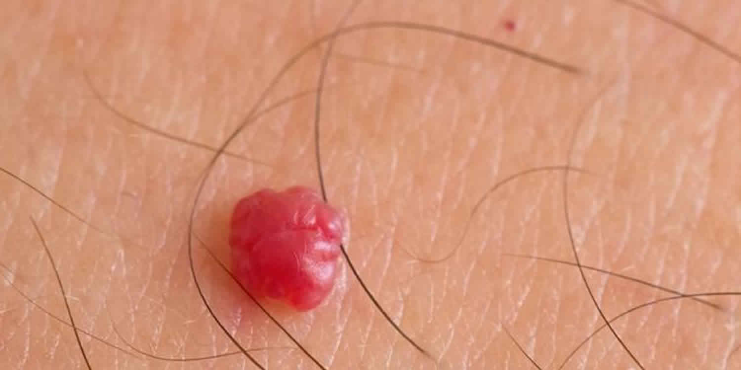
As the name suggests, cherry angiomas appear as tiny cherry red domes measuring 1 to 4 mm in diameter. Early angiomas are flat. They may occur as solitary lesions or number in the hundreds. They may be found on all body sites.
When thrombosed, cherry angiomas can appear black in color until evaluated with a dermatoscope when the red or purple colour is more easily seen. Cherry angioma may develop on any part of the body but most often appear on the scalp, face, lips and trunk.
Figure 1. Cherry angioma
Figure 2. Cherry angioma on breast
Cherry angioma causes
The cause is unknown, but cherry angiomas tend to be inherited (genetic). Cherry angiomas are most common after age 30. A cherry angioma is a harmless overgrowth of blood vessels in the skin due to proliferation of the endothelial cells that line the blood vessels. It does not cause any symptoms but may bleed due to friction or trauma.
Cherry angioma symptoms
A cherry angioma is:
- Bright cherry-red
- Small — pinhead size to about one quarter inch (0.5 centimeter) in diameter
- Smooth, or can stick out from the skin
What is the likely outcome of cherry angiomas?
Cherry angiomas in adults tend to persist unless treated. Some people will continue to accumulate these lesions with age. However, in young children, they frequently spontaneously disappear.
How are cherry angiomas diagnosed?
No investigations are required to diagnose cherry angiomas. However, your dermatologist may examine these with a dermatoscope if angiomas co-exist with moles or other suspicious skin lesions.
When there is uncertainty about the diagnosis, a biopsy may be performed. The angioma is composed of venules in a thickened papillary dermis. Collagen bundles may be prominent between the lobules.
Cherry angioma treatment
Cherry angiomas are benign and will not grow into a skin cancer. They are treated for cosmetic reasons or if the lesions frequently bleed due to friction or trauma.
If cherry angiomas affect your appearance or bleed often, they may be removed by:
- Burning (electrosurgery/cautery)
- Freezing (cryotherapy)
- Laser
- Shave excision
Cherry angioma removal
Vascular lasers such as KTP or pulsed dye laser provide the best treatment outcomes. A KTP laser is a solid-state laser that uses a potassium titanyl phosphate (KTP) crystal as its frequencing doubling device. The KTP crystal is engaged by a beam generated by a neodynium:yitrium aluminium garnet (Nd: YAG) laser. This is directed through the KTP crystal to produce a beam in the green visible spectrum with a wavelength of 532 nm. Intense pulsed light can also be used.
Fine wire diathermy, excision, ablative laser and cryotherapy can be effective but carry a higher risk of scarring.


