What is cholesteatoma
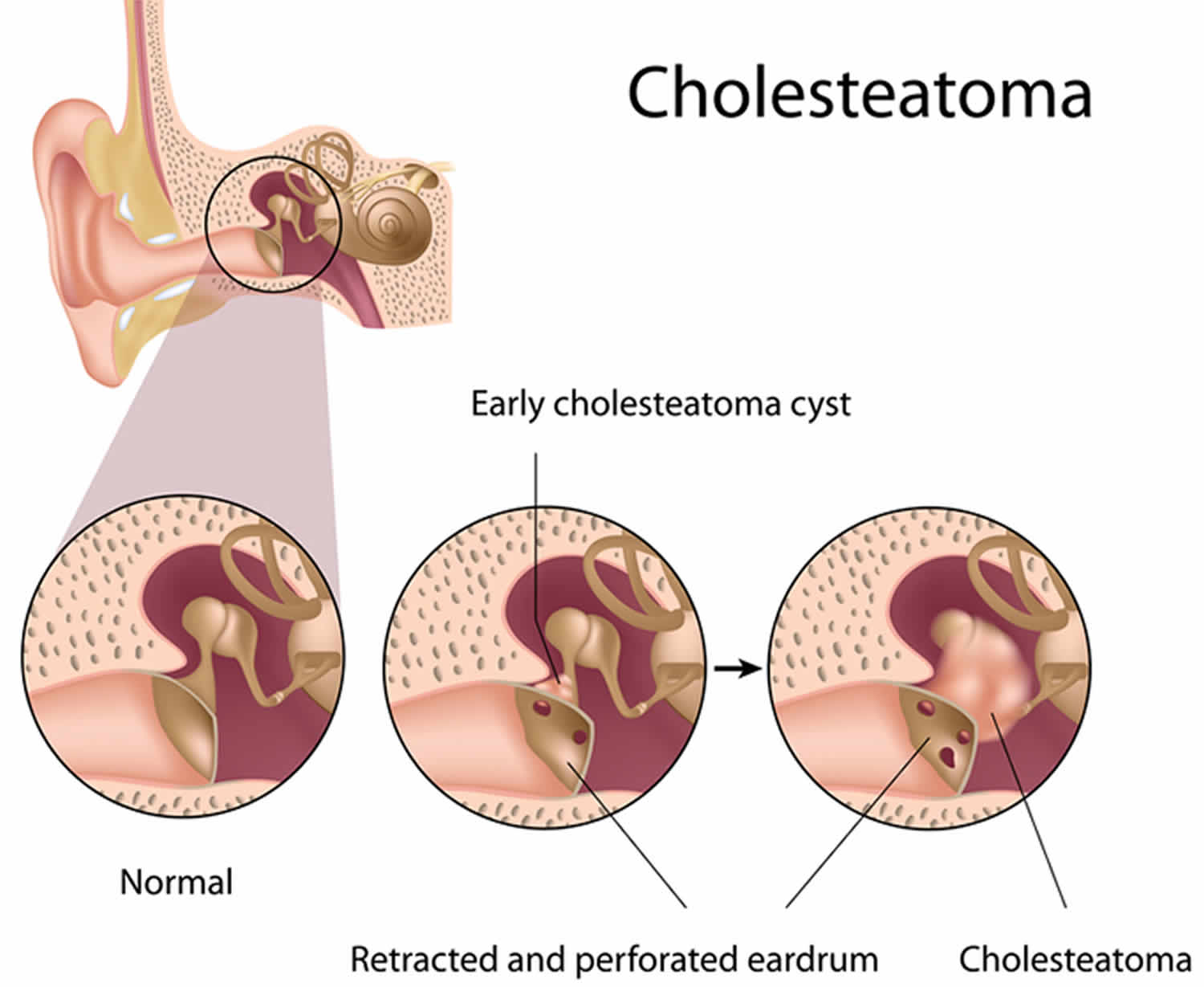
Cholesteatoma is an abnormal skin growth where a sac of dead skin cells forms in a pocket in your middle ear or skin cyst trapped behind the eardrum or the bone behind the ear. Cholesteatomas begin as a build-up of ear wax and skin, which causes either a lump on the eardrum or an eardrum retraction pocket. The cholesteatoma will slowly get larger and eventually fill your middle ear and mastoid bone. Over time, the skin collects and eventually causes problems like infection, drainage, and hearing loss. The skin may take a long time to accumulate and can spread to the area behind the eardrum (the middle ear space) or to the bone behind the ear, called the mastoid bone.
The cholesteatoma can cause an unpleasant-smelling discharge and loss of hearing.
Cholesteatoma can be a birth defect (congenital cholesteatoma). However, cholesteatoma is more commonly occurs as a result of chronic ear infection.
Cholesteatoma is rare but, if left untreated, they can damage the delicate structures inside your ear that are essential for hearing and balance.
A cholesteatoma can also lead to:
- an ear infection – causing discharge from the ear
- hearing loss – this can be permanent
- vertigo – the sensation that you, or the world around you, is spinning
- tinnitus – hearing sounds coming from inside the body, rather than from an outside source
- damage to your facial nerve – this can cause weakness in half your face
In very rare cases, an infection can spread into the inner ear and brain, leading to a brain abscess or meningitis.
Cholesteatoma may cause these symptoms:
- Hearing loss in one ear
- Ear drainage, often with a bad smell
- Recurrent ear infections
- Sensation of ear fullness or pressure
- Dizziness
- Facial muscle weakness on the side of the infected ear
- Ear ache/pain
To remove a cholesteatoma, you usually need to have surgery under general anaesthetic.
After the cholesteatoma has been taken out, your ear may be packed with a dressing. This will need to be removed a few weeks later, and you’ll be told how to look after it in the meantime.
As well as removing the cholesteatoma, the surgeon may be able to improve your hearing. This can be done in a number of ways.
For example, a tiny artificial hearing bone (prosthesis) can be inserted to bridge the gap between your eardrum and the cochlea (hearing organ). In some cases, it may not be possible to reconstruct the hearing or a further operation may be needed.
The benefits of removing a cholesteatoma usually far outweigh the complications. However, as with any type of surgery, there’s a small risk associated with having anaesthetic, and a very small chance of facial nerve damage resulting in weakness of the side of the face.
Discuss the risks with your surgeon before having the operation.
See your doctor if you have problems with your hearing or a watery discharge from your ear.
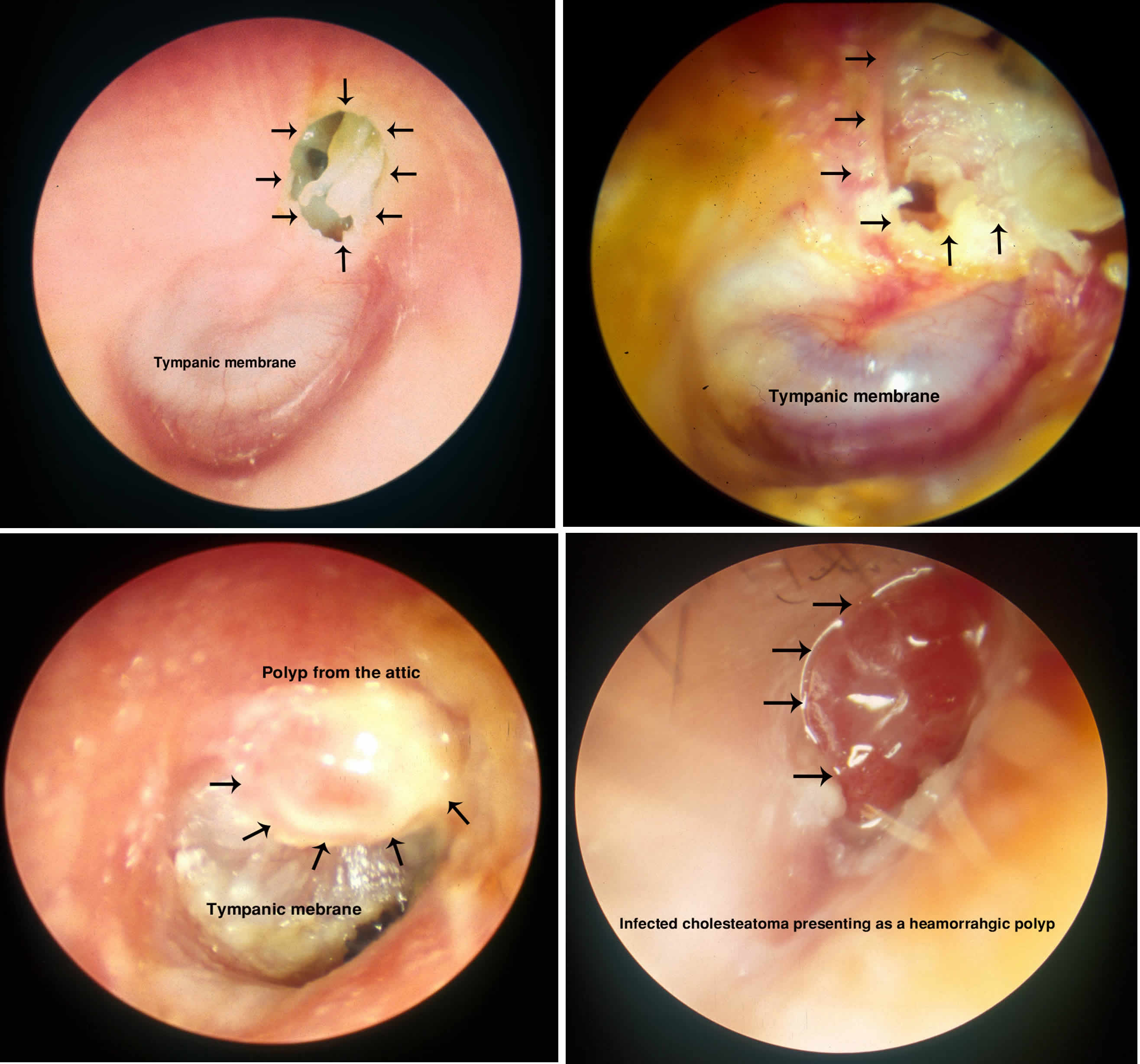
Your doctor may examine your ear with an otoscope – an instrument with a light and magnifying glass.
They may suspect a cholesteatoma from your symptoms, but it can be difficult to confirm because a build-up of pus inside the ear often blocks it from view.
If your doctor thinks your symptoms could just be an ear infection, they may offer you treatment for this first and ask to see you again once you’ve completed it.
If they think you have a cholesteatoma, they should refer you to an ear, nose and throat (ENT) specialist, or otolaryngologist, for further tests.
This may include a CT scan to see whether the cholesteatoma has spread and which parts of your ear are affected.
Figure 1. Ear structure
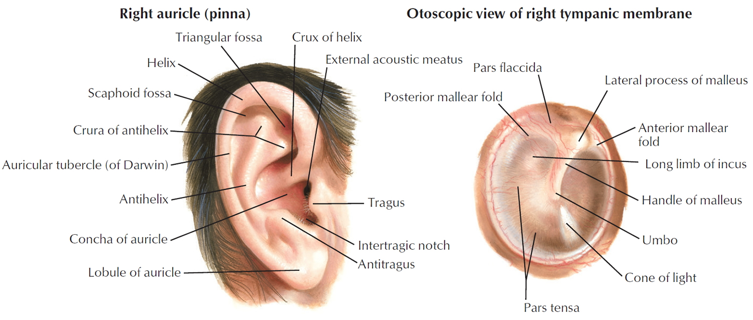
Figure 2. Ear anatomy
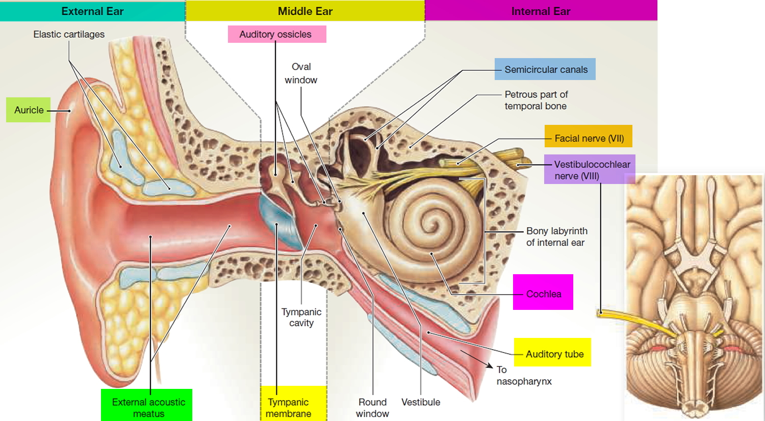
Figure 3. Middle ear and auditory ossicles
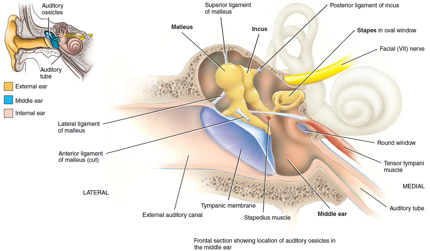
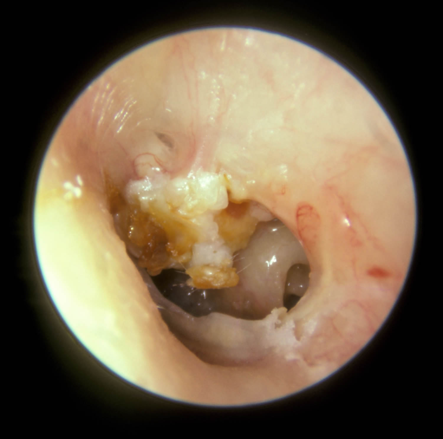
Figure 4. Cholesteatoma
What causes cholesteatoma?
There are different reasons why a cholesteatoma may develop. Some people are born with a cholesteatoma, called congenital cholesteatoma. The most common cause is poor ventilation of the middle ear space, which is called “eustachian tube dysfunction.” The eustachian tube (auditory tube) is the natural tube that connects your middle ear space to your nose and sinuses, and helps regulate the pressure behind your eardrum. If the eustachian tube is not working properly, the middle ear space does not get ventilated. This creates negative pressure and ultimately causes the weakened eardrum to retract. This retraction collects skin and earwax, which leads to a cholesteatoma. Seasonal allergies, upper respiratory infections (cough/cold), or sinusitis may contribute to eustachian tube dysfunction.
A cholesteatoma can develop when skin of the ear canal passes through a hole in the eardrum and into the middle ear space. Finally, another rare type of cholesteatoma is present at birth (congenital) and is related to how the ear develops.
Cholesteatoma prevention
Prompt and thorough treatment of chronic ear infection may help prevent cholesteatoma.
Cholesteatoma symptoms
A cholesteatoma usually only affects one ear. In the early cholesteatoma stages, symptoms are minimal and often ignored. Patients usually present when the cholesteatoma gets infected. Typically the symptoms are, ear pain and ear discharge which is often scanty and foul smelling. The discharge may be blood stained. Patients may present with a complication such as mastoiditis, abscess, facial palsy and meningitis.
The two most common symptoms of cholesteatoma are:
- a persistent or recurring watery, often smelly, discharge from the ear, which can come and go or may be continuous
- a gradual loss of hearing in the affected ear
Some people may experience slight discomfort in their ear.
Cholesteatoma may also cause these symptoms:
- Hearing loss in one ear
- Ear drainage, often with a bad smell
- Recurrent ear infections
- Sensation of ear fullness or pressure
- Dizziness
- Facial muscle weakness on the side of the infected ear
- Ear ache/pain
If you experience any of these symptoms, you should see an ENT (ear, nose, and throat) specialist, or otolaryngologist, as soon as possible.
Cholesteatoma complications
Without proper treatment cholesteatoma will cause recurrent ear infections. Chronic infection of the ear can lead to progressive hearing loss and even deafness. Cholesteatoma can erode bone, including the three bones of hearing, which may cause infection to spread to the inner ear or brain. These infections can lead to meningitis, brain abscess, facial paralysis, dizziness (vertigo), and even death.
Complications may include:
- Brain abscess (rare)
- Erosion into the facial nerve (causing facial paralysis)
- Meningitis
- Spread of the cyst into the brain
Cholesteatoma treatment
Cholesteatoma can be managed in a variety of ways, but definitive removal of the skin or cyst typically requires surgical intervention. Before surgery, your ENT specialist may need to carefully clean your ear and prescribe medications to help stop the drainage. These medications (oral antibiotics) may be taken by mouth, applied directly to the ear (topical antibiotics), or both. It is advised that you keep the ear dry while treating these infections.
The specific type of surgery depends on what part of the ear is involved with the cholesteatoma. Sometimes the extent of cholesteatoma is clearly seen on the office exam. Other times imaging, often a CT scan, helps to define where the cholesteatoma is located. CT scans are a collection of X-rays that provide good detail on the bony anatomy of the ear. A hearing test, or audiogram, should be obtained. Other tests like an MRI or balance testing are less commonly required.
Cholesteatoma surgery
The primary goal of cholesteatoma surgery is to remove the skin, clear the infection, and create a dry, safe ear. It may be possible to improve your hearing at the same time. This may involve reconstructing the eardrum, removing bone behind the ear, or reconstructing the hearing bones. In some cases, a second surgery may be required to make sure all the cholesteatoma has been removed before the hearing bones can be rebuilt.
Cholesteatoma surgery is performed under a general anaesthetic and usually takes between 2 to 3 hours.
Your surgeon will make a cut in front of or behind your ear. They will remove bone from around the cholesteatoma to see where it has spread to, and remove it.
Your surgeon may need to remove the bone of your ear canal. If this happens, they will shape the bone behind your ear (mastoid bone) into a cavity that opens into your ear.
A second surgery will typically be performed six to 12 months after your first surgery, if necessary. Your hearing might temporarily worsen after the first surgery if the reconstruction of your hearing bones is delayed. There are many factors that contribute to how well you hear after surgery, and these should be discussed with your ENT specialist.
Surgery is generally performed in an outpatient setting, but some patients may require an overnight stay. In rare cases of serious infection, a prolonged hospitalization for antibiotic treatment may be required. Interventions for facial nerve weakness or to control dizziness are rarely needed.
Cholesteatoma surgery complications and risks
General complications of surgery:
- pain
- bleeding
- infection of the surgical site (wound)
- unsightly scarring
- blood clots
Specific complications of cholesteatoma surgery:
- hearing loss
- numbness of your ear
- damage to the facial nerve
- change of taste
- dizziness
- tinnitus
- ear discharge
- allergic reaction
Cholesteatoma surgery recovery
You may need to stay in hospital overnight after the operation.
You should be able to return to work after about 3 weeks. Time off from work is typically one to two weeks.
After surgery, follow-up office visits will be needed to clean your ear, recheck your hearing, and evaluate the results. Cholesteatoma requires long-term surveillance to check for recurrence.
If your surgeon needed to shape your mastoid bone into a cavity, you will probably need to come back to the clinic several times in the first few months until the cavity has healed completely.
When you get home, you’ll need to keep the affected ear dry. You should be able to wash your hair after a week, provided you don’t get water inside the ear. To avoid this, you can plug the ear with Vaseline-coated cotton wool. DO NOT swim until your surgeon has told you that your ear has healed.
You may be advised to avoid flying, swimming and doing strenuous activities or sports for a few weeks after surgery. At your follow-up appointment, ask when it will be safe to return to your usual activities.
Regular exercise should help you to return to normal activities as soon as possible. Before you start exercising, ask your ENT doctor for advice.
If the bone of your ear canal was not removed, some cholesteatoma may be left behind.
Contact your ENT specialist or your hospital ENT department if you have:
- discharge or significant bleeding from your ear or wound
- a high temperature (fever)
- severe or increasing pain
These symptoms could be a sign of a complication, such as an infection.
Follow-up appointments
If your stitches aren’t dissolvable, they may need to be removed by your practice nurse after a week or two.
Most people have a follow-up appointment in a clinic within a few weeks of the operation, when any dressings in your ear will be removed.
A cholesteatoma can come back, and you could get one in your other ear, so you’ll need to attend regular follow-up appointments to monitor this.
Sometimes a second operation is needed after about a year to check for any skin cells left behind. However, MRI scans are now often used instead of surgery to check for this.


