What is clubfoot
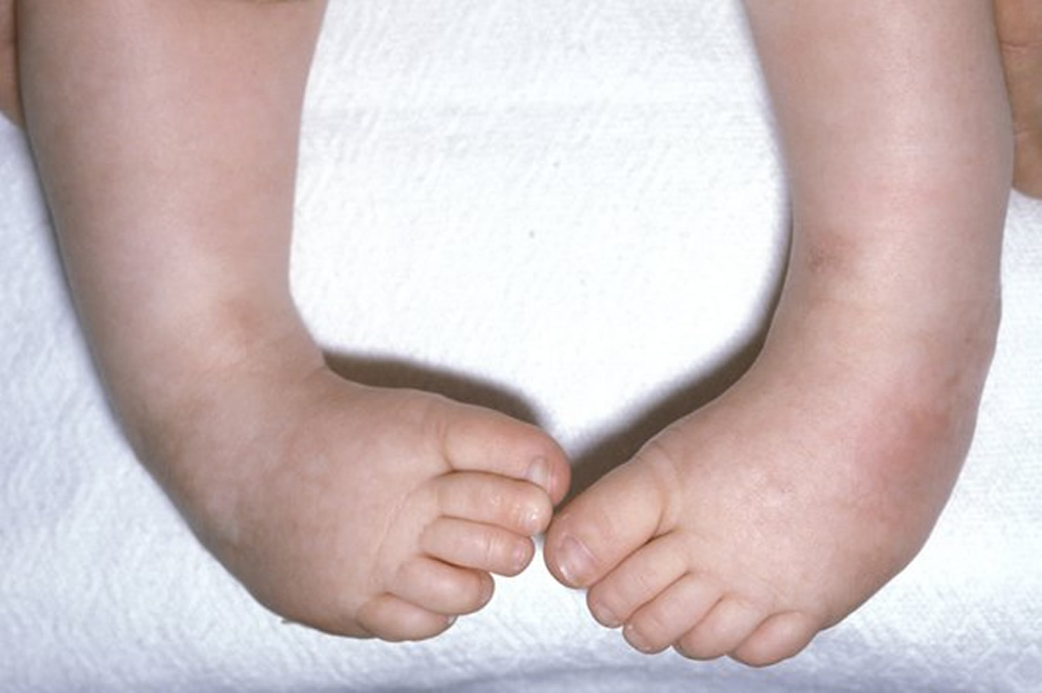
Club foot also known to doctors as congenital talipes equinovarus, is a common birth defect (congenital clubfoot) that can affect one or both feet. The child is born with a foot pointing the wrong way – turned down and in – that cannot be placed flat on the ground in the position needed for walking (Figure 1). Clubfoot is not painful during infancy. However, if your child’s clubfoot is not treated, the foot will remain deformed, and he or she will not be able to walk normally.
In clubfoot, the tendons that connect the leg muscles to the foot bones are short and tight, causing the foot to twist inward.
Around 1-2 babies per 1,000 are born with the clubfoot, making it one of the more common congenital (present at birth) foot deformities 1. Although clubfoot is diagnosed at birth, many cases are first detected during a prenatal ultrasound. In about half of the children with clubfoot, both feet are affected. Boys are twice more likely than girls to have the deformity.
Clubfoot can range from mild to severe, but typically has the same general appearance. The foot is turned inward and there is often a deep crease on the bottom of the foot.
Clubfoot can often cause wasting of the calf muscle. In limbs affected by clubfoot, the foot and leg are slightly shorter than normal, and the calf is thinner due to underdeveloped muscles. These differences are more obvious in children with clubfoot on only one side.
Early treatment usually helps correct congenital clubfoot. With proper treatment the majority of children are able to enjoy a wide range of physical activities with little trace of the deformity.
Most cases of clubfoot are successfully treated with nonsurgical methods that may include a combination of stretching, multiple plaster casts and bracing. Treatment usually begins shortly after birth.
Figure 1. Clubfoot baby

If your baby has club foot, one or both feet points down and inwards with the sole of the foot facing backwards.
Club foot isn’t painful for babies, but if it isn’t treated, it can become painful and make it difficult to walk as they get older.
Types of clubfoot
Clubfoot is often broadly classified into two major groups:
- Isolated (idiopathic) clubfoot is the most common form of the deformity and occurs in children who have no other medical problems.
- Nonisolated clubfoot occurs in combination with various health conditions or neuromuscular disorders, such as arthrogryposis and spina bifida. If your child’s clubfoot is associated with a neuromuscular condition, the clubfoot may be more resistant to treatment, require a longer course of nonsurgical treatment, or even multiple surgeries.
Regardless of the type or severity, clubfoot will not improve without treatment. A child with an untreated clubfoot will walk on the outer edge of the foot instead of the sole, develop painful calluses, be unable to wear shoes, and have lifelong painful feet that often severely limit activity.
Parents of infants born with clubfeet and no other significant medical problems should be reassured that with proper treatment their child will have feet that permit a normal, active life.
Clubfoot outcomes (prognosis)
Your baby’s clubfoot will not get better on its own. With treatment, your child should have a nearly normal foot, and he or she can run and play and wear normal shoes.
The affected foot is usually 1 to 1 1/2 sizes smaller and somewhat less mobile than the normal foot. The calf muscles in your child’s clubfoot leg will also stay smaller, so your child may complain of “sore legs” or getting tired sooner than peers. The affected leg may also be slightly shorter than the unaffected leg, but this is rarely a significant problem.
Clubfoot complications
Clubfoot typically doesn’t cause any problems until your child starts to stand and walk. If the clubfoot is treated, your child will most likely walk fairly normally.
He or she may have some difficulty with:
- Movement. Mobility may be slightly limited.
- Shoe size. The affected foot may be up to 1 1/2 shoe sizes smaller than the unaffected foot.
- Calf size. The muscles of the calf on the affected side may always be smaller than the other side.
However, if not treated, clubfoot causes more-serious problems. These can include:
- Arthritis. Your child is likely to develop arthritis.
- Poor self-image. The unusual appearance of the foot may make your child’s body image a concern during the teen years.
- Inability to walk normally. The twist of the ankle may not allow your child to walk on the sole of the foot. To compensate, he or she may walk on the ball of the foot, the outside of the foot or even the top of the foot in severe cases.
- Problems stemming from walking adjustments. Walking adjustments may prevent natural growth of the calf muscles, cause large sores or calluses on the foot, and result in an awkward gait.
Clubfoot causes
What causes clubfoot
Researchers are still uncertain about the cause of most cases of clubfoot. The most widely accepted theory is that clubfoot is caused by a combination of genetic and environmental factors. What is known, however, is that there is an increased risk in families with a history of clubfeet.
- If you have one child with club foot, your chance of having a second child with the condition is about 1 in 35.
- If one parent has club foot, there’s about a 1 in 30 chance of your baby having it.
- If both parents have the condition, this increases to about a 1 in 3 chance.
- In rare cases, clubfoot is linked to more serious conditions, such as spina bifida.
In 20016 researchers from the University of Aberdeen were able to show for the first time that variation in a gene that processes folate in the body may be part of the cause of clubfoot. They found that babies with the less common variant of the C677T variant in the gene methyltetrahydrofolate reductase (MTHFR) are less likely to develop clubfoot.The scientists say more work needs to be done to determine the level of this effect, the effects of other genes, and to establish the best advice on folic acid supplementation for pregnant women who have a child or relative with clubfoot. The researchers emphasize that women should continue to take recommended levels of folic acid while planning a pregnancy or during the first three months of pregnancy.
If you have an methyltetrahydrofolate reductase (MTHFR) variant, taking 400 mcg of folic acid every day before and during early pregnancy can help prevent neural tube defects in your baby 2.
Methyltetrahydrofolate reductase (MTHFR) is an enzyme (protein) that helps your body break down and use folate. One MTHFR variant (called MTHFRTT or CT genotype) is a change in your body’s MTHFR gene that makes you use folate more slowly than usual. Genes are parts of your body’s cells that store instructions for how your body grows and works. MTHFR variants are inherited (passed from parents to children) through genes. If you know you have an MTHFR variant or you think it runs in your family, talk to your healthcare provider.
Your doctor may want to test you for an MTHFR variant if you have high levels of a substance in your blood called homocysteine. Too much homocysteine in your blood can cause heart conditions, blood clots and stroke. You can find out your homocysteine levels with a blood test. If your level is high, you can have a genetic test (test that checks your genes) to see if you have an MTHFR variant.
You may have heard not to take folic acid if you have an MTHFR variant because it can increase your risk of pregnancy complications and your baby having health problems. The Centers for Disease Control and Prevention 2 recommends that all women take 400 mcg of folic acid every day before and during early pregnancy. If you have an MTHFR variant, talk to your provider.
Risk factors for clubfoot
Risk factors include:
- Family history. If either of the parents or their other children have had clubfoot, the baby is more likely to have it as well.
- Congenital conditions. In some cases, clubfoot can be associated with other abnormalities of the skeleton that are present at birth (congenital), such as spina bifida, a serious birth defect that occurs when the tissue surrounding the developing spinal cord of a fetus doesn’t close properly.
- Environment. If a woman with a family history of clubfoot smokes during pregnancy, her baby’s risk of the condition may be double that of nonsmokers. Also, getting an infection or using recreational drugs during pregnancy can increase the risk of clubfoot.
- Not enough amniotic fluid during pregnancy. Too little of the fluid that surrounds the baby in the womb may increase the risk of clubfoot.
Clubfoot prevention
Because doctors don’t know what causes clubfoot, you can’t completely prevent it. However, if you’re pregnant, you can do things to limit your baby’s risk of birth defects, such as:
- Not smoking or spending time in smoky environments
- Not drinking alcohol
- Avoiding drugs not approved by your doctor
- Make sure you take enough folic acid before and during early pregnancy can help prevent neural tube defects like spina bifida in your baby. Before pregnancy, take a multivitamin that has 400 micrograms (400 mcg) of folic acid in it every day. Most multivitamins have this amount, but do check the label to be sure. During pregnancy, take a prenatal vitamin that has 600 mcg of folic acid in it every day.
You need more folic acid during pregnancy to help your baby grow and develop. Your health care provider can prescribe a prenatal vitamin for you. Or you can get prenatal vitamins over the counter without a prescription.
You also can get folic acid from food. Some foods have folic acid added to them. Look for the word “fortified” or “enriched” on the package label on foods like:
- Bread
- Breakfast cereal
- Cornmeal
- Flour
- Pasta
- Products made from a kind of flour called corn masa, like tortillas, tortilla chips, taco shells, tamales and pupusas
- White rice
Some fruits and vegetables are good sources of folic acid. When folic acid is naturally in a food, it’s called folate. Foods that are good sources of folate are:
- Beans, like lentils, pinto beans and black beans
- Leafy green vegetables, like spinach and Romaine lettuce
- Asparagus
- Broccoli
- Peanuts (But don’t eat them if you have a peanut allergy.)
- Citrus fruits, like oranges and grapefruit
- Orange juice (100 percent juice is best. This means one serving of juice is equal to one serving of fruit.)
It’s hard to get all the folic acid you need from food. Even if you eat foods that have folic acid in them, take your multivitamin each day, too.
Do some women need extra folic acid?
Yes. Most women don’t need more than 1,000 mcg of folic acid each day, but some may need more. Talk to your provider to make sure you take the right amount. You may need extra folic acid before and during pregnancy if:
- You’ve had a pregnancy affected by an neural tube defect in the past.
- You have diabetes. This is a medical condition in which your body has too much sugar (called glucose) in your blood.
- You’re obese. This means you have an excess amount of body fat and your body mass index (also called BMI) is 30 or higher.
- You have a hemoglobin disorder, like sickle cell disease. Hemoglobin disorders are rare blood conditions that are caused by problems with hemoglobin. Hemoglobin is a protein in the blood that carries oxygen.
- You take antiseizure medicine.
Club foot symptoms
If your child has clubfoot, here’s what it might look like
- The top of the foot is usually twisted downward and inward, increasing the arch and turning the heel inward.
- The foot may be turned so severely that it actually looks as if it’s upside down.
- The calf muscles in the affected leg are usually underdeveloped.
- The affected foot may be up to 1/2 inch (about 1 centimeter) shorter than the other foot.
Despite its look, however, clubfoot itself doesn’t cause any discomfort or pain.
Club foot diagnosis
Club foot is usually diagnosed after a baby is born, although it may be spotted in pregnancy during the routine ultrasound scan carried out between 18 and 21 weeks.
Club foot can’t be treated before birth, but picking up the problem during pregnancy means you can talk to doctors and find out what to expect after your baby is born.
Some babies are born with normal feet that are in an abnormal position because they have been squashed in the womb.
The feet usually correct themselves by 3 months, but some babies may need a few sessions of physiotherapy.
Clubfoot treatment
The goal of treatment is to obtain a functional, pain-free foot that enables standing and walking with the sole of the foot flat on the ground.
Nonsurgical Treatment
The initial treatment of clubfoot is nonsurgical, regardless of how severe the clubfoot deformity is.
Treatment for club foot usually starts within a week or two of your baby being born.
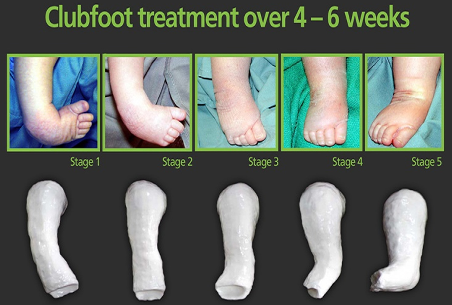
The Ponseti method – Stretching and casting
A technique known as the Ponseti method is the most widely used technique in North America and throughout the world, which uses gentle stretching and casting to gradually correct the deformity.
Treatment should ideally begin shortly after birth, but older babies have also been treated successfully with the Ponseti method. Elements of the method include:
- Manipulation and casting. Your baby’s foot is gently stretched and manipulated into a corrected position and held in place with a long-leg cast (toes to thigh) (see Figure 2). Each week this process of stretching, re-positioning, and casting is repeated until the foot is largely improved. For most infants, this improvement takes about 5 to 8 weeks.
Figure 2. Ponseti method
- Achilles tenotomy. After the last cast comes off, most babies need a minor operation to loosen the tendon at the back of their ankle (Achilles tendon). It helps to release their foot into a more natural position. This is done using a local anesthetic. During this quick procedure (called a tenotomy), your doctor will use a very thin instrument to cut the tendon. The cut is very small and does not require stitches. A new cast will be applied to the leg to protect the tendon as it heals. This usually takes about 3 weeks. By the time the cast is removed, the Achilles tendon has regrown to a proper, longer length, and the clubfoot has been fully corrected.
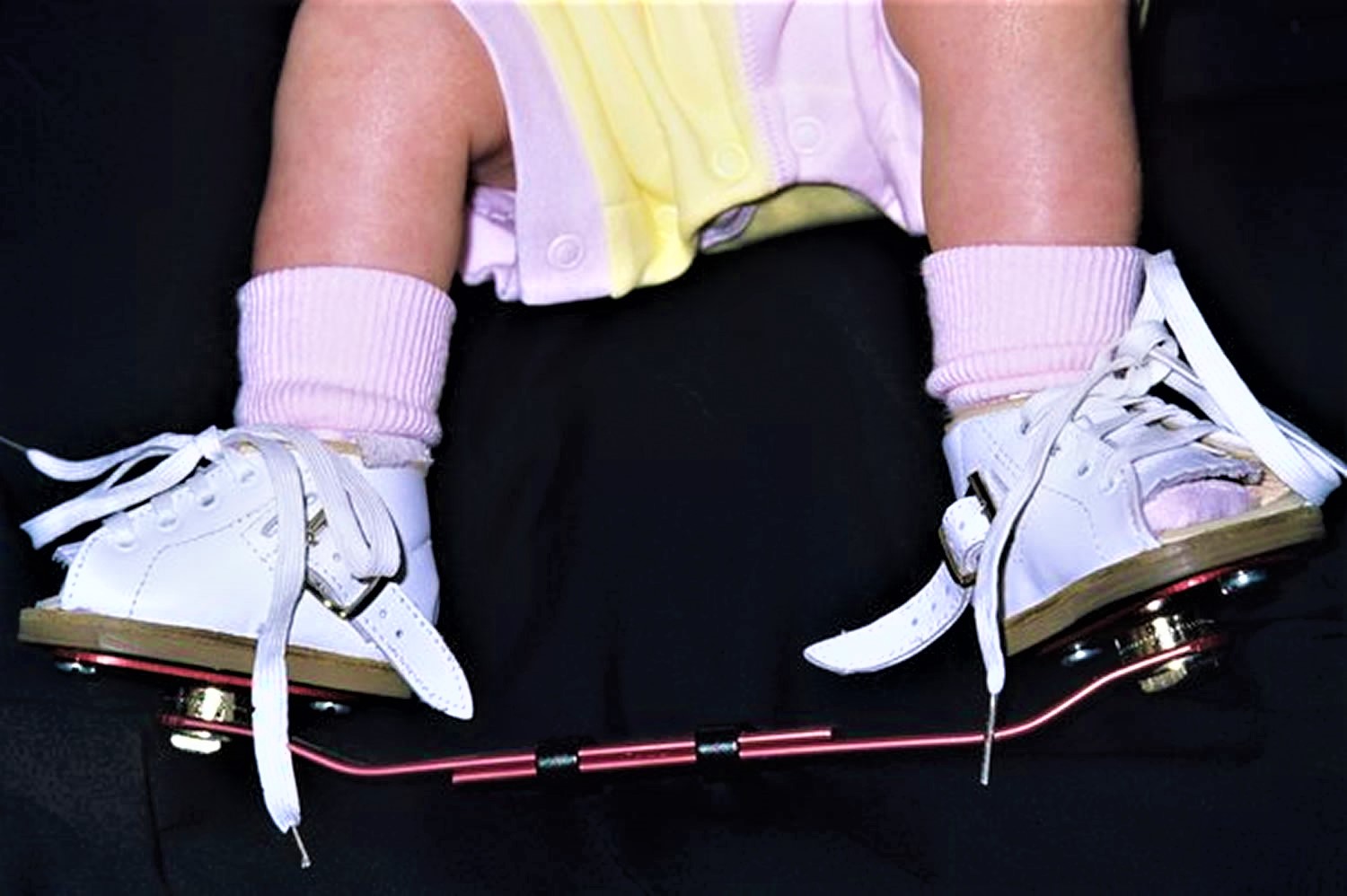
Club foot brace
- Even after successful correction with casting, clubfeet have a natural tendency to recur. To ensure that the foot will permanently stay in the correct position, your baby will need to wear a brace (commonly called “boots and bar”) for a few years (see Figure 3). The brace keeps the foot at the proper angle to maintain the correction. This bracing program can be demanding for parents and families, but is essential to prevent relapses.For the first 3 months, your baby will wear the brace essentially full-time (23 hours a day). Your doctor will gradually decrease the time in the brace to just overnight and nap time (about 12 to 14 hours per day). Most children will follow this overnight bracing regimen until they’re 4 or 5 years old.There are several different types of braces — all of which consist of shoes, sandals, or custom-made footwear attached to the ends of a bar. The bar can be solid (both legs move together) or dynamic (each leg moves independently). Your doctor will talk with you about the type of brace that would best meet your baby’s needs.
Babies might be fussy during the first few days of wearing a brace and will need time to adjust.
Helpful Tips for Clubfoot Bracewear
- Play with Your Child in the Brace
This is the key to getting over the irritability quickly. If your child is using the solid bar, he or she can kick and swing the legs simultaneously with the brace on. You can help facilitate this by gently bending and straightening the knees by pushing and pulling on the bar of the brace. If your child is using the dynamic bar, it is also helpful to gently move the legs up and down as your child adjusts to the brace.
- Make It a Routine
Children do better if you develop a fixed routine for the bracewear. During the years of night and naptime wear, put the brace on anytime your child goes to the “sleeping spot.” Your child will soon figure out that when it is sleep time, it is time to wear the brace. Your child is less likely to fuss if this is a consistent routine.
- Pad the Bar
A bicycle handle bar pad works well for this. By padding the bar, you will protect your child, yourself and your furniture from the metal bar.
- Never Use Lotion on the Skin
Lotion will make the problem worse. Some redness is normal with use. Bright red spots or blisters, especially on the back of the heel, usually indicate that the heel is slipping. Ensure that the heel stays down in the shoe by securing the straps and/or buckles. It is important to check your child’s feet several times a day after starting bracing to make sure no blisters are developing.
- Prevent Escapes
If your child continues to escape from the brace, try the tips below. After each step, check to see if the heel is down. If not, proceed to the next step.
- In boots or sandals with a single strap, tighten it by one more hole, using your thumb to hold the foot and tongue in place. In boots with multiple straps, tighten the middle one first, using your thumb to hold the foot and tongue in place.
- Try double socks. In boots with a removable insert, place one sock directly over the foot, and a second sock over the insert to help take up excess room.
- Remove the tongue of the shoe — this will not harm your child.
- Try lacing the shoes from top to bottom, so that the bow is by the toes.
- Use 40-inch round shoelaces.
- Try thinner or thicker cotton socks, or the ones with non-slip soles.
Figure 3. Clubfoot brace
The Ponseti method has proven extremely effective for many children. It does, however, require the family to be highly committed to applying the braces properly every day. If the brace is not worn as prescribed, the clubfoot will recur.
A small percentage of children develop relapses despite proper bracing. If the child’s foot slips out of the boot on a regular basis, it may be the first sign of a mild recurrence of the deformity. If addressed promptly, this can usually be corrected with a few serial casts and possibly a minor surgery.
In addition, applying the Ponseti method correctly requires training, experience, and practice. Be sure to ask your pediatrician for a referral to an orthopedic surgeon with expertise in the nonsurgical correction of clubfoot.
French method
Another nonsurgical method to correct clubfoot incorporates stretching, mobilization, and taping. The French method — also called the functional or physical therapy method — is typically directed by a physical therapist who has specialized training and experience.
Like the Ponseti method, the French method is begun soon after birth and requires family involvement. Each day, the baby’s foot must be stretched and manipulated, then taped to maintain the range of motion gained by the manipulation. After taping, a plastic splint is put on over the tape to maintain the improved range of motion.
This method requires approximately three visits to the physical therapist each week. Because this is a daily regimen, the therapist will teach the parents how to do it correctly at home.
After 3 months, most babies have significant improvement in foot position, and visits to the physical therapist are required less often. Like children treated with the Ponseti method, babies treated with the French method commonly require an Achilles tenotomy to improve dorsiflexion of the ankle.
To prevent recurrence of the clubfoot, the daily regimen of stretching, taping, and splinting must be continued by the family until the child is 2 to 3 years old.
Club foot prognosis (outlook)
Nearly all children treated with the Ponseti method will have pain-free, normal-looking feet.
Most learn to walk at the usual age and can enjoy physical activities, including sports, when they’re older.
Children who only have one affected foot may be left with a slightly shorter leg and smaller foot on one side.
This may mean your child is slightly less mobile and gets tired more quickly than other children.
Before the Ponseti method, club foot was often treated with surgery to alter the position of the foot. This wasn’t always effective, and led to long-term pain and stiffness for some adults.
Clubfoot relapses
Sometimes club foot can come back, especially if treatment isn’t followed exactly.
If it comes back, some of the treatment stages may need to be repeated.
Clubfoot surgery
Although many cases of clubfoot are successfully corrected with nonsurgical methods, sometimes the deformity cannot be fully corrected or it returns, often because parents have difficulty following the treatment program. In addition, some infants have very severe deformities that do not respond to stretching. When this happens, surgery may be needed to adjust the tendons, ligaments, and joints in the foot and ankle.
Because surgery typically results in a stiffer foot, particularly as a child grows, every effort is made to correct the deformity as much as possible through nonsurgical methods. Even an infant with severe deformities or clubfeet associated with neuromuscular conditions can improve without surgery. If a child’s foot has been partially corrected with stretching and casting, then the surgery required to fully correct the clubfoot will be less extensive.
- Less extensive surgery will target only those tendons and joints that are contributing to the deformity. In many cases, this involves releasing the Achilles tendon at the back of the ankle or moving the tendon that travels from the front of the ankle to the inside of the midfoot (this procedure is called an anterior tibial tendon transfer).
- Major reconstructive surgery for clubfoot involves extensive release of multiple soft tissue structures of the foot. Once the correction is achieved, the joints of the foot are usually stabilized with pins and a long-leg cast while the soft tissue heals.After 4 to 6 weeks, the doctor will remove the pins and cast, and typically apply a short-leg cast, which is worn for an additional 4 weeks. After the last cast is removed, it is still possible for the muscles in your child’s foot to try to return to the clubfoot position, so special shoes or braces will likely be used for up to a year or more after surgery.The most common complications of extensive soft tissue release are overcorrection of the deformity, stiffness, and pain.


