What is a dilated pore of Winer
A dilated pore of Winer is a commonly occurring benign adnexal tumor of follicular differentiation 1. Dilated pore of Winer lesion falls within the group of benign follicular tumors. Dilated pore of winer occurs as a small solitary papule centered by a follicular pore on the face, neck or back, mimicking a giant comedo 2. Dilated pore of Winer was first described by Louis H. Winer in 1954 3. Although most commonly located on the head and neck, a dilated pore of Winer can also be found on the trunk of middle-aged and elderly individuals. These clinically present as an asymptomatic, solitary, enlarged pore with a keratin plug and normal surrounding skin. Prognosis is excellent for these lesions as they are benign and do not require any further testing or work-up. Histopathologic evaluation can confirm the diagnosis in uncertain cases. Removal can be performed via excision for cosmetic purposes.
Although it occurs in both sexes, a dilated pore of Winer occurs more frequently in males compared to females and is also more frequent in whites. Most cases occur at the age of 40 and older; however, there are reports of dilated pores occurring as early as 20 years of age.
Dilated pore of winer causes
Some scientists have considered the dilated pore of Winer to simply be an epidermal inclusion cyst with reactive hyperplasia of its epithelial lining and others proposed it to be a variant of nevus comedonicus 4. However, this lesion has been shown to be a distinct entity as an adnexal neoplasm of the follicular infundibulum 5. The exact cause and pathophysiology of a dilated pore of Winer are unknown 1. Winer, in his original article, noted an association with a history of inflammatory cystic acne and other cystic processes 3. Actinic damage has also been attributed to the development of these lesions.
Dilated pore of Winer histopathology
A dilated pore of Winer is characterized histopathologically by a markedly dilated follicular infundibulum extending deeply into the dermis. The cavity is filled with lamellar keratin material. It is lined by epithelium that is atrophic near the ostium and acanthotic at the deeper portion of the invagination. Radiating off of the epithelium are regularly spaced, small, finger-like epithelial projections pushing into the surrounding dermis. These finger-like projections do not contain keratin cysts, ducts, or hair shafts 4.
Dilated pore of Winer signs and symptoms
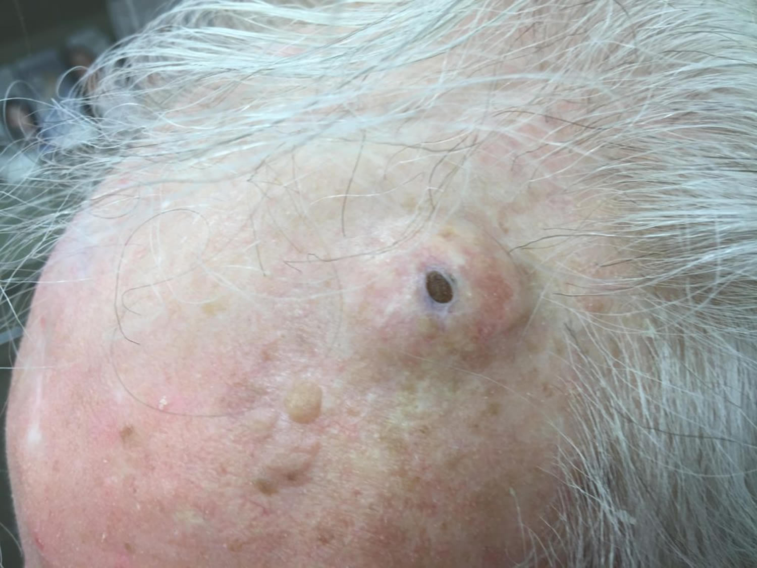
A dilated pore of Winer will present as a single, enlarged pore. The pore may be occluded by a keratin plug with softer, white, keratin material beneath. They are asymptomatic, and the surrounding skin appears normal however a background of actinic damage may be noted. Inflammation or infection along with pain and swelling can occur with manipulation. Although they have a predilection for the head and neck, particularly the face, they can also present on the trunk, most commonly the back. Patients are usually middle-aged or older and may report a previous history of severe acne.
Dilated pore of Winer diagnosis
A dilated pore of Winer is usually a clinical diagnosis. Histopathologic examination is not required but can be performed if the diagnosis is uncertain or in cases where the lesion is excised for cosmetic purposes. No other testing or work-up is needed.
Dilated pore of Winer treatment
No treatment is required for a dilated pore of Winer 1. Removal can be performed for cosmetic concern. Excision in an elliptical fashion or by punch biopsy is usually curative 6. Merely removing the keratin material can be done using a comedone extractor. However, keratin will gradually re-accumulate within the lesion. Destructive techniques such as electrodesiccation, electrocautery, laser surgery, dermabrasion, and cryotherapy are less effective due to the deeply situated base of the invagination 3. There are no effective medical treatments for these lesions.
Dilated pore of Winer complications
Complications of surgical removal include scarring, infection, and bleeding. completely after surgery can be minimized by using proper aseptic or clean technique. Infection prior to or following surgery can be treated using topical or oral antibiotics depending on severity.
Dilated pore of Winer prognosis
Complete excision of the lesion is curative. Incomplete excision can result in recurrence from the remaining infundibular lining. To date, there have been no reports of death associated with a dilated pore of Winer. Also, there have been no reports of syndromes associated with this entity. There have been rare case reports of other malignant cutaneous neoplasms such as basal cell carcinoma and squamous cell carcinomas arising in dilated pores of Winer. However, these are most likely coincidental 7. There has been one case report of a trichoblastoma arising in a dilated pore of Winer 8. Inflammation or infection of the surrounding skin can result from manipulation of the lesion by the patient in an attempt to remove the keratin plug.
References- Benedetto CJ, Athalye L. Dilated Pore Of Winer (Black Head) [Updated 2019 Jun 4]. In: StatPearls [Internet]. Treasure Island (FL): StatPearls Publishing; 2019 Jan-. Available from: https://www.ncbi.nlm.nih.gov/books/NBK532967
- Tellechea O, Cardoso JC, Reis JP, et al. Benign follicular tumors. An Bras Dermatol. 2015;90(6):780–798. doi:10.1590/abd1806-4841.20154114 https://www.ncbi.nlm.nih.gov/pmc/articles/PMC4689065
- WINER LH. The dilated pore, a tricho-epithelioma. J. Invest. Dermatol. 1954 Sep;23(3):181-8.
- Tellechea O, Cardoso JC, Reis JP, Ramos L, Gameiro AR, Coutinho I, Baptista AP. Benign follicular tumors. An Bras Dermatol. 2015 Nov-Dec;90(6):780-96; quiz 797-8.
- Morikawa T, Takizawa H, Ohnishi T, Watanabe S. Dilated pore: a case report and an immunohistochemical study of cytokeratin expression. J. Dermatol. 2003 Jul;30(7):556-8.
- Jakobiec FA, Bhat P, Sutula F. Winer’s dilated pore of the eyelid. Ophthalmic Plast Reconstr Surg. 2009 Sep-Oct;25(5):411-3
- Zhao L, Xu J, Fang F, Qian G, Wang Y, Wang QQ. Squamous cell carcinoma found in a dilated pore. J Eur Acad Dermatol Venereol. 2007 Feb;21(2):277-8.
- Misago N, Sada A, Narisawa Y. Trichoblastoma with a dilated pore. J. Am. Acad. Dermatol. 2006 Feb;54(2):357-8.


