What is a papilloma on the eyelid
An eyelid papilloma is any lesion on the eyelid that is papillomatous, that is, of smooth, rounded, or pedunculated elevation. The lesion that most commonly fits this description is a benign squamous papilloma. Squamous papilloma is the most common benign (non-cancerous) lesion of the eyelid. Squamous papilloma frequency increases steadily with age, but they may occur at any age and are seen most frequently in patients older than 30 years. However, it is possible that other benign eyelid lesions may take on the same appearance, as well as malignant skin lesions, especially squamous carcinoma (SCC). Malignant eyelid skin conditions are covered in other articles; this article outlines benign skin lesions.
Squamous cell papilloma may be defined as a small benign growth that begins in squamous cells (thin, flat cells) that are found in the tissue that forms the surface of the skin (epidermis), the passages of the respiratory and digestive tract and in the lining of hollow organs of the body.
Squamous cell papilloma is caused by infection with the human papillomavirus (HPV). When the papillomas are found on the skin they are more commonly referred to as warts or verrucas. And papillomas occurring on the genital tract are known as genital warts. Squamous cell papillomas may also occur on many other parts of the body. Squamous cell papilloma has been investigated as a disease entity on their own in the mouth and throat, esophagus (digestive tract), respiratory tract and conjunctiva (the membrane that covers the eye).
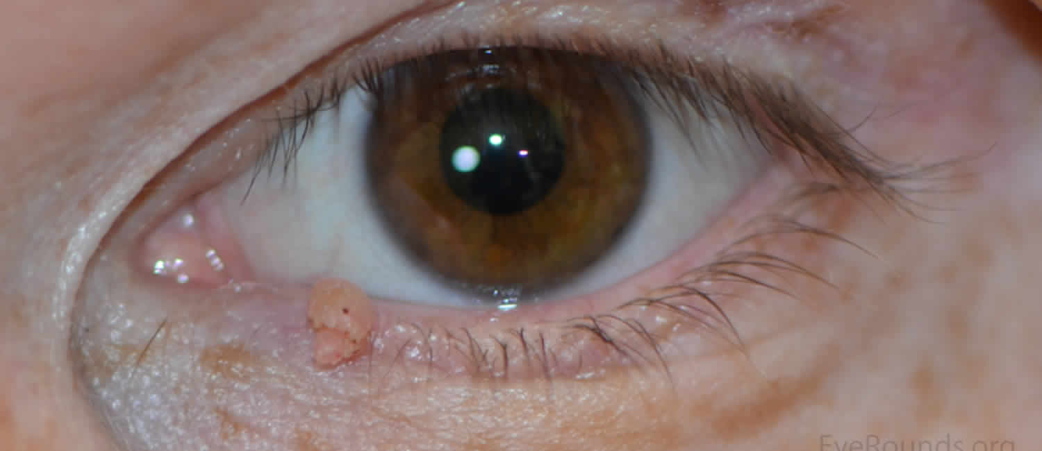
A single lesion is most common and appears as a soft, pedunculated mass (supported on a stem or stalk) with numerous finger-like projections. The projections may be long and pointy or short and rounded if keratin (skin-forming protein) has built-up around the lesion. Less keratinised lesions are pink or red in colour and resemble a raspberry, whilst heavily keratinised lesions are white and look like the head of a cauliflower.
Squamous papilloma prognosis is excellent. However, the lesions can recur in the same or different location.
Eyelid papilloma cause
The human papillomavirus (HPV) subtypes most often found in oral squamous cell papilloma are HPV-6 and HPV-11. These subtypes are not associated with malignancy or precancer. However, for most malignant lesions, UV (sun) exposure is the main etiologic factor.
Premalignant, malignant, or benign eyelid skin lesions may be papillomatous.
Cysts – Epidermal inclusion, sudoriferous, sebaceous
Seborrheic keratosis usually occurs in middle-aged or elderly patients and appears as brown-black, “stuck on,” well-circumscribed, crustlike lesions. Usually, the lesions are slightly elevated and uninflamed. The lesions can be removed with a shave biopsy, if desired. In black adults, a heavily pigmented variant, dermatosis papulosa nigra, occurs involving the malar region and often the eyelids.
Keratoacanthoma appears similar to basal cell carcinoma and squamous cell carcinoma because it is elevated with a central ulcer crater. However, these dome-shaped tumors with rolled margins usually appear and rapidly grow in size (up to 1-2 cm) over a few weeks to months and then often spontaneously involute. The tumors can be destructive, especially if they involve the eyelash margin. To be sure of the diagnosis, surgical excision and biopsy often is performed. These tumors can occur in individuals who are immunosuppressed (eg, after renal transplantation).
Nevus is usually light to dark brown, but it can be amelanotic and indistinguishable from a squamous papilloma. Usually, it is well circumscribed, sometimes with hair growing from its surface. It does not grow in size.
Inverted follicular keratosis usually presents as a solitary nodular or wartlike keratotic mass, it may be pigmented simulating a melanocytic lesion. It also can present as a cutaneous horn. If incompletely excised, it has a tendency to recur.
Pseudoepitheliomatous hyperplasia often clinically and histopathologically is confused with carcinoma. Usually, it is elevated with an irregular, ulcerated, or crusted surface, mimicking squamous cell carcinoma or basal cell carcinoma. It can occur anywhere on the eyelid and usually is of short duration (weeks to months). They may be associated with mycotic infections, insect bites, drugs, burns, radiation therapy, and underlying malignant lymphoma.
Eyelid papilloma diagnosis
Usually, squamous papillomas are sessile or pedunculated and have a color similar to the surrounding skin. They often are multiple and tend to involve the lid margin. A small keratin crust often can be palpated on the surface (keratotic papilloma). Microscopically, these lesions are composed of fingerlike projections of vascularized connective tissue covered by hyperplastic epithelium (papillae). The epidermis usually is acanthotic, with elongation of the rete ridges, and shows areas of hyperkeratosis and focal parakeratosis.
A biopsy of the lesion may be performed 1. Accurate diagnosis of an eyelid lesion requires histologic examination.
Eyelid papilloma treatment
Surgical excision usually is a simple procedure for these benign skin lesions. Lee et al 2 studied eyelid margin papillomas for which complete excision was cosmetically unacceptable. The study reported a case in which interferon was an effective treatment for a conjunctival papilloma.
Surgical scarring and possibly lid notching are the only likely complications. Usually, the eyelid papillomas are so small that bleeding and infection rarely occur post excision.
Patients should receive follow-up care as needed.
References- Eshraghi B, Torabi HR, Kasaie A, Rajabi MT. The use of a radiofrequency unit for excisional biopsy of eyelid papillomas. Ophthal Plast Reconstr Surg. 2010 Nov-Dec. 26(6):448-9.
- Lee BJ, Nelson CC. Intralesional Interferon for Extensive Squamous Papilloma of the Eyelid Margin. Ophthal Plast Reconstr Surg. 2011 Jun 8.


