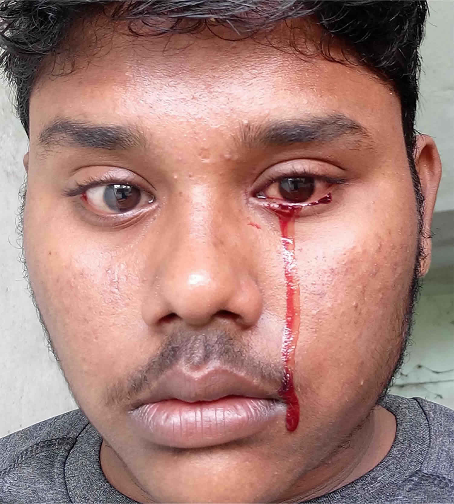
Haemolacria
Haemolacria (hemolacria) also known as bloody epiphora, bloody tears, blood-stained tears, dacryohemorrhea, hematodacryorrhea, hemolacrimia, sanguineous tears, sanguineous lacrimation, hematic epiphora, dacryohemorrhysis, lacrimae cruentae or tears of blood, is the presence of blood in the tear 1.
Haemolacria causes
The source of blood in tears may be:
Bleeding from the conjunctiva – Conjunctiva is a vascular tissue with limbal, bulbar, forniceal, and palpebral parts. The conjunctival vessels lie at the ocular surface and may bleed spontaneously or after eye-rubbing usually causing subconjunctival hemorrhage. Subconjunctival hemorrhage without a breach in conjunctiva is usually not associated with frank blood in the tears, though red blood corpuscles may be seen on microscopic examination of tears 2. Causes of haemolacria due to the involvement of the conjunctiva include:
- Trauma – Conjunctival laceration or rupture of conjunctival vessels may cause hemolacria. Surgery involving incision or excision of conjunctiva may also cause hemolacria. The surgeries include small incision cataract surgery, pterygium surgery, squint surgery, scleral buckling, trabeculectomy, pars plana vitrectomy, and other ocular surgeries. After manipulation of the conjunctival follicle in trachoma, there may be haemolacria.
- Inflammation – Severe conjunctivitis including hemorrhagic conjunctivitis, membranous or pseudomembranous conjunctivitis, follicular conjunctivitis with congested semilunar fold and caruncle, severe viral or bacterial conjunctivitis cause blood stained tears. Conjunctival ulcer or granulation due to various reasons may cause haemolacria. Postoperative sclera buckle infection can also cause bloody tears.
- Vascular lesions – Hemangioma, lymphangioma, inflammatory papilloma of the conjunctival sac, telangiectasia of conjunctival vessels, pyogenic granuloma 3, conjunctival varies, pathological vasodilation of the conjunctival vessels, severely vascular papillae, giant papillary conjunctivitis may bleed leading to bloody tears. Malignant melanoma of the conjunctiva may be associated with haemolacria.
- Vicarious menstruation – The conjunctiva may periodically/cyclically bleed (vicarious menstruation) during menstruation or hormonal disturbances. This phenomenon is usually seen around menarche or rarely around the menopause. Sir Duke-Elder proposed that ‘instability of the nervous system’ and not hormonal disturbance is the cause of vicarious menstruation from the conjunctiva 4. The source of blood in hemolacria in vicarious menstruation has been thought to be the lacrimal gland, lower forniceal conjunctiva, episclera, dilation of forniceal and bulbar conjunctival vessels, and hemorrhages in upper tarsal conjunctiva. The ophthalmic examination may be normal with normal appearing conjunctiva in some cases. Vicarious menstruation may cause subconjunctival hemorrhage. The vicarious menstruation is usually painless, may last for seconds to minutes, and may involve one or both eyes. Islands of ectopic endometrial tissue may be present outside the uterus. This is called endometriosis, and such aberrant endometrial tissue in conjunctiva may cause vicarious menstruation. Primary localized conjunctival amyloidosis may cause recurrent subconjunctival bleed due to vicarious menstruation 5. Other possible explanations of vicarious menstruation include 2:
- Normal conjunctiva stimulated by hormonal or other factors.
- Estrogenic premenstrual light blood hypertension.
- Foreign body – Foreign body at the upper fornix can cause chronic irritation, erosion of the conjunctiva and hemolacria. Subconjunctival metallic splinter after trauma may cause bleeding into the tear.
- Chemical injury – Application of silver nitrate over the conjunctiva is another cause of bloody tears.
Bleeding from the lid margin – Inflammed lid margin due to blepharitis may cause erosion of the surface of the eyelid and lead to bleeding. Trauma causing laceration of the lid margin may lead to haemolacria. Infestation of the lid with Phthirus pubis may also cause mild staining of blood in the tear 6.
Hemorrhage from the lacrimal puncta – The source of the hemorrhage in tears may be the puncta. There is one lacrimal punctum each at the medial side of upper and lower lid respectively. These are situated at the inner margin of the lid. Punctum connects medially to a lacrimal canaliculus on both the upper and lower lid. Both of these canaliculi may meet to form a common canaliculus and then connect to the lacrimal sac, or they may open into the lacrimal sac separately. The lacrimal sac connects to the lower nasal cavity below the inferior nasal concha via the nasolacrimal duct. Thus, the lacrimal puncta drain tear to the nasal cavity normally. If the nasal cavity fills with any fluid under pressure, that fluid may get released through the puncta; this happens when profuse epistaxis is managed with nasal packing. Sometimes, bleeding through the puncta may occur in epistaxis without nasal packing also. When the pressure within the nasal cavity increases, the blood can pass retrogradely through the nasolacrimal duct, lacrimal sac, and canaliculi to the puncta. Rarely, the blood may come to the puncta from connected body cavities including upper digestive tract or trachea 2. The bleeding from the lacrimal puncta may be evident on pinching or blowing the nose in case of epistaxis 7. Fracture of the nasal bone or walls of the sinuses, and Le Fort fracture type 1 can cause epistaxis which may retrogradely flow to the puncta. Haemolacria has been reported after orbital floor fracture due to displaced implant eroding the nasolacrimal drainage system in a patient on warfarin and aspirin 8. The bleeding through the lacrimal puncta due to epistaxis or a local pathology of the lacrimal sac or nasolacrimal duct usually occurs through the lower punctum, sometimes from both puncta, and rarely through the upper punctum alone 2. Other causes of bleeding from the lacrimal punctum include:
- Trauma
- Infection
- Tumor- angioma, meningioma of the lacrimal sac
- Vascular lesions including varices 9 and dacryolith, rupture of dilated/distended vessels within lacrimal sac
Tear glands – In some rare cases, the source of bleeding remains undetermined. In such cases, some authors have hypothesized that the hemorrhage may come from the lacrimal gland or accessory lacrimal gland. Other hypothesized origins of hemolacria include meibomian glands or conjunctival goblet cells 2. Dacryoadenitis or inflammation of the lacrimal gland may cause bloody tears.
Orbit – orbital varix at the junction of the angular vein and superior ophthalmic has been reported to be associated with bloody tears due to bleeding from the medial conjunctiva and overlying skin after jugular compression 10. Rarely, fracture of the orbital roof, orbital hemangioma, and epibulbar dermoid may cause haemolacria if the conjunctiva is open.
Hematologic diseases which cause increased bleeding diathesis may lead to hemorrhages at multiple organs including haemolacria. The disorders include hemophilia, thrombocytopenic purpura, deficiency of clotting factors including factor VII deficiency.
Other systemic diseases which have correlations with bloody tears include anemia, jaundice, exanthematous fever (may also cause epistaxis, and bleeding through the mouth), and severe disturbances of the autonomic nervous system.
Vascular disorders may play an important role in the pathogenesis of hemolacria. Hypertension is an important factor which has been reported to cause epistaxis and retrograde haemolacria through the leak of hemorrhage via the lacrimal puncta. Causes of haemolacria include Hereditary hemorrhagic telangiectasia or Osler Weber Rendu disease, Henoch-Schönlein purpura, Gardner Diamond syndrome (autoerythrocyte sensitization or psychogenic purpura, or painful bruising syndrome), and conjunctival varicose vessels. Osler Weber Rendu disease or hereditary hemorrhagic telangiectasia is an autosomal dominant disease characterized by mucocutaneous telangiectasia and visceral involvement leading to hemorrhage which may be life-threatening 11. A patient with occlusion of the posterior inferior cerebellar artery causing unilateral Horner’s syndrome and vasodilation has been reported 12. This patient had episodes of haemolacria after slight abrasion or inflammation of the conjunctiva.
Medications:
- The mecholyl test uses subcutaneous methacholine to evaluate the autonomic nervous system in psychiatric patients. This test has also been used for the diagnosis of achalasia cardia. Mecholyl test may cause haemolacria 13.
- Systemic drugs like aspirin, clopidogrel, heparin, and warfarin should be ruled out in haemolacria.
- Acetylcholine may cause bloody tears.
Psychiatric disorders – Attention seeking behavior or other psychiatric disorders may lead to bloody tears. An 11-year-old girl has been reported whose ocular exam was otherwise normal but had recurrent episodes of haemolacria 14. On further evaluation, she was noted to have multiple needle pricks over the fingertips which could possibly explain her symptoms. She was diagnosed with Munchausen syndrome (factitious disorder imposed on self). In this syndrome, the affected person repeatedly feign disease/trauma/illness to draw attention, sympathy, or to fulfill other psychological needs. Risk factors for haemolacria include stress, emotion, anger, fright, anxiety, hysteria. Some patients may malinger as haemolacria by putting red colored liquid over the conjunctival sac.
Bloody sweats or hemathydrosis – A prepubertal 9-year-old female has been reported who had bloody sweats at the skin between the lacrimal sac and medial canthus, and haemolacria. The symptoms resolved 8 days after application of iron perchloride over the affected skin 2.
Other causes of haemolacria include 15:
- Cranial trauma
- Post-traumatic epilepsy
- In otherwise normal individuals after stooping, or muscular effort
- In children after ‘copious weeping’
- Coughing
- Hypertensive crisis
- Acute hemorrhagic edema of infancy
Unknown cause/idiopathic haemolacria – In some cases, despite a thorough search for ocular, systemic, or psychiatric causes, no obvious etiology or source is found.
Haemolacria complications
Haemolacria as such will not lead to complications. But it can be a complication of a multitude of conditions, as explained in the cause section.
Haemolacria differential diagnosis
Differential diagnosis of haemolacria includes:
- Red colored tears may be noted in patients on therapy with rifampicin.
- Occult haemolacria is the term used for apparently clear tears if microscopic or chemical evidence of red blood corpuscles is present in tears. Microscopy showed erythrocytes in the tear of 10% of the normal population, and chemically blood was detected by chemical stix method in 3% of normal persons 16. Occult blood was found commonly in acute infectious conjunctivitis and was also noted in chronic and subacute infectious conjunctivitis though it was less frequent 16.
- False bloody tears – Malingerers may feign red colored liquids and may claim that tear is bloodstained. Rats treated with muscarinic agents may secrete reddish brown tears (chromodacryorrhea) due to protoporphyrin and coproporphyrin secreted from Harderian glands. Melanodacryorrhea or black tears may be a feature in conjunctival argyrosis or necrotic uveal melanoma.
Haemolacria diagnosis
All patients presenting with haemolacria should undergo comprehensive ocular examination and systemic examination to look for organic causes and source of the hemorrhage. Specifically, the history of systemic diseases (hematological disorders, coagulopathies, hypertension) and medications like warfarin, aspirin or clopidogrel is necessary.
When no ocular or systemic causes are present, psychiatric diseases and vicarious menstruation must be ruled out before labeling the etiology as ‘unknown’ or ‘idiopathic.’
When needed, imaging may give a clue to the cause of haemolacria. Dacryoendoscopy promises to be an important tool to rule out bleeding lesions within the tear drainage system which may be undetected without this modality 9.
Haemolacria treatment
The treatment of haemolacria depends on the cause. Close observation is needed when no apparent cause of haemolacria is detected to rule out malingering.
Haemolacria prognosis
Severe bleeding through tears may even be fatal in some cases especially in a patient with coagulopathy. Ruling out hemorrhage from other sources including visceral bleeding is of utmost importance.
References- Tripathy K, Salini B. Hemolacria (Haemolacria) [Updated 2019 Nov 13]. In: StatPearls [Internet]. Treasure Island (FL): StatPearls Publishing; 2019 Jan-. Available from: https://www.ncbi.nlm.nih.gov/books/NBK539774
- Murube J. Bloody tears: historical review and report of a new case. Ocul Surf. 2011 Jul;9(3):117-25.
- Iovieno A, Coassin M, Piana S, De Luca M, Giunta P, Fontana L. A case of unilateral hemolacria. Int Ophthalmol. 2016 Apr;36(2):273-4.
- Abboud IA, Hanna LS. Bleeding from the conjunctiva. Br J Ophthalmol. 1971 Jul;55(7):487-91.
- Gauba V, Cooper M, Liu C. Vicarious menstruation in primary localized conjunctival amyloidosis. Arch. Ophthalmol. 2006 Sep;124(9):1361-2.
- Singh A, Tripathy K, Gupta N, Kale P, Verma N, Mirdha BR. Phthirus pubis in the eye. Indian J Med Microbiol. 2016 Jul-Sep;34(3):405-6.
- Wiese MF. Bloody tears, and more! An unusual case of epistaxis. Br J Ophthalmol. 2003 Aug;87(8):1051.
- Chon BH, Zhang R, Bardenstein DS, Coffey M, Collins AC. Bloody Epiphora (Hemolacria) Years After Repair of Orbital Floor Fracture. Ophthalmic Plast Reconstr Surg. 2017 Sep/Oct;33(5):e118-e120
- Ali MJ, Naik MN. Dacryoendoscopy in a Case of Unexplained Hemolacria. Ophthalmic Plast Reconstr Surg. 2018 Nov/Dec;34(6):e213
- Bonavolontà G, Sammartino A. Bloody tears from an orbital varix. Ophthalmologica. 1981;182(1):5-6.
- Macri A, Wilson AM, Shafaat O, Sharma S. StatPearls [Internet]. StatPearls Publishing; Treasure Island (FL): Jun 19, 2019. Osler-Weber-Rendu Disease (Hereditary Hemorrhagic Telangiectasia, HHT).
- Ziegler LH. Bloody tears. JAMA 1938;110 (17):1387
- JIRICKA Z, VOLICER L. PRESENCE OF BLOOD IN TEARS DURING THE MECHOLYL TEST. Med Pharmacol Exp Int J Exp Med. 1965;12:56-60.
- Audelan T, Best AL, Ameline V. [Hemolacria: A pediatric clinical case report]. J Fr Ophtalmol. 2019 Jan;42(1):e15-e17.
- Manzano G, Shantharam R, Webb E, Finelt N, Hengel K. Case 2: Hemolacria, Hematochezia, and Hematuria in an 11-month-old Boy. Pediatr Rev. 2018 Aug;39(8):418-420.
- Norn MS. Microscopically and chemically detected haemolacria. Acta Ophthalmol (Copenh). 1977 Feb;55(1):132-40.


