
Hirschberg test
Hirschberg test also known as the corneal light reflex test, is a quick and simple way to check ocular alignment 1. Hirschberg test is particularly useful for testing for strabismus (misalignment of the eyes) in newborns, young children, patients with poor vision, patients that are not able to fixate or track well or in any situation where a full motility evaluation is not feasible.
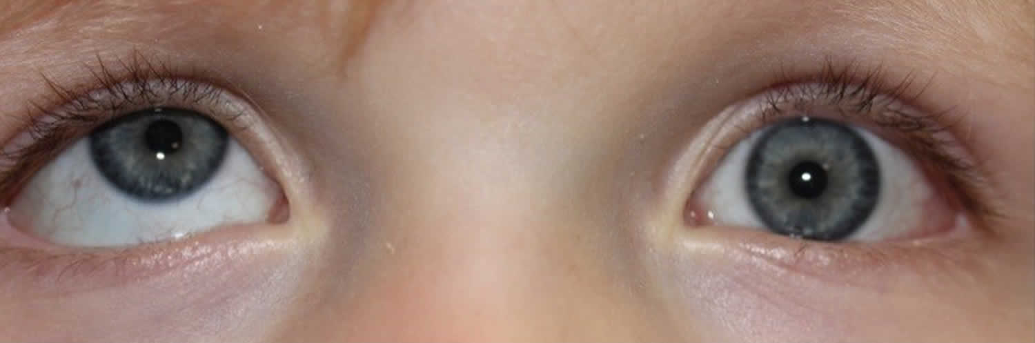
Hirschberg corneal light reflex test involves directing the patient to look at a point of light held about three feet from the patient’s face. The corneal light reflex is observed. The Hirschberg test is considered normal when the corneal light reflexes are slightly decentered nasally (about 5º, due to angle kappa). If the light reflexes fall asymmetrically in the pupils, strabismus may be present. In the case of a hypertropia, the light reflex of the deviated eye is located below the light reflex of the fixing eye. The amount of deviation can be grossly estimated by multiplying the mm of deviation by 15PD 2.
Hirschberg testing estimates the size of the strabismus by determining how far the deviated light reflex is off-center.
Krimsky light reflex testing involves holding a prism over one eye to center the deviated light reflex until the reflexes are symmetric. The amount of the prism needed to center the deviated light reflex estimates the size of the eye misalignment. Light reflex testing is the least accurate way to measure strabismus but may be the only means possible in young children and in those with vision too poor to fixate well on a target.
Young children and patients of Asian descent can appear “cross-eyed” without a true misalignment. This pseudostrabismus is most commonly due to a flat nasal bridge with prominent epicanthal skin folds (see Figure 3). These facial features can obscure the medial sclera and create an optical illusion of misalignment. In this case, the eyes may appear to be esotropic, but the corneal reflexes will appear symmetric and in the center of each pupil.
Figure 1. Normal Hirschberg corneal reflex test
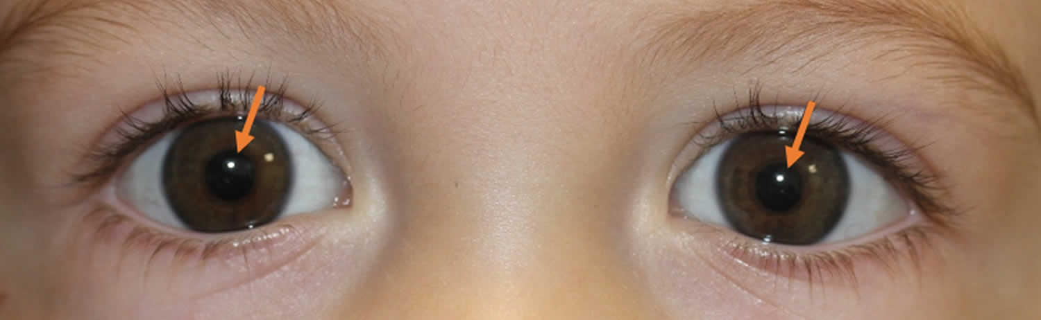
Figure 2. Abnormal Hirschberg corneal reflex test
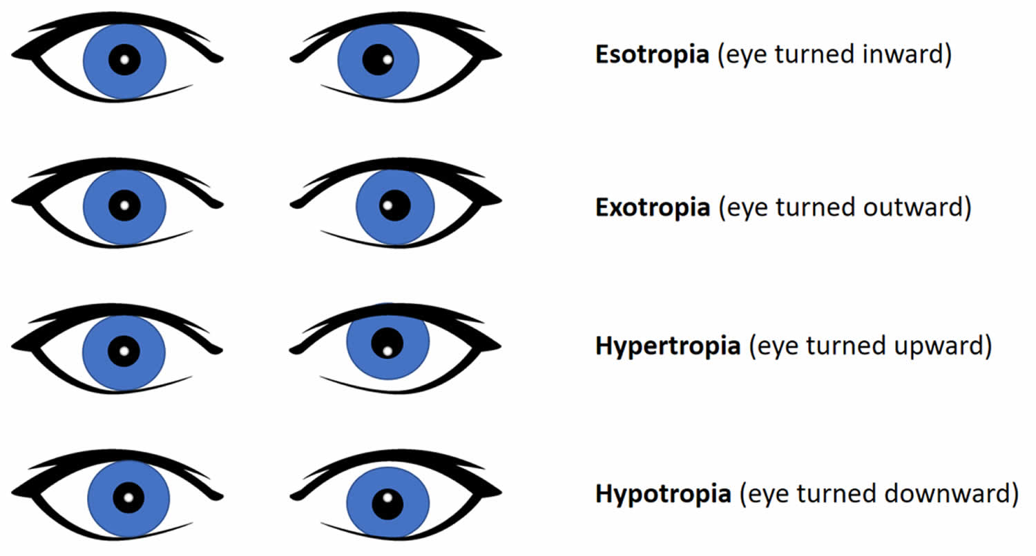
Figure 3. Pseudostrabismus

Hirschberg corneal light reflex test
To perform the Hirschberg light reflex test:
- Use a light source, such as a penlight or finhoff transilluminator.
- Instruct the patient to focus their gaze on your light source.
- From a distance of 2 feet, shine your light source equally into the patient’s eyes at midline.
- Observe the reflection of light off the cornea, which should appear as a pin-point white light near the center of the pupil in each eye.
If there is normal alignment, the reflection will appear in the same position in each pupil (see Figure 1). If there is misalignment of the eyes, the location of the corneal reflex will appear asymmetric and “off center” of the pupil in the deviating eye. The relative difference in the position of the reflex will be in the opposite direction as the eye deviation. For example, in an esotropia (where there is inward deviation of the eye) (Figure 5), the light reflex will appear outwardly displaced from the center of the pupil; in a hypertropia (where there is an upward deviation of the eye) the light reflex will appear inferiorly displaced from the center of the pupil (see Figure 7).
For every 1 millimeter of deviation from the center of the pupil, it will take approximately 15 diopters of prism to correct the misalignment (Figure 4).
This can be visually estimated with the Hirschberg test or physically measured with the Krimsky test, where a prism is placed in front of the deviating eye until the corneal reflex is re-centered in the pupil.
Figure 4. Hirschberg test degrees
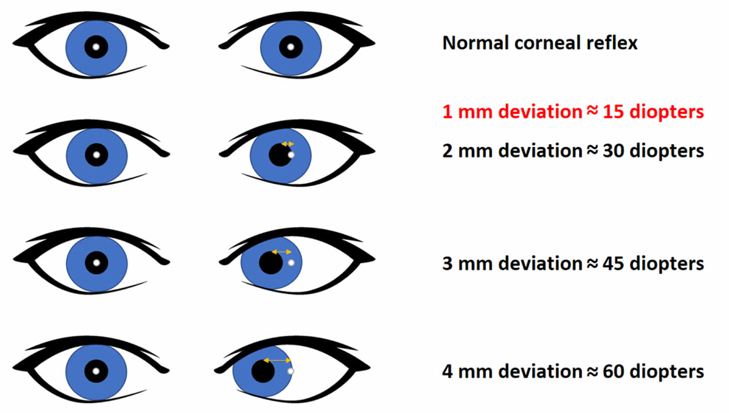
Figure 5. Esotropia (in this case an inward deviation of the left eye)
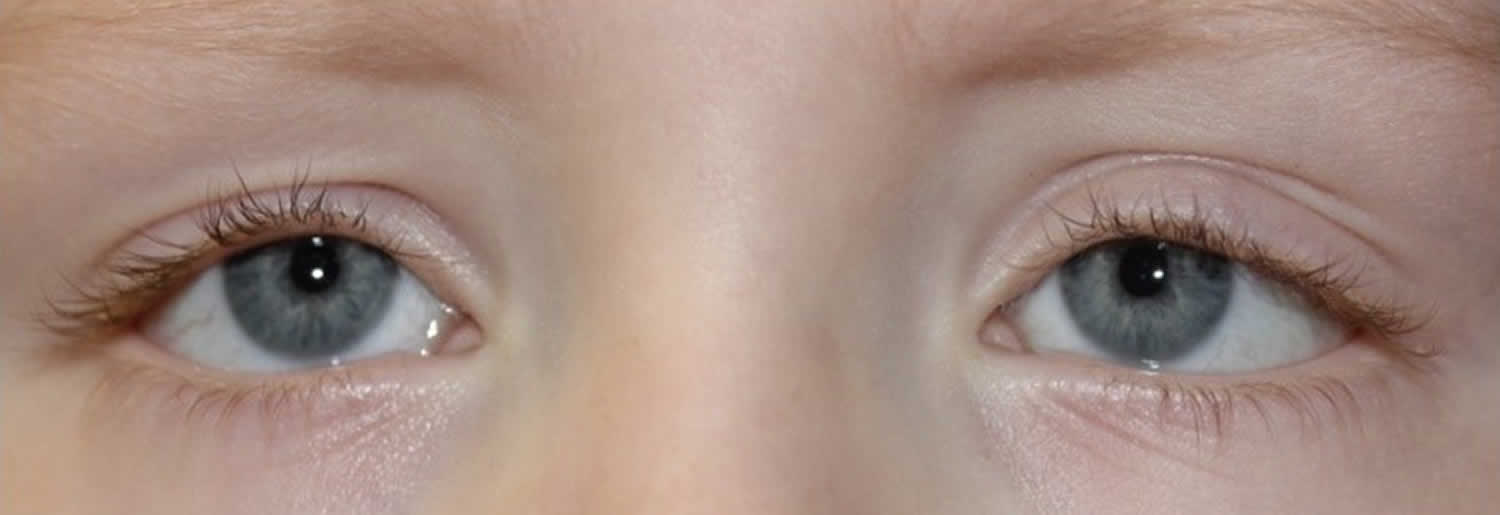
Figure 6. Exotropia (in this case an outward deviation of the left eye)

Figure 7. Hypertropia (patient with hypertropia of the right eye)


