
What is toenail fungus
Toenail fungus infection or onychomycosis is a fungal infection of the nail bed, matrix or plate, which causes discoloration, thickening, and separation of the toenail from the nail bed. Toenails fungus infection occurs in 10% of the general population but is more common in older adults; the prevalence is 20% in those older than 60 years and 50% in those older than 70 years 1. The increased prevalence in older adults is related to peripheral vascular disease, immunologic disorders, and diabetes mellitus. The risk of toenail fungus infection is 1.9 to 2.8 times higher in persons with diabetes compared with the general population 2. In patients with human immunodeficiency virus infection, the prevalence ranges from 15% to 40% 3.
Fungal nail infection affects toenails more often than fingernails because of their slower growth, reduced blood supply, and frequent confinement in dark, moist environments. It may occur in patients with distorted nails, a history of nail trauma, genetic predisposition, hyperhidrosis, concurrent fungal infections, and psoriasis. It is also more common in smokers and in those who use occlusive footwear and shared bathing facilities 4.
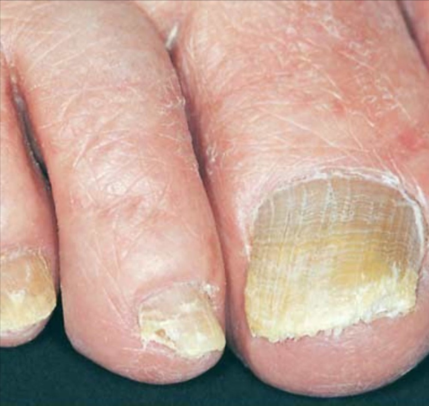
Fungal toenail infection is caused by various organisms, most often dermatophytes of the genus Trichophyton. Dermatophytes are fungi that require keratin for growth. These fungi can cause superficial infections of the skin, hair, and nails. Other organisms include Candida, which is more common in fingernail infections (Figure 2) and in patients with chronic mucocutaneous candidiasis 1. Nondermatophyte molds are a less common cause in the general population. Recent studies, however, have demonstrated that they are the predominant organisms in patients with fungal nail infection and human immunodeficiency virus infection 3.
Toenail Fungus Infection in Children
Onychomycosis in children is rare, with an estimated prevalence of 0.2 percent 5. Most often, onychomycosis develops in children with immunosuppression (e.g., acquired immunodeficiency syndrome, chemotherapy, congenital immunodeficiency syndromes), a strong familial history of onychomycosis or extensive cutaneous mycosis (tinea capitis or pedis).
Although griseofulvin remains the mainstay of onychomycosis treatment in children, the efficacy of this drug is variable, and relapse is common. Newly available medications may improve the traditionally mediocre treatment outcomes in this age group.
Some studies have shown terbinafine to be safe and quite effective in the treatment of tinea capitis, and it is licensed for this purpose in several countries 6. In more limited trials, itraconazole has also been shown to be safe and efficacious in the treatment of tinea capitis. If the safety and effectiveness of terbinafine and itraconazole are established over the longer courses needed to treat nail infections, they may become potent first-line therapies for onychomycosis in children.
Figure 1. Toenail anatomy
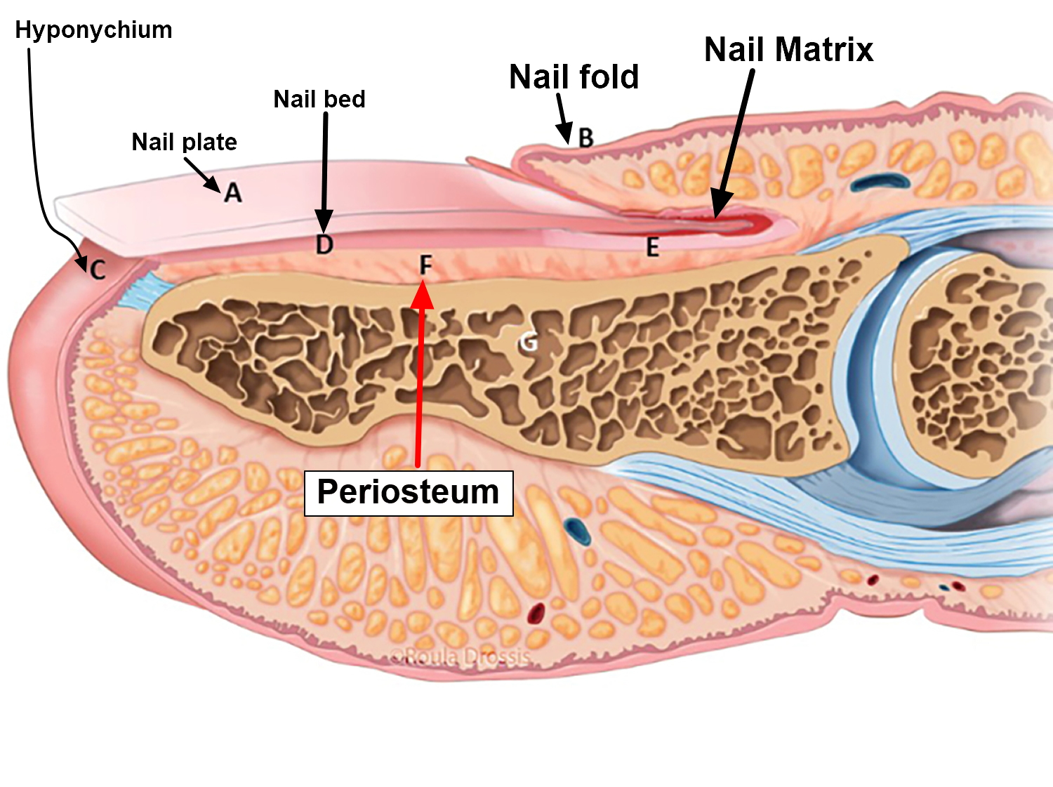
Figure 2. Toenail fungus infection
What Causes Toenail Fungus Infection
The most common form of toenail fungus is distal subungual onychomycosis, which can also be distal and lateral (Figures 3 and 4). Distal subungual onychomycosis may develop in the toenails, fingernails or both. Some degree of tinea pedis (tinea foot infection) is almost always present. The infection is usually caused byTrichophyton rubrum, which invades the nail bed and the underside of the nail plate, beginning at the hyponychium and then migrating proximally through the underlying nail matrix 7 (Figure 1). Susceptibility to distal superficial onychomycosis may occur in an autosomal dominant pattern within families 8.
White superficial onychomycosis (Figure 6) accounts for only 10 percent of onychomycosis cases 9. White superficial onychomycosis is caused by certain fungi that directly invade the superficial layers of the nail plate and form well-delineated opaque “white islands” on the plate. As the disease progresses, these patches coalesce to involve the entire nail plate. The nail becomes rough, soft and crumbly. The most common causative agent isTrichophyton mentagrophytes 8.
Proximal subungual onychomycosis is the least common form of tinea unguium in healthy persons (Figure 5). It occurs when the infecting organism, usually T. rubrum, invades the nail unit through the proximal nail fold, penetrates the newly formed nail plate and then migrates distally. Fingernails and toenails are equally affected 8. This form of onychomycosis usually occurs in immunocompromised persons and is considered a clinical marker of human immunodeficiency virus infection 8. Proximal subungual onychomycosis can also arise secondary to local trauma 9.
Patients with chronic mucocutaneous candidiasis may develop candidal infection of the nails. Candida species may invade nails previously damaged by infection or trauma 9. Candidal paronychia more commonly affects the hands and usually occurs in persons who frequently immerse their hands in water 10.
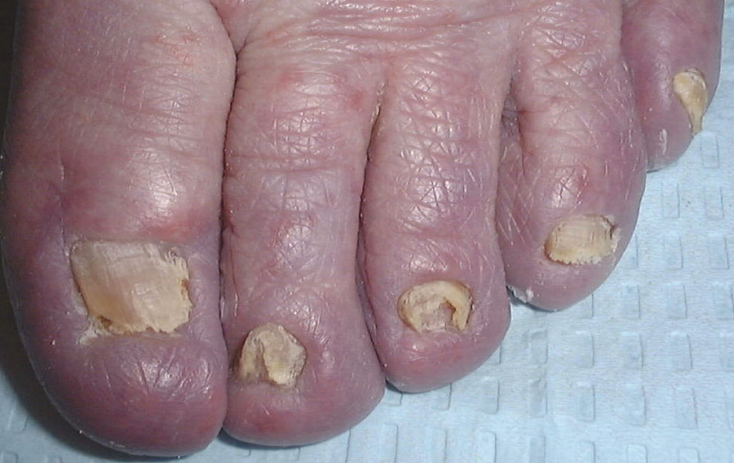
Total dystrophic onychomycosis may be the end result of any of the four main forms of onychomycosis. This condition is characterized by total destruction of the nail plate 9.
Table 1. Common Pathogens in Toenail Fungus Infection (Onychomycosis)
Dermatophytes (80% to 90%) |
Epidermophyton floccosum |
Microsporum species |
Trichophyton interdigitale |
Trichophyton mentagrophytes |
Trichophyton rubrum |
Trichophyton tonsurans |
Nondermatophyte molds (2% to 10%)* |
Acremonium species |
Alternaria species |
Aspergillus species |
Cladosporium carrionii |
Fusarium species |
Geotrichum candidum |
Lasiodiplodia theobromae |
Onychocola species |
Scopulariopsis species |
Scytalidium species |
Yeast (2% to 11%) |
Candida albicans |
Candida guilliermondii |
Candida parapsilosis |
*—Nondermatophyte molds are the predominant organism in patients with human immunodeficiency virus infection.
Classification of Toenail Fungus Infection
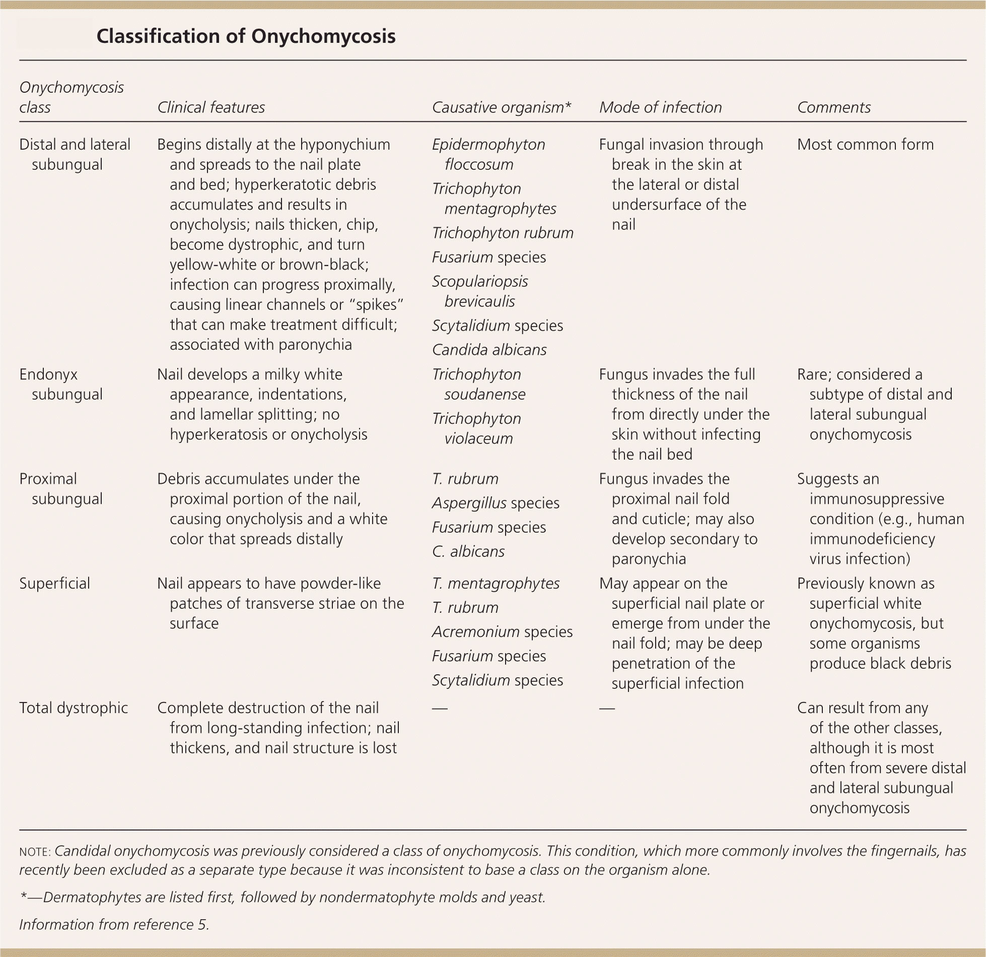
Onychomycosis is divided into several classes based on morphologic patterns and mode of invasion of the toenail (Table 2) 12. Classification provides a framework for diagnosis and expected response to treatment, and can help predict the prognosis. The classes include distal and lateral subungual onychomycosis (Figures 3 and 4), proximal subungual onychomycosis (Figure 5), superficial onychomycosis (Figure 6), and total dystrophic onychomycosis (Figure 7). A fifth class, endonyx subungual onychomycosis, is rare. Some nails have features from a combination of classes.
Table 2. Classification of Fungal Nail Infection
[Source 11]
Figure 3. Distal and lateral subungual onychomycosis
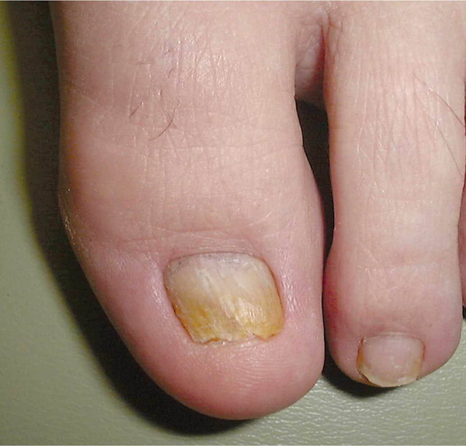
Figure 4. Distal and lateral subungual onychomycosis with spike deformity
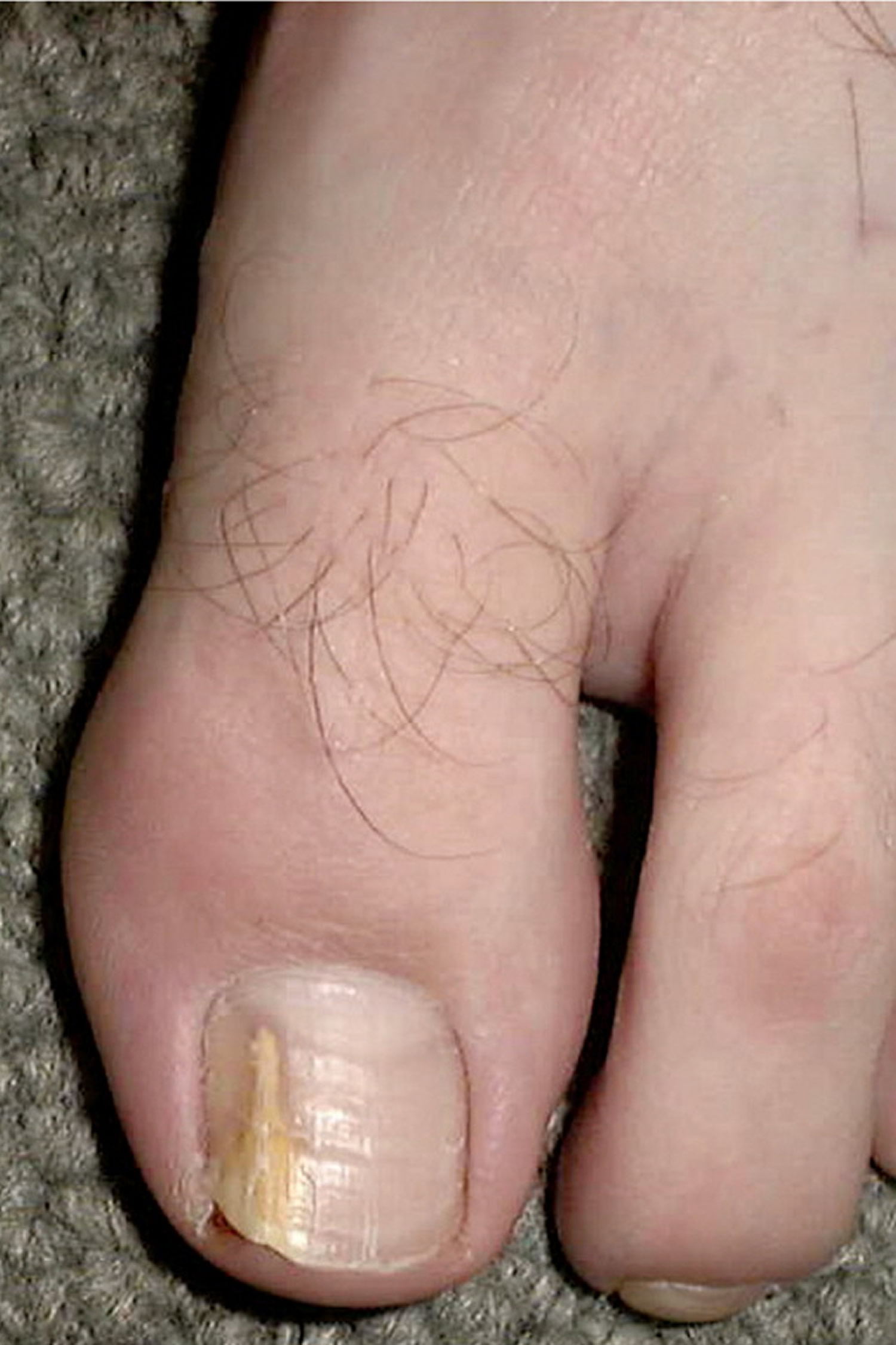
Figure 5. Proximal subungual onychomycosis
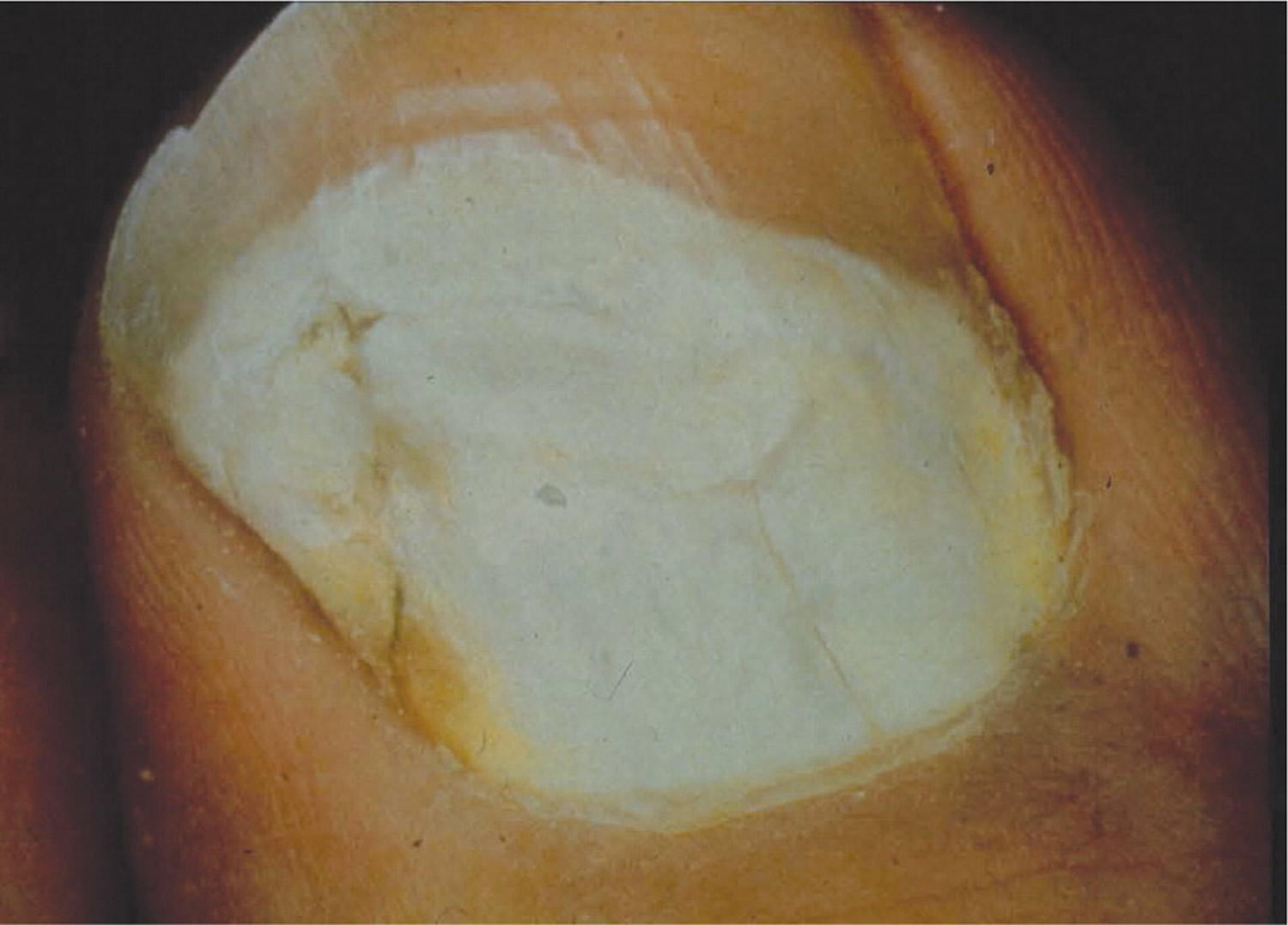
Figure 6. Superficial onychomycosis
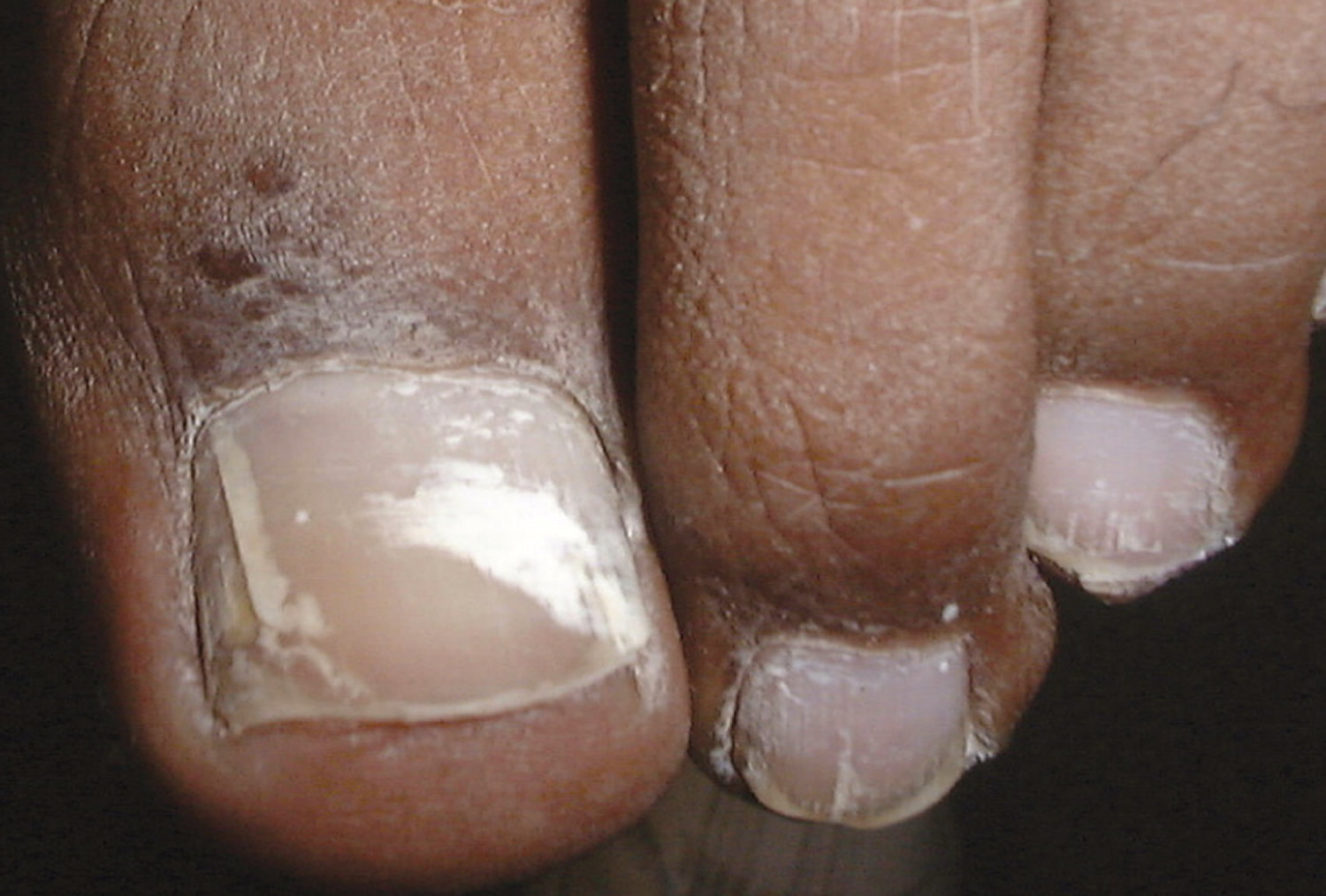
Figure 7. Total dystrophic onychomycosis
Accurate diagnosis is crucial for successful treatment and requires identification of physical changes and positive laboratory analysis. Only 50% of nail problems are caused by onychomycosis 13, and clinical diagnosis by physical examination alone can be inaccurate. Psoriasis, chronic nail trauma, and other causes must also be considered. The differential diagnosis of onychomycosis is presented in Table 3.
Table 3. Common Conditions That Can Mimic Onychomycosis
| Condition | Features |
|---|---|
Infections | |
Chronic paronychia | Chronic inflammation of the proximal paronychium; cross-striations of the nail; Streptococcus, Staphylococcus, or Candida found on smear and culture; common in children |
Viral warts | Localized in nail folds and subungual tissue; longitudinal depressed grooves in the nail plate |
Skin disorders | |
Chronic dermatitis | Subungual dermatitis, hyperkeratosis, Beau lines, and pitting; thickened nail with corrugated surface |
Lichen planus | Longitudinal grooves and fissures; usually affects fingernails |
Psoriasis | Nail pitting, splinter hemorrhages, “oil staining,” yellow-gray or silvery white nails |
Twenty-nail dystrophy | Dystrophy of all 20 nails; usually resolves in childhood; associated with the lesions of lichen planus |
Trauma | |
Footwear | Oncholysis, ingrown toenails, subungual keratosis, nail plate discoloration and irregularities; caused by friction against the shoe |
Manipulation (e.g., manicures, pedicures, rubbing) | Horizontal parallel nail plate grooves, inflammation from Staphylococcus aureus or Pseudomonas infection |
Tumors | |
Bowen disease | Squamous cell carcinoma; bleeding, pain, nail deformity, and nail discoloration |
Fibroma | Oval or spherical, white or yellow nodule; causes tunnel-like melanonychia; fibrous dermatofibroma or periungual fibroma |
Melanoma | Brown-yellow nail with dark pigment extending into the periungual skin folds; poor prognosis |
Treatment of fungal nail infection
Onychomycosis is widely believed to be only a cosmetic problem, but it can be uncomfortable and can lead to cellulitis in older adults and foot ulcers in patients with diabetes. Eradication of the infection is key to improving appearance and avoiding these complications, but it is not easily accomplished because nails are made of keratin, which is nonvascular and impermeable to many agents 14. Because of poor drug delivery to nails, results of treatment may not be apparent for a year.
Treatment varies depending on the severity of nail changes, the organism involved, and concerns about adverse effects and drug interactions. Treatments also have varying effectiveness, based on cure parameters that are defined differently among studies. Mycotic cure denotes that no organism is identified on microscopy and culture. Clinical cure refers to improvement in the appearance of the nail, often defined as a normal appearance in 80% to 100% of the nail. It is a subjective measure that is difficult to compare across studies 15. Complete cure indicates that mycotic and clinical cure have been achieved.
Topical Agents
Several topical agents are used for the treatment of onychomycosis. These agents have few contraindications and no drug-drug interactions.
Ciclopirox 8% solution is the only topical prescription medication available in the United States for the treatment of onychomycosis. It is a synthetic hydroxypyridine antifungal formulated as a nail lacquer. Adverse effects include burning, itching, and stinging at the application site 16. It may be used in patients who cannot take oral antifungals and in those with less than 50% of the distal nail affected and no lunular involvement 17. It has been used in children, although it is not approved for use in patients younger than 12 years 18. When used alone, ciclopirox has a mycotic cure rate of 29% to 36%, and a clinical cure rate of 6% to 9% 17. A Cochrane review noted that the treatment failure rate was 61% to 64% after 48 weeks of use 19.
Ciclopirox has also been used in combination with oral agents to improve effectiveness. In one comparative study, a combination of ciclopirox and oral terbinafine had a mycotic cure rate of 88% and a complete cure rate of 68%, whereas terbinafine alone had a mycotic cure rate of 65% and a complete cure rate of 50% 20.
Nonprescription agents have also been used for treatment of onychomycosis. These therapies have been evaluated in only a small number of studies involving few patients.
Topical mentholated ointment (Vicks Vaporub) was used in a small study involving 18 patients. After 48 weeks, 28% had mycotic and clinical cure, 56% had partial clearance, and 17% had no improvement 21.
Tea tree oil (Melaleuca alternifolia) has been evaluated in two studies. Although one trial was favorable, combined data from both studies did not demonstrate significant benefit 18, 22.
Snakeroot extract (Ageratina pichinchensis) is an antifungal derived from plants of the sunflower family. It was studied in a randomized trial involving 96 patients who applied the extract or ciclopirox for six months to nails with confirmed infections 23. Mycotic cure occurred in 59% of patients receiving the extract and in 64% of those receiving ciclopirox. Clinical cure occurred in 71% and 81% of patients, respectively. Differences between the two treatments were not statistically significant.
A small study showed that a combination of cyanoacrylate, undecylenic acid, and hydroquinone (marketed as Renewed Nail) demonstrated mycotic cure in 78 of 154 participants (50%) 24.
Nail trimming and debridement are often performed concomitantly with other treatments and appear to offer benefit. Study groups that received nail debridement with oral terbinafine had higher clinical cure rates than those who received oral terbinafine alone 25. When debridement was performed with concurrent administration of ciclopirox, the mycotic cure rate was 77%, higher than that for ciclopirox alone 26. Improvement in nail appearance was reported, but clinical cure rates were not.
Table 4. Nonprescription Treatments for Toenail Fungus Infection
| Agent | Administration | Clinical cure rate (%) | Mycotic cure rate (%) | Comments |
|---|---|---|---|---|
Ageratina pichinchensis (snakeroot) extract | Apply every third day for the first month, twice per week for the second month, then once per week for the third month | 71 | 59 | Study of 110 patients; therapeutic effectiveness was similar to that in the control group, which used ciclopirox |
Cyanoacrylate, undecylenic acid, and hydroquinone (Renewed Nail) | Soak and debride affected nails, then apply solution every two weeks for three to four visits; patients may also apply at home | NA | 50 to 65 (mild to moderate cases) | Study of 154 patients with cure rates reported after three months |
35 (severe cases) | ||||
Dual-wavelength near-infrared laser (Noveon) | Treatment on days 1, 14, 42, and 120 | Mild cases: 65 (3 mm of nail clearance) 26 (4 mm of nail clearance) | 30 | Toenails were evaluated on day 180 |
Moderate to severe cases: 63 (3 mm of nail clearance) | ||||
Melaleuca alternifolia (tea tree) oil | Apply twice per day | NA | NA | Cochrane review found no evidence of benefit |
Mentholated ointment (Vicks Vaporub) | Apply small amount with cotton swab daily | 28 | 28 | Pilot study of 18 patients; 56% had partial clearance, and 17% had no clearance |
Neodymium: yttrium-aluminum-garnet laser (Patholase Pinpointe) | One to three sessions four to six weeks apart | NA | 61 (complete cure) | Study of 37 toenails with onychomycosis |
19 (significant improvement) | ||||
11 (moderate improvement) |
NA = not available.
Laser and photodynamic therapies
Although they are expensive, laser and photodynamic therapies have become popular based on the success of in-vitro studies. Several neodymium:yttrium-aluminum-garnet (Nd:YAG) laser therapies have been approved by the U.S. Food and Drug Administration for treatment of onychomycosis 27. The Pinpointe Foot-laser, Cutera GenesisPlus laser, and Cooltouch Varia laser are short-pulse laser systems, whereas the Light Age Q-Clear laser is a Q-switched laser. However, there are only limited data about the use of these therapies in patients. In one study, Nd:YAG laser light was used to treat 37 nails, with one to three treatments given four to eight weeks apart. At 16 weeks, 61% were completely cured, 19% had significant improvement in the nail appearance, and 11% had moderate improvement in the nail appearance 28.
Another laser treatment, the dual-wavelength near-infrared laser (Noveon), is approved for dermatologic use, but not specifically for treatment of onychomycosis 29. This treatment was used on 26 nails on days 1, 14, 42, and 120. After 180 days, 91% of nails with mild infection showed clinical improvement (3 to 4 mm of the nail free of clinical infection); however, only 30% had mycotic cure 30.
Photodynamic therapy using photosensitizing drugs and light to destroy fungal cells has shown some success in the treatment of onychomycosis, but further evaluation is needed 31.
Oral Anti-Fungal Medications
Antifungals from the azole and allylamine classes are the most widely used oral medications for the treatment of onychomycosis. The azole class includes itraconazole (Sporanox), fluconazole (Diflucan), and ketoconazole; however, ketoconazole is rarely prescribed because of drug interactions and hepatotoxicity. The allylamine class is represented by terbinafine (Lamisil).
A meta-analysis of treatments for toenail onychomycosis determined that mycotic cure rates were 76% for terbinafine, 63% for itraconazole with pulse dosing, 59% for itraconazole with continuous dosing, and 48% for fluconazole 32. Clinical cure rates were 66% for terbinafine, 70% for itraconazole with pulse dosing, 70% for itraconazole with continuous dosing, and 41% for fluconazole. Common adverse effects included headache, gastrointestinal problems, and rash; these drugs also have been associated with Stevens-Johnson syndrome, prolonged QT interval, and ventricular dysfunction. The use of these agents is discouraged in patients with liver, renal, or heart disease, and in those receiving medications with which there may be significant drug-drug interactions 33. Liver function studies are recommended before beginning treatment and after one month of therapy. A meta-analysis concluded that the risk of asymptomatic elevation of transaminase levels in immunocompetent patients receiving oral antifungal agents was 2%, and that the risk of elevations requiring termination of therapy was 1% 34. Although these medications are not approved for use in children, they have been used in children with positive results 18.
Table 5. Commonly Prescribed Medications for Treatment of Toenail Fungus Infection in Adults
Medication | Dosing | Cure rates (%) | Organisms targeted | |
|---|---|---|---|---|
Clinical | Mycotic | |||
Ciclopirox 8% solution (nail lacquer) | Apply once daily to affected nails and to the underside of the nail | 6 to 9 | 29 to 36 (77 when used in combination with debridement) | Candida species, dermatophytes |
Potential adverse effects: Periungual erythema, erythema of the proximal nail fold, burning sensation, nail shape changes, ingrown toenails, nail discoloration | ||||
Potential drug interactions* – | ||||
FDA pregnancy category: B | ||||
Estimated monthly cost† $11 for 3.3-mL bottle | ||||
Comments: Indicated for use in immunocompetent patients with mild to moderate onychomycosis without lunular involvement; patients should not bathe for eight hours after applying nail lacquer; lacquer should be removed once per week, and as much of the damaged nail as possible should be removed using scissors, nail clippers, or a nail file | ||||
Medication | Dosing | Cure rates (%) | Organisms targeted | |
| Clinical | Mycotic | |||
Fluconazole (Diflucan) | 100 to 300 mg orally every week for three to six months (fingernails) or six to 12 months (toenails) | 41 | 48 | Candida species |
Potential adverse effects: Nausea, vomiting, abdominal pain, diarrhea, headache, rash | ||||
| Potential drug interactions* – Benzodiazepines, calcium channel blockers, statins | ||||
| FDA pregnancy category: C | ||||
| Estimated monthly cost† $13 for 30 100-mg tablets ($492 brand) | ||||
| Comments: Not FDA approved for treatment of onychomycosis in children or adults; prescribing guidelines recommend periodic monitoring of liver function, renal function, and potassium levels; use with caution in breastfeeding women and in patients with hepatic or renal disease or porphyria. | ||||
Medication | Dosing | Cure rates (%) | Organisms targeted | |
| Clinical | Mycotic | |||
Itraconazole (Sporanox) | Pulse dosing: 200 mg orally two times per day for one week per month, for two months (fingernails) or three months (toenails) Continuous dosing: 200 mg orally once per day for six weeks (fingernails) or 12 weeks (toenails) | 70 | 63 (pulse dosing) 69 (continu ous dosing) | Candida species, dermatophytes, nondermatophyte molds, Aspergillus species |
Potential adverse effects: Nausea, vomiting, hypokalemia, elevated transaminase and triglyceride levels, rash | ||||
| Potential drug interactions* – Benzodiazepines, calcium channel blockers, proton pump inhibitors, statins, warfarin (Coumadin), zolpidem (Ambien) | ||||
| FDA pregnancy category: C | ||||
| Estimated monthly cost† $195 for 30 100-mg capsules ($523 brand) | ||||
| Comments: Liver function should be monitored in patients with preexisting hepatic dysfunction, and in all patients being treated for longer than one month; serum drug levels should be monitored because of erratic bioavailability with capsule formulation; renal function should be monitored; use with caution in breastfeeding women and in patients with hepatic or renal disease or porphyria; contraindicated in patients with ventricular dysfunction or congestive heart failure. | ||||
| Medication | Dosing | Cure rates (%) | Organisms targeted | |
| Clinical | Mycotic | |||
Terbinafine (Lamisil) | 250 mg orally once per day for six weeks (fingernails) or 12 weeks (toenails) | 66 | 76 | Some yeasts, dermatophytes, nondermatophyte molds |
Potential adverse effects: Gastrointestinal upset, rash, headache | ||||
| Potential drug interactions* – Antiarrhythmic agents, beta blockers, selective serotonin reuptake inhibitors, tricyclic antidepressants, warfarin. | ||||
| FDA pregnancy category: C | ||||
| Estimated monthly cost† $4 for 30 250-mg tablets ($607 brand) | ||||
| Comments: Liver transaminase levels should be checked before therapy is started; if treatment continues beyond six weeks, complete blood count and liver function testing should be performed; use with caution in breastfeeding women and in patients with hepatic or renal disease, psoriasis, or porphyria | ||||
Treatment Failure
Despite the number of available treatments, not all patients with onychomycosis are cured 11. Numerous factors have been cited to explain the lack of response to therapy, such as nonadherence to treatment, incorrect diagnosis, or advanced disease. Factors contributing to poor response or nonresponse to treatment are listed in Table 6.
For those who appear to be cured, recurrent infection is a risk, with a number of factors increasing the chance of recurrence. Risk factors include concomitant disease, genetic factors, immunosuppression, incorrect dosing or duration of treatment, moisture, occlusive footwear, older age, poor hygiene, tinea pedis, and trauma 35. Recurrence can be caused by lack of mycotic cure or reinfection, and the reported rate of clinical recurrence of onychomycosis ranges from 10% to 53%, regardless of the treatment method used 36. Many patients tire of continued unsuccessful treatments or recurrences, and ultimately elect to undergo permanent nail removal.
Table 6. Risk Factors for Poor Response or Nonresponse to Treatment for Toenail Fungus Infection
| Risk factor | Example |
|---|---|
Diagnostic problem | Another cause of nail dystrophy; mixed disease (e.g., onychomycosis and psoriasis) |
Fungal problem | Infection with drug-resistant or multiple organisms |
Nail condition that is difficult to treat | Dermatophytoma (i.e., a mass of hyphae and necrotic keratin below the nail plate); involvement of lateral aspect of the nail; more than 50% of nail affected; “spikes” extending from distal to proximal nail; subungual hyperkeratosis greater than 2 mm |
Patient characteristic or condition | Older age; diabetes mellitus; immunosuppression; impaired peripheral circulation |
Problem with medication adherence | Early termination of therapy; incorrect dosing; missed doses |
- Thomas J, Jacobson GA, Narkowicz CK, Peterson GM, Burnet H, Sharpe C. Toenail onychomycosis: an important global disease burden. J Clin Pharm Ther. 2010;35(5):497–519.
- Mayser P, Freund V, Budihardja D. Toenail onychomycosis in diabetic patients: issues and management. Am J Clin Dermatol. 2009;10(4):211–220.
- Surjushe A, Kamath R, Oberai C, et al. A clinical and mycological study of onychomycosis in HIV infection. Indian J Dermatol Venereol Leprol. 2007;73(6):397–401.
- Gupta AK, Gupta MA, Summerbell RC, et al. The epidemiology of onychomycosis: possible role of smoking and peripheral arterial disease. J Eur Acad Dermatol Venereol. 2000;14(6):466–469.
- Elewski BE. Cutaneous mycoses in children. Br J Dermatol. 1996;134(suppl 46):7–11.
- Krafchik B, Pelletier J. The use of oral terbinafine (Lamisil) in children. Dermatology. 1997;194(suppl 1):43–4.
- Crissey JT. Common dermatophyte infections. A simple diagnostic test and current management. Postgrad Med. 1998;103(2):191–1,197–200,205.
- Scher RK, Coppa LM. Advances in the diagnosis and treatment of onychomycosis. Hosp Med. 1998;34:11–20.
- Elewski BE. Onychomycosis: pathogenesis, diagnosis, and management. Clin Microbiol Rev. 1998;11:415–29.
- Evans EG. Causative pathogens in onychomycosis and the possibility of treatment resistance: a review. J Am Acad Dermatol. 1998;38(5 pt 3):S32–56.
- Onychomycosis: Current Trends in Diagnosis and Treatment. Am Fam Physician. 2013 Dec 1;88(11):762-770. http://www.aafp.org/afp/2013/1201/p762.html
- Hay RJ, Baran R. Onychomycosis: a proposed revision of the clinical classification. J Am Acad Dermatol. 2011;65(6):1219–1227.
- Faergemann J, Baran R. Epidemiology, clinical presentation and diagnosis of onychomycosis. Br J Dermatol. 2003;149(suppl 65):1–4.
- Baran R, Kaoukhov A. Topical antifungal drugs for the treatment of onychomycosis: an overview of current strategies for monotherapy and combination therapy. J Eur Acad Dermatol Venereol. 2005;19(1):21–29.
- Scher RK, Tavakkol A, Sigurgeirsson B, et al. Onychomycosis: diagnosis and definition of cure. J Am Acad Dermatol. 2007;56(6):939–944.
- Rotta I, Sanchez A, Gonçalves PR, Otuki MF, Correr CJ. Efficacy and safety of topical antifungals in the treatment of dermatomycosis: a systematic review. Br J Dermatol. 2012;166(5):927–933.
- Gupta AK, Fleckman P, Baran R. Ciclopirox nail lacquer topical solution 8% in the treatment of toenail onychomycosis. J Am Acad Dermatol. 2000;43(4 suppl):S70–S80.
- Gupta AK, Skinner AR. Onychomycosis in children: a brief overview with treatment strategies. Pediatr Dermatol. 2004;21(1):74–79.
- Crawford F, Hollis S. Topical treatments for fungal infections of the skin and nails of the foot. Cochrane Database Syst Rev. 2007;(3):CD001434.
- Avner S, Nir N, Henri T. Combination of oral terbinafine and topical ciclopirox compared to oral terbinafine for the treatment of onychomycosis. J Dermatolog Treat. 2005;16(5–6):327–330.
- Derby R, Rohal P, Jackson C, Beutler A, Olsen C. Novel treatment of onychomycosis using over-the-counter mentholated ointment: a clinical case series. J Am Board Fam Med. 2011;24(1):69–74.
- Buck DS, Nidorf DM, Addino JG. Comparison of two topical preparations for the treatment of onychomycosis: Melaleuca alternifolia (tea tree) oil and clotrimazole. J Fam Pract. 1994;38(6):601–605.
- Romero-Cerecero O, Zamilpa A, Jiménez-Ferrer JE, Rojas-Bribiesca G, Román-Ramos R, Tortoriello J. Double-blind clinical trial for evaluating the effectiveness and tolerability of Ageratina pichinchensis extract on patients with mild to moderate onychomycosis. A comparative study with ciclopirox [published correction appears in Planta Med. 2008;74(14):1767]. Planta Med. 2008;74(12):1430–1435.
- Rehder P, Nguyen TT. A new concept in the topical treatment of onychomycosis with cyanoacrylate, undecylenic acid, and hydroquinone. Foot Ankle Spec. 2008;1(2):93–96.
- Jennings MB, Pollak R, Harkless LB, Kianifard F, Tavakkol A. Treatment of toenail onychomycosis with oral terbinafine plus aggressive debridement: IRON-CLAD, a large, randomized, open-label, multicenter trial. J Am Podiatr Med Assoc. 2006;96(6):465–473.
- Malay DS, Yi S, Borowsky P, Downey MS, Mlodzienski AJ. Efficacy of debridement alone versus debridement combined with topical antifungal nail lacquer for the treatment of pedal onychomycosis: a randomized, controlled trial. J Foot Ankle Surg. 2009;48(3):294–308.
- Gupta AK, Simpson F. Newly approved laser systems for onychomycosis. J Am Podiatr Med Assoc. 2012;102(5):428–430.
- Kimura U, Takeuchi K, Kinoshita A, Takamori K, Hiruma M, Suga Y. Treating onychomycoses of the toenail: clinical efficacy of the sub-millisecond 1,064 nm Nd:YAG laser using a 5 mm spot diameter. J Drugs Dermatol. 2012;11(4):496–504.
- 510(k) summary: Noveon (Model LS1100-01-0968) dual wavelength leser instrument. http://www.accessdata.fda.gov/cdrh_docs/pdf7/K071815pdf
- Landsman AS, Robbins AH. Treatment of mild, moderate, and severe onychomycosis using 870- and 930-nm light exposure: some follow-up observations at 270 days. J Am Podiatr Med Assoc. 2012;102(2):169–171.
- Sotiriou E, Koussidou-Eremonti T, Chaidemenos G, Apalla Z, Ioannides D. Photodynamic therapy for distal and lateral subungual toenail onychomycosis caused by Trichophyton rubrum: preliminary results of a single-centre open trial. Acta Derm Venereol. 2010;90(2):216–217.
- Gupta AK, Ryder JE, Johnson AM. Cumulative meta-analysis of systemic antifungal agents for the treatment of onychomycosis. Br J Dermatol. 2004;150(3):537–544.
- Antifungal drugs. Treat Guidel Med Lett. 2009;7(88):95–102.
- Chang CH, Young-Xu Y, Kurth T, Orav JE, Chan AK. The safety of oral antifungal treatments for superficial dermatophytosis and onychomycosis: a meta-analysis. Am J Med. 2007;120(9):791–798.
- Scher RK, Baran R. Onychomycosis in clinical practice: factors contributing to recurrence. Br J Dermatol. 2003;149(suppl 65):5–9.
- Piraccini BM, Sisti A, Tosti A. Long-term follow-up of toenail onychomycosis caused by dermatophytes after successful treatment with systemic antifungal agents. J Am Acad Dermatol. 2010;62(3):411–414.


