
Impetigo
Impetigo also called “school sores” or pyoderma, is a highly contagious skin infection caused by bacteria 1. Impetigo is characterized by pustules and honey-colored crusted erosions (“school sores”). More than 90 percent of impetigo cases are caused by Staphylococcus aureus or “golden staph” bacteria, while the rest are caused by Streptococcus pyogenes (group A beta-hemolytic Streptococcus) bacteria (which also are responsible for “strep throat” and “scarlet fever“) 2, 3, 4. Generally, those who are affected are carriers of these bacteria, Staphylococcus aureus and Streptococcus pyogenes, meaning that their nostrils are colonized by the bacteria. If Staphylococcus aureus bacteria are to blame, the infection may cause blisters filled with clear fluid (bullous impetigo). These can break easily, leaving a raw, glistening area that soon forms a scab with a honey colored crust. By contrast, infections with Streptococcus pyogenes (group A beta-hemolytic Streptococcus) bacteria usually are not associated with blisters (non bullous impetigo or impetigo contagiosa), but they do cause crusts over larger sores and ulcers. Methicillin-resistant Staphylococcus aureus (MRSA) bacteria is also becoming an important cause of impetigo.
Impetigo is most common in children between the ages of two and six; however, adults can be affected by impetigo too. Impetigo usually starts when bacteria get into a break in the skin, such as a cut, scratch, or insect bite.
Young children often develop impetigo around the nose or mouth as a result of colonization of the nostrils, but the disease can occur anywhere – particularly if the skin barrier is disrupted in another part of the body such as with eczema or a dermatitis.
Symptoms start with red or pimple-like sores surrounded by red skin. These sores can be anywhere, but usually they occur on your face (around the nose, mouth, and ears), arms and legs. The sores fill with pus, then break open after a few days and form a thick crust. They are often itchy, but scratching them can spread the sores to other parts of their body. Impetigo can spread to anyone who touches infected skin or items that have been touched by infected skin (such as clothing, towels, and bed linens).
In the United States, impetigo is more common in the summer 5. There are more than 3 million cases of impetigo in the United States every year. Doctors typically see impetigo with kids 2 to 6 years old, probably because they get more cuts and scrapes and scratch more. The World Health Organization (WHO) estimates that 111 million children in less developed countries have streptococcal impetigo at any one time 6. Higher rates of impetigo are found in crowded and impoverished settings, in warm and humid conditions, and among populations with poor hygiene.
Complications related to impetigo can include deeper skin infection (cellulitis), meningitis, or a kidney inflammation (post streptococcal glomerulonephritis, which is not prevented by treatment) with few people going to develop kidney failure 7. The kidney dysfunction appears 7-14 days after the infection 7. The transient blood in urine (hematuria) and protein in urine (proteinuria) may last a few weeks or months. Other complications include septic arthritis, scarlet fever, sepsis, and staphylococcal scalded skin syndrome.
Impetigo is contagious and it can spread by contact with sores or nasal discharge from an infected person. You can treat impetigo with antibiotics.
Your doctor will look at your skin to determine if you have impetigo. Your doctor may take a sample of bacteria from your skin to grow in the lab. This can help determine if MRSA is the cause. Specific antibiotics are needed to treat this type of bacteria.
Impetigo will generally resolve on its own in a matter of weeks, but the use of topical or oral antibiotics prescribed by your doctor can hasten resolution of the infection.
Who gets impetigo?
Impetigo is most common in children (especially boys), but may also affect adults if they have low immunity to the bacteria. It is prevalent worldwide. Peak onset is during summer and it is more prevalent in developing countries.
The following factors predispose to impetigo.
- Atopic dermatitis or eczema
- Scabies
- Skin trauma: chickenpox, insect bite, abrasion, laceration, thermal burn, dermatitis, surgical wound.
How does impetigo spread?
Streptococcal impetigo is most commonly spread through direct contact with other people with impetigo, including through contact with drainage from impetigo lesions. Lesions can be spread (by fingers and clothing) to other parts of the body. People with impetigo are much more likely to transmit the bacteria than asymptomatic carriers. Crowding, such as found in schools and daycare centers, increases the risk of disease spread from person to person.
Humans are the primary reservoir for Streptococcus pyogenes (group A beta-hemolytic Streptococcus). There is no evidence to indicate that pets can transmit the bacteria to humans.
What is the incubation period for impetigo?
The incubation period of impetigo, from colonization of the skin to development of the characteristic lesions, is about 10 days 5. It is important to note not everyone who becomes colonized will go on to develop impetigo.
How long is impetigo contagious?
Impetigo is contagious until the rash clears, or until at least two days of antibiotics have been given and there is evidence of improvement 2. Your child should avoid close contact with other children during this period, and you should avoid touching the rash. If you or other family members do come in contact with it, wash your hands and the exposed site thoroughly with soap and water. Also, keep the infected child’s washcloths and towels separate from those of other family members.
Types of impetigo
The three types of impetigo are non-bullous (crusted), bullous (large blisters) and ecthyma (ulcers) 4:
Non-bullous impetigo
Non-bullous also known as crusted impetigo or impetigo contagiosa is most common. Non-bullous impetigo is caused by either Staphylococcus aureus, Streptococcus pyogenes, or both bacteria conjointly 8, 9. It begins as tiny blisters that eventually burst and leave small wet patches of red skin that may weep fluid. Gradually, a yellowish-brown or tan crust covers the area, making it look like it has been coated with honey or brown sugar.
Intact skin is usually resistant to colonization from bacteria. Disruption in skin integrity allows for invasion of bacteria via the interrupted surface.
Bullous impetigo
Bullous impetigo causes larger fluid-containing blisters that look clear, then cloudy. Bullous impetigo is caused by Staphylococcus aureus bacteria which produces exfoliative toxins (exfoliatins A and B) 4, 8. Exfoliative toxins target intracellular adhesion molecules (desmoglein – 1) present in the epidermal granular layer. Results in dissociation of epidermal cells which causes blister formation. These blisters are more likely to stay longer on the skin without bursting.
Bullous impetigo can occur on areas of intact skin.
Ecthyma or ulcerated impetigo
Ecthyma or ulcerated impetigo is usually due to Streptococcus pyogenes, but co-infection with Staphylococcus aureus may occur. Ecthyma is a deep form of impetigo, as the same bacteria causing the infection are involved. Ecthyma causes deeper erosions of the skin into the dermis. Ecthyma is a skin infection characterized by crusted sores beneath which ulcers form.
Impetigo causes
Impetigo is caused by Streptococcus pyogenes (group A beta-hemolytic Streptococcus) or Staphylococcus aureus (staph) bacteria 3, 4. Methicillin-resistant staph aureus (MRSA) is becoming a common cause. Impetigo can be “bullous impetigo” or “non-bullous impetigo”. Toxin-producing Staphylococcus aureus cause bullous impetigo, Streptococcus pyogenes (group A beta-hemolytic Streptococcus) or both cause non-bullous impetigo, which is also called “impetigo contagiosa.”
Streptococcus pyogenes (group A beta-hemolytic Streptococcus) are Gram-positive cocci that grow in chains. They exhibit beta-hemolysis (complete hemolysis) when grown on blood agar plates. Streptococcus pyogenes belong to group A in the Lancefield classification system for beta-hemolytic Streptococcus, and thus are also called group A streptococci.
Skin normally has many types of bacteria on it; Streptococcus pyogenes (group A beta-hemolytic Streptococcus) and Staphylococcus aureus, intermittently colonizing the nasal, armpit, throat, or perineal areas 10, 11. When there is a break in the skin, the bacteria can enter the body and grow there. This causes inflammation and infection. Breaks in the skin may occur from injury or trauma to the skin or from insect (e.g., infected mosquito bites), animal, or human bites 12, 13. However, impetigo may also occur on skin where there is no visible break.
Many bacteria inhabit healthy skin; some types, such as Streptococcus pyogenes (group A beta-hemolytic Streptococcus) or Staphylococcus aureus (staph) can lead to infection of susceptible skin 11. Other factors that predispose to impetigo are skin trauma; hot, humid climates; poor hygiene; day care settings; crowding; malnutrition; and diabetes mellitus or other medical comorbidities 10, 11. Autoinoculation via fingers, towels, or clothing often leads to the formation of satellite lesions in adjacent areas 11. The highly contagious nature of impetigo also allows spread from patients to close contacts. Although impetigo is considered a self-limited infection, antibiotic treatment is often initiated for a quicker cure and to prevent the spread to others 10. This can help decrease the number of school and work days lost 11. Hygienic practices such as cleaning minor injuries with soap and water, handwashing, regular bathing, and avoiding contact with infected children can help prevent infection 14.
Impetigo is most common in children who live in unhealthy conditions.
In adults, it may occur following another skin problem. It may also develop after a cold or other virus.
Impetigo can spread to others. You can catch the infection from someone who has it if the fluid that oozes from their skin blisters touches an open area on your skin.
Because impetigo spreads by skin-to-skin contact, there often are small outbreaks within a family or a classroom. Avoid touching objects that someone with impetigo has used, such as utensils, towels, sheets, clothing and toys. If you have impetigo, keep your fingernails short so the bacteria can’t live under your nails and spread. Also, don’t scratch the sores.
See your health care provider if the symptoms don’t go away or if there are signs the infection has worsened, such as fever, pain, or increased swelling.
Risk factors for impetigo
Factors that increase the risk of impetigo include:
- Age. Impetigo most commonly occurs in children ages 2 to 5.
- Poor personal hygiene. Lack of proper handwashing, body washing, and facial cleanliness can increase someone’s risk of getting impetigo.
- People with weakened immune system or immunosuppressed (eg, diabetes, neutropenia, immunosuppressive medication, cancer, HIV and AIDS)
- Crowded conditions. Impetigo spreads easily in schools and child care settings. Close contact with another person with impetigo is the most common risk factor for infection.
- Warm, humid weather. Impetigo infections are more common in areas with hot, humid summers and mild winters (subtropics), or wet and dry seasons (tropics).
- Certain sports. Participation in sports that involve skin-to-skin contact, such as football or wrestling, increases your risk of developing impetigo.
- Skin trauma. The bacteria that cause impetigo often enter your skin through a small skin injury, thermal burns, abrasions, insect bite or rash.
- Skin conditions such as atopic dermatitis, contact dermatitis and chickenpox
- People with scabies infection are at increased risk for impetigo 15, 16.
Adults and people with diabetes or a weakened immune system are more likely to develop ecthyma. Ecthyma is a deep form of impetigo, as the same bacteria causing the infection are involved. Ecthyma causes deeper erosions of the skin into the dermis. Ecthyma is a skin infection characterized by crusted sores beneath which ulcers form.
Impetigo prevention
The bacteria that cause impetigo thrive in breaks in the skin. The best ways to prevent impetigo rash are to keep your child’s fingernails clipped and clean and to teach him not to scratch minor skin irritations. When he does have a scrape, cleanse it with soap and water, and apply an antibiotic cream or ointment. Be careful not to use washcloths or towels that have been used by someone else who has an active skin infection.
When certain types of strep bacteria cause impetigo, a rare but serious complication called glomerulonephritis can develop. This disease injures the kidney and may cause high blood pressure and blood to pass in the urine. Therefore, if you notice any blood or dark brown color in your child’s urine, let your pediatrician know so he can evaluate it and order further tests if needed.
Prevent the spread of impetigo.
- If you have impetigo, always use a clean washcloth and towel each time you wash.
- DO NOT share towels, clothing, razors, and other personal care products with anyone.
- Avoid touching blisters that are oozing.
- Wash your hands thoroughly after touching infected skin.
Keep your skin clean to prevent getting the infection. Wash minor cuts and scrapes well with soap and clean water. You can use a mild antibacterial soap.
Impetigo symptoms
Symptoms of impetigo are:
- One or many blisters that are filled with pus and easy to pop. In infants, the skin is reddish or raw-looking where a blister has broken.
- Blisters that itch, are filled with yellow or honey-colored fluid, and ooze and crust over. Rash that may begin as a single spot, but spreads to other areas with scratching.
- Skin sores on the face, lips, arms, or legs that spread to other areas.
- Swollen lymph nodes near the infection.
- Patches of impetigo on the body (in children).
In general, impetigo is a mild infection that can occur anywhere on the body. Impetigo most often affects exposed skin, such as:
- Around the nose and mouth
- On the arms or legs
Impetigo symptoms include red, itchy sores that break open and leak a clear fluid or pus for a few days. Next, a crusty yellow or “honey-colored” scab forms over the sore, which then heals without leaving a scar.
What are the clinical features of impetigo?
Primary impetigo mainly affects exposed areas such as the face and hands, but may also affect trunk, perineum and other body sites. It presents with single or multiple, irregular crops of irritable superficial plaques. These extend as they heal, forming annular or arcuate lesions.
Although many children are otherwise well, lymphadenopathy (enlarged lymph gland), mild fever and malaise may occur.
Figure 1. Impetigo on nose
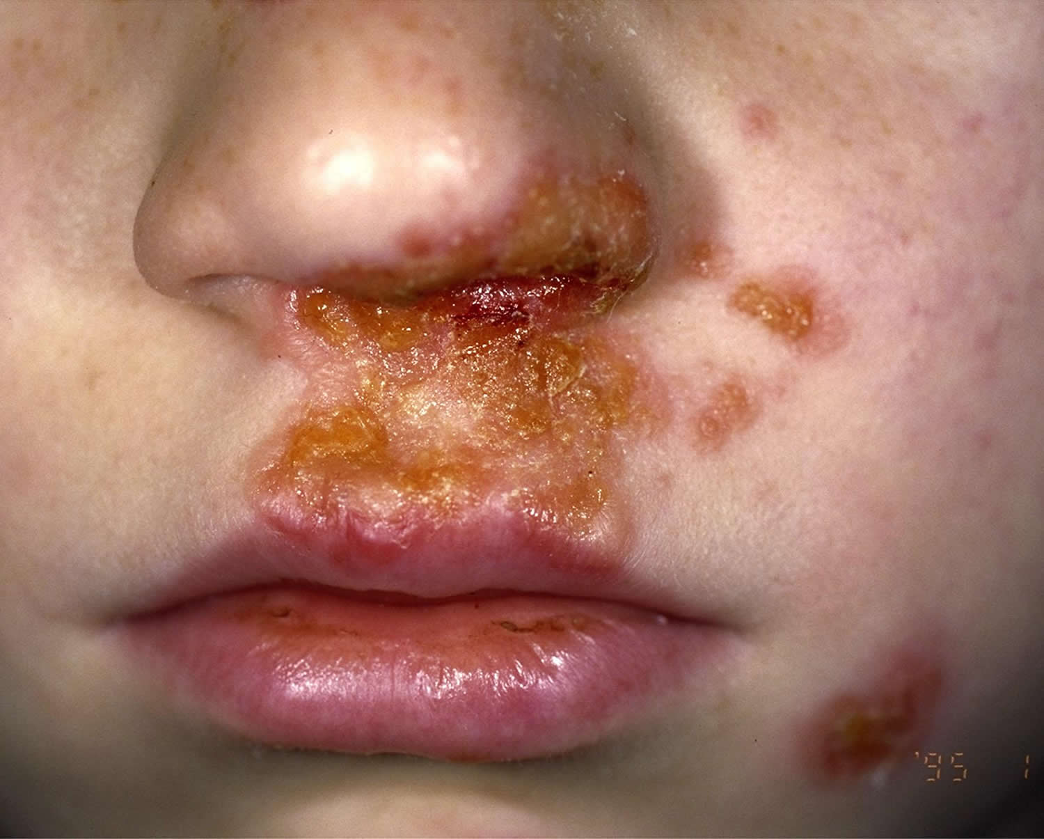
Figure 2. Impetigo on face
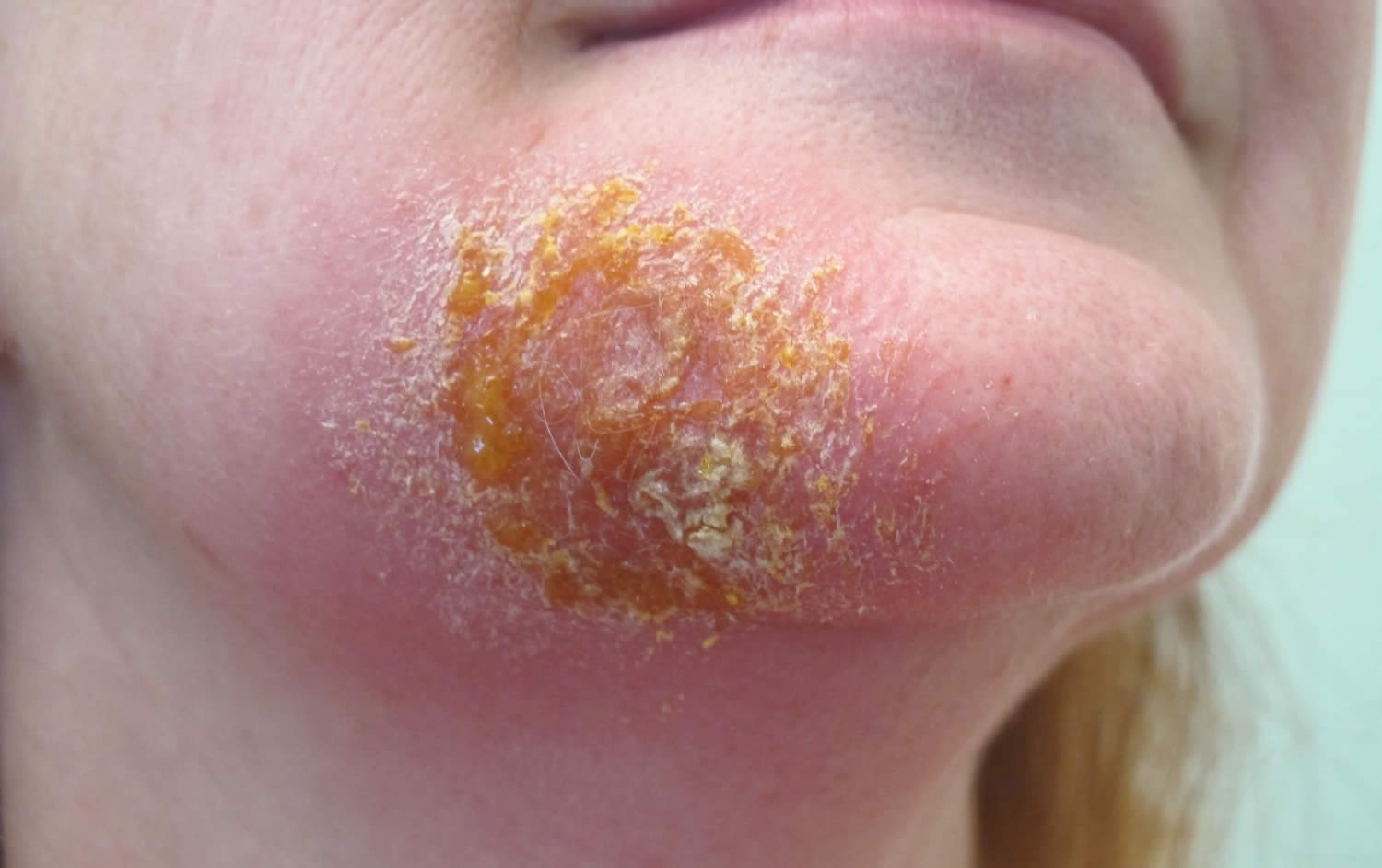
Figure 3. Impetigo on legs
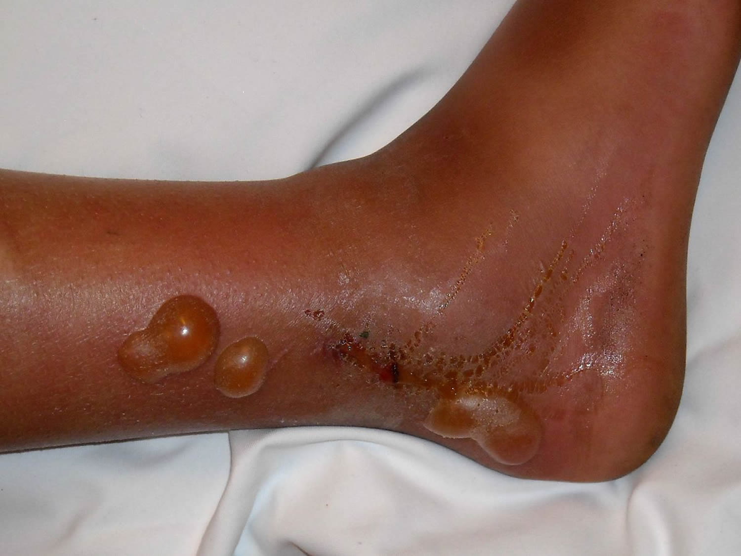
Figure 4. Impetigo on scalp
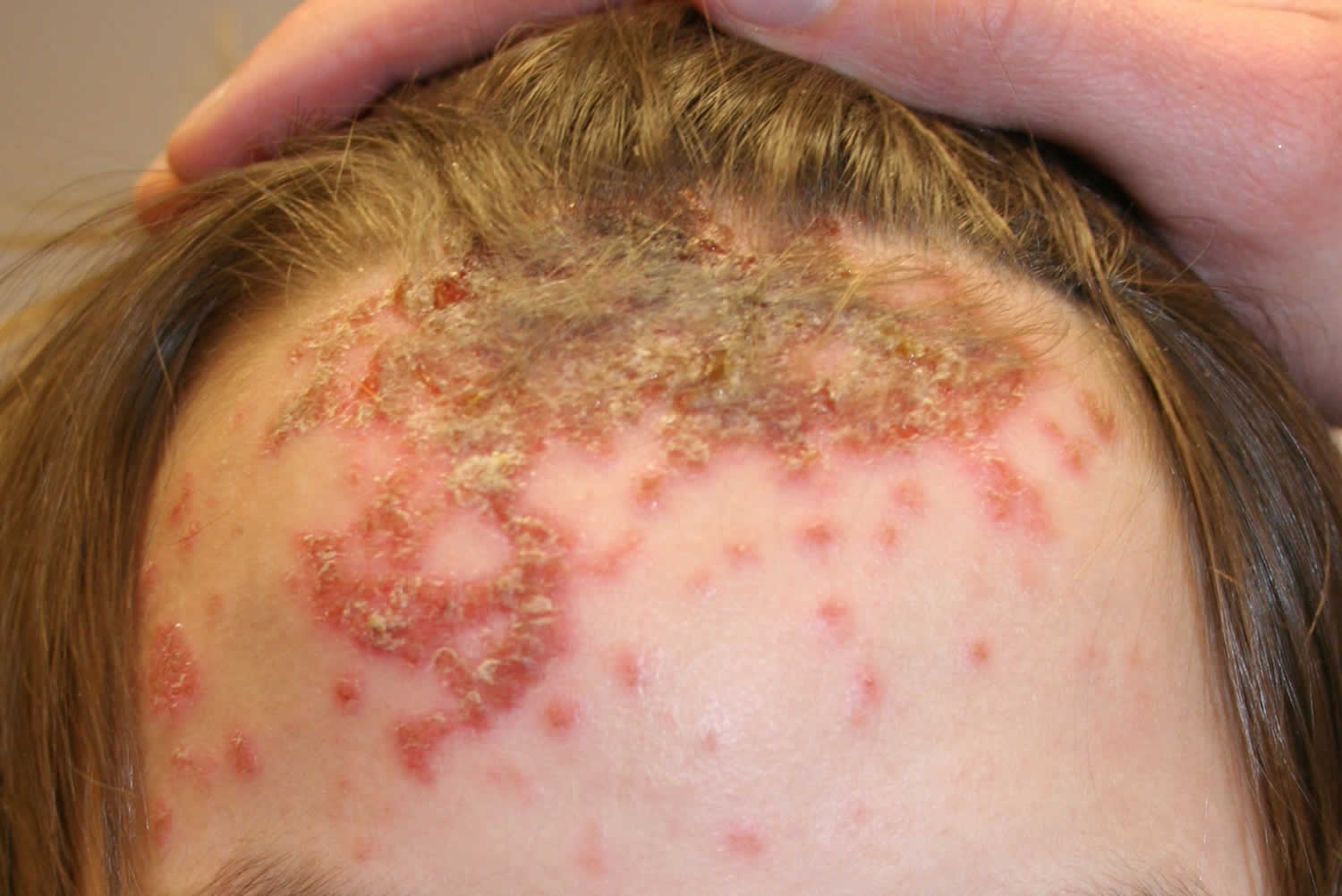
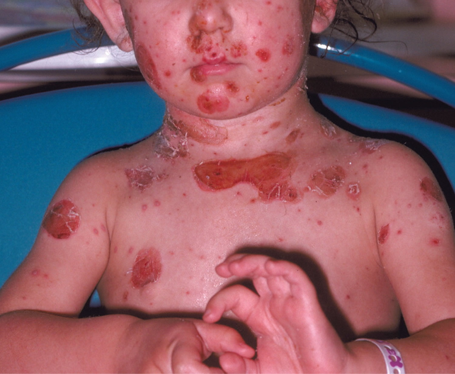
Figure 5. Impetigo on children
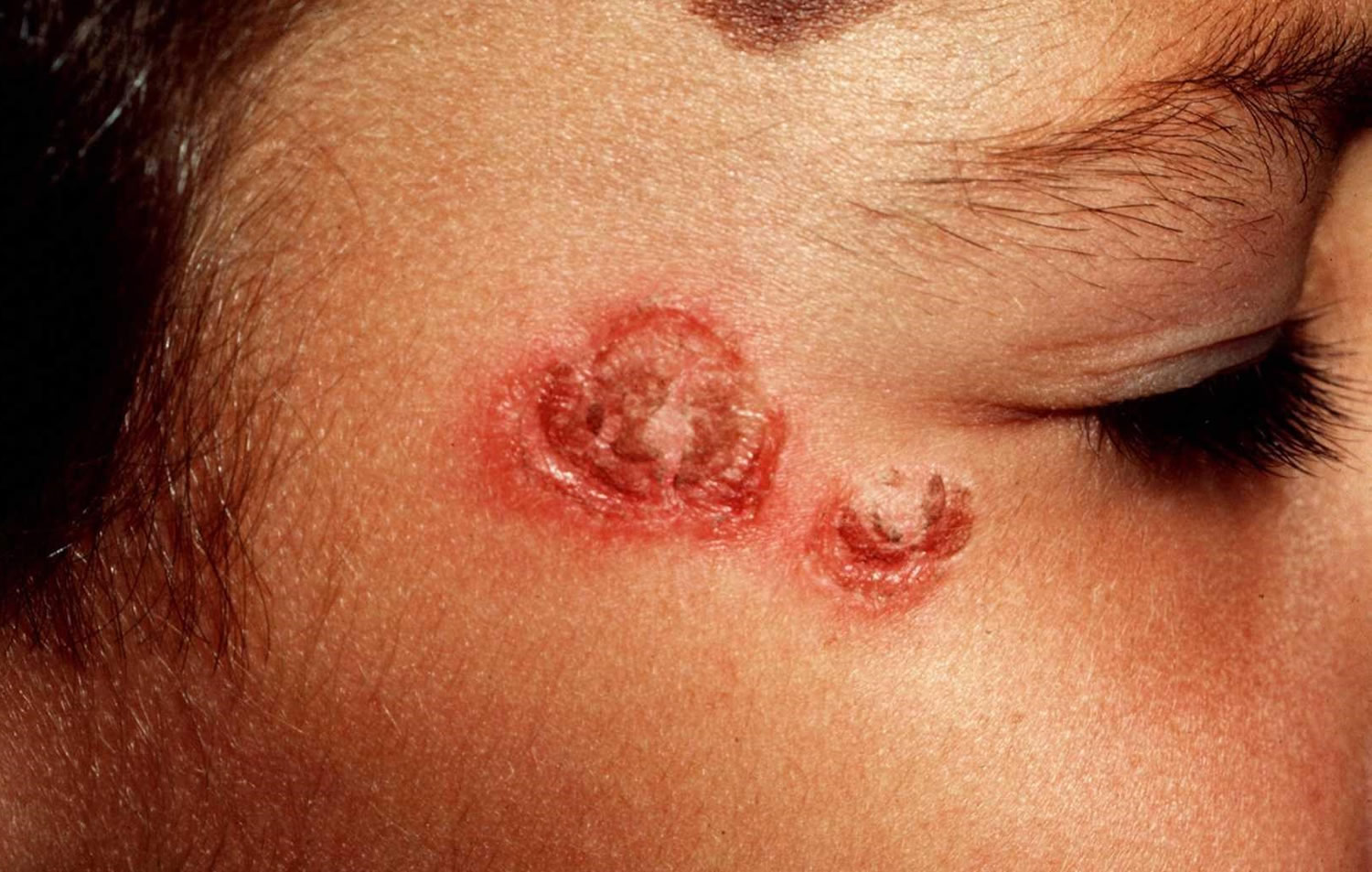
Figure 6. Impetigo rash
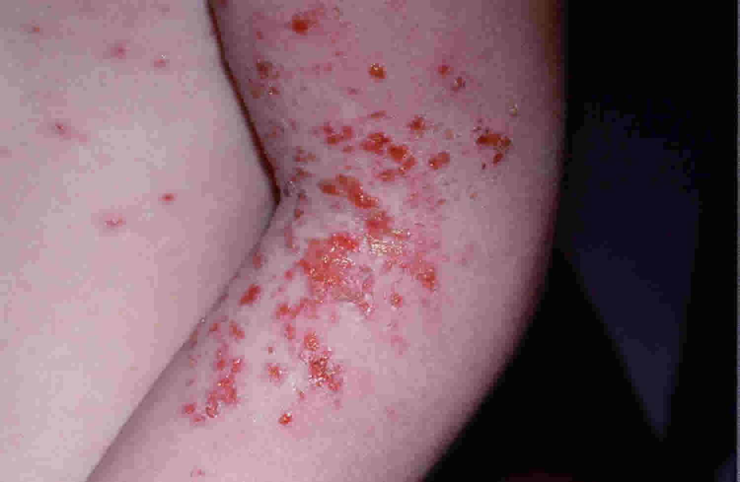
Figure 7. Bullous impetigo
Nonbullous impetigo
Nonbullous impetigo starts as a pink macule that evolves into a vesicle or pustule. Pustule or vesicle ruptures releasing serous contents which dries leaving a typical honey-colored crust. There’s minimal or no surrounding redness (erythema). Can spread rapidly with satellite lesions due to autoinoculation. “Kissing lesions” arise where two skin surfaces are in contact. People with non-bullous impetigo are typically otherwise well; they may experience some itching and regional enlarged lymph node (lymphadenopathy).
Non-bullous impetigo most commonly found on the face or extremities but skin on any part of the body can be involved.
Untreated nonbullous impetigo usually resolves within 2 to 4 weeks without scarring.
Bullous impetigo
Bullous impetigo presents with small vesicles that evolve quickly into flaccid transparent superficial, small or large thin roofed bullae which tend to spontaneously rupture and ooze yellow fluid leaving a scaley rim (collarette). It heals without scarring. People with bullous impetigo are more likely to have systemic symptoms of malaise, fever, and enlarged lymph node (lymphadenopathy).
Bullous impetigo is usually found on the face, trunk, extremities, buttocks, and perineal regions. Can spread distally due to autoinoculation.
Ecthyma
Ecthyma is a deep form of impetigo, as the same bacteria causing the infection are involved. Ecthyma causes deeper erosions of the skin into the dermis. Ecthyma starts as nonbullous impetigo but develops into a punched-out necrotic ulcer. This heals slowly, leaving a scar.
Streptococcus pyogenes and Staphylococcus aureus are the bacteria responsible for ecthyma.
Impetigo complications
Impetigo is usually a self-limited condition, and rarely, complications can occur after impetigo. These include cellulitis (nonbullous form), septicemia, osteomyelitis, septic arthritis, lymphangitis, lymphadenitis, guttate psoriasis, staphylococcal scalded skin syndrome, and acute poststreptococcal glomerulonephritis, with post streptococcal glomerulonephritis being the most serious 17. Post streptococcal glomerulonephritis is thought to be the result of an immune response that is triggered by Streptococcus pyogenes (group A beta-hemolytic Streptococcus) infection. The number of possible causes, incidence, and clinical severity of acute post streptococcal glomerulonephritis have decreased, because the causative organism of impetigo has shifted from Streptococcus pyogenes (group A beta-hemolytic Streptococcus) to Staphylococcus aureus 18. Most cases of poststreptococcal glomerulonephritis in the United States are associated with pharyngitis. The strains of Streptococcus pyogenes (group A beta-hemolytic Streptococcus) implicated in impetigo are thought to have minimal nephritogenic potential 18. There are no data to indicate that antibiotic treatment of impetigo has any effect on preventing the development of acute poststreptococcal glomerulonephritis, which can occur in up to 5% of patients with nonbullous impetigo 19, 20, 21, 22. Rheumatic fever does not appear to be a complication of impetigo 21.
Impetigo may lead to:
- Spread of the infection to other parts of the body (common)
- Kidney inflammation or failure (post-streptococcal glomerulonephritis) (rare). Acute kidney condition following infection with Streptococcus pyogenes (group A streptococcus). This is due to a type III hypersensitivity reaction and presents 2–6 weeks post-skin infection.
- Cellulitis. This potentially serious infection affects the tissues underlying your skin and eventually may spread to your lymph nodes and bloodstream. Untreated cellulitis can quickly become life-threatening.
- Wider spread infection: lymphangitis, and bacteremia.
- Staphylococcal scalded skin syndrome.
- Scarlet fever.
- Streptococcal toxic shock syndrome: a rare complication causing diffuse erythematous rash, hypotension, and pyrexia.
- Postinflammatory pigmentation.
- Permanent skin damage and scarring, particularly with ecthyma (very rare)
Soft tissue infection
The bacteria causing impetigo can become invasive, leading to cellulitis and lymphangitis; subsequent bacteremia might result in osteomyelitis, septic arthritis or pneumonia.
Staphylococcal scalded skin syndrome
In infants under 6 years of age or adults with renal insufficiency, localized bullous impetigo due to certain Staphylococcal serotypes can lead to a sick child with generalized staphylococcal scalded skin syndrome. Superficial crusting then tender cutaneous denudation on face, in skin folds, and elsewhere is due to circulating exfoliatin/epidermolysin, rather than direct skin infection. It does not scar.
Toxic shock syndrome
Toxic shock syndrome is rare and rarely preceded by impetigo. It causes fever, diffuse erythematous then desquamating rash, hypotension and involvement of other organs.
Post-streptococcal glomerulonephritis
Group A streptococcal infection may rarely lead to acute post-streptococcal glomerulonephritis 3–6 weeks after the skin infection. It is associated with anti-DNase B and antistreptolysin O (ASO) antibodies.
Rheumatic fever
Group A streptococcal skin infections have rarely been linked to cases of rheumatic fever and rheumatic heart disease. It is thought that this occurs because strains of group A streptococci usually found on the skin have moved to the throat (the more usual site for rheumatic fever-associated infection).
Impetigo diagnosis
Impetigo is usually diagnosed clinically but can be confirmed by bacterial swabs sent for microscopy (Gram positive cocci are observed), culture and sensitivity.
Blood count may reveal neutrophil leucocytosis when impetigo is widespread.
Skin biopsy is rarely necessary. Histological features are characteristic.
Non-bullous impetigo
- Gram-positive cocci
- Intraepidermal neutrophilic pustules,
- Dense inflammatory infiltrate in upper dermis
Bullous impetigo
- Split through granular layer of epidermis without inflammation or bacteria
- Acantholytic cells
- Minimal inflammatory infiltrate in upper dermis
- Resembles pemphigus foliaceus
Ecthyma
- Full thickness skin ulceration
- Gram stain shows cocci within dermis
Impetigo treatment
Impetigo needs to be treated with antibiotics, either topically (antibiotics rubbed onto the sores) or by mouth (oral antibiotics), and your doctor may order a culture in the lab to determine which bacteria are causing the rash. Gram stain and culture of the pus or exudates from skin lesions of impetigo and ecthyma are recommended to help identify whether Staphylococcus aureus and/or a Streptococcus pyogenes (group A beta-hemolytic Streptococcus) is the cause 20.
Make sure your child takes the medication for the full prescribed course or the impetigo could return.
- When it just affects a small area of the skin (and especially if it’s the non-bullous form), impetigo is treated with antibiotic ointment. Treatment of bullous and nonbullous impetigo should be with either mupirocin or retapamulin twice daily for 5 days 20.
- If the infection has spread to other areas of the body or the antibiotic ointment isn’t working, the doctor may prescribe an antibiotic pill or liquid to be taken for 7–10 days. Oral antibiotic therapy for ecthyma or impetigo should be a 7-day regimen with an agent active against Staphylococcus aureus unless cultures yield streptococci alone (when oral penicillin is the recommended agent) 20. Because Staphylococcus aureus isolates from impetigo and ecthyma are usually methicillin susceptible, dicloxacillin or cephalexin is recommended. When MRSA is suspected or confirmed, doxycycline, clindamycin, or sulfamethoxazole-trimethoprim (SMX-TMP) is recommended 20.
There is no over-the-counter (OTC) treatment for impetigo 23.
Quality evidence-based research for the most effective treatment of impetigo is lacking 24. In 2012, an updated Cochrane review on impetigo interventions evaluated 68 randomized controlled trials, including 26 on oral treatments and 24 on topical treatments. There was no clear evidence as to which intervention is most effective 25. Topical antibiotics are more effective than placebo and preferable to oral antibiotics for limited impetigo 24. Systemic antibiotics are often reserved for more generalized or severe infections in which topical therapy is not practical. Clinicians sometimes may choose both topical and systemic therapy 25. The ideal treatment should be effective, be inexpensive, have limited adverse effects, and should not promote bacterial resistance 26.
Untreated, impetigo often clears up on its own after a few days or weeks 27. The key is to keep the infected area clean with soap and water and not to scratch it. The downside of not treating impetigo is that some people might develop more lesions that spread to other areas of their body.
And you can infect others. To spread impetigo, you need fairly close contact — not casual contact — with the infected person or the objects they touched. Avoid spreading impetigo to other people or other parts of your body by:
- Cleaning the infected areas with soap and water (DO NOT scrub).
- Loosely covering scabs and sores until they heal.
- Gently removing crusty scabs.
- Washing your hands with soap and water after touching infected areas or infected persons.
- To prevent impetigo from spreading among family members, make sure everyone uses their own clothing, sheets, razors, soaps, and towels. Separate the bed linens, towels, and clothing of anyone with impetigo, and wash them in hot water. Keep the surfaces of your kitchen and household clean.
Table 1. Topical antibiotics for impetigo
| Medication | Instructions | Cost* |
|---|---|---|
| Fusidic acid 2% ointment† | Apply to affected skin three times daily for seven to 12 days | Available in Canada and Europe |
| Mupirocin 2% cream (Bactroban)‡ | Apply to affected skin three times daily for seven to 10 days; reevaluate after three to five days if no clinical response Approved for use in persons older than three months | 15-g tube: $48 ($89) 30-g tube: $50 ($144) |
| Mupirocin 2% ointment‡ | Apply to affected skin three times daily for seven to 14 days Dosing in children is same as adults Approved for use in persons older than two months | 22-g tube: $14 ($103) |
| Retapamulin 1% ointment (Altabax)§ | Apply to affected skin twice daily for five days Total treatment area should not exceed 100 cm2 in adults or 2% of total body surface area in children Approved for use in persons nine months or older | 15-g tube: NA ($130) 30-g tube: NA ($245) |
Footnotes: NA = not available
*Estimated retail price
† Coverage for Staphylococcus aureus (methicillin-susceptible) and streptococcus
‡ Coverage for Staphylococcus aureus (methicillin-susceptible) and streptococcus. Mupirocin resistant streptococcus has now been documented.
§ First member of the pleuromutilin class of antibiotics. Coverage for Staphylococcus aureus (methicillin-susceptible) and streptococcus.
[Source 28 ]Table 2. Oral antibiotics for impetigo
| Drug | Adult seven-day dose | Cost (for a typical course of treatment)* | Children seven-day dose | Cost* |
|---|---|---|---|---|
| Amoxicillin/clavulanate (Augmentin)† | 875/125 mg every 12 hours | $19 ($193) | Younger than three months: 30 mg per kg per day Three months or older: 25 to 45 mg per kg per day for those weighing less than 40 kg (88 lb); 875/125 mg every 12 hours for those weighing 40 kg or more Based on mg per kg per day of the amoxicillin component in divided doses every 12 hours | 1 bottle, 400/57 mg per 5 mL (100-mL oral suspension): $30 ($125) |
| Cephalexin (Keflex) | 250 mg every six hours or 500 mg every 12 hours | $5 ($90) | 25 to 50 mg per kg per day in divided doses every six to 12 hours | 1 bottle, 250 mg per 5 mL (100-mL oral suspension): $14 (NA) |
| Clindamycin‡ | 300 to 600 mg every six to eight hours | $18 ($200) | 10 to 25 mg per kg per day in divided doses every six to eight hours | 1 bottle, 75 mg per 5 mL (100-mL oral solution): $47 (pricing varies by region) |
| Dicloxacillin | 250 mg every six hours | $14 (NA) | 12.5 to 25 mg per kg per day in divided doses every six hours | See adult pricing: no liquid formulation available |
| Doxycycline§ | 50 to 100 mg every 12 hours | $15 ($95) | 2.2 to 4.4 mg per kg per day in divided doses every 12 hours Not recommend in children younger than eight years | 1 bottle, 25 mg per 5 mL (60-mL oral suspension): $20 (pricing varies by region) |
| Minocycline (Minocin)§ | 100 mg every 12 hours | $36 ($185) | Loading dose of 4 mg per kg for first dose (maximum dose of 200 mg), then 4 mg per kg per day in divided doses every 12 hours Maximum of 400 mg per day Not recommend in children younger than eight years | See adult pricing: no liquid formulation available |
| Trimethoprim/sulfamethoxazole§ | 160/800 mg every 12 hours | $4 (NA) | 8 to 10 mg per kg per day based on the trimethoprim component in divided doses every 12 hours | 1 bottle, 40/200 mg per 5 mL (100-mL oral suspension): $4 (pricing varies by region) |
Footnote: Because of emerging resistance, penicillin and erythromycin are no longer recommended treatments.
NA = not available
* Estimated retail price based on information obtained at http://www.goodrx.com. Generic price listed first; brand listed in parentheses.
† Good coverage for Staphylococcus aureus (methicillin-susceptible) and streptococcus.
‡ If methicillin-resistant Staphylococcus aureus (MRSA) is suspected or proven.
§ If methicillin-resistant Staphylococcus aureus (MRSA) is suspected or proven. There is no activity against streptococcus.
[Source 28 ]Home remedies for impetigo
Topical disinfectants
There are some studies on the benefits of nonantibiotic treatments, such as disinfectant soaps, but they lack statistical power 24. Disinfectants appear to be less effective than topical antibiotics and are not recommended 25. Studies comparing hexachlorophene (not available in the United States) with bacitracin and hydrogen peroxide with topical fusidic acid found the topical antibiotic to be more effective 29.
Natural therapies
The evidence is insufficient to recommend or dismiss popular herbal treatments for impetigo 30. Natural remedies such as tea tree oil; tea effusions; olive, garlic, and coconut oils; and Manuka honey have been anecdotally successful. The fact that impetigo is self-limited means that many “cures” could appear to be helpful without being superior to placebo. In one study, tea leaf ointment and oral cephalexin (Keflex) were similarly effective, with a cure rate of 81% vs. 79% 31. Tea tree oil (derived from Melaleuca alternifolia) appeared to be equivalent to mupirocin 2% for topical decolonization of MRSA 32.
Impetigo prognosis
Without treatment, impetigo heals in 14-21 days 7. About 20% of cases resolve spontaneously. Scarring is rare but some patients may develop pigmentation changes. Some patients may develop ecthyma. With treatment, cure occurs within 10 days 7. Neonates may develop meningitis. A rare complication is acute post streptococcal glomerulonephritis, which occurs 2-3 weeks after the skin infection 7.
References- Impetigo. Medline Plus. https://medlineplus.gov/impetigo.html
- Impetigo Care. American Academy of Pediatrics. https://www.healthychildren.org/English/health-issues/conditions/skin/pages/Impetigo-Care.aspx
- Bowen AC, Mahé A, Hay RJ, Andrews RM, Steer AC, Tong SY, Carapetis JR. The Global Epidemiology of Impetigo: A Systematic Review of the Population Prevalence of Impetigo and Pyoderma. PLoS One. 2015 Aug 28;10(8):e0136789. doi: 10.1371/journal.pone.0136789
- Pereira LB. Impetigo – review. An Bras Dermatol. 2014 Mar-Apr;89(2):293-9. doi: 10.1590/abd1806-4841.20142283
- Bryant AE, Stevens DL. Streptococcus pyogenes. In Bennett J, Dolin R, Blaser M, editors. 8th Mandell, Douglas, and Bennett’s Principles and Practice of Infectious Diseases. Philadelphia (PA): Elsevier; 2015:2:2285–300.
- Carapetis JR. The current evidence for the burden of group A streptococcal diseases. World Health Organization. Geneva. 2005. https://apps.who.int/iris/handle/10665/69063
- Nardi NM, Schaefer TJ. Impetigo. [Updated 2022 Oct 19]. In: StatPearls [Internet]. Treasure Island (FL): StatPearls Publishing; 2022 Jan-. Available from: https://www.ncbi.nlm.nih.gov/books/NBK430974
- D’Cunha N.M., Peterson G.M., Baby K.E., Thomas J. Impetigo: A need for new therapies in a world of increasing antimicrobial resistance. J. Clin. Pharm. Ther. 2017;43:150–153. doi: 10.1111/jcpt.12639
- Steer A.C., Danchin M.H., Carapetis J.R. Group A streptococcal infections in children. J. Paediatr. Child Health. 2007;43:203–213. doi: 10.1111/j.1440-1754.2007.01051.x
- Feaster T, Singer JI. Topical therapies for impetigo. Pediatr Emerg Care. 2010 Mar;26(3):222-7; quiz 228-31. doi: 10.1097/PEC.0b013e3181d1e71b
- Bangert, S., Levy, M. and Hebert, A.A. (2012), Bacterial Resistance and Impetigo Treatment Trends: A Review. Pediatric Dermatology, 29: 243-248. https://doi.org/10.1111/j.1525-1470.2011.01700.x
- Cole C, Gazewood J. Diagnosis and treatment of impetigo. Am Fam Physician. 2007 Mar 15;75(6):859-64. https://www.aafp.org/pubs/afp/issues/2007/0315/p859.html
- Hochedez P, Canestri A, Lecso M, Valin N, Bricaire F, Caumes E. Skin and soft tissue infections in returning travelers. Am J Trop Med Hyg. 2009 Mar;80(3):431-4.
- Luby SP, Agboatwalla M, Feikin DR, Painter J, Billhimer W, Altaf A, Hoekstra RM. Effect of handwashing on child health: a randomised controlled trial. Lancet. 2005 Jul 16-22;366(9481):225-33. doi: 10.1016/S0140-6736(05)66912-7
- Taiaroa G, Matalavea B, Tafuna’i M, Lacey JA, Price DJ, Isaia L, Leaupepe H, Viali S, Lee D, Gorrie CL, Williamson DA, Jack S. Scabies and impetigo in Samoa: A school-based clinical and molecular epidemiological study. Lancet Reg Health West Pac. 2020 Dec 29;6:100081. doi: 10.1016/j.lanwpc.2020.100081
- Mitchell E, Bell S, Thean LJ, Sahukhan A, Kama M, Koroivueti A, Kaldor J, Steer A, Romani L. Community perspectives on scabies, impetigo and mass drug administration in Fiji: A qualitative study. PLoS Negl Trop Dis. 2020 Dec 4;14(12):e0008825. doi: 10.1371/journal.pntd.0008825
- Weinberg JM, Tyring SK. Retapamulin: an antibacterial with a novel mode of action in an age of emerging resistance to Staphylococcus aureus. J Drugs Dermatol. 2010 Oct;9(10):1198-204.
- Ilyas M, Tolaymat A. Changing epidemiology of acute post-streptococcal glomerulonephritis in Northeast Florida: a comparative study. Pediatr Nephrol. 2008 Jul;23(7):1101-6. doi: 10.1007/s00467-008-0778-1
- George A, Rubin G. A systematic review and meta-analysis of treatments for impetigo. Br J Gen Pract. 2003 Jun;53(491):480-7. https://www.ncbi.nlm.nih.gov/pmc/articles/PMC1314624/pdf/12939895.pdf
- Dennis L. Stevens, Alan L. Bisno, Henry F. Chambers, E. Patchen Dellinger, Ellie J. C. Goldstein, Sherwood L. Gorbach, Jan V. Hirschmann, Sheldon L. Kaplan, Jose G. Montoya, James C. Wade, Executive Summary: Practice Guidelines for the Diagnosis and Management of Skin and Soft Tissue Infections: 2014 Update by the Infectious Diseases Society of America, Clinical Infectious Diseases, Volume 59, Issue 2, 15 July 2014, Pages 147–159, https://doi.org/10.1093/cid/ciu444
- Bisno AL, Stevens DL. Streptococcal infections of skin and soft tissues. N Engl J Med. 1996 Jan 25;334(4):240-5. doi: 10.1056/NEJM199601253340407
- Brown, J., Shriner, D.L., Schwartz, R.A. and Janniger, C.K. (2003), Impetigo: an update. International Journal of Dermatology, 42: 251-255. https://doi.org/10.1046/j.1365-4362.2003.01647.x
- How to Treat Impetigo and Control This Common Skin Infection. U.S. Food and Drug Administration. https://www.fda.gov/forconsumers/consumerupdates/ucm048837.htm
- George A, Rubin G. A systematic review and meta-analysis of treatments for impetigo. Br J Gen Pract. 2003;53(491):480–487.
- Koning S, van der Sande R, Verhagen AP, et al. Interventions for impetigo. Cochrane Database Syst Rev. 2012;(1):CD003261.
- Feaster T, Singer JI. Topical therapies for impetigo. Pediatr Emerg Care. 2010;26(3):222–227, quiz 228–231.
- How to Treat Impetigo and Control This Common Skin Infection. U.S. Food and Drug Administration. https://www.fda.gov/ForConsumers/ConsumerUpdates/ucm048837.htm
- Impetigo: Diagnosis and Treatment. Am Fam Physician. 2014 Aug 15;90(4):229-235. http://www.aafp.org/afp/2014/0815/p229.html
- Christensen OB, Anehus S. Hydrogen peroxide cream: an alternative to topical antibiotics in the treatment of impetigo contagiosa. Acta Derm Venereol. 1994;74(6):460–462.
- Martin KW, Ernst E. Herbal medicines for treatment of bacterial infections: a review of controlled clinical trials. J Antimicrob Chemother. 2003;51(2):241–246.
- Sharquie KE, al-Turfi IA, al-Salloum SM. The antibacterial activity of tea in vitro and in vivo (in patients with impetigo contagiosa). J Dermatol. 2000;27(11):706–710.
- Caelli M, Porteous J, Carson CF, Heller R, Riley TV. Tea tree oil as an alternative topical decolonization agent for methicillin-resistant Staphylococcus aureus. J Hosp Infect. 2000;46(3):236–237.


