
What is a karyotype
A karyotype is an individual’s collection of chromosomes. The term also refers to a laboratory technique that produces an image of an individual’s chromosomes. The karyotype is used to look for abnormal numbers or structures of chromosomes.
As you know, your body is made up of billions of cells. You have skin cells, heart cells, brain cells, etc. These cells all have special functions in your body, but one thing they have in common is that each cell has a set of 46 chromosomes, or 23 pairs. The first 22 pairs of chromosomes are the same in men and women and the 23rd pair is different. Women have two ‘X’ chromosomes (XX) for their 23rd pair, and men have one ‘X’ chromosome and one ‘Y’ chromosome (XY). See pictures below of a set of male/female chromosomes, also called a karyotype.
When you hear the word “karyotype”, think about a picture of chromosomes. When somebody has their blood studied to look at how many chromosomes they have and whether the chromosomes are complete, scientists come up with a picture in which they can line up all the chromosomes and count them. That way scientists can tell whether or not somebody has all the proper number of chromosomes, which is 46, and that way the scientists can look at the X and the Y chromosomes and determine if it’s a female or male. Your doctor might order a chromosome study and look at a karyotype if he/she were worried that a child might have an extra or missing bit of chromosome material. So one of the most common things you can see on karyotyping is an extra chromosome 21 (Trisomy 21), which is associated with Down syndrome. Doctors also get karyotypes when pregnant women choose to have testing on their unborn fetus, and the karyotype allows your doctor to look at and count the chromosomes to determine whether or not the child is affected by having an extra chromosome.
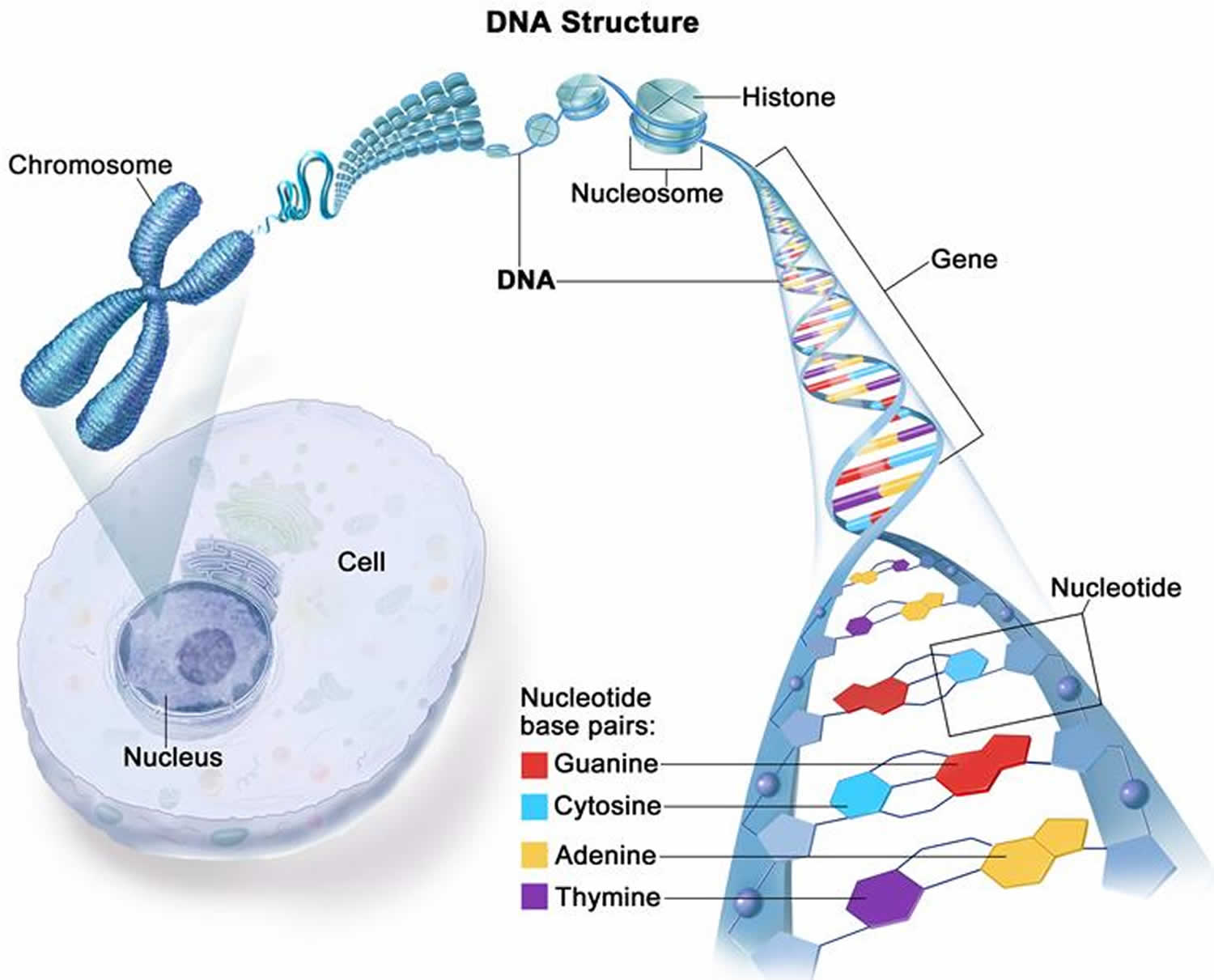
Along the chromosomes are the individual instructions, or genes, that tell your body how to grow and function; instructions for things such as hair color, eye color, height, etc. Since you have two copies of every chromosome, you also have two copies of every gene. All in all it is estimated that you have about 20,000 pairs of genes, one set form your mother and one set from your father. Males only have one X chromosome though, so they only have one copy of all of the genes on the X chromosome, and one copy of the genes on the Y chromosome. Genes are specific instructions which contain your genetic code or DNA (deoxyribonucleic acid). DNA is made up of four similar chemicals, called bases: adenine (A), thymine (T), cytosine (C ), and guanine (G). The specific order of these letters (bases) is what makes the genes (instructions) work properly. If one or more of these letters is changed, deleted, or duplicated and this change causes the gene to not work as usual, this is called a mutation. Mutations in your genes may lead to genetic conditions in you or your children.
In order for your body to grow and function as usual, it is important that you have the typical 46 chromosomes. It is also critical that you have only two of each. Many of the genetic conditions that are discussed in prenatal genetics are due to a baby having an extra (trisomy) or missing (monosomy) chromosome. An important thing to remember is that you have no control over how many chromosomes are packaged in your egg or sperm. If a baby has an extra or missing chromosome, there is nothing that could have done to cause or prevent it.
Other types of genetic conditions involve changes at the gene level, not the chromosome level. Some of the more common genetic conditions involving a single gene can be categorized into 3 main ways that they can be passed down in a family: autosomal recessive, autosomal dominant and X-linked. These three terms are also referred to as “patterns of inheritance” and understanding the basics behind these terms will help you learn about the chances of your baby having certain genetic conditions.
Figure 1. Human karyotype (normal karyotype)
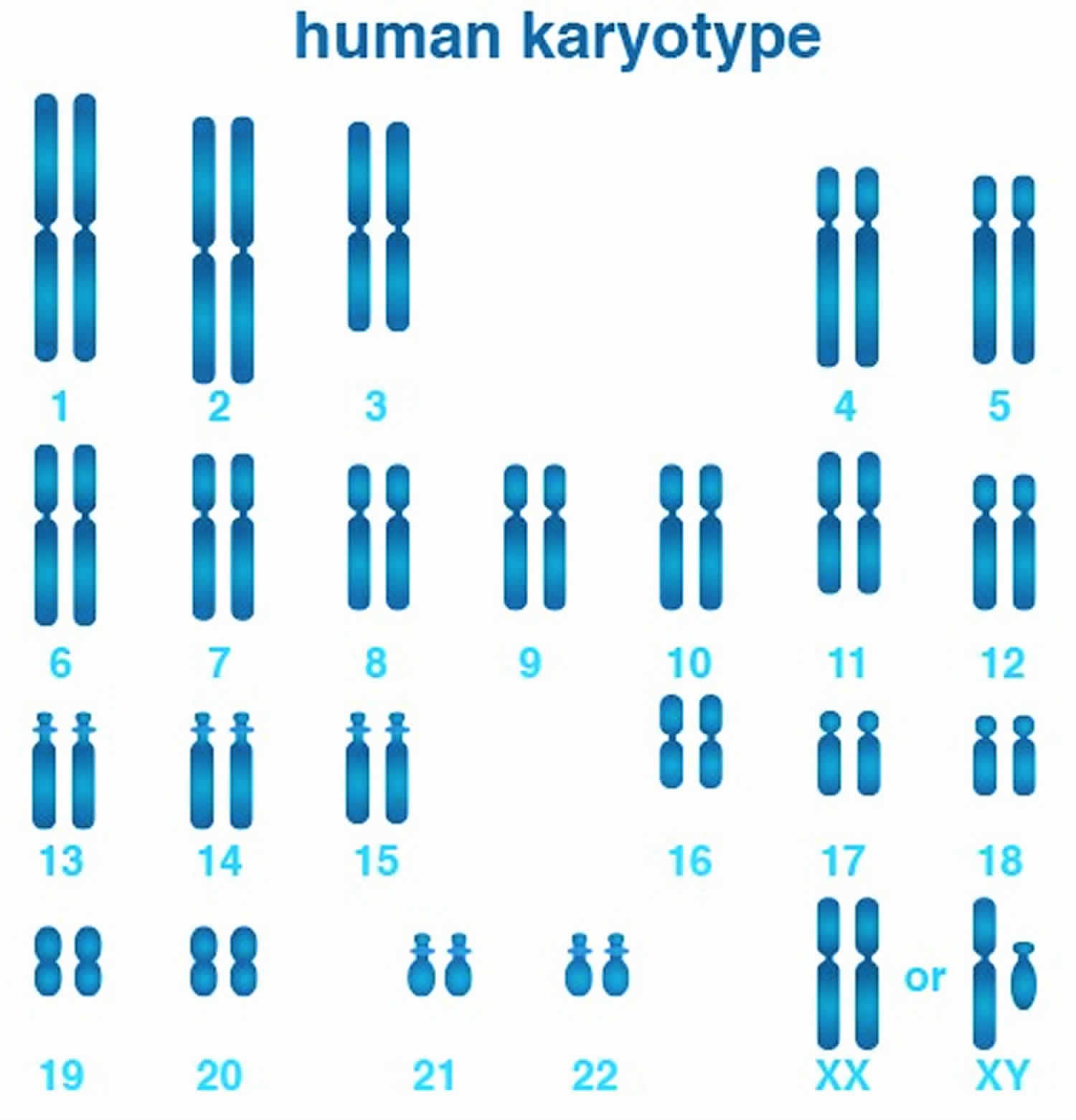 Figure 2. Female karyotype (normal female karyotype)
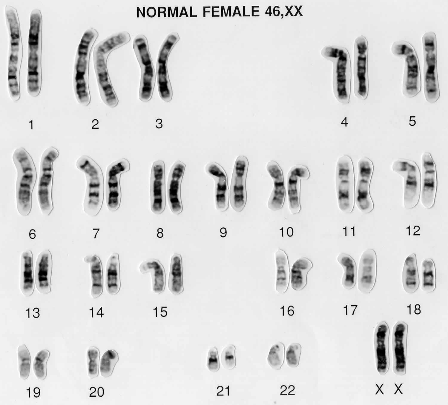
Figure 3. Male karyotype (normal male karyotype)
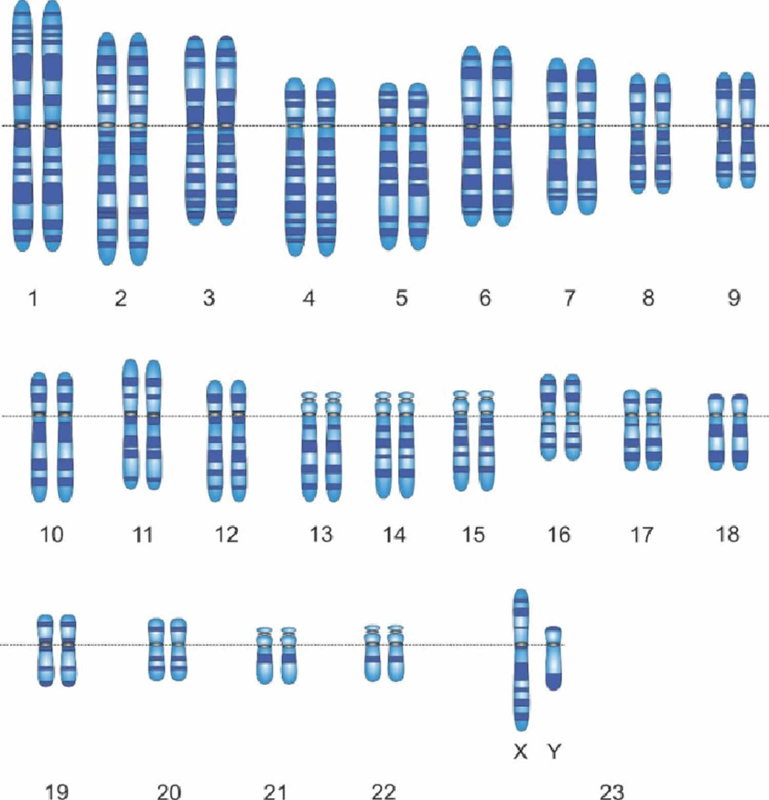
Figure 4. DNA structure
What is a chromosome?
The total of an individual’s genetic information is called their genome. The genome consists of structures called chromosomes that are composed of very long double strands of DNA. In the nucleus of each cell, the DNA molecule is packaged into thread-like structures called chromosomes. Each chromosome is made up of DNA tightly coiled many times around proteins called histones that support its structure.
Chromosomes are not visible in the cell’s nucleus—not even under a microscope—when the cell is not dividing. However, the DNA that makes up chromosomes becomes more tightly packed during cell division and is then visible under a microscope. Most of what researchers know about chromosomes was learned by observing chromosomes during cell division.
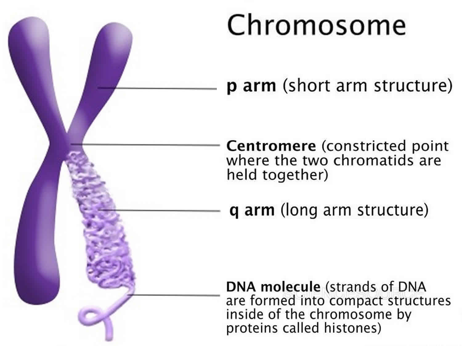
Each chromosome has a constriction point called the centromere, which divides the chromosome into two sections, or “arms.” The short arm of the chromosome is labeled the “p arm.” The long arm of the chromosome is labeled the “q arm.” The location of the centromere on each chromosome gives the chromosome its characteristic shape, and can be used to help describe the location of specific genes.
Figure 5. Chromosome structure
How many chromosomes do people have?
In humans, each cell normally contains 23 pairs of chromosomes, for a total of 46 chromosomes. One-half of each pair of chromosomes is inherited from an individual’s mother and the other half of the pair is inherited from an individual’s father. Twenty-two of the 23 pairs of chromosomes are called autosomes, look the same in both males and females. The 23rd pair, are the sex chromosomes, differ between males and females. Females have two copies of the X chromosome (XX), while males have one X and one Y chromosome (XY). The sex chromosomes determine a person’s sex; normal males have one X and one Y chromosome (XY) (see Figure 3 above) while normal females have two X chromosomes (XX) (see Figure 2 above).
The 22 autosomes are numbered by size. The other two chromosomes, X and Y, are the sex chromosomes. This picture of the human chromosomes lined up in pairs is called a karyotype (see Figure 1).
What is a gene?
A gene is the basic physical and functional unit of inheritance. Genes are passed from parents to offspring and contain the information needed to specify traits. Genes are pieces of DNA and most genes contain the information for making a specific protein. Some genes act as instructions to make molecules called proteins. However, many genes do not code for proteins. In humans, genes vary in size from a few hundred DNA bases to more than 2 million bases. Genes are arranged, one after another, on structures called chromosomes. Each chromosome contains many genes. A chromosome contains a single, long DNA molecule, only a portion of which corresponds to a single gene. The Human Genome Project estimated that humans have between 20,000 and 25,000 genes arranged on their chromosomes.
Every person has two copies of each gene, one inherited from each parent. Most genes are the same in all people, but a small number of genes (less than 1 percent of the total) are slightly different between people. Alleles are forms of the same gene with small differences in their sequence of DNA bases. These small differences contribute to each person’s unique physical features.
Scientists keep track of genes by giving them unique names. Because gene names can be long, genes are also assigned symbols, which are short combinations of letters (and sometimes numbers) that represent an abbreviated version of the gene name. For example, a gene on chromosome 7 that has been associated with cystic fibrosis is called the cystic fibrosis transmembrane conductance regulator; its symbol is CFTR.
Karyotype test
Karyotyping or chromosome analysis, is a test that evaluates the number and structure of a person’s chromosomes in order to detect abnormalities. Chromosomes are thread-like structures within each cell nucleus and contain the body’s genetic blueprint. Each chromosome contains thousands of genes in specific locations. These genes are responsible for a person’s inherited physical characteristics and they have a profound impact on growth, development, and function.
Humans have 46 chromosomes, present as 23 pairs. Twenty-two pairs are found in both sexes (autosomes) and one pair (sex chromosomes) is present as either XY (in males) or XX (in females). Normally, all cells in the body that have a nucleus will contain a complete set of the same 46 chromosomes, except for the reproductive cells (eggs and sperm), which contain a half set of 23. This half set is the genetic contribution that will be passed on to a child. At conception, half sets from each parent combine to form a new set of 46 chromosomes in the developing fetus.
Chromosomal abnormalities include both numerical and structural changes. For numerical changes, anything other than a complete set of 46 chromosomes represents a change in the amount of genetic material present and can cause health and development problems. For structural changes, the significance of the problems and their severity depends upon the chromosome that is altered. The type and degree of the problem may vary from person to person, even when the same chromosome abnormality is present.
A chromosomal karyotyping examines a person’s chromosomes to determine if the right number is present and to determine if each chromosome appears normal. It requires experience and expertise to perform properly and to interpret the results. While theoretically almost any cells could be used to perform testing, in practice it is usually performed on amniotic fluid to evaluate a fetus and on lymphocytes (a white blood cell) from a blood sample to test all other ages. Alternately, white blood cells may be obtained from bone marrow aspirations to look for changes in individuals suspected of having hematologic or lymphoid diseases (e.g., leukemia, lymphoma, myeloma, refractory anemia).
What is a karyotype used for?
A chromosomal karyotype is used to detect chromosome abnormalities and thus used to diagnose genetic diseases, some birth defects, and certain disorders of the blood or lymphatic system.
A chromosomal karyotype may be performed for:
- A fetus, using amniotic fluid or chorionic villi (tissue from the placenta):
- If one or more of a woman’s pregnancy screening tests, such as the first trimester Down syndrome screen or the second trimester maternal serum screening, are abnormal.
- If a pregnant woman is having amniotic fluid analysis performed because she is considered at higher than normal risk of having a baby with a birth defect.
- If fetal structural and/or developmental abnormalities are detected, such as during an ultrasound.
- If there is a known chromosomal abnormality in the family line.
- A woman or a couple, prior to pregnancy, to evaluate her or their chromosomes, especially if a woman has experienced previous miscarriages or infertility.
- Tissue from a miscarriage or stillbirth, to help determine if the cause was due to a chromosomal abnormality in the fetus.
- An infant who is born with congenital abnormalities, including physical birth defects, mental retardation, delayed growth and development, or signs of a specific genetic disorder.
- A person with infertility or one who shows signs of a genetic disorder.
- Family members, to detect specific chromosomal abnormalities when they have been detected in a child or another family member.
- A person who has been diagnosed with certain types of leukemia, lymphoma, refractory anemia, or cancer as these conditions can lead to acquired changes in chromosomes; this testing may be performed on blood or a bone marrow sample.
When is karyotype analysis ordered?
A chromosome analysis may be ordered when a fetus is suspected of having a chromosomal abnormality, when an infant has congenital abnormalities, when a woman experiences miscarriages or infertility, and when an adult shows signs of a genetic disorder.
It may also be ordered to detect the presence of a chromosomal abnormality in family members when it has been detected in a child or in another family member.
It may be ordered to detect acquired chromosomal abnormalities when an individual has leukemia, lymphoma, myeloma, refractory anemia, or another cancer.
What is abnormal karyotype?
Interpretation of karyotype test results must be done by a person with specialized training in cytogenetics. Some findings are relatively straightforward, such as an extra chromosome 21 (Trisomy 21) indicating Down syndrome, but others may be very complex.
Although there will be typical signs with specific chromosomal abnormalities, the effects and the severity may vary from person to person and often cannot be reliably predicted.
Table 1. Some examples of abnormalities that chromosome analysis may reveal
| Trisomy | This is the presence of an extra chromosome, a third instead of a pair. Diseases associated with trisomies include Down syndrome (associated with a Trisomy of chromosome 21), Patau syndrome (Trisomy 13), Edward syndrome (Trisomy 18), and Klinefelter syndrome (a male with an extra X chromosome – XXY instead of XY). |
| Monosomy | This is the absence of one of the chromosomes. An example of monosomy is Turner syndrome (a female with a single X chromosome – X instead of XX). Most other monosomies are not compatible with life. |
| Deletions | These are missing pieces of chromosomes and/or genetic material. Some may be small and difficult to be detected. |
| Duplications | These represent extra genetic material and may be present on any chromosome, such as the presence of two horizontal bands at a specific location instead of one. |
| Translocations | With translocations, pieces of chromosomes break off and reattach to another chromosome. If it is a one-to-one switch and all of the genetic material is present (but in the wrong place), it is said to be a balanced translocation. If it is not, then it is called an unbalanced translocation. |
| Genetic Rearrangement | With this, genetic material is present on a chromosome but not in its usual location. A person could have both rearrangement and duplication or deletion. An almost infinite number of rearrangements are possible. Interpreting the affects of the changes can be challenging. |
Duplications, deletions, translocations, and genetic rearrangements can cause a myriad of health and development issues. It depends upon what genes are missing or are present in too many copies.
Some genetic rearrangements will be variations that do not cause noticeable symptoms. Balanced translocations (where two chromosomes have swapped portions of themselves but all of the genetic material is present) may cause no problems for the person who has them but may cause problems in their children.
Many hematologic and lymphoid malignancies (e.g., leukemia, lymphoma, myeloma, myelodysplasia) are associated with chromosomal abnormalities, which can help diagnose the disease and/or predict the clinical course of the disease.
Abnormal karyotype (chromosomal disorders) that may be detected include:
- Down syndrome (Trisomy 21), caused by an extra chromosome 21; this may occur in all or most cells of the body.
- Edwards syndrome (Trisomy 18), a condition associated with severe mental retardation; caused by an extra chromosome 18.
- Patau syndrome (Trisomy 13), caused by an extra chromosome 13.
- Klinefelter syndrome, the most common sex chromosome abnormality in males; caused by an extra X chromosome (XXY).
- Turner syndrome, caused by missing one X chromosome in females.
- Chronic myelogenous leukemia, a classic 9;22 translocation that is diagnostic of the disease.
Is there anything else I should know about the karyotype test?
Since the sex chromosomes (XX or XY) are identified during the chromosome analysis, this test will also, as a byproduct, definitely determine the sex of a fetus.
Some chromosome alterations are too small or subtle to detect with karyotyping. Other testing technique such as fluorescent in situ hybridization (FISH) or a microarray may sometimes be performed to further investigate chromosomal abnormalities.
It is possible for people to have cells in their body with differing genetic material. This happens because of changes early in the development of a fetus that lead to the development of distinctly different cell lines and is called mosaicism. An example of this is some cases of Down syndrome. The affected person can have some cells with an extra third chromosome 21 and some cells with the normal pair.
Why does the karyotype test take several days to perform?
The cells that are tested must be cultured and cell division promoted. The amount of time that this takes will vary from sample to sample. Highly complex, abnormal karyotypes may require a longer time to evaluate.
Should everyone have karyotype testing done?
Chromosome analysis is frequently performed, but it is not indicated as a general screening test. The majority of people will never need to have one done.
Can a karyotype analysis be performed in my home using a test kit?
No, karyotype analysis requires specialized equipment to perform and expertise to interpret. In most cases, samples will be sent to a reference laboratory.
How is a karyotype made?
Early studies of the human karyotype simply stained chromosomes within cells with Giemsa and “squashed” them between the cover slip and slide 1. Most cells were not at the proper mitotic phase for chromosomes to be observed, and chromosome separation was poor. The exact count was uncertain: most scientists accepted the number 48. The breakthrough came in 1952, when a technician in the lab of T. C. Hsu accidentally substituted distilled water for the normal saline solution used in washing the cells just before “squashing”. This “hypotonic” treatment caused the cell nuclei to swell, and allowed the chromosomes to separate before squashing. A further refinement was “dropping” the cells onto the slide at arm’s length, which caused the nuclei to burst on impact, further separating them. Finally, the use of a plant spindle-poison Colchicine allows chromosomes to be arrested at mitotic metaphase, during their maximum state of compaction. These experiments quickly established the human chromosome number as 2n = 46 chromosomes.
Classification of chromosomes into seven groups by size and relative centromere position established the so-called “Denver System” in 1960. Chromosomes within groups B – G were not readily distinguishable from each other. The X chromosome is in the C group, and the Y is in the G group: males are recognizable by five small G-type chromosomes. Modern banding techniques allow each chromosome in the karyotype to be distinguished individually.
How is the sample collected for karyotype testing?
- A blood sample is obtained by inserting a needle into a vein in the arm.
- Amniotic fluid and chorionic villi are collected from a pregnant woman by a healthcare practitioner using amniocentesis or chorionic villus sampling procedures.
- Bone marrow or tissue sample collections require a biopsy procedure to be performed.
How is the karyotype test is performed?
- Taking a sample of a person’s cells, culturing them in nutrient-enriched media to promote cell division in vitro. This is done in order to select a specific time during the cells’ growth phase when the chromosomes are easiest to distinguish.
- Isolating the chromosomes from the nucleus of the cells, placing them on a slide, and treating them with a special stain.
- Taking microphotographs of the chromosomes.
- In jigsaw puzzle fashion, rearranging the pictures of the chromosomes to match up pairs and arrange them by size, from largest to smallest, numbers 1 to 22, followed by the sex chromosomes as the 23rd pair.
- The pictures also allow the chromosomes to be vertically oriented. Each chromosome looks like a striped straw. It has two arms that differ in length [a short arm (p) and a long arm (q)] (see Figure 5), a pinched-in area between the arms called a centromere, and a series of light and dark horizontal bands. The length of the arms and the location of the bands help determine top from bottom.
- Once the chromosome photo arrangement is completed, a laboratory specialist evaluates the chromosome pairs and identifies any abnormalities that may be present.


