Lichenoid keratosis
Lichenoid keratosis also known as benign lichenoid keratosis, solitary lichen planus, lichen planus-like keratosis or involuting lichenoid plaque, is a common, benign (harmless) and often solitary skin lesion. Lichenoid keratosis appears to result from the inflammatory destruction of a pre-existing epidermal lesion such as a solar lentigo or seborrheic keratosis.
Lichenoid keratosis is usually a small, solitary, inflamed macule or thin pigmented plaque 1. Multiple eruptive lichenoid keratoses in sun-exposed sites are also described. Their color varies from an initial reddish brown to a greyish purple/brown as the lesion resolves several weeks or months later.
Lichenoid keratosis generally develops in fair-skinned patients aged 30–80 years. It is twice as common in females as than males. It is most commonly seen in Caucasians and rarely affects Asians, African Americans or Hispanics.
Lichenoid keratosis key points
- Generally asymptomatic.
- Lesions are benign, no treatment is required.
- More common in Caucasians.
- More common on UV-exposed areas, but can affect any part of the body.
- Predominantly affect adults.
- Equal incidence in males and females.
- Morphology:
- Usually solitary
- Generally less than 1 cm in diameter
- Flat or slightly elevated
- The surface tends to be smooth, although occasionally scaly or warty
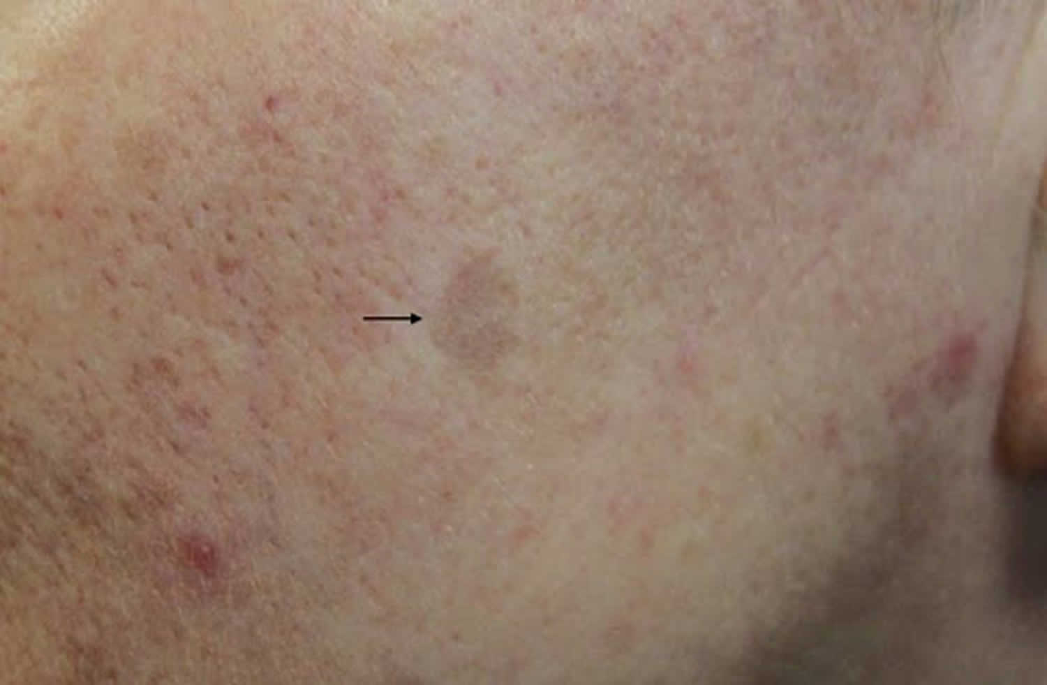
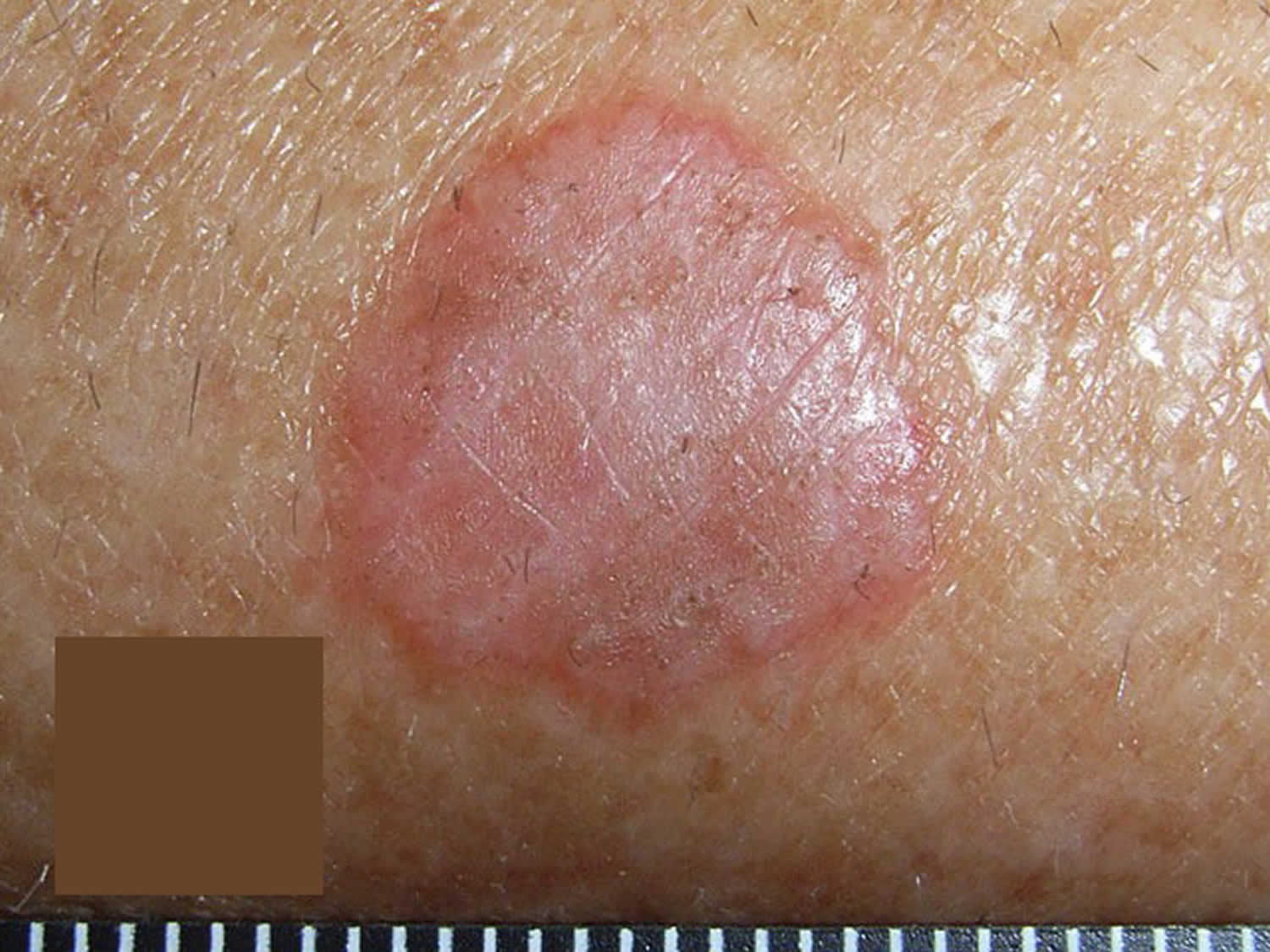
- Lesions typically progress from an inflammatory phase, where there may be some pink-red discolouration, to a pigmented phase, where the lesion becomes grey-brown in color.
- Dermoscopic appearance:
- Grey granules uniformly distributed throughout the lesion
Figure 1. Lichenoid keratosis
Lichenoid keratosis causes
Lichenoid keratosis is an inflammatory reaction arising in a regressing existing solar lentigo or seborrheic keratosis. It is not known what causes the reaction, but triggers include minor trauma such as friction, drugs, dermatitis, and sun exposure.
Lichenoid keratosis symptoms
The clinical features of lichenoid keratosis vary depending on the inflammatory stage of the lesion (see Table 1).
Other features of lichenoid keratosis are:
- A solitary lesion is present in 90% of cases of lichenoid keratosis, with other patients presenting with few to many lesions.
- It is most commonly found on the upper trunk, followed by the distal upper extremities, and less commonly on the head and neck.
- Size ranges from a few millimeters to one centimeter or more in size.
- The skin surface may be smooth, scaly or warty.
- The lesion is often symptomless or it may be itchy or have a mild stinging sensation.
Table 1. Clinical features of lichenoid keratosis
| Classic, bullous or atypical subtype | |
|---|---|
| Clinical features |
|
| Histopathology |
|
| Early or interface subtype | |
| Clinical features |
|
| Histopathology |
|
| Late regressed or atrophic subtype | |
| Clinical features |
|
| Histopathology |
|
Lichenoid keratosis diagnosis
Lichenoid keratosis is diagnosed by its clinical and dermoscopic appearance, which reveals uniform clusters of grey dots and, depending on the stage of the lesion, may show signs of an original pre-existing lentigo or seborrheic keratosis. In time, signs of the original lesion disappear. Later on the grey dots also disappear, as the lesion resolves to reveal normal skin.
Because clinical examination and dermatoscopy may not be able to differentiate between lichenoid keratosis and other solitary erythematous lesions that could be melanocytic, non-melanocytic benign, malignant or inflammatory, a punch or shave skin biopsy may be necessary.
Histopathology of resembles that of lichen planus or lichenoid drug eruption, with some slight differences. Remnants of the original solar lentigo or seborrheic keratosis may be evident.
Lichenoid keratosis treatment
Lichenoid keratosis is harmless and resolves spontaneously. If there is any doubt about the diagnosis, dermatoscopic digital images can be taken and used in follow-up a few months later.
Lichenoid keratosis can be removed if desired by liquid nitrogen, electrosurgery or curettage.
Multiple eruptive lichenoid keratoses may be effectively treated with the oral retinoid, acitretin.
To date there have been no reports of lichenoid keratosis turning into malignant skin tumors.
References


