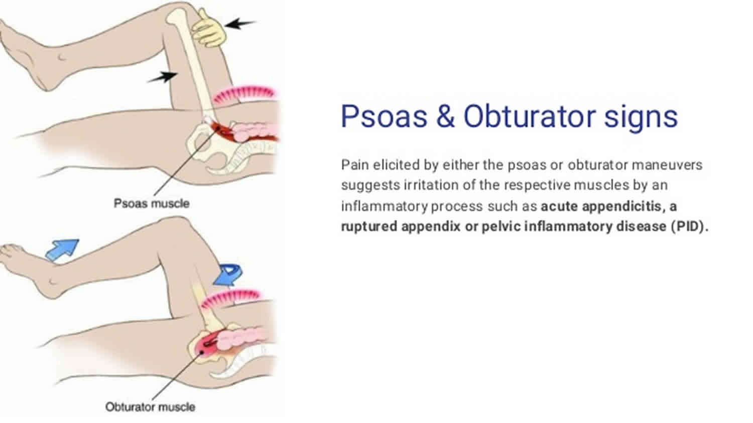
Obturator sign
Obturator sign also known as Cope’s obturator test, is an indicator of irritation to the obturator internus muscle 1. The technique for detecting the obturator sign, called the obturator test, is carried out on each leg in succession. The patient lies on her/his back with the hip and knee both flexed at ninety degrees. The examiner holds the patient’s ankle with one hand and knee with the other hand. The examiner internally rotates the hip by moving the patient’s ankle away from the patient’s body while allowing the knee to move only inward. This is flexion and internal rotation of the hip.
Obturator sign technique 2:
- Patient lies supine with right thigh flexed 90 degrees
- Examiner immobilizes right ankle with right hand
- Left hand rotates right hip by:
- Pull right knee laterally (hip external rotation)
- Pull right knee medially (hip internal rotation)
- Left obturator/pelvis examined in similar fashion
In the clinical practice, the obturator test is performed when acute appendicitis is suspected. In this condition, the appendix becomes inflamed and enlarged. The appendix may come into physical contact with the obturator internus muscle, which will be stretched when obturator test maneuver is performed on the right leg. This causes pain and is evidence in support of an inflamed pelvic appendix. However, evidence shows that the obturator test does not adequately diagnose appendicitis, but can be used in conjunction with other signs and symptoms to make a diagnosis 3.
Psoas sign
Psoas sign also known as Cope’s psoas test 4 or Obraztsova’s sign 5, is a medical sign that indicates irritation to the iliopsoas group of hip flexors in the abdomen, and consequently indicates that the inflamed appendix is retrocaecal in orientation (as the iliopsoas muscle is retroperitoneal).
The technique for detecting the psoas sign is carried out on the patient’s right leg. The patient lies on his/her left side with the knees extended. The examiner holds the patient’s right thigh and passively extends the hip. Alternatively, the patient lies on their back, and the examiner asks the patient to actively flex the right hip against the examiner’s hand 6.
If abdominal pain results, it is a “positive psoas sign”. The pain results because the psoas borders the peritoneal cavity, so stretching (by hyperextension at the hip) or contraction (by flexion of the hip) of the muscles causes friction against nearby inflamed tissues. In particular, the right iliopsoas muscle lies under the appendix when the patient is supine, so a positive psoas sign on the right may suggest appendicitis. A positive psoas sign may also be present in a patient with a psoas abscess. It may also be positive with other sources of retroperitoneal irritation, e.g. as caused by hemorrhage of an iliac vessel.
References- Allan B Wolfson (1 September 2009). Harwood-Nuss’ Clinical Practice of Emergency Medicine. Lippincott Williams & Wilkins. pp. 1222–. ISBN 978-0-7817-8943-1
- Diagnostic Accuracy of History, Physical Examination, Laboratory Tests, and Point‐of‐care Ultrasound for Pediatric Acute Appendicitis in the Emergency Department: A Systematic Review and Meta‐analysis. Acad Emerg Med. 2017 May;24(5):523-551. doi: 10.1111/acem.13181. https://doi.org/10.1111/acem.13181
- McGee, S. (2007). Evidence based physical diagnosis (2nd ed.). Philadelphia: Saunders.
- Bhat, Sriram; M, Sriram Bhat (30 December 2012). SRB’s Manual of Surgery. JP Medical Ltd. p. 1279. ISBN 978-93-5025-944-3.
- Augustin, Goran (12 May 2014). Acute Abdomen During Pregnancy. Springer. p. 8. ISBN 978-3-319-05422-3.
- Bickley, Lynn S. Bates’ Guide to Physical Exam and History Taking (9th ed.). Lippincott, Williams, and Wilkins. p. 390.


