Ophthalmic herpes zoster
Ophthalmic herpes zoster also known as herpes zoster ophthalmicus, is defined as herpes zoster involvement of the ophthalmic division of the trigeminal nerve. Herpes zoster commonly called shingles, results from reactivation of varicella-zoster virus (VZV) infection, the virus that causes chicken pox and shingles 1. Ophthalmic herpes zoster is an ocular disease which usually manifests as a unilateral painful skin rash in a dermatomal distribution of the trigeminal nerve shared by the eye and ocular adnexa. Ophthalmic herpes zoster occurs typically in older adults but can present at any age and occurs after reactivation of latent varicella-zoster virus (VZV) present within the sensory spinal or cerebral ganglia 2. The varicella-zoster virus remains dormant in the dorsal root or other sensory ganglia after the primary varicella (chickenpox) infection 3. The trigeminal ganglion is the most frequent site of latency (65–90%) for varicella-zoster virus 4. In the United States, 1 million new cases are reported per year 5. Typically, the incidence of herpes zoster increases with age, as well as with diseases and drugs, which can lead to immunosuppression.
Ophthalmic herpes zoster is the second most common type of herpes zoster. Ophthalmic herpes zoster accounts for 20% of all cases and approximately 50% of patients will have ocular involvement 6. Herpes zoster can affect virtually every eye tissue, resulting in conjunctivitis, scleritis, keratitis, uveitis, keratouveitis, and endotheliitis. Nearly two-thirds of patients with ophthalmic herpes zoster demonstrate a keratitis that often is associated with loss of corneal sensation. Corneal complications can occur due to inflammatory and immune reaction to the virus, vasculopathy, and neuropathy, and may commonly present with neurotrophic keratitis. Neurotrophic keratitis then results in dry eye disease, persistent corneal epithelial defects, inflammation, corneal melting, and potentially perforation, possibly leading to significant vision loss or legal blindness.
Corneal nerve damage is likely to occur after viral infections (herpes simplex and herpes zoster) 7. Nearly two-thirds of patients with ophthalmic herpes zoster will develop loss of corneal sensation due to nerve damage, necrotic ganglionitis, or damage to the mesencephalic nucleus in the brainstem 8. Corneal nerve fibers exert important trophic influences on the ocular surface, and a large number of nerves contain substance P and/or calcitonin gene-related peptide. Cornea sensory nerves interact with the epithelium through soluble mediators, such as substance P, and are essential to the ocular surface homeostasis and function 9. Recent reviews have shown the correlation between corneal nerve alterations and sensation 10. Thus, the loss of sensation as a result of nerve damage can lead to neurotrophic keratopathy, which represents one of the most challenging ocular diseases. The prognosis of neurotrophic keratopathy depends mainly on the level of hypo- or anesthesia and its consequences, which can result in other conditions, such as dry eye disease, exposure keratopathy, neurotrophic ulcers, and limbal stem cell deficiency.
Ophthalmic herpes zoster key points
- The eye is affected in about half of cases of ophthalmic division of the trigeminal nerve varicella-zoster virus reactivation.
- Keratitis and/or uveitis can be severe and cause morbidity.
- Appearance of the typical herpes zoster rash is usually diagnostic.
- Treatment is with oral antivirals and usually topical corticosteroids and pupillary dilation.
- Give the recombinant herpes zoster vaccine to all immunocompetent adults ≥ 50 years.
Figure 1. Ophthalmic herpes zoster
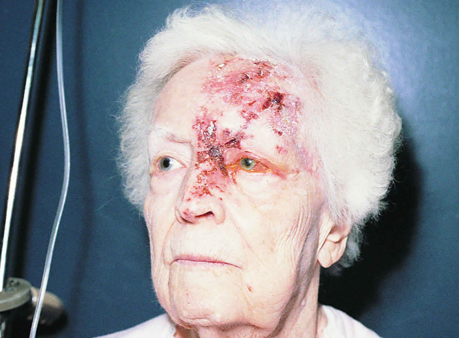
Footnote: Vesicular lesions developed over the V1 distribution of the ophthalmic branch of the trigeminal nerve in this patient with acute herpes zoster. Lesions on the tip of her nose suggest involvement of the nasociliary nerve that supplies the globe.
Ophthalmic herpes zoster cause
Ophthalmic herpes zoster is caused by the varicella-zoster virus which has re-activated from its dormant status in the dorsal ganglion cells of the central nervous system. From there it may travel along neurons to the sensory axons of the skin to form vesicular lesions.
Virulence of the varicella-zoster virus and the immune status of the host are primary factors leading to the development of ophthalmic herpes zoster. The incidence and severity of herpes zoster increases with advancing age with patients over the age of 60 at the highest risk 11. One study showed that racial factors may play role since elderly black patients were one fourth as likely as elderly white patients to develop herpes zoster 12. Further supporting the theory that immune system status plays a role, patients that are treated with immunosuppressive drugs have a significantly increased risk for herpes zoster 13. An immunocompromised patient is more likely to have a prolonged illness, more likely to recur, and more likely to develop myelitis and vasculopathy 14. The risk of herpes zoster is 15 times greater in men with HIV than in men without HIV 15.
Ophthalmic herpes zoster pathophysiology
Unless the immune system is compromised the varicella-zoster virus is usually suppressed. However, for reasons that are not fully understood, the virus reactivates from its dormant state in the sensory ganglion, replicates in the nerve cells, and sheds virions from the cells that are carried down the axons to the skin served by that ganglion. The local immune response results in skin blisters or ocular inflammation depending on which tissues are affected. Perineuritis causes intense pain along the nerve distribution 16. Paresthesia and segmental pain at the area supplied by trigeminal nerve may be noted before the onset of rash. Aging, immunosupression therapy, and psychological stress all could be factors resulting in reactivation of the virus 17.
Ophthalmic herpes zoster prevention
A varicella-zoster shingles vaccination (recombinant herpes zoster vaccine) is now recommended for patients over the age of 60, regardless of whether they have had herpes zoster or been given the older, live-attenuated vaccine. Although 90% of the population has prior exposure to varicella-zoster virus, there appears to be a benefit to booster immunity especially since the community incidence of native varicella-zoster virus exposure has decreased. This recombinant vaccine decreases the chance of getting herpes zoster by 97% for adults 50 to 69 years and 91% for adults ≥ 70 years. During a recent study, a 50% decreased incidence of zoster and 66% reduction of postherpetic neuralgia was demonstrated 18.
Ophthalmic shingles symptoms
Tingling of the forehead may occur before any other symptoms called a prodrome. The skin of the forehead and sometimes the tip of the nose are covered with small, extremely painful, red blisters. Infection of the eye causes ache, redness, light sensitivity, and eyelid swelling. The cornea (the clear layer in front of the iris and pupil) can get infected and inflamed. Months and years later, the cornea can become swollen, severely damaged, and scarred. The structures behind the cornea can become inflamed (uveitis), the pressure in the eye can increase (glaucoma), and the cornea can become numb, which leaves it vulnerable to injuries.
Many cases of ophthalmic herpes zoster exhibit a prodromal period of fever, malaise, headache, and eye pain prior to eruption of the skin rash. The patient may describe eye pressure, tearing, eye redness, or decreasing vision. Pain in the distribution of the trigeminal nerve may be severe.
Erythematous skin lesions with macules, papules, vesicles, pustules, and crusting lesions in the distribution of the trigeminal nerve. Hutchinson’s sign is defined as skin lesions at the tip, side, or root of the nose. This is a strong predictor of ocular inflammation and corneal denervation in ophthalmic herpes zoster, especially if both branches of the nasociliary nerve are involved 19.
Ophthalmic shingles complications
Herpes zoster skin manifestations in the eyelids can affect the deep dermis. Therefore, scars can result in ptosis, lid scarring, ectropion, and entropion.
Keratitis and/or uveitis may be severe and followed by scarring. Late complications—glaucoma, cataract, chronic or recurrent uveitis, corneal scarring, corneal neovascularization, and hypesthesia—are common and may threaten vision. Postherpetic neuralgia may develop late. Patients may develop episcleritis (without increased risk of visual loss) and/or retinitis (with risk of severe visual loss).
Scleritis can cause scleral, limbal and corneal atrophy. Inflammation in the cornea, optic nerve, retina and choroid could result in permanent vision loss. Corneal scars commonly affect the vision requiring hard contact lens or cornea transplantation interventions. Postherpetic neuraligia occurs in 36.6% of patients over the age of 60 and in 47.5% over the age of 70 19.
Ophthalmic shingles diagnosis
Ophthalmic shingles diagnosis is based on a typical acute herpes zoster rash on the forehead, eyelid, or both or on a characteristic history plus signs of previous zoster rash (eg, atrophic hypopigmented scars). Vesicular or bullous lesions in this distribution that do not yet involve the eye suggest significant risk and should prompt an ophthalmologic consultation to determine whether the eye is involved. Culture and immunologic or polymerase chain reaction (PCR) studies of skin at initial evaluation or serial serologic tests are done only when lesions are atypical and the diagnosis uncertain.
History
Herpes zoster is an acute, painful, vesicular eruption distributed along a single dermatome and is associated with a prodrome of fever, malaise, headache, and pain in the dermatome. The vesicles typically crust and will heal within 2-6 weeks.
Physical examination
- Visual acuity with best correction
- External examination of eyelids, periocular skin, and scalp.
- Measurement of intraocular pressure
- Slit-lamp biomicroscopy of the anterior segment with special attention to any staining cornea defects, stroma opacities, cornea vascularization, keratic precipitates, and anterior chamber cell and flare.
- Dilated examination of the lens, macula, peripheral retina, optic nerve, and vitreous.
Clinical diagnosis
Dermatome distribution pain and rash with associated ocular findings strongly suggest ophthalmic herpes zoster. Cornea epithelial defects, decreased corneal sensation, and ocular inflammation in any of the layers of the eye also correlate with the diagnosis. ophthalmic herpes zoster iritis is frequently associated with high intraocular pressure.
Diagnostic procedures
Cornea sensation should be tested prior to instillation of anesthetic drops. This can be accomplished with a Cachet-Bonnet anesthesiometer or with a fine wisp of a cotton-tip applicator. Decreased sensation is very suspicious for herpes simplex virus (HSV). Using fluorescein, cornea epithelial defects should be ruled out.
Laboratory test
Cornea scrapings of any skin lesions may be sent to the laboratory for a Tzanck smear. However, this test will not differentiate between herpes simplex virus (HSV) and Varicella. Alternatively, cultures may be sent for immunoflourescence assays to look for IgM specific to varicella-zoster virus. Viral cultures and polymerase chain reaction testing may also be obtained to diagnose varicella-zoster virus 20.
Ophthalmic herpes zoster differential diagnosis
Not many disease processes produce a painful vescicular rash. However, other conditions that create vescicular rashes should be considered especially in the absence of pain: for example, contact dermatitis and vaccinia dermatitis. Other disease entities that can mimic cornea findings include recurrent erosion, noninfectious cornea melts, infectious keratitis. There are numerous infectious and non-infectious entities that can exhibit ocular inflammation in the aqueous, vitreous, optic nerve, retina, and choroid.
Ophthalmic herpes zoster treatment
Skin rash treatment should prevent bacterial superinfection. With careful examination inflammation in all layers of the eye should be ruled out and treated with antivirals and steroids if indicated. When a skin rash is the only clinical sign, follow-up care must be directed to ruling out any ocular manifestations that may develop.
Medical therapy
Oral acyclovir 800 mg oral five times daily for 7 to 10 days is the standard treatment. Alternatively, your doctor could use famciclovir 500 mg oral three times daily or valacyclovir 1000mg oral three times daily. If the systemic condition warrants or if the patient is unable to tolerate food by mouth then acyclovir 5-10 mg/kg IV every 8 hours for 5 days may be utilized.
Topical steroids (e.g. prednisolone acetate 1%) should be used for interstitial keratitis and uveitis. For episodes of scleritis, retinitis, choroiditis, and optic neuritis, systemic steroids by mouth or intravenous administration should be strongly considered.
For increased intraocular pressure commonly found in herpes trabeculitis, topical steroids should be administered as well as aqueous suppressants (e.g. timolol, brimonidine, dorzolamide, acetazolamide).
Pain should be treated with narcotics if warranted. Neuropathic pain responds well to amitriptyline 25 mg oral each night at bedtime and can decrease the incidence of postherpetic neuralgia. Capsaicin cream applied to the rash may decrease pain as well 21. Pregabalin 150mg /day in divided doses may alleviate pain due to acute herpetic neuralgia.
Medical follow up
Depending on the ocular findings and severity, the patient should be monitored every 1 to 7 days during the acute episode. Monitoring every 3-12 months afterwards may be helpful to monitor for delayed complications such as ocular hypertension, cataract, and cornea scarring. If there is any concern of future exacerbations, viral prophylaxis should be considered with acyclovir 400 mg oral twice daily.
Surgery
Cornea transplantation and/or hard contact lenses is sometimes required for lesions that cause severe cornea thinning and loss of structural integrity of the eye. Scars that are visually significant and refractory to medical therapy may require transplantation. Vitrectomy or retina detachment surgery may be performed especially in cases of acute retina necrosis. Glaucoma filtration surgery is sometimes performed if there are difficulties with maintaining optimum intraocular pressure. If the intraocular inflammation and/or steroid treatment causes a cataract then cataract surgery may be performed when the disease process is quiescent.
Surgical follow up
Depending on the type of surgery performed, the patient should be closely monitored for severe inflammation commonly associated with herpes after surgical procedures. Viral prophylaxis with antiviral therapy and steroids should be strongly considered.
Ophthalmic herpes zoster prognosis
Prognosis is greatly variable and dependent on long-term complications. Long-term vision loss, need for surgery, and long-term antiviral prophylaxis are all possible.
References- Cavalcanti BM, Cruzat A, Sahin A, Pavan-Langston D, Samayoa E, Hamrah P. In vivo confocal microscopy detects bilateral changes of corneal immune cells and nerves in unilateral herpes zoster ophthalmicus. Ocul Surf. 2018;16(1):101–111. doi:10.1016/j.jtos.2017.09.004 https://www.ncbi.nlm.nih.gov/pmc/articles/PMC5756670
- Liesegang TJ. Herpes Zoster Ophthalmicus. Ophthalmology 2008;115:S3-S12.
- Gnann JW, Jr, Whitley RJ. Clinical practice. Herpes zoster. N Engl J Med. 2002;347:340–6.
- Liesegang TJ. Varicella-zoster virus eye disease. Cornea. 1999;18:511–31.
- Oxman MN, Levin MJ, Johnson GR, Schmader KE, Straus SE, Gelb LD, et al. A vaccine to prevent herpes zoster and postherpetic neuralgia in older adults. N Engl J Med. 2005;352:2271–84.
- Schmader KE, Dworkin RH. Natural history and treatment of herpes zoster. J Pain. 2008;9:S3–9.
- Bonini S, Rama P, Olzi D, Lambiase A. Neurotrophic keratitis. Eye (Lond) 2003;17:989–95.
- Pavan-Langston D. Herpes zoster ophthalmicus. Neurology. 1995;45:S50–1.
- Muller LJ, Marfurt CF, Kruse F, Tervo TM. Corneal nerves: structure, contents and function. Exp Eye Res. 2003;76:521–42.
- Pritchard N, Edwards K, Shahidi AM, Sampson GP, Russell AW, Malik RA, et al. Corneal markers of diabetic neuropathy. The ocular surface. 2011;9:17–28.
- Chapman RS, Cross KW, Fleming DM. The incidence of shingles and its implications for vaccination policy. Vaccine 2003;21:2541-2547.
- Schmader K, George LK, Burchett BM, et al. Racial differences in the occurrence of herpes zoster. J Infect Dis 1995;171:701-704.
- Cohen PR, Grossman ME. Clinical features of human immunodeficiency virus-associated disseminated herpes zoster virus infection –a review of the literature. Clin Exp Dermatol 1989;14:273-276.
- Hilt DC, Bucholz D, Krumholz A, et al. Herpes zoster ophthalmicus and delayed contralateral hemiparesis caused by cerebral angiitis: diagnosis and management approaches. Ann Neurol 1983;14:543-553.
- Buchbinder SP, Katz MH, Hessol NA, et al. Herpes zoster and human immunodeficiency virus infection. J Infect Dis 1992;166:1153-1156.
- Schmader K. Herpes zoster and postherpetic neuralgia in older adults. Clin Geriatr Med 2007;23(3):615-632.
- Thomas SL, Hall AJ. What does epidemiology tell us about risk factors for herpes zoster?. Lancet Infect Dis 2004;4(1):26-33.
- Oxman MN, Levin MJ, Johnson GR. Shingles Prevention Study Group. A vaccine to prevent herpes zoster and postherpetic neuralgia in older adults. N Engl J Med. 2005;352(22):2271-2284.
- Zaal MJ, Volker-Dieben HJ, D’Amarao J. Prognostic value of Hutchinson’s sign in acute herpes zoster ophthalmicus. Graefes Arch Clin Exp Ophthalmol 2003;241:187-191.
- Burns DA, et al. Rook’s Textbook of Dermatology, 7th Edition.
- Kunimoto DY, et al. Wills Eye Manual, 4th Edition.


