What is sebaceous hyperplasia
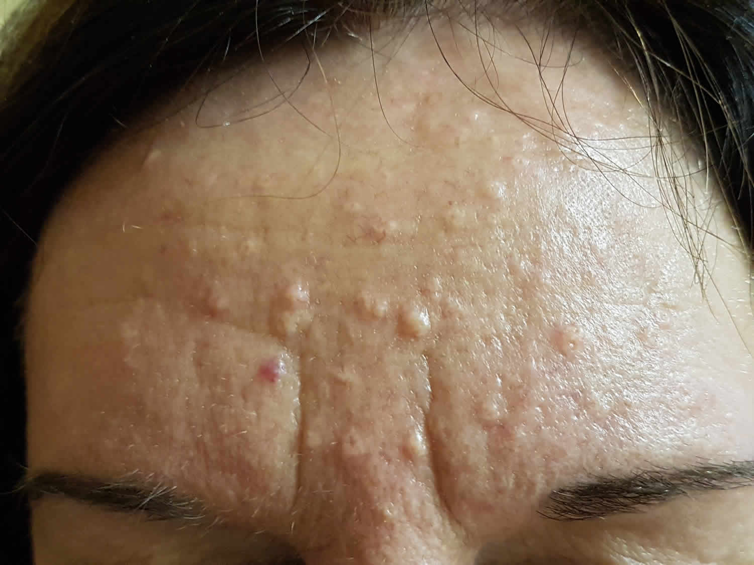
Sebaceous hyperplasia is the term used for enlarged sebaceous glands (skin oil glands) seen on the forehead or cheeks of the middle-aged and elderly (usually fifth or sixth decade) and continue to appear into later life and is seen in about 1% of the US population. Sebaceous hyperplasia is also common in newborns 1. Of 1000 consecutive neonates examined in an Iranian prospective cohort study, 43.7% had sebaceous hyperplasia 2. Sebaceous hyperplasia is a form of benign hair follicle tumor, but they may be associated with non-melanoma skin cancer in transplantation patients 3. Sebaceous hyperplasia may be more prevalent in immunosuppressed patients: for example, in a patient following organ transplantation. About 10–16% of people on long-term cyclosporin A for organ transplants also develop sebaceous hyperplasia. There are a few families where multiple lesions begin to occur during puberty. It is also frequently prominent in the rare Torre-Muir syndrome. Sebaceous hyperplasia lesions are sometimes confused with basal cell carcinoma. Sebaceous hyperplasia appears as small yellow bumps up to 3 mm in diameter. Close inspection reveals a central hair follicle surrounded by yellowish lobules. There are often prominent blood vessels, best seen using dermoscopy.
Sebaceous hyperplasia can be single or multiple and manifest as yellowish, soft, small papules on the face (particularly nose, cheeks, and forehead). Sebaceous hyperplasia occasionally also occurs on the chest, areola (nipple), mouth, scrotum, foreskin, shaft of penis, and vulva 4. Rarely reported variants have included a giant form 5, a linear 6 or zosteriform arrangement, a diffuse form, a nevoid form 7 and a familial form.
Sebaceous hyperplasia is harmless and does not require any treatment. However, for cosmetic reasons or if they are bothersome if irritated, individual lesions may be removed by light electrocautery or laser vaporisation.
When the lesions are severe, extensive or disfiguring, oral isotretinoin is effective in clearing lesions but these may recur when treatment is stopped. In females, antiandrogens may help improve the appearance.
Sebaceous hyperplasia causes
Facial papular sebaceous hyperplasia is thought to be caused by a decrease in the circulating levels of androgen associated with aging 4. Ultraviolet radiation is considered only a cofactor, because sebaceous hyperplasia occasionally occurs on areas of the body where sunlight is not a relevant issue, including the buccal mucosa, areolae, and vulva.
A decrease in circulating androgen results in smaller sebocytes with larger nuclei and lower lipid content, which migrate slowly through the sebaceous gland. As migration and disintegration of the sebocytes slows, the gland becomes enlarged, with a widened sebaceous duct and an increased number of basal cells.
Sebaceous hyperplasia has also been linked to long-term immunosuppression in post-transplantation patients taking cyclosporin A. Although the mechanism for this reaction is poorly understood, it is thought to be specific to the lipophilic cyclosporin A, considering that other immunosuppressants have not been strongly associated with an increased prevalence of sebaceous hyperplasia. Occasionally, presentation is delayed for months after completion of cyclosporin therapy 8. In 2017, a case of eruptive sebaceous hyperplasia was reported in a renal transplant patient treated with the immunosuppressant tacrolimus 9.
Although more commonly found in the older population, premature or familial cases have been reported in which younger individuals are affected with multiple lesions, suggesting a genetic predisposition. In these cases of premature familial sebaceous hyperplasia, extensive eruptions appear at puberty and tend to progress with age 10.
Sebaceous hyperplasia pathophysiology
Sebaceous glands are found throughout the skin except on the palms and soles. They exist as a component of the pilosebaceous unit or, less frequently, open directly to the epithelial surface in areas of modified skin, including the lips and buccal mucosa (as Fordyce spots), glans penis or clitoris (as Tyson glands), areolae (as Montgomery glands), and eyelids (as meibomian glands). The largest and greatest numbers of sebaceous glands are found on the face, chest, back, and the upper outer arms.
These holocrine glands are composed of acini attached to a common excretory duct. The life cycle of a sebocyte, the cells that form the sebaceous gland, begins at the periphery of the gland in the highly mitotic basal cell layer. As sebocytes differentiate and mature, they accumulate increasing amounts of lipid and migrate toward the central excretory duct. The mature sebocytes complete their life cycle at the central duct and disintegrate, releasing their lipid contents as sebum. The turnover time from sebocyte production to disintegration is approximately 1 month.
Sebaceous glands are highly androgen sensitive, and, although the number of sebaceous glands remains approximately the same throughout life, their activity and size vary according to age and circulating hormone levels. Together, sebaceous and sweat glands account for the vast majority of androgen metabolism in the skin.
Sebocytes contain androgen-metabolizing enzymes, including 5-alpha-reductase type I, 3-beta-hydroxysteroid dehydrogenase, and 17-beta-hydroxysteroid dehydrogenase type II. These enzymes metabolize relatively weak circulating androgens, such as dehydroepiandrosterone-sulfate, into the more potent androgens, such as dihydrotestosterone. These, in turn, bind to receptors within the sebocytes, causing an increase in the size and metabolic rate of the sebaceous gland. Studies have shown that the activity of 5-alpha-reductase is higher in the scalp and facial skin than in other areas, so that testosterone and dihydrotestosterone stimulate more sebaceous gland proliferation in these areas. Estrogens, on the other hand, have been found to decrease sebaceous gland secretion.
In the perinatal period, the sebaceous glands are initially large and are likely responsible for the production of vernix caseosa often seen in newborns. The activity and size of the sebaceous glands regress shortly after birth, due to withdrawal of maternal hormones, and remain small throughout infancy and childhood. At puberty, sebaceous glands enlarge and become increasingly active due to increased production of androgens, reaching their maximum by the third decade of life. As androgen levels decrease with advancing age, the sebocyte turnover begins to slow down.
This decrease in cellular turnover results in crowding of primitive sebocytes within the gland, resulting in enlargement. In contrast to normal sebocytes that are engorged with lipid, the hyperplastic sebaceous glands contain small undifferentiated sebocytes with large nuclei and scant cytoplasmic lipid 10.
Sebaceous hyperplasia signs and symptoms
Sebaceous hyperplasia lesions may be single or multiple. They are seen in areas where many oil glands are found – the face (nose, cheeks, and forehead), chest, upper arms, mouth lining, vulvar area, and around the nipples.
They are small (2–9 mm), painless, whitish-yellow-to-pink or skin-colored bumps, often with a central depression or dimple.
Sebaceous hyperplasia diagnosis
No laboratory studies are necessary. Biopsy is occasionally indicated to exclude basal cell carcinoma.
Sebaceous hyperplasia treatment
Sebaceous hyperplasia is harmless and does not require any treatment. However, for cosmetic reasons or if they are bothersome if irritated, individual lesions may be removed by light electrocautery or laser vaporization.
Many types of treatment can remove sebaceous hyperplasia, with a small risk of leaving scars:
- Burning (cautery)
- Freezing (cryosurgery)
- Applying topical chemicals
- Applying a drug activated by light (photodynamic therapy)
- Laser treatment
- Cutting out the lesions (excision)
When the lesions are severe, extensive or disfiguring, oral isotretinoin is effective in clearing lesions but these may recur when treatment is stopped. In females, antiandrogens may help improve the appearance.
Sebaceous hyperplasia prognosis
Sebaceous hyperplasia has no direct association with malignant degeneration and is not a cause of morbidity beyond cosmetic concerns. Sebaceous hyperplasia has been reported in association with internal malignancy in the setting of Muir-Torre syndrome. Muir-Torre syndrome is a rare autosomal dominant disorder in which visceral malignancies, sebaceous neoplasms (eg, sebaceous adenoma, sebaceous carcinoma), and keratoacanthoma coincide. Colon cancer is the leading internal malignancy associated with Muir-Torre syndrome, followed by hematologic malignancies 11. Sebaceous hyperplasia alone does not signify a predisposition to cancer or represent a sign of Muir-Torre syndrome.
One study reported that 29.9% of patients with a renal transplant had sebaceous hyperplasia, and that 45.7% of these patients had a history of nonmelanoma skin cancer, compared with 7.3% of patients without sebaceous hyperplasia. In this population, the association of nonmelanoma skin cancer with sebaceous hyperplasia remained significant after correction of factors such as age, sex, skin type, and duration since transplantation 3. Thus, the presence of sebaceous hyperplasias in the setting of renal transplantation may serve as a marker for an elevated risk of nonmelanoma skin cancer.
References- Kanada KN, Merin MR, Munden A, Friedlander SF. A prospective study of cutaneous findings in newborns in the United States: correlation with race, ethnicity, and gestational status using updated classification and nomenclature. J Pediatr. 2012 Aug. 161(2):240-5.
- Moosavi Z, Hosseini T. One-year survey of cutaneous lesions in 1000 consecutive Iranian newborns. Pediatr Dermatol. 2006 Jan-Feb. 23(1):61-3.
- Salim A, Reece SM, Smith AG, et al. Sebaceous hyperplasia and skin cancer in patients undergoing renal transplant. J Am Acad Dermatol. 2006 Nov. 55(5):878-81.
- Sebaceous hyperplasia. https://emedicine.medscape.com/article/1059368-overview
- Kato N, Yasuoka A. “Giant” senile sebaceous hyperplasia. J Dermatol. 1992 Apr. 19(4):238-41.
- Sato T, Tanaka M. Linear sebaceous hyperplasia on the chest. Dermatol Pract Concept. 2014 Jan. 4 (1):93-5.
- Mandal RK, Das A, Chakrabarti I, Agarwal P. Nevoid sebaceous hyperplasia mistaken as nevus sebaceous: Report of four cases. Indian J Dermatol Venereol Leprol. 2017 Mar-Apr. 83 (2):213-216.
- Wilken R, Fung MA, Shi VY, Cheng MY, Patel F, Sultani H, et al. Cyclosporine-induced sebaceous hyperplasia in a hematopoetic stem cell transplant patient: delayed onset of a common adverse event. Dermatol Online J. 2016 Jan 15. 22, 1
- Levandoski KA, Girardi NA, Loss MJ. Eruptive sebaceous hyperplasia as a side effect of oral tacrolimus in a renal transplant recipient. Dermatol Online J. 2017 May 15. 23, 5
- Wang Q, Liu JM, Zhang YZ. Premature sebaceous hyperplasia in an adolescent boy. Pediatr Dermatol. 2011 Mar-Apr. 28(2):198-200.
- Johnson PJ, Heckler F. Muir-Torre syndrome. Ann Plast Surg. 1998 Jun. 40(6):676-7.


