What is spider angioma
A spider angioma is also called spider telangiectasia, spider nevus, arterial spider, vascular spider and naevus araneus, is composed of dilated blood vessels, and is clinically characterized by its spider-like appearance. Spider angioma is given that name because it has a central red papule (the body of the ‘spider’) from which fine red lines (smaller blood vessels) resembling the spider’s legs extend radially. An alternative explanation for the name explains that the red lines form a spider-like network.
Some of these names are Latin: ‘araneus’ for ‘spider’ and ‘angioma’ for ‘blood vessel’. ‘Nevus’ means an increase in normal or healthy tissue within the skin.
Spider angioma can develop at any age, but are more common in children. The vast majority affect healthy people, and most people have only one spider angioma or a just a few. Solitary spider angiomas are seen in 15% of young adults who usually have fewer than 3 lesions 1. Spider angiomas also can appear in other physiologic conditions like pregnancy or severe malnutrition. Multiple spider angiomas are characteristic of chronic liver disease with a specificity of 95% 1.
In one study, 38% of healthy children had at least single spider telangiectasia. They are also visible in about 60% of pregnant women. Physiological spider angiomas in younger adults usually disappear as the age advances, although in few, it may take several years to disappear completely. In women who take oral contraceptives and present with lesions, they may resolve after the patient discontinues the hormonal preparations. If spider angiomas are associated with pregnancy, they will disappear after delivery of the baby. There is no racial predilection for spider angiomas, but lesions are more apparent in light-skinned patients. Spider angiomas are more common in women than in men, and this is thought to be due to the role of steroid hormones in their formation.
The majority of spider angiomas are on the face, upper chest, back and upper arms. In children, the back of the hands are also often affected.
Spider angiomas in children may disappear after puberty, and often disappear after a woman gives birth. Untreated, spider angiomas tend to last in adults.
Spider angiomas are of cosmetic concern only and so are not usually treated. In cosmetic clinics, the central artery can be treated with an electric current (‘electrodessication’), causing it to dry up. A vascular laser such as the pulsed dye laser or KTP (potassium titanyl phosphate) laser can target the blood in the central small artery, causing it to shrink.
These treatments may hurt but do not usually need any local anesthetic. They may leave a small permanent scar like a dent in the skin, which is less common after laser treatment than after electrodessication.
About a third of spider angioma come back after treatment.
Cosmetic camouflage can be useful if there are many spider angioma which are causing cosmetic concern. Camouflage is a type of special make-up, which is matched to the color of the person’s skin and which is water resistant.
- See your doctor if the area bleeds repeatedly or begins to grow in size or change in color.
- If you suddenly develop many lesions, tell your physician.
- If you have any sign or particular risk of liver disease (such as yellow skin color, swollen belly, or a history of heavy alcohol use), seek medical care.
Who gets spider telangiectasia
A solitary spider angioma is common in children and adults, affecting 10–15% of the population. Although spider angiomas can affect people of any race, spider angiomas are more easily seen in fair skin compared to skin of color. Multiple spider angiomas arise most frequently in pregnancy, in women taking a combined oral contraceptive pill, in patients with liver disease (particularly, in cirrhosis due to alcohol abuse), and in those with thyrotoxicosis.
Is spider angioma hereditary?
Spider angiomas are very common and affect at least one in ten of healthy adults and are even more common in children. Spider angiomas do not run in families.
A spider angioma is not contagious or cancerous.
Can a spider angioma be cured?
In children and some adults, spider angioma may go away on their own, which can take several years. Treatment is usually not necessary.
If spider angiomas are related to increased estrogen hormones and the levels then go back to normal (after a pregnancy or on stopping an oral contraceptive pill), the spider angioma may go away within about nine months.
A spider angioma can also completely disappear after treatment, but sometimes repeated treatments may be required. A spider angioma may come back a few months later after treatment.
What does a spider angioma look like?
Spider angiomas are often located on the face, neck, and upper chest (this has been postulated to relate to the distribution of a large vein draining the heart, the superior vena cava). Spider angiomas may also occur on the hands, arms, or other sites. Spider angiomas vary in size and number, tending to be larger and more numerous in people with severe liver disease when other cutaneous signs of liver disease may be present such as palmar erythema, leukonychia, and jaundice. A central dilated arteriole may be present without radial capillaries, and the capillaries may vary in diameter, length, and number. They can also be star-shaped.
A spider angioma has 3 features: a body, legs, and surrounding erythema 1. The body appears as a 1 to 10 mm central arteriole visible as a punctum or eminence. It is typically painless, resembles a spider’s body), and is surrounded by attenuated capillaries radiating in a spider-legged fashion, decreasing in size toward the margins.
Spider angioma lesions may briefly bleed on trauma but otherwise do not cause any symptoms or complications.
Figure 1. Spider angioma
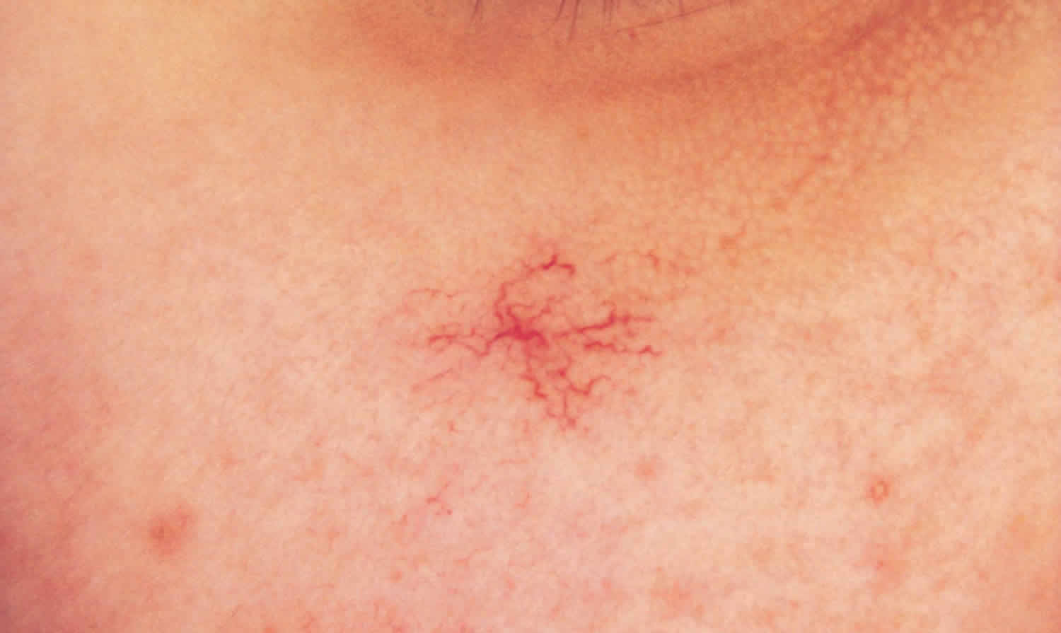
Figure 2. Spider angioma on face
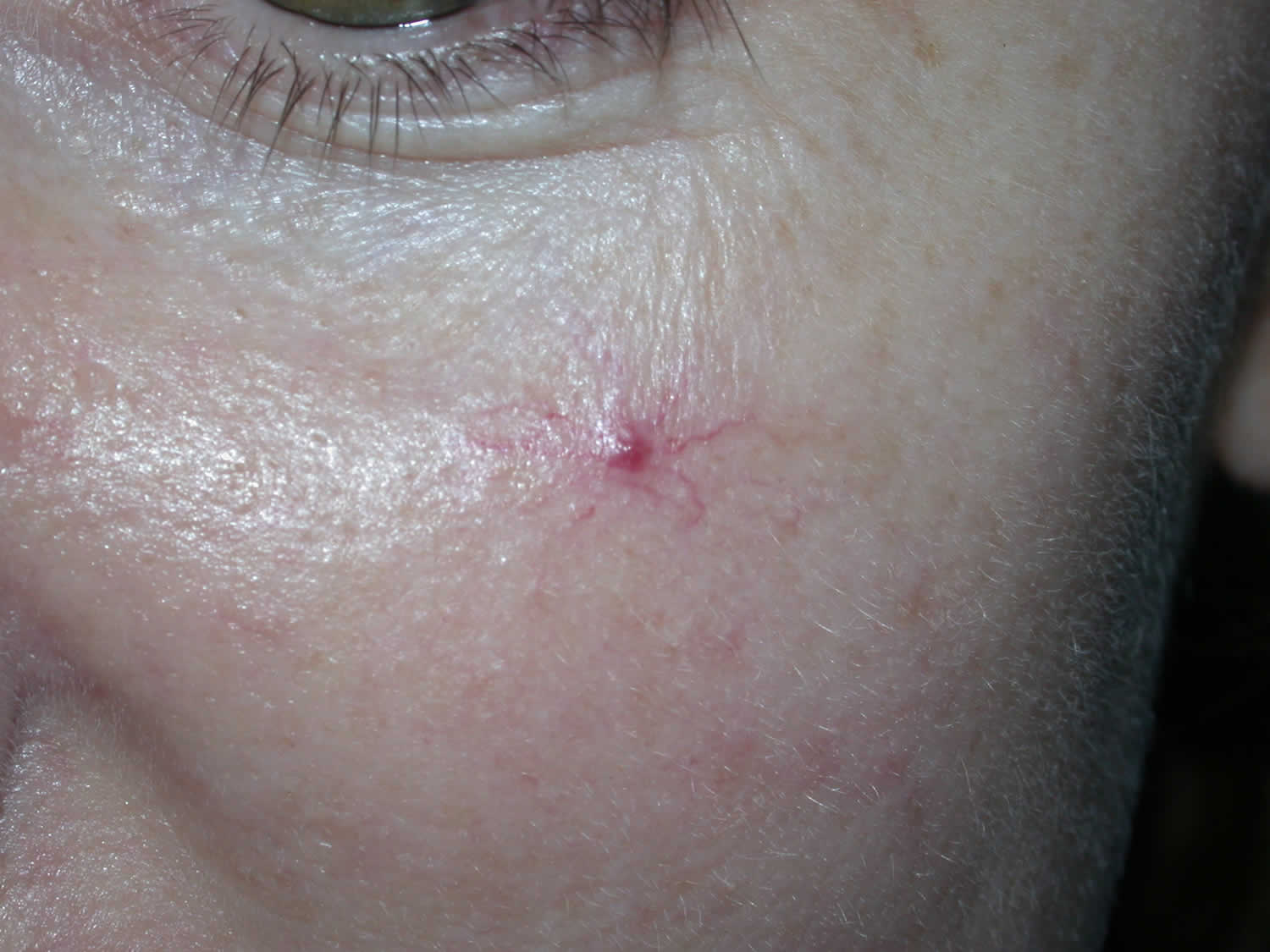
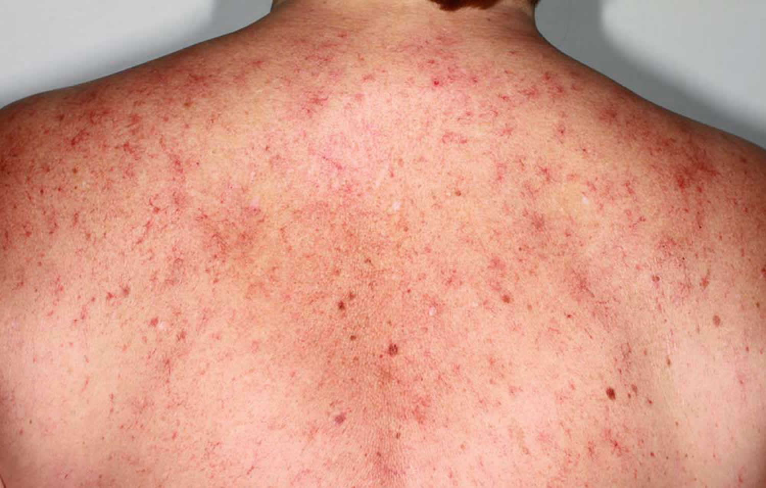
Figure 3. Spider angioma of chronic liver disease
Spider angioma causes
The cause of spider angioma is not known. Spider angioma is an acquired vascular malformation. Spider angioma occurs because of the failure of a tiny smooth muscle restricting the size of an arteriole. Increased pulsating flow through the vessel (the central papule) results in the dilatation of distal vessels (the red lines).
Spider angioma may arise spontaneously or may be induced by circulating estrogen, which is increased in pregnancy, women taking combined oral contraceptive pill and in those with cirrhosis, rheumatoid arthritis, or thyrotoxicosis. Various vascular endothelial growth factors may be involved. For decades, there have been many hypotheses of possible mechanisms that lead to the arteriolar vasodilation. Important among those are direct vasodilatory effects of alcohol, substance P, hyperestrogenism, and inadequate hepatic metabolism of steroid hormones. Angiogenesis as a possible mechanism in the pathogenesis of spider nevi has been proposed due to elevated serum vascular growth factors, such as vascular endothelial growth factor (VEGF) and basic fibroblast growth factor (bFGF) levels in patients with liver cirrhosis. Sex hormone imbalance predominantly hyperestrogenism has also been implicated for the development spider angiomas. This is also suggested by the occurrence of spider angioma in individuals with a hyperestrogenic state, like pregnancy.
Prevalence of spider angiomas is highest among patients with cirrhosis, alcoholic hepatitis, and hepatopulmonary syndrome 1. Spider angiomas correspond with a higher risk of mortality among patients with the alcoholic liver disease. Spider angiomas also suggest a high likelihood of esophageal varices and are indicative of the extent of hepatic fibrosis 1. The reported prevalence of spider angiomas in cirrhosis is 33%.
Spider angioma symptoms
Apart from its appearance, a spider angioma does not usually cause any symptoms. Bleeding from a spider angioma is unusual but may occur if picked or scratched.
The main symptom of a spider angioma is a blood vessel spot that:
- May have a red dot in the center
- Has reddish extensions that reach out from the center
- Disappears when pressed on and comes back when pressure is released
Spider angiomas are characteristically found on the face, neck, upper chest, and arms in adults, corresponding to the distribution of superior vena cava. In children, lesions are common on the upper extremities. They may also be present on the backs of the hands and fingers. However, it must be emphasized that spider angiomas can also be seen in locations other than the skin, such as the mucosa of the oral cavity and gastrointestinal (GI) tract. Its characteristic appearance diagnoses spider angioma. Large spider angiomas may be pulsatile with blood flow toward the periphery secondary to the local increase in arterial blood supply. Bleeding from these lesions is unusual unless picked or scratched. Skin temperature over a spider angioma is higher than surrounding skin. The blood pressure measures 50 to 70 mm Hg in these small arterioles.
Spider angioma diagnosis
A spider angioma is diagnosed by its typical appearance. Compression of the central arteriole results in the disappearance of the radial capillaries, which rapidly refill when the compression is relieved. This is best seen through a transparent object, such as a glass slide or the lens of a contact dermatoscope.
The direction of blood flow can be illustrated by applying pressure over the body of spiders with a glass slide (diascopy), leading to pallor with refilling following the release of pressure. No other angiomas show this phenomenon. Due to the varying sizes of spider angioma and for the ease of description, they can be graded from grade 1+ (readily recognizable containing a body, legs, and surrounding erythema) to grade 4+ (visible pulsations with a hand lens, and raised central punctum with many obvious “spider legs” radiating from it). Pregnant patients may present with numerous spider nevi which are harmless and usually resolve after childbirth. Patients with the chronic liver disease will typically have symptoms like jaundice, fluid retention, confusion, and on examination, shifting dullness, icterus, findings related to cause of cirrhosis, and stigmata of liver cell failure.
Most of the time, you DO NOT need tests to diagnose spider angioma. But sometimes, a skin biopsy may be needed to confirm the diagnosis. Blood tests may be done if a liver problem is suspected. As a general rule, number and size of spider angioma correlate with the severity of liver disease. In some occasions, history and exam might not point towards spider angiomas and can lead to concerns of skin malignancy, such as basal cell carcinoma. Rarely, spider angiomas can be a disguised basal cell carcinoma and may warrant a biopsy.
Spider angioma treatment
A spider angioma is harmless and does not require treatment. Patients with an underlying systemic disease like liver cirrhosis should be managed as the standard of care. However, when spider angiomas are present, these patients may already have the advanced liver disease.
A spider angioma that is unsightly can be removed by destroying the feeding central arteriole, but this may result in a small scar. Treatments to remove spider angioma include:
- Cryotherapy
- Electrocautery
- Intense pulsed light
- Vascular laser.
Rarely, fine-needle electrocautery, 585 nm pulsed, dye laser, 532 nm KTP (potassium-titanyl-phosphate) laser, or electro desiccation have been used to clear spider angioma for cosmetic concerns. The results of the procedure are generally good except for the small risk of the scar.
Surgical excision is rarely necessary and inevitably leaves a scar.
Spider angiomas can recur after treatment. Spider angiomas in healthy individuals usually disappear in a few years, in pregnancy following childbirth and those related to oral contraceptive pills after discontinuation of medication. Cirrhotic patients note the disappearance of nevi following liver transplantation.
Spider angioma prognosis
Spider angiomas can persist or disappear. In women, estrogen-induced spider angioma often disappear within 6 months after having a baby or of stopping the combined contraceptive pill. Spider angioma can also reappear after initially successful treatment.
References


