
What is the amygdala
The amygdala is an almond-shaped structure that lies in the temporal lobe, lying just beneath the uncus 1. The amygdala is diverse and complex in structure and comprises approximately 13 nuclei. They further subdivide into extensive internuclear and intranuclear connections. These nuclei functionally sort into five major groups: basolateral nuclei, cortical-like nuclei, central nuclei, other amygdaloid nuclei, and extended amygdala 2. Amygdala is one of the components of the limbic system, which is responsible for the control of emotions and behavior besides memory formation. The amygdala has been implicated in many diseases, such as depression 3, sleep debt and anger 4, as well as other neuropsychiatric diseases.
Anatomically, the amygdala lies at the anterior border of the hippocampal formation and the anterior aspect of lateral ventricle’s inferior horn where it merges with the peri-amygdaloid cortex, which forms part of the surface of the uncus 2.
Figure 1. Limbic system
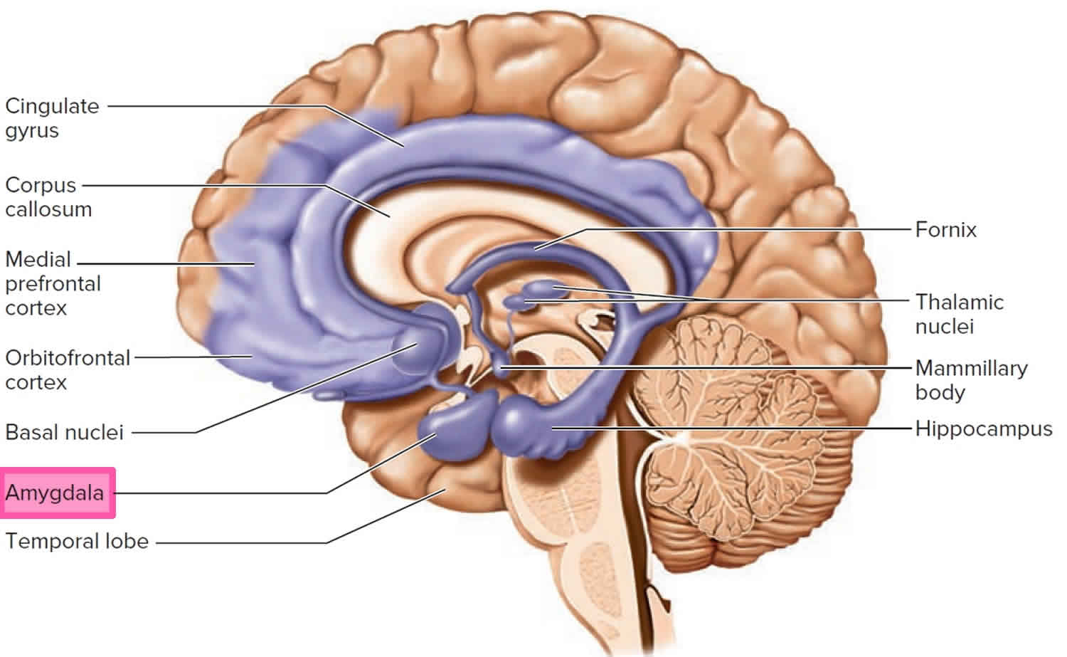
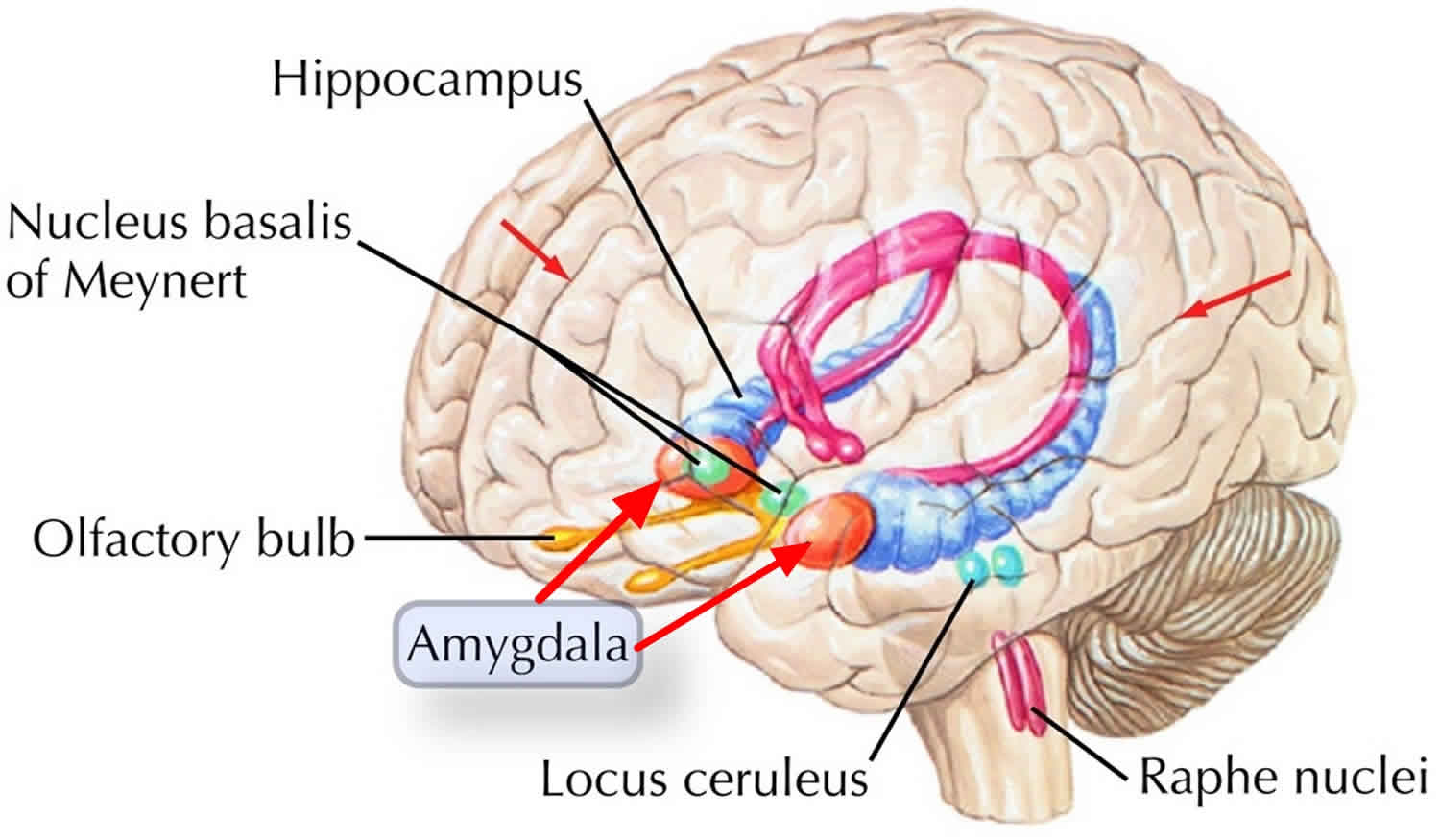
Figure 2. Amygdala
What does the amygdala do?
The amygdala plays a central role in emotion recognition, which is essential for social interaction and communication 5. The amygdala has been associated with many diseases, mainly neuropsychiatric 1. Many studies showed its effects on depression 3, 6. Others stressed the involvement of the amygdala in post-traumatic stress disorder (PTSD) as there is a bilateral reduction of the hippocampus and amygdala in PTSD 7. Neural functioning among patients with PTSD is characterized by attenuated prefrontal inhibition on the limbic system, resulting in emotional dysregulation, and suggests that amygdala neurofeedback may not only be therapeutic for this patient group but may also be used as a future adjunctive treatment 8. The amygdala and the limbic system might also be involved in chronic pain and has an association with the emotional effects of such pain 9. The amygdala also plays a key role in emotional hyperreactivity in response to social threat in patients with social anxiety disorder 10.
The amygdala combines many different sensory inputs. Like the hippocampus it combines external and internal stimuli. Every sensory modality has input. These are integrated with somatosensory and visceral inputs—this is where you get your “gut reaction”. The link between prefrontal cortex, septal area, hypothalamus, and amygdala likely gives you your gut feelings, those subjective feelings, about what is good and what is bad.
It is also where memory and emotions are combined. When the reward is particularly sweet, that behavior and association may last a lifetime. Likewise, the trauma and humiliation of punishment may be remembered for a long time too.
Fear Conditioning
Another example of emotion being linked to some perceptual experience is fear conditioning. In this example the sensory experience is auditory rather than visual as in the emotion of faces. Much of what scientists know about the amygdala and its role in emotional learning and memory comes from fear conditioning, mostly but not exclusively conducted with animals. This is an example of classical conditioning or Pavlovian conditioning. In the classic experiments conducted by Pavlov just after the turn of the century, a neutral stimulus—a bell—was sounded and after a brief interval food powder—the unconditioned stimulus—was placed in the dog’s mouth. After a few such pairings the dog would salivate to the sound of the bell. The crucial aspect of classical conditioning is that it is a pairing between two stimuli. No response is required to get the reward. In fear conditioning, an organism hears a noise or sees a visual stimulus. A few seconds, later it receives a mild shock. The reactions involve freezing, elevated blood pressure and heart rate, and it gets twitchy—startles easily.
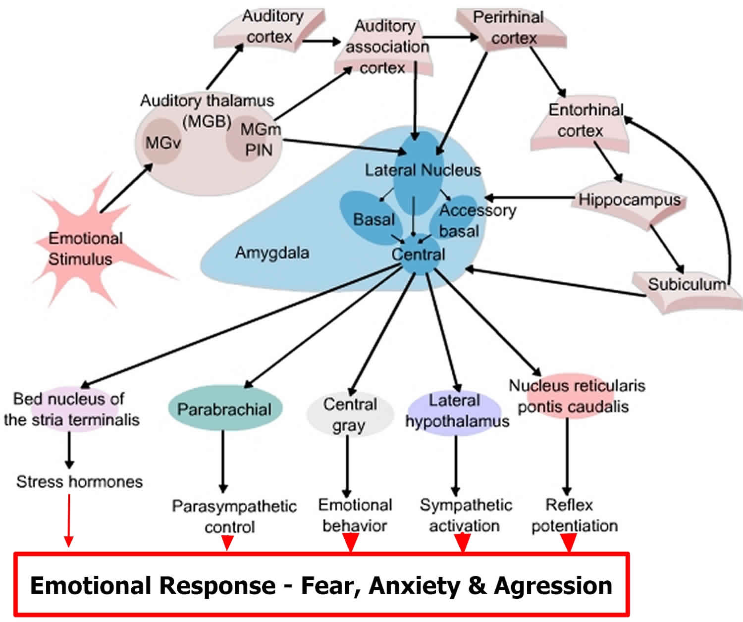
Pathways from the thalamus to the amygdala are particularly important in emotional learning. Output pathways from the central nucleus of the amygdala make extensive connections with the brain stem for emotional responses and extensive connections with cortical areas through the nucleus basalis. Cholinergic projections from the nucleus basalis to the cortex are thought to arouse the cortex.
The following diagram provides additional information on outputs controlled by the amygdala during fear conditioning.
Figure 3. Amygdala function
Some pathways of fear conditioning have been discovered and this is a hot research topic in neuroscience. If the auditory cortex pathway is lesioned, for example, basic fear conditioning is unaltered, but discrimination is altered. In the discrimination procedure one sound is paired with shock and another sound is not paired with shock. The animals had to rely solely on the thalamus and amygdala for learning and they could not learn the discrimination; apparently the two stimuli were indistinguishable.
So, the cortex is not needed for simple fear conditioning; instead it allows you to recognize an object by sight or sound— to interpret the environment.
Thus, pathways from the sensory thalamus provide only a crude perception of the world, but because they involve only one neural link they are fast pathways. Why might FAST be important? You need a quick reaction to potential danger. The thalamus—amygdala pathway provides you with this and may also prepare the amygdala to receive more highly processed information from the cortex.
On the other hand, pathways from the cortex offer detailed and accurate representations of the environment. Because these pathways have multiple neural links they are slow by comparison.
If for example you see a slender curled shape behind a tree its much better to jump back and later recognize its a garden hose than to fail to quickly jump back if it were a snake. There is plenty of time later to reflect that it was foolish to be startled in your own secure garden where there are no snakes.
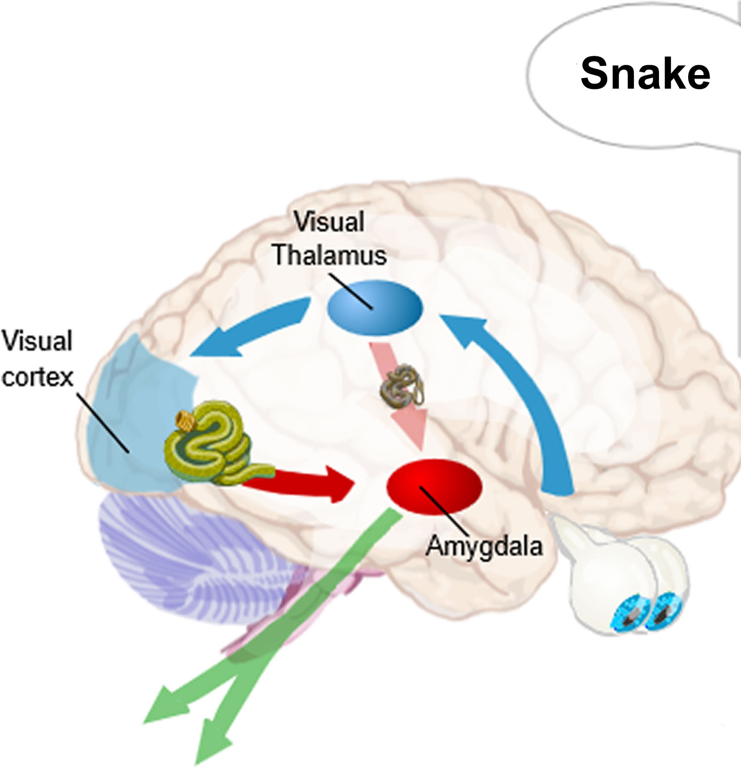
Fear producing visual stimuli is quickly processed by the thalamus and this information is passed to the amygdala (red) producing a quick response (green) to danger (see Figure 4). The thalamus also passes the information to the cortex so that more careful (and slower) judgments can be made about the real potential danger.
Figure 4. Amygdala function in fear conditioning
The amygdala is involved in pleasureful emotional learning as well as fearful emotional learning. Consider instrumental learning. Unlike classical conditioning where two stimuli are paired, in instrumental conditioning responses are followed by reward and stimulus-response associations are learned. There are thus three events: a stimulus, a response, and a reward. It has become clear that all three pairwise combinations are learned in instrumental conditioning. Where the amygdala comes in is that lesions of the basolateral nuclei of the amygdala disrupt the association the stimulus and rewarding attributes of the food.
This amygdala memory system serves as an example of memory systems generally. The establishment of memories is a function of the entire network, not any single component. The amygdala is involved in a kind of primitive emotional memory, one that is likely preserved by evolution. According to the diagram of memory systems (e.g., Nolte, p.577), declarative memory is mediated by the hippocampus and the cortex. But like the cortex, lesions of the hippocampus have little effect on fear conditioning except in discriminating environmental stimuli.
A study of patients with damage to the amygdala, hippocampus, or both clearly demonstrates the distinctive roles of these two structures in memory. These patients were shown slides of green, blue, yellow, or red colors. After some colors, a loud and frightening horn blast was sounded. Autonomic responses were recorded (via GSR recordings) to determine learning. Amygdala patients did not become conditioned to colors followed by the loud horn. But when asked how many colors were presented and which were followed by the horn, their recall was correct. That is, they had explicit memory about the events. On the other hand, hippocampal patients showed learning and conditioning to the colors followed by the horn, but could not recall which they were. That is, they had implicit memory about the events. Patients with both types of lesions showed no conditioning and had no explicit memory about which colors were followed by the horn.
Amygdala function
Amygdala function is to manage the processing of information between prefrontal-temporal association cortices and the hypothalamus. Amygdala has neural circuits to carry out its different functions with two major output pathways; dorsal route via stria terminalis that projects to the septal area and hypothalamus, and the ventral route via the ventral amygdalofugal pathway which terminates in the septal area, hypothalamus, and the medial dorsal thalamic nucleus 2. The amygdala also has connections with the basal ganglia circuit via its projections to the ventral pallidum and ventral striatum; these projections are relayed back to the cortex via the dorsomedial nucleus of the thalamus 2. The basolateral circuit includes amygdala (especially the basolateral amygdala), the orbitofrontal and anterior temporal cortex, and in the thalamus, the magnocellular division of the dorsomedial nucleus (frontothalamic pathway), which serves as a relay back to the orbitofrontal cortex 11. The circuit has been proposed as a substrate for the human ability to infer the intentions of others from their language, gaze, and gestures (theory of mind and social cognition) 12 and helps with social interactions.
The amygdala also functions in regulating anxiety, aggression, fear conditioning, emotional memory, and social cognition 2. Electrical stimulation of the amygdala evokes intense emotion, such as aggression, fear and anxiety responses in humans. Irritative lesions of temporal lobe epilepsy have the effect of stimulating the amygdala. In its extreme form irritative lesions of temporal lobe epilepsy can cause a panic attack. Panic attacks are brief spontaneously recurrent episodes of terror that generate a sense of impending disaster without a clearly identifiable cause. PET scans have shown an increase in blood flow to the parahippocampal gyri, beginning with the right parahippocampal gyrus. Similar but attenuated blood flow increases occurs during anxiety attacks.
Destructive lesions such as ablation of the amygdala cause an effect opposite to the irritative lesions of temporal lobe epilepsy. Destructive lesions of the amygdala block certain types of unconditioned fear and cause tameness in animals, and a placid calmness in humans characterized as a flatness of affect. For example, rats with lesions in the amygdala show reduced freezing in response to cats, or cat hair, attenuated analgesia, heart rate responses to loud noise, and have reduced taste neophobia. However, amygdala lesions do not affect other measures of fear such as an open arm avoidance in an elevated plus maze in rats or analgesia to shock. Lesions of the amygdala can occur as a result of Urbach-Wiethe disease where calcium is deposited in the amygdala. If this disease occurs early in life then these patients with bilateral amygdala lesions cannot discriminate emotion in facial expressions, but their ability to identify faces remains. The anatomical area for face recognition and memory is in the multimodal association area of the inferotemporal cortex. This is a good example of how emotion in one area (amygdala) is linked with perception in another area (inferotemporal cortex) to create an intense emotionally charged memory.
The amygdala is also necessary for learning by fear, amygdala lesions disrupt the acquisition of both active avoidance (escape from fear), and passive avoidance of conditioned responses, but does not affect retention. The amygdala processes not only emotions of fear and aversive stimuli, but it is also involved in conditioning using stimuli of appetite such as food, sex, and drugs. As for its role in memory, the activation of the amygdala has a modulatory effect on the acquisition and consolidation of memories that evoke an emotional response 13.
Some parts of the amygdala have even more specific functions. The basolateral nucleus (BLA) is a cortical-like structure in the dorsal amygdala, and it regulates behavioral and physiological responses to stress 14. The central amygdala (CeA) plays a crucial role in physiological responses to stressors, such as fearful stimuli, stressful stimuli, and some drug-related stimuli 15. Meanwhile, the extended amygdala, named the bed nucleus of the stria terminalis (BNST), is involved in anxiety and stress 16.
Where is the amygdala located?
The amygdala is an almond-shaped structure that lies immediately in front of the hippocampus in the temporal lobe, lying just beneath the uncus.
Amygdala damage
Amygdala damage is associated with impaired recognition of social emotions 17 and impairs eye contact during conversations 18. Recent studies conducted in patients with amygdala lesions reported impairments in the recognition of facial expressions 19, emotional words 20, or vocal emotions 21. Furthermore, studies in both human patients 22 and monkeys 23 showed significant changes in visual cortical activations to facial expressions following lesions of the amygdala. These changes in cortical processing are assumed to be remotely driven by the impaired emotional processing in the amygdala 24. Distant effects of amygdala damage have also been observed for visual stimuli in cats 25 and for auditory stimuli in rats 26. However, other results have challenged this view, with some studies reporting no impairment in recognition 27 or changes in cortical processing for emotional stimuli in patients with amygdala lesions 28. Notably, Edmiston et al. 28 observed normal visual increases in response to emotional scenes for patients with unilateral amygdala resection, arguing against a direct role for the amygdala in modulating activity in sensory cortical areas. However, in that study 28, such increases could be related to attentional effects driven by greater interest or complexity of emotional scenes 29.
- AbuHasan Q, Siddiqui W. Neuroanatomy, Amygdala. [Updated 2019 Feb 20]. In: StatPearls [Internet]. Treasure Island (FL): StatPearls Publishing; 2019 Jan-. Available from: https://www.ncbi.nlm.nih.gov/books/NBK537102[↩][↩]
- Rajmohan V, Mohandas E. The limbic system. Indian J Psychiatry. 2007 Apr;49(2):132-9.[↩][↩][↩][↩][↩]
- Ruiz NAL, Del Ángel DS, Olguín HJ, Silva ML. Neuroprogression: the hidden mechanism of depression. Neuropsychiatr Dis Treat. 2018;14:2837-2845[↩][↩]
- Saghir Z, Syeda JN, Muhammad AS, Balla Abdalla TH. The Amygdala, Sleep Debt, Sleep Deprivation, and the Emotion of Anger: A Possible Connection? Cureus. 2018 Jul 02;10(7):e2912[↩]
- Habel U, Windischberger C, Derntl B, Robinson S, Kryspin-Exner I, Gur RC, et al. . Amygdala activation and facial expressions: explicit emotion discrimination versus implicit emotion processing. Neuropsychologia (2007) 45:2369–77. 10.1016/j.neuropsychologia.2007.01.023[↩]
- Helm K, Viol K, Weiger TM, Tass PA, Grefkes C, Del Monte D, Schiepek G. Neuronal connectivity in major depressive disorder: a systematic review. Neuropsychiatr Dis Treat. 2018;14:2715-2737[↩]
- Thompson JM, Neugebauer V. Cortico-limbic pain mechanisms. Neurosci. Lett. 2018 Nov 29[↩]
- Nicholson AA, Rabellino D, Densmore M, Frewen PA, Paret C, Kluetsch R, Schmahl C, Théberge J, Neufeld RW, McKinnon MC, Reiss J, Jetly R, Lanius RA. The neurobiology of emotion regulation in posttraumatic stress disorder: Amygdala downregulation via real-time fMRI neurofeedback. Hum Brain Mapp. 2017 Jan;38(1):541-560.[↩]
- Ahmed-Leitao F, Spies G, van den Heuvel L, Seedat S. Hippocampal and amygdala volumes in adults with posttraumatic stress disorder secondary to childhood abuse or maltreatment: A systematic review. Psychiatry Res Neuroimaging. 2016 Oct 30;256:33-43[↩]
- Jung YH, Shin JE, Lee YI, Jang JH, Jo HJ, Choi SH. Altered Amygdala Resting-State Functional Connectivity and Hemispheric Asymmetry in Patients With Social Anxiety Disorder. Front Psychiatry. 2018;9:164. Published 2018 Apr 26. doi:10.3389/fpsyt.2018.00164 https://www.ncbi.nlm.nih.gov/pmc/articles/PMC5932339/[↩]
- Deakin JF, Slater P, Simpson MD, Gilchrist AC, Skan WJ, Royston MC, Reynolds GP, Cross AJ. Frontal cortical and left temporal glutamatergic dysfunction in schizophrenia. J. Neurochem. 1989 Jun;52(6):1781-6[↩]
- Frith C. Brain mechanisms for ‘having a theory of mind’. J. Psychopharmacol. (Oxford). 1996 Jan;10(1):9-15[↩]
- Sah P, Faber ES, Lopez De Armentia M, Power J. The amygdaloid complex: anatomy and physiology. Physiol. Rev. 2003 Jul;83(3):803-34[↩]
- Bhatnagar S, Vining C, Denski K. Regulation of chronic stress-induced changes in hypothalamic-pituitary-adrenal activity by the basolateral amygdala. Ann. N. Y. Acad. Sci. 2004 Dec;1032:315-9[↩]
- Gilpin NW, Herman MA, Roberto M. The central amygdala as an integrative hub for anxiety and alcohol use disorders. Biol. Psychiatry. 2015 May 15;77(10):859-69.[↩]
- Li C, Pleil KE, Stamatakis AM, Busan S, Vong L, Lowell BB, Stuber GD, Kash TL. Presynaptic inhibition of gamma-aminobutyric acid release in the bed nucleus of the stria terminalis by kappa opioid receptor signaling. Biol. Psychiatry. 2012 Apr 15;71(8):725-32[↩]
- Adolphs R, Baron-Cohen S, Tranel D. Impaired recognition of social emotions following amygdala damage. J Cogn Neurosci. (2002) 14:1264–74. 10.1162/089892902760807258[↩]
- Spezio ML, Huang PY, Castelli F, Adolphs R. Amygdala damage impairs eye contact during conversations with real people. J Neurosci. (2007) 27:3994–7. 10.1523/JNEUROSCI.3789-06.2007[↩]
- Meletti S, et al. Impaired facial emotion recognition in early-onset right mesial temporal lobe epilepsy. Neurology. 2003;60(3):426–431[↩]
- Anderson AK, Phelps EA. Lesions of the human amygdala impair enhanced perception of emotionally salient events. Nature. 2001;411(6835):305–309[↩]
- Sprengelmeyer R, et al. Knowing no fear. Proc Biol Sci. 1999;266(1437):2451–2456[↩]
- Vuilleumier P, Richardson MP, Armony JL, Driver J, Dolan RJ. Distant influences of amygdala lesion on visual cortical activation during emotional face processing. Nat Neurosci. 2004;7(11):1271–1278.[↩]
- Hadj-Bouziane F, et al. Amygdala lesions disrupt modulation of functional MRI activity evoked by facial expression in the monkey inferior temporal cortex. Proc Natl Acad Sci USA. 2012;109(52):E3640–E3648[↩]
- Kumar S, von Kriegstein K, Friston K, Griffiths TD. Features versus feelings: Dissociable representations of the acoustic features and valence of aversive sounds. J Neurosci. 2012;32(41):14184–14192[↩]
- Chen Y, Li H, Jin Z, Shou T, Yu H. Feedback of the amygdala globally modulates visual response of primary visual cortex in the cat. Neuroimage. 2014;84:775–785[↩]
- Armony JL, Quirk GJ, LeDoux JE. Differential effects of amygdala lesions on early and late plastic components of auditory cortex spike trains during fear conditioning. J Neurosci. 1998;18(7):2592–2601.[↩]
- Bach DR, Hurlemann R, Dolan RJ. Unimpaired discrimination of fearful prosody after amygdala lesion. Neuropsychologia. 2013;51(11):2070–2074[↩]
- Edmiston EK, et al. Enhanced visual cortical activation for emotional stimuli is preserved in patients with unilateral amygdala resection. J Neurosci. 2013;33(27):11023–11031[↩][↩][↩]
- Mourão-Miranda J, et al. Contributions of stimulus valence and arousal to visual activation during emotional perception. Neuroimage. 2003;20(4):1955–1963[↩]


