What is bifid uvula
Bifid uvula also called cleft uvula, is a split in the soft flap of tissue that hangs from the back of the mouth. The term bifid uvula means the partial or full bifurcation of the uvula. Bifid uvula is often regarded as a marker for submucous cleft palate although this relationship has not been fully confirmed 1. Bifid uvula, although looks benign, apparently sometimes may be associated with anomalies leading to catastrophic complications. Cornelia de Lange syndrome is a rare congenital syndrome associated with bifid uvula and sub mucous cleft palate that causes problems in airway due anatomical distortion 2. Bifid uvula may be associated with increased risk of schizophrenia, mild mental retardation, and chromosomal disorder, diagnosed by fluorescent in situ hybridization technique 3. Loeys–Dietz syndrome (autosomal dominant) is a genetic syndrome with clinical features overlapped with Marfan syndrome, but due to mutations in the genes encoding transforming growth factor beta receptor 1. Hypertelorism, cleft palate, or bifid uvula are the major findings. Arterial aneurysms/dissections, arterial tortuosity involving aortic and its branches, carotid, vertebral, extracranial artery, abdominal aorta and its branches, common iliac, and popliteal arteries are reported in this syndrome 4.
Bifid uvula is apparent in 0.44%–3.3% of normal individuals 5. The incidence of bifid uvula is higher in Indians and Mongols, average in Caucasians and less frequent in blacks 6. As several studies 7 showed a higher incidence of cleft palate in females, it is possible to assume a higher prevalence of bifid uvula in females as well. However, there are also other studies 8 that have shown a higher occurrence of bifid uvula in males.
Bifid uvula has been reported to represent the least severe form of cleft palate 9. Various studies have shown the prevalence of bifid uvula associated with cleft palate to ranging between 1.5 and 10% but isolated bifid uvula has rarely been reported. It is a symptom of several chromosomal syndromes (eg. Trisomy). Bifid uvula may possibly be genetically inherited. Bifid uvula is more common in males than in females 10. Formation of soft palate and uvula takes a slightly different course than that of the regions of the secondary palate which give rise to the hard palate 11. The soft palate and uvula develop from two separate masses found at the most posterior portions of the secondary palatine shelves. Unlike the fusion mechanism which is in place along much of the length of the palatine shelves, the consolidation of these two separate masses is brought about by a selective proliferation of mesenchymal cells located deep in the valley between the masses 12. As that proliferation, called merging, continues the valley between the two distal shelf masses is obliterated, this results in the smoothening of the contour of the soft palate and uvula 12. Failure of the merging process in the soft palate and uvula development can result in complete or partial clefts of the soft palate and uvula 13.
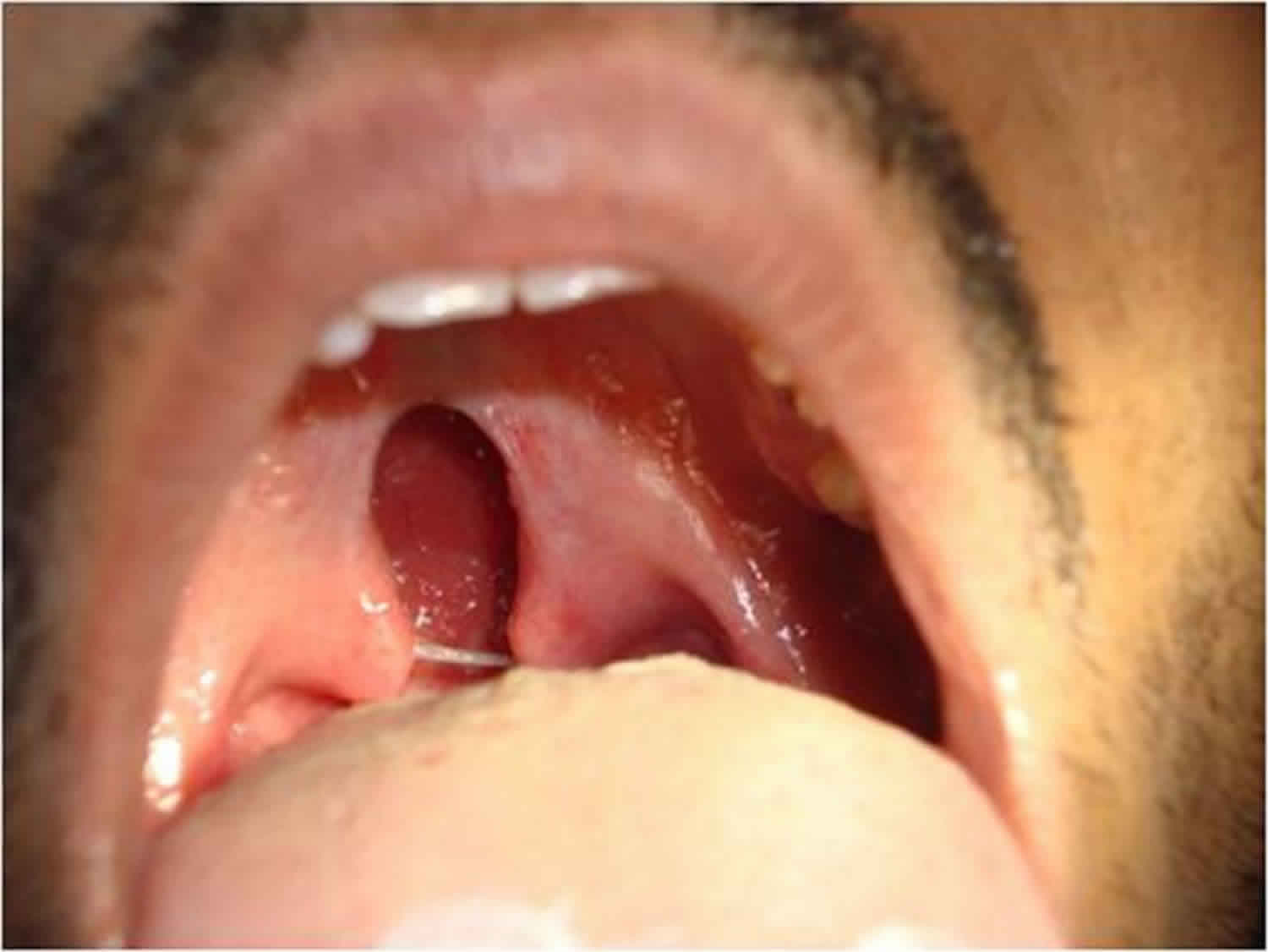
Classification of the types of bifid uvula according to Meskin et al 14 based on morphology are:
- Type A: Normal uvula,
- Type B: Uvula bifurcated up to one-fourth of its total length,
- Type C: Uvula bifurcated from one-fourth to three-fourths of its length,
- Type D: Uvula bifurcated from three-fourths to its total length.
Bifid uvula causes
Isolated bifid uvula cause is unknown 15, but may possibly be genetically inherited 12. It is hypothesized that bifid uvula is inherited as an autosomal dominant trait with about 40% penetrance 12. It could be expected that of 50 families having a bifid uvula proband, 40% or 20 parents would demonstrate a similar defect. If bifid uvula is a true palatal cleft it should follow the hereditary pattern of severe cleft palate 16. Meskin et al. 17 reported that about 1% of Caucasians and 10% of American Indians and Japanese show bifid uvula in some degree. The frequency in sibling and parents of affected persons is said to be about 18% 18.
Recent articles have shown the occurrence of bifid uvula in rare conditions like Loeys–Dietz syndrome and inflammatory linear verrucous epidermal nevus syndrome 19.
Bifid uvula complications
Children with bifid uvula may have changes in speech, hearing and swallowing. Bifid uvula is associated with an increased incidence of otitis media with effusion 20.
Children in whom bifid uvula is evident upon oral examination during regular health checkups should be examined by a specialist 21.
- A.C. Suryadevara, S.A. Tatum. Floating the uvula: an intraoperative method for detecting bifidity. Int J Pediatr Otorhinolaryngol, 71 (2007), pp. 175-177[↩]
- Callea M, Montanari M, Radovich F, Clarich G, Yavuz I. Bifid uvula and submucous cleft palate in cornelia de lange syndrome. J Int Dent Med Res. 2011;4:74.[↩]
- [A bifid uvula in a patient with schizophrenia as a sign of 22q11 deletion syndrome]. Vorstman JA, de Ranitz AG, Udink ten Cate FE, Beemer FA, Kahn RS. Ned Tijdschr Geneeskd. 2002 Oct 26; 146(43):2033-6.[↩]
- Loeys-Dietz syndrome: MDCT angiography findings. Johnson PT, Chen JK, Loeys BL, Dietz HC, Fishman EK. AJR Am J Roentgenol. 2007 Jul; 189(1):W29-35.[↩]
- R.J. Shprintzen, R.H. Schwartz, A. Daniller, L. Hoch. Morphologic significance of bifid uvula. Pediatrics, 75 (1985), pp. 553-561[↩]
- E.R. Richardson. Cleft uvula incidence in Negroes. Cleft Palate J, 7 (1970), pp. 669-672[↩]
- D.R. Martelli, P.R. Bonan, M.C. Soares, L.R. Paranaíba, H. Martelli-Júnior. Analysis of familial incidence of non-syndromic cleft lip and palate in a Brazilian population. Med Oral Patol Oral Cir Bucal, 15 (2010), pp. 898-901[↩]
- R.P. Rivron. Bifid uvula: prevalence and association in otitis media with effusion in children admitted for routine otolaryngological operations. J Laryngol Otol, 103 (1989), pp. 249-252[↩]
- Sperber GH, Tobias PV. Craniofacial Embryology. 4th edn. Wright; p. 141.[↩]
- Reichert PA, Philipsen HP. Color Atlas of Dental Medicine Oral Pathology. 128.[↩]
- Burdi AR. Distribution of midpalatine cysts: a re-evaluation of human palatal closure mechanism. J Oral Surg. 1968;26:41–5.[↩]
- Achalli S, Bhat S, Ram Shetty S, Babu SG, Suvarna R. Deformities of the uvula in the oral cavity- a case series. Iran Red Crescent Med J. 2012;14(10):676–679. https://www.ncbi.nlm.nih.gov/pmc/articles/PMC3518986/[↩][↩][↩][↩]
- Berkowitz S. Cleft And Palate Diagnosis And Management. 2nd edn. Springer; p. 9.[↩]
- Meskin LH, Gorlin RJ, Isaacson RJ. Abnormal Morphology Of The Soft Palate: I. The Prevalence of Cleft Uvula. Cleft Palate J. 1964;35:342–6.[↩]
- M.J. Dixon, M.L. Marazita, T.H. Beaty, J.C. Murray. Cleft lip and palate: understanding genetic and environmental influences. Nat Rev Genet, 12 (2011), pp. 167-178[↩]
- Meskin LH, Gorlin RJ, Isaacson RJ. Abnormal Morphology of The Soft Palate: II. The Genetics of Cleft Uvula. Cleft Palate J. 1964;45:40–5.[↩]
- Meskin, L. H., Gorlin, R. J., Isaacson, R. J. Abnormal morphology of the soft palate. II. The genetics of cleft uvula. Cleft Palate J. 2: 40-45, 1965.[↩]
- Bifid uvula. https://omim.org/entry/192100?search=bifid%20uvula&highlight=bifid%20uvula[↩]
- Yang JH, Ki CS, Han H, Song BG, Jang SY, Chung TY, Sung K, Lee HJ, Kim DK. Clinical features and genetic analysis of Korean patients with Loeys–Dietz syndrome. Journal of Human Genetics. 2012;57:52–56.[↩]
- Bifid uvula: prevalence and association in otitis media with effusion in children admitted for routine otolaryngological operations. J Laryngol Otol. 1989 Mar;103(3):249-52. https://doi.org/10.1017/S002221510010862X[↩]
- T. Oji, Y. Sakamoto, H. Ogata, I. Tamada, K. Kishi. A 25-year review of cases with submucous cleft palate. Int J Pediatr Otorhinolaryngol, 77 (2013), pp. 1183-1185[↩]


