close

Procedures

Procedures

Sigmoidoscopy

sigmoidoscopy

What is a sigmoidoscopy

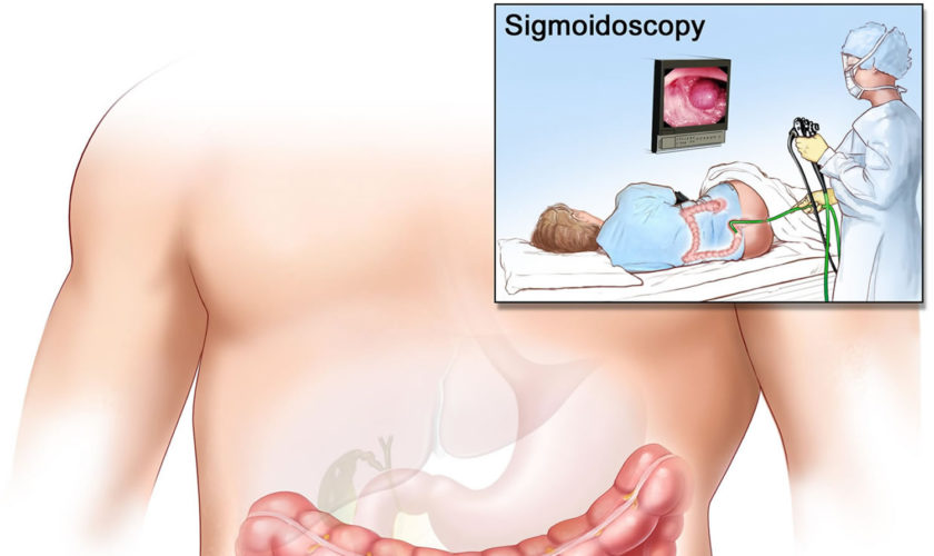
A sigmoidoscopy is a medical procedure that allows examination of the mucosal surface of the end-region of the colon (large intestine) –  the rectum and sigmoid (lower) – for lesions including cancer or pre-cancerous polyps (growths). Using sigmoidoscopy to screen average-risk populations for colon cancer and rectal cancer has been associated with a 50-80% reduction in cancer death. A sigmoidoscope is a thin, tube-like instrument with a light and a camera for viewing. A sigmoidoscope may also have a tool to remove polyps or tissue samples, which are checked under a microscope for signs of cancer. The sigmoidoscope is inserted via the anus through the rectum into the sigmoid colon. The doctor is able to see any inflammation, internal bleeding or other things that may be abnormal. A sigmoidoscopy is a short procedure, lasting only about 15-25 minutes. Sigmoidoscopy doesn’t usually require sedation and people can go straight home after the procedure. Special training is required to carry out the procedure but it is performed routinely by a variety of practitioners including physicians, physician’s assistants, nurses and gastroenterologists.

There are two types of sigmoidoscopes:

  • The rigid sigmoidoscope – This is about 25cm long and allows examination of up to about 20cm of the rectum and distal sigmoid colon, though it is best suited for rectal assessments only.
  • The flexible sigmoidoscope – This is made of a flexible fiber optic tube and can be up to 60cm long. It is able to reach further than the rigid sigmoidoscope. Depending on its length it is also able to view the descending colon. The upper parts of the colon however, are out of its reach and require a colonoscopy.

A special camera on the sigmoidoscope allows the person carrying out the procedure to monitor the examination on a video screen.

Why is a sigmoidoscopy done?

A sigmoidoscopy is able to diagnose a variety of conditions including colorectal cancer or colonic polyps, inflammatory bowel disease (eg. ulcerative colitis, Crohns disease), bowel obstruction and causes of bleeding, abdominal pain or diarrhoea. As well as being able to see the rectum and lower colon, the sigmoidoscope is able to take samples of or even remove lesions or polyps of interest.

A sigmoidoscopy may be used to examine or diagnose certain conditions or structures in your lower colon, including the following:

  • Polyps
  • Tumors
  • Ulcers (sores)
  • Inflammation (redness and swelling)
  • Hemorrhoids
  • Diverticula (pouches on your colon wall)
  • Strictures (narrowing of your lower colon)

Sigmoidoscopy can also be used to investigate the following:

  • Changes in your bowel habits
  • Lower belly pain
  • Itching around your anus
  • Blood or mucus in your stool
  • Low iron levels
  • Low blood counts

A tissue biopsy is usually required for the accurate diagnosis of colon or rectal cancers. The sigmnoidoscope is able to take a biopsy or even surgically remove a polyp in the therapeutic treatment of bowel cancer. A polyp may be removed, even if it is benign (non-cancerous) because it may be prone to becoming malignant (cancerous). Patients with a family history of colon or rectal cancer may be done regularly to check for the development of polyps. After surgery to remove any cancerous lesions, sigmoidoscopy may also be carried out regularly in order to monitor the disease.

The sigmoidscope can be useful in the identification of the cause of bowel obstruction and may also be used to rectify the obstruction. Its ability to closely examine the wall of the rectum and lower colon allows the sigmoidoscope to identify any areas of inflammation, or sources of bleeding.

Sigmoidoscopy risks and limitations

The sigmoidoscopy procedure is considered safe and the risk of complication very low, when performed by a trained practitioner. During three sigmoidoscopy studies carried out in the UK, Italy and Norway the procedure showed a complication rate of 3 per 100 000 examination. Although the procedure is able to identify a large number of pathologies, it does not have the reach of a colonoscope, which is able to examine right to the end of the small intestine.

Sigmoidoscopy procedure

Before a sigmoidoscopy your doctor will inform you about everything that is involved and you will be asked to sign a written consent form. The sigmoidoscopy procedure is quite short with duration of 5-10 minutes. It can be carried out in a hospital or in a physician’s office. The sigmoidoscope is inserted through the anus to gain access to the rectum and distal colon. You will not need to be sedated for the procedure but you may feel some discomfort or a bloated feeling. As well as an onboard camera, the sigmoidoscope also has a number of tools that are used to take samples of anything the doctor may be interested in analysing.

Sigmoidoscopy prep

Before your procedure, be sure to tell your health care provider:

  • If you are pregnant or think you may be pregnant
  • If you are sensitive to or allergic to any medicines, latex, tape or anesthesia medicines (local and general)
  • About all the medicines you take, including over-the-counter drugs, prescription medicines, vitamins, herbs and other supplements
  • If you have a history of bleeding disorders
  • If you are taking any blood-thinning medicines, aspirin, ibuprofen or other medicines that affect blood clotting; these drugs may need to be stopped before the procedure

Like most forms of endoscopy, for a sigmoidoscopy to work properly, it needs to have a clear view of the walls of the rectum and large intestine. It is therefore necessary that the colon is completely empty before the procedure. As preparation, your doctor will put you on a special diet to reduce the amount of solid food you eat in the days leading up to the procedure. You will be asked to take in only water on the day of the sigmoidoscopy and you may also be given laxatives in order to ensure that everything is cleared out of your system. On the morning of the examination you may also be given a saline enema to flush out the rectum.

Follow any other instructions your provider gives you to get ready for your sigmoidoscopy.

After the sigmoidoscopy procedure

Following a sigmoidoscopy some people experience a bloated feeling, this is a normal side effect of the procedure. A sigmoidoscopy is relatively non-invasive and usually doesn’t require any anaesthesia so you should be able to go straight home after the procedure. Your doctor will discuss the results with you once they have been analysed. If there was a biopsy taken during the procedure, some small amounts of blood may appear in the next stool. If you develop a fever or pain in your abdomen you should contact your doctor immediately.

What do the results mean?

Any samples can be analysed in a laboratory to see whether further examination or surgery is needed. A positive sigmoidoscopy result (cancerous polyps are found) will usually prompt examination of the full colon, requiring a colonoscopy. Patients with a family history of colorectal cancer or familial adenomatous polyposis (FAP) should be screened at regular intervals in order to monitor the onset or development of disease. If results suggest that you may have an irritable bowel disease such as Crohn’s disease, your doctor will inform you about the condition and treatments available.

What is a flexible sigmoidoscopy

Flexible sigmoidoscopy is a procedure in which a trained medical professional uses a flexible, narrow tube with a light and tiny camera on one end, called a sigmoidoscope or scope, to look inside your rectum and lower colon, also called the sigmoid colon and descending colon. Flexible sigmoidoscopy can show irritated or swollen tissue, ulcers, polyps, and cancer. However, on the negative side, flexible sigmoidoscopy doesn’t allow the doctor to see the entire colon. As a result, any cancers or polyps farther into the colon can’t be detected with flexible sigmoidoscopy alone.

A tiny video camera at the tip of the flexible sigmoidoscope allows the doctor to view the inside of the rectum and most of the sigmoid colon — about the last two feet (61 centimeters) of the large intestine. If necessary, tissue samples (biopsies) can be taken through the scope during a flexible sigmoidoscopy exam.

Why do doctors use flexible sigmoidoscopy?

A flexible sigmoidoscopy can help a doctor find the cause of unexplained symptoms, such as:

  • bleeding from your anus
  • changes in your bowel activity such as diarrhea
  • pain in your abdomen
  • unexplained weight loss

Doctors also use flexible sigmoidoscopy as a screening tool for colon polyps and colon and rectal cancer. Screening may find diseases at an early stage, when a doctor has a better chance of curing the disease. Sigmoidoscopy is one option for colon cancer screening, but there are other options like colonoscopy that allow visualization of the whole colon. Talk with your doctor about your options.

Colorectal cancer is the second-leading cause of cancer death in the United States. In 2016, an estimated 134,000 persons will be diagnosed with the disease, and about 49,000 will die from it 1. Colorectal cancer is most frequently diagnosed among adults aged 65 to 74 years; the median age at death from colorectal cancer is 73 years 2.

Screening for colon and rectal cancer

Your doctor will recommend screening for colon and rectal cancer at age 50 if you don’t have health problems or other factors that make you more likely to develop colon cancer 1.

Factors that make you more likely to develop colorectal cancer include:

  • someone in your family has had polyps or cancer of the colon or rectum
  • a personal history of inflammatory bowel disease, such as ulcerative colitis or Crohn’s disease
  • other factors, such as if you weigh too much or smoke cigarettes

If you are more likely to develop colorectal cancer, your doctor may recommend screening at a younger age, and you may need to be tested more often.

If you are older than age 75, talk with your doctor about whether you should be screened. The current colorectal cancer screening guidelines from the U.S. Preventive Services Task Force 1 are as follow:

  • Adults aged 50 to 75 years: The U.S. Preventive Services Task Force 1 recommends screening for colorectal cancer starting at age 50 years and continuing until age 75 years. The risks and benefits of different screening methods vary.
  • Adults aged 76 to 85 years: The decision to screen for colorectal cancer in adults aged 76 to 85 years should be an individual one, taking into account the patient’s overall health and prior screening history.
    • Adults in this age group who have never been screened for colorectal cancer are more likely to benefit.
    • Screening would be most appropriate among adults who 1) are healthy enough to undergo treatment if colorectal cancer is detected and 2) do not have comorbid conditions that would significantly limit their life expectancy.

Evidence from randomized clinical trials demonstrates that annual or every 2 years screening with guaiac-based fecal occult blood test (gFOBT) as well as 1-time and every 3- to 5-year flexible sigmoidoscopy reduces colorectal cancer deaths 3. The Cancer Intervention and Surveillance Modeling Network (CISNET) models found that several screening strategies were estimated to yield comparable life-years gained (i.e., life-years gained with the noncolonoscopy strategies were within 90% of those gained with the colonoscopy strategy) among adults aged 50 to 75 years and an efficient balance of benefits and harms 4. These screening strategies include 1) annual screening with fecal immunochemical test (FIT), 2) screening every 10 years with flexible sigmoidoscopy and annual screening with fecal immunochemical test (FIT), 3) screening every 10 years with colonoscopy, and 4) screening every 5 years with CT colonography. The findings for CT colonography depend on the proxy measure used for the burden of screening (number of lifetime colonoscopies or lifetime cathartic bowel preparations). Two of the 3 Cancer Intervention and Surveillance Modeling Network models found that fecal immunochemical test (FIT)-DNA screening every 3 years (as recommended by the manufacturer) was estimated to yield life-years gained less than 90% of the colonoscopy screening strategy (84% and 87%, respectively). Another way to conceptualize these findings is to note that Cancer Intervention and Surveillance Modeling Network (CISNET) modeling found that fecal immunochemical test (FIT)-DNA screening every 3 years was estimated to provide about the same amount of benefit as screening with flexible sigmoidoscopy alone every 5 years.

Table 1. Characteristics of Colorectal Cancer Screening Strategies

Screening MethodFrequencybEvidence of EfficacyOther Considerations
Stool-Based Tests
gFOBTEvery yearRCTs with mortality end points:
High-sensitivity versions (eg, Hemoccult SENSA) have superior test performance characteristics than older tests (eg, Hemoccult II)
Does not require bowel preparation, anesthesia, or transportation to and from the screening examination (test is performed at home)
FITcEvery yearTest characteristic studies:
Improved accuracy compared with gFOBTCan be done with a single specimen
Does not require bowel preparation, anesthesia, or transportation to and from the screening examination (test is performed at home)
FIT-DNAEvery 1 or 3 yearsdTest characteristic studies:
Specificity is lower than for FIT, resulting in more false-positive results, more diagnostic colonoscopies, and more associated adverse events per screening testImproved sensitivity compared with FIT per single screening test
There is insufficient evidence about appropriate longitudinal follow-up of abnormal findings after a negative diagnostic colonoscopy; may potentially lead to overly intensive surveillance due to provider and patient concerns over the genetic component of the test
Direct Visualization Tests
ColonoscopycEvery 10 yearsProspective cohort study with mortality end pointRequires less frequent screening. Screening and diagnostic followup of positive results can be performed during the same examination.
CT colonographyeEvery 5 yearsTest characteristic studiesThere is insufficient evidence about the potential harms of associated extracolonic findings, which are common
Flexible sigmoidoscopyEvery 5 yearsRCTs with mortality end points:
Modeling suggests it provides less benefit than when combined with FIT or compared with other strategies
Test availability has declined in the United States
Flexible sigmoidoscopy with FITcFlexible sigmoidoscopy every 10 years plus FIT every yearRCT with mortality end point (subgroup analysis)Test availability has declined in the United States. Potentially attractive option for patients who want endoscopic screening but want to limit exposure to colonoscopy

Abbreviations: FIT=fecal immunochemical test; FIT-DNA=multitargeted stool DNA test; gFOBT=guaiac-based fecal occult blood test; RCT=randomized clinical trial.

a Although a serology test to detect methylated SEPT9 DNA was included in the systematic evidence review, this screening method currently has limited evidence evaluating its use (a single published test characteristic study met inclusion criteria, which found it had a sensitivity to detect colorectal cancer of <50%). It is therefore not included in this table.
b Applies to persons with negative findings (including hyperplastic polyps) and is not intended for persons in surveillance programs. Evidence of efficacy is not informative of screening frequency, with the exception of gFOBT and flexible sigmoidoscopy alone.
c Strategy yields comparable life-years gained (ie, the life-years gained with the noncolonoscopy strategies were within 90% of those gained with the colonoscopy strategy) and an efficient balance of benefits and harms in CISNET modeling.
d Suggested by manufacturer.
e Strategy yields comparable life-years gained (ie, the life-years gained with the noncolonoscopy strategies were within 90% of those gained with the colonoscopy strategy) and an efficient balance of benefits and harms in CISNET modeling when lifetime number of colonoscopies is used as the proxy measure for the burden of screening, but not if lifetime number of cathartic bowel preparations is used as the proxy measure.

[Source: U.S. Preventive Services Task Force 1]

Several randomized clinical trials have shown that flexible sigmoidoscopy alone reduces deaths from colorectal cancer 3. Flexible sigmoidoscopy combined with fecal immunochemical test (FIT) has been studied in a single trial and was found to reduce the colorectal cancer–specific mortality rate more than flexible sigmoidoscopy alone 5. Modeling studies conducted by Cancer Intervention and Surveillance Modeling Network (CISNET) also consistently estimate that combined testing yields more life-years gained and colorectal cancer deaths averted compared with flexible sigmoidoscopy alone 4. Flexible sigmoidoscopy can result in direct harms, such as colonic perforations and bleeding, although the associated event rates are much lower than those observed with colonoscopy 3. Harms can also occur as a result of follow-up colonoscopy.

Completed trials of flexible sigmoidoscopy provide indirect evidence that colonoscopy—a similar endoscopic screening method—reduces colorectal cancer mortality. A prospective cohort study also found an association between patients who self-reported being screened with colonoscopy and a lower colorectal cancer mortality rate 6. Colonoscopy has both indirect and direct harms. Harms may be caused by bowel preparation prior to the procedure (e.g, dehydration and electrolyte imbalances), the sedation used during the procedure (eg, cardiovascular events), or the procedure itself (e.g, infection, colonic perforations, or bleeding).

Most doctors recommend colonoscopy to screen for colon cancer because colonoscopy shows the entire colon and can remove colon polyps. However, preparing for and performing a flexible sigmoidoscopy may take less time and you may not need anesthesia. Health care providers may combine flexible sigmoidoscopy with other tests.

If your doctor finds abnormal tissue or one or more polyps during a flexible sigmoidoscopy, you should have a colonoscopy to examine the rest of your colon.

Government health insurance plans, such as Medicare, and private health insurance plans sometimes change whether and how often they pay for cancer screening tests. Check with your insurance plan to find out how often your insurance will cover a screening flexible sigmoidoscopy.

Benefits of Colorectal Cancer Screening and Early Intervention

The U.S. Preventive Services Task Force 1 found convincing evidence that screening for colorectal cancer in adults aged 50 to 75 years reduces colorectal cancer mortality. The U.S. Preventive Services Task Force 1 found no head-to-head studies demonstrating that any of the screening strategies it considered are more effective than others, although the tests have varying levels of evidence supporting their effectiveness, as well as different strengths and limitations (see Table 1). About one-third of eligible adults in the United States have never been screened for colorectal cancer 7, and offering choice in colorectal cancer screening strategies may increase screening uptake 8. As such, the screening tests are not presented in any preferred or ranked order; rather, the goal is to maximize the total number of persons who are screened because that will have the largest effect on reducing colorectal cancer deaths.

The benefit of early detection of and intervention for colorectal cancer declines after age 75 years. Among older adults who have been previously screened for colorectal cancer, there is at best a moderate benefit to continuing screening during the ages of 76 to 85 years. However, adults in this age group who have never been screened for colorectal cancer are more likely to benefit than those who have been previously screened.

The time between detection and treatment of colorectal cancer and realization of a subsequent mortality benefit can be substantial. As such, the benefit of early detection of and intervention for colorectal cancer in adults 86 years and older is at most small.

To date, no method of screening for colorectal cancer has been shown to reduce all-cause mortality in any age group 9.

Harms of Colorectal Cancer Screening Screening and Early Intervention

The harms of screening for colorectal cancer in adults aged 50 to 75 years are small. The majority of harms result from the use of colonoscopy, either as the screening test or as follow-up for positive findings detected by other screening tests. The rate of serious adverse events from colorectal cancer screening increases with age 3. Thus, the harms of screening for colorectal cancer in adults 76 years and older are small to moderate.

Flexible sigmoidoscopy risks

A flexible sigmoidoscopy exam poses few risks. Rarely, complications of a flexible sigmoidoscopy exam may include:

  • bleeding
  • perforation of the colon or rectum wall
  • severe pain in your abdomen
  • death, although this risk is rare

Bleeding and perforation are the most common complications from flexible sigmoidoscopy. Most cases of bleeding occur in patients who have polyps removed. The doctor can treat bleeding that occurs during the flexible sigmoidoscopy right away. However, you may have delayed bleeding up to 2 weeks after the procedure. The doctor diagnoses and treats delayed bleeding with a colonoscopy or repeat flexible sigmoidoscopy. The doctor may need to treat perforation with surgery.

Figure 1. Large intestine (colon)

large intestine

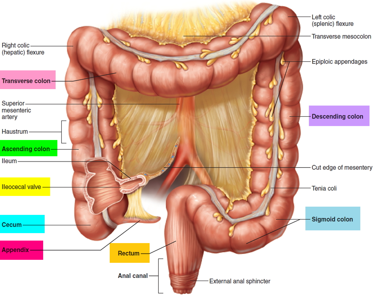
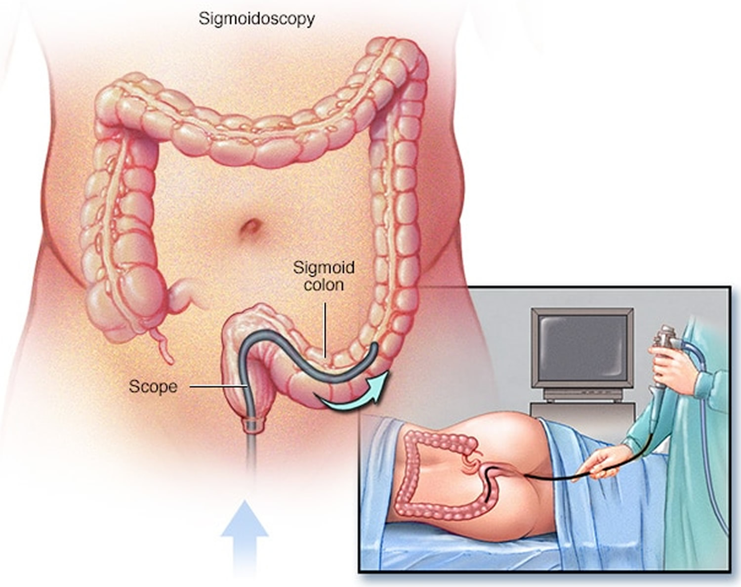
Figure 2. Flexible sigmoidoscopy

flexible-sigmoidoscopy

Flexible sigmoidoscopy prep

To prepare for a flexible sigmoidoscopy, you will need to talk with your doctor, change your diet, and clean out your bowel.

Before a flexible sigmoidoscopy exam, you’ll need to clean out (empty) your colon. Any residue in your colon may obscure the view of your colon and rectum during the exam.

Talk with your doctor

You should talk with your doctor about any medical conditions you have and all prescribed and over-the-counter medicines, vitamins, and supplements you take, including:

  • arthritis medicines
  • aspirin or medicines that contain aspirin
  • blood thinners
  • diabetes medicines
  • nonsteroidal anti-inflammatory drugs, such as ibuprofen or naproxen
  • vitamins that contain iron or iron supplements

Change your diet and clean out your bowel

A health care professional will give you written bowel prep instructions to follow at home before the procedure. A health care professional orders a bowel prep so that little or no stool is present in your intestine. A complete bowel prep lets you pass stool that is clear and liquid. Stool inside your colon can prevent your doctor from clearly seeing the lining of your intestine.

You may need to follow a clear liquid diet the day before the procedure. The instructions will provide specific direction about when to start and stop the clear liquid diet. In most cases, you may drink or eat the following:

  • fat-free bouillon or broth
  • gelatin in flavors such as lemon, lime, or orange
  • plain coffee or tea, without cream or milk
  • sports drinks in flavors such as lemon, lime, or orange
  • strained fruit juice, such as apple or white grape—doctors recommend avoiding orange juice and red or purple liquids.
  • water

Your doctor will tell you how long before the procedure you should have nothing by mouth.

  • Typically, you won’t be able to eat the day before the exam. Drinks may be limited to clear liquids — plain water, broth, carbonated beverages, and tea and coffee without milk or cream. You may not be able to eat or drink anything after midnight the night before the exam.
  • Take a laxative the night before the exam. The laxative will be in either pill or liquid form.
  • Use an enema kit. In some cases, you may need to use an over-the-counter enema kit — either the night before the exam or a few hours before the exam — to empty your colon. Or you may be asked to take two enemas at home the morning of the procedure.
  • Adjust your medications. Remind your doctor of your medications at least a week before the exam — especially if you have diabetes, if you take medications or supplements that contain iron, or if you take aspirin or other blood thinners. You may need to adjust your dosages or stop taking the medication temporarily.

A health care professional will ask you to follow the directions for a bowel prep before the procedure. The bowel prep will cause diarrhea, so you should stay close to a bathroom.

Different bowel preps may contain different combinations of laxatives—pills that you swallow or powders that you dissolve in water and other clear liquids—and enemas. Some people will need to drink a large amount, often a gallon, of liquid laxative over a scheduled amount of time—most often the night before the procedure.

You may find this part of the bowel prep difficult; however, completing the prep is very important. Your doctor will not be able to see your sigmoid colon clearly if the prep is incomplete.

Call a health care professional if you have side effects that prevent you from finishing the prep.

Flexible sigmoidoscopy procedure

A trained medical professional performs a flexible sigmoidoscopy during an office visit or at a hospital or an outpatient center. You typically do not need sedatives or anesthesia, and the procedure takes about 15 to 20 minutes. Flexible sigmoidoscopy may require slightly more time if biopsies are taken. Sedation and pain medications usually aren’t necessary. If a polyp is found, your doctor will likely recommend a full colonoscopy to look at your whole colon.

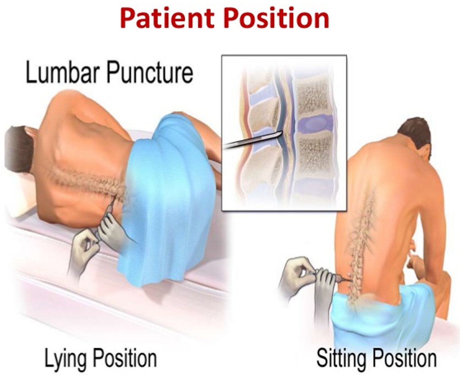
Wearing a gown, you’ll begin the exam lying on your side on the exam table, usually with your knees drawn toward your chest. For the procedure, you’ll be asked to lie on a table while the doctor inserts a sigmoidoscope into your anus and slowly guides it through your rectum and into your sigmoid colon. The scope pumps air into your large intestine to give the doctor a better view. The camera sends a video image of your intestinal lining to a monitor, allowing the doctor to examine the tissues lining your sigmoid colon and rectum. The doctor may ask you to move several times on the table to adjust the scope for better viewing. Once the scope has reached your transverse colon, the doctor slowly withdraws it and examines the lining of your sigmoid colon again.

During the procedure, your doctor may remove polyps and send them to a lab for testing. Colon polyps are common in adults and are harmless in most cases. However, most colon cancer begins as a polyp, so removing polyps early is an effective way to prevent cancer.

If your doctor finds abnormal tissue, he or she may perform a biopsy. You won’t feel the biopsy.

If your doctor found polyps or other abnormal tissue during a flexible sigmoidoscopy, your doctor may suggest you return for a colonoscopy.

After a flexible sigmoidoscopy

After a flexible sigmoidoscopy, you can expect the following:

  • You may have cramping in your abdomen or bloating during the first hour after the procedure. You may feel bloated or pass gas for a few hours as you clear the air from your colon. Walking may help relieve any discomfort.
  • You can resume regular activities right away after the procedure.
  • You can return to a normal diet.
  • You may also notice a small amount of blood with your first bowel movement after the exam, which usually isn’t cause for alarm. Consult your doctor if you continue to pass blood or blood clots or if you have persistent abdominal pain or a fever of 100 °F (37.8 °C) or higher.

A health care professional will give you written instructions on how to take care of yourself after the procedure and will review them with you. You should follow all instructions.

If the doctor removed polyps or performed a biopsy, you may have light bleeding from your anus. This bleeding is normal. Some results from a flexible sigmoidoscopy are available right after the procedure, and your doctor will share these results with you. A pathologist will examine the biopsy tissue. Biopsy results take a few days or longer to come back.

Seek care right away

If you have any of the following symptoms after a flexible sigmoidoscopy, seek care right away:

  • severe pain in your abdomen
  • fever
  • continued bloody bowel movements or continued bleeding from your anus
  • dizziness
  • weakness

Flexible sigmoidoscopy results

Your doctor will review the results of the flexible sigmoidoscopy exam and then share the results with you.

  • Negative result. A flexible sigmoidoscopy exam is considered negative if the doctor doesn’t find any abnormalities in the colon. If you’re at average risk of colon cancer — you have no colon cancer risk factors other than age — your doctor may recommend waiting five years and then repeating the exam.
  • Positive result. A flexible sigmoidoscopy exam is considered positive if the doctor finds polyps or abnormal tissue in the colon. Depending on the findings, you may need additional testing — such as a colonoscopy — so that any abnormalities can be examined more thoroughly, biopsied or removed. During colonoscopy, your doctor can also screen the entire colon for other abnormalities.

How much of your colon and rectum can be viewed during a flexible sigmoidoscopy depends on the experience of the doctor doing your exam and the success of the colon preparation. If your doctor is concerned about the quality of the view through the scope, he or she may recommend a repeat flexible sigmoidoscopy exam or another screening test.

References
  1. Final Recommendation Statement. Colorectal Cancer: Screening. https://www.uspreventiveservicestaskforce.org/Page/Document/RecommendationStatementFinal/colorectal-cancer-screening2
  2. Howlader N, Noone AM, Krapcho M, et al (eds). SEER Cancer Statistics Review, 1975–2015. National Cancer Institute. https://seer.cancer.gov/csr/1975_2015/
  3. Lin JS, Piper M, Perdue LA, et al. Screening for Colorectal Cancer: A Systematic Review for the U.S. Preventive Services Task Force. Evidence Synthesis No. 135. AHRQ Publication No. 14-05203-EF-1. Rockville, MD: Agency for Healthcare Research and Quality; 2016.
  4. Zauber A, Knudsen A, Rutter CM, Lansdorp-Vogelaar I, Kuntz KM. Evaluating the Benefits and Harms of Colorectal Cancer Screening Strategies: A Collaborative Modeling Approach. AHRQ Publication No. 14-05203-EF-2. Rockville, MD: Agency for Healthcare Research and Quality; 2015.
  5. Holme Ø, Løberg M, Kalager M, et al. Effect of flexible sigmoidoscopy screening on colorectal cancer incidence and mortality: a randomized clinical trial. JAMA. 2014;312(6):606-15.
  6. Nishihara R, Wu K, Lochhead P, et al. Long-term colorectal-cancer incidence and mortality after lower endoscopy. N Engl J Med. 2013;369(12):1095-105.
  7. Shapiro JA, Klabunde CN, Thompson TD, Nadel MR, Seeff LC, White A. Patterns of colorectal cancer test use, including CT colonography, in the 2010 National Health Interview Survey. Cancer Epidemiol Biomarkers Prev. 2012;21(6):895-904.
  8. Inadomi JM, Vijan S, Janz NK, et al. Adherence to colorectal cancer screening: a randomized clinical trial of competing strategies. Arch Intern Med. 2012;172(7):575-82.
  9. Lin JS, Piper M, Perdue LA, et al. Screening for colorectal cancer: updated evidence report and systematic review for the U.S. Preventive Services Task Force. JAMA. doi:10.1001/jama.2016.3332
read more
Procedures

Lumbar puncture

lumbar puncture

What is a lumbar puncture

A lumbar puncture also called a spinal tap, is a common medical test where a spinal needle is advanced between two lumbar bones (vertebrae) into the subarachnoid space in your lower back in order to collect a small sample of cerebrospinal fluid (CSF) for examination. Lumbar puncture procedure is done to measure pressures within the CSF and to collect a sample of the cerebrospinal fluid (CSF) for further testing. Cerebral spinal fluid (CSF) is a clear, colorless liquid that delivers nutrients and “cushions” the brain and spinal cord, or central nervous system.

In a lumbar puncture, a needle is carefully inserted into the lower spine to collect the cerebrospinal fluid (CSF) sample.

Cerebral spinal fluid (CSF) is a clear fluid that circulates in the space surrounding the spinal cord and brain. Cerebrospinal fluid (CSF) protects the brain and spinal cord from injury by acting like a liquid cushion. Cerebrospinal fluid (CSF) is usually obtained through a lumbar puncture (spinal tap). During the procedure, a needle is inserted usually between the 3rd and 4th lumbar vertebrae and the cerebrospinal fluid (CSF) is collected for testing.

Healthcare professionals perform lumbar punctures and test the CSF to detect or rule out suspected diseases or conditions through analysis of the white blood cell count, glucose levels, protein, and bacteria.

Most lumbar punctures are done to test for infections (such as meningitis), but they also can detect bleeding in the brain and certain conditions affecting the nervous system (such as Guillain-Barré syndrome and multiple sclerosis or cancers of the brain or spinal cord). Lumbar punctures also can deliver chemotherapeutic medications or to inject anesthetic medications.

Special testing can look for certain bacteria and viruses, or find the presence of abnormal cells that can help identify specific diseases in the central nervous system.

Cerebrospinal fluid (CSF) analysis can also be used to diagnose certain neurologic disorders. These may include brain or spinal cord damage. A spinal tap may also be done to establish the diagnosis of normal pressure hydrocephalus.

Depending on the doctor’s recommendations, you might have to lie on your back for a few hours after the procedure. You might feel tired and have a mild backache the day after the lumbar puncture procedure.

Up to 1 to 43 percent of people who have undergone a lumbar puncture develop a headache afterward due to a leak of CSF fluid into nearby tissues 1. Lumbar puncture headache after the test that can last a few hours or days. If headaches last more than a few days (especially when you sit, stand or walk) you might have a CSF-leak. You should talk to your physician if this occurs.

Post-dural (post-lumbar or post-spinal) puncture headache is defined as any headache after a lumbar puncture that worsens within 15 minutes of sitting or standing and that is relieved within 15 minutes of lying down 2. Ninety per cent of post-lumbar puncture headaches occur within three days of the procedure, and 66% start within the first 48 hours 3.

A lumbar puncture takes around 30 to 45 minutes but you’ll need to stay lying down at the hospital for at least another hour while the nurses monitor you.

You’ll be able to go home the same day if you feel well enough – but you won’t be able to drive yourself home.

Lumbar puncture side effects

A lumbar puncture is generally a safe procedure and serious side effects are uncommon.

The most common side effects are:

  • headaches, which can last for up to a week – you’ll be given painkillers at the hospital if you need them
  • swelling and lower back pain where the needle was inserted – this should get better on its own after a few days and is normally nothing to worry about

Is lumbar puncture painful?

While some people notice a brief pinch and some discomfort, most people don’t consider a lumbar puncture to be painful.

Sometimes, sedation medication may be helpful for you in order to perform the lumbar puncture procedure. If sedation is necessary, be sure to discuss the risks and benefits with your doctor.

When to get medical attention after a lumbar puncture

Contact the hospital team or a doctor if:

  • your headaches are severe or don’t go away
  • you’re feeling or being sick
  • you have a very high temperature or feel hot and shivery
  • it’s painful to look at bright lights
  • the swelling in your back lasts for more than a few days or keeps getting worse
  • you see blood or clear fluid leaking from your back

Why is lumbar puncture done

There are many indications for lumbar puncture (Table 1), but obtaining CSF may be the only way of confirming or refuting subarachnoid haemorrhage (SAH), meningitis and neuro-inflammatory diseases. Lumbar puncture is still required to obtain indirect measurements of intracranial pressure, although non-invasive methods of intracranial pressure estimation are undergoing validation 4.

A lumbar puncture may be done to:

  • Collect cerebrospinal fluid for laboratory analysis
  • Measure the pressure of your cerebrospinal fluid
  • Remove some fluid to reduce pressure in the skull or spine
  • Inject spinal anesthetics, chemotherapy drugs or other medications
  • Inject dye (myelography) or radioactive substances (cisternography) into cerebrospinal fluid to make diagnostic images of the fluid’s flow

Table 1. Indications for Lumbar Puncture

To exclude subarachnoid haemorrhage in acute severe headache
To investigate or exclude meningitis

  • Bacterial
  • Viral
  • Tuberculous
  • Cryptococcal
  • Chemical
  • Carcinomatous
To investigate neurological disorders

  • Multiple Sclerosis
  • Sarcoidosis
  • Guillian Barre, Chronic Inflammatory Demyelinating Polyneuropathy
  • Mitochondrial Disorders
  • Leukencephalopathies
  • Paraneoplastic Syndromes
To demonstrate and manage disorders of Intracranial Pressure

  • Idiopathic Intracranial Hypertension
  • Spontaneous Intracranial Hypotension
To administer therapeutic or diagnostic agents

  • Spinal anaesthesia
  • Intrathecal chemotherapy
  • Intrathecal antibiotics
  • Intrathecal baclofen
  • Contrast media in myelography or cisternography
[Source 5]

A lumbar puncture procedure may be helpful in diagnosing many diseases and disorders, including:

  • Meningitis. An inflammation of the membrane covering the brain and spinal cord. The inflammation is usually the result of a viral, bacterial, or fungal infection.
  • Encephalitis. An inflammation of the brain that is usually caused by a virus.
  • Certain cancers involving the brain and spinal cord
  • Bleeding in the area between the brain and the tissues that cover it (subarachnoid space)
  • Reye syndrome. A sometimes fatal disease that causes severe problems with the brain and other organs. Although the exact cause of the disease is not known, it has been linked to giving aspirin to children. It is now advised not to give aspirin to children during illnesses, unless prescribed by your child’s healthcare provider.
  • Myelitis. An inflammation of the spinal cord or bone marrow.
  • Neurosyphilis. A stage of syphilis during which the bacteria invades the central nervous system.
  • Guillain-Barré syndrome. A disorder in which the body’s immune system attacks part of the nervous system.
  • Demyelinating diseases. Diseases that attack the protective coating that surrounds certain nerve fibers – for example, multiple sclerosis or acute demyelination polyneuropathy.
  • Headaches of unknown cause. After evaluation and head imaging if necessary, a lumbar puncture may be done to diagnose certain inflammatory conditions that can result in a headache.
  • Pseudotumor cerebri (also called idiopathic intracranial hypertension, or IIH). In this condition,pressure within the subarachnoid space is elevated for reasons that are not clear. A lumbar puncture is only done in this condition after evaluation and head imaging.
  • Normal pressure hydrocephalus. A rare condition affecting mainly older people in which there is a triad of loss of urinary control, memory problems, and an unsteady gait. A lumbar puncture is done to see if the pressure of the CSF is elevated or not.

In addition, a lumbar puncture may be used to measure the pressure of the CSF. The healthcare provider uses a special tube (called a manometer) to measures the pressure during a lumbar puncture.

Finally, a lumbar puncture may be done to inject medicine directly into the spinal cord. These include:

  • Spinal anesthetics before a surgical procedure
  • Contrast dye for X-ray studies – for example, myelography
  • Chemotherapy drugs used to treat cancer

Your healthcare provider may have other reasons to recommend a lumbar puncture.

Information gathered from a lumbar puncture can help diagnose:

  • Serious bacterial, fungal and viral infections, including meningitis, encephalitis and syphilis
  • Bleeding around the brain (subarachnoid hemorrhage)
  • Certain cancers involving the brain or spinal cord
  • Certain inflammatory conditions of the nervous system, such as multiple sclerosis and Guillain-Barre syndrome

Getting the results

Some results from a lumbar puncture are available within 45 to 60 minutes. However, to look for specific bacteria growing in the sample, a bacterial culture is sent to the lab and these results are usually available in 48 hours. If it’s determined there might be an infection, the doctor will start antibiotic treatment while waiting for the results of the culture.

Lumbar puncture can allow the physician to determine:

  • the cause of meningitis
  • if the patient has a subarachnoid hemorrhage in the setting of a normal non-contrast head CT scan
  • the presence of a malignancy affecting the central nervous system (e.g., leptomeningeal carcinomatosis)
  • the presence of a demyelinating disease (e.g., multiple sclerosis)
  • if the patient has symptoms concerning for Guillain-Barre syndrome

Caution should be taken in performing the lumbar puncture in patients with:

  • increased intracranial pressure
  • the patient runs the risk of brain herniation
  • thrombocytopenia (low platelets count) or other bleeeding diathesis
  • signs and symptoms concerning for a spinal epidural abscess

Lumbar puncture complications

  • post-lumbar puncture headache
  • infection
  • bleeding
  • cerebral herniation

Spinal Cord Anatomy

Knowledge of the anatomy of the lumbar spine 6 is essential for anyone performing lumbar puncture. The lumbar puncture needle pierces in order: skin, subcutaneous tissue, supraspinous ligament, interspinous ligament, ligamentum flavum, epidural space containing the internal vertebral venous plexus, dura, arachnoid, and finally the subarachnoid space (Figure 4).

The most important bony landmark is the L4 spinous process, which is located at the intersection of the ‘intercristal’ or ‘Tuffier’s’ line (the line between the top of the iliac crests) and the lumbar spine midline (Figure 2). Radiological studies have shown that this clinical landmark is accurate in over 95% of the population 7, although in females or obese people Tuffier’s Line tends to be found at a higher level than L4 8. Note the conus medullaris can extend as low as to the upper aspect of the L3 vertebral segment 9.

Figure 1. Spinal cord

spinal cord

Figure 2. Spinal cord segments

spinal cord nerves and segments

Figure 3. Lumbar puncture site L3-L4 (a spinal needle is inserted, usually between the third and fourth lumbar vertebrae in the lower spine. Once the needle is properly positioned in the subarachnoid space [the space between the spinal cord and its covering, the meninges], pressures can be measured and fluid can be collected for testing.)

lumbar puncture site

Figure 4. Lumbar puncture site L4-L5. Note: Sagittal section of lumbar vertebrae illustrating the course of the lumbar puncture needle through skin, subcutaneous tissue, supraspinous ligament, interspinous ligament between the spinous processes, ligamentum flavum, dura mater, into the subarachnoid space and between the nerve roots of the cauda equina. Lumbar vertebral bodies, intervertebral disc , and lumbar puncture needle.

lumbar puncture siteFigure 5. Lumbar puncture anatomical landmarks – the lines referred to in the study: the palpated intercristal line (a), the imaged Intercristal Line (eponymously Tuffier’s Line) (b) and the palpated posterior superior iliac spine line (c).

Lumbar puncture anatomical landmarks

Figure 6. Lumbar puncture locations – Blue dots-Iliac crests, and the line connecting them is the Intercristal Line (eponymously Tuffier’s Line). Red Dots are either side of the palpated L4 spinous process, the right hand one is in the L4/5 interspinal space and the left hand one in the L3/4 interspinal space. A diagnostic Lumbar Puncture should be performed at the L3/4 interspinal space, marked ‘x’.

Lumbar puncture locations

Lumbar puncture contraindications

Severe thrombocytopenia, bleeding diathesis or anticoagulant therapy are contraindications to lumbar puncture, although it can be performed safely at platelet counts of 50 x 10/l or above 10.

Aspirin therapy is not associated with a high risk of bleeding in spinal anaesthetic interventions 11, but data on clopidogrel and other GP IIa/IIIb receptor antagonists are lacking. GP IIb/IIIa–Receptor Antagonists affect platelet-fibrinogen and platelet–von Willebrand factor binding to inhibit platelet aggregation. This class of antiplatelet drugs includes abciximab (ReoPro), eptifibatide (Integrilin), and tirofiban (Aggrastat) 12. Guidance on spinal anaesthetic procedures suggest that procedures should be avoided until platelet function has recovered 13. Warfarin should be stopped 5-7 days in advance of procedures and the INR should be less than 1.2. Low molecular weight heparin can be used in the interim but treatment dose heparin should be stopped for twenty-four hours prior to spinal procedures. Prophylactic doses should be avoided within twelve hours 13.

Lumbar puncture procedure

There are different ways to get a sample of cerebrospinal fluid (CSF). Lumbar puncture (spinal tap) is the most common method.

A lumbar puncture is usually done in an outpatient facility or a hospital. Your doctor will talk to you about the potential risks, and any discomfort you might feel during the procedure.

If a child is having a lumbar puncture, a parent is usually allowed to stay in the room. Talk to your child’s doctor about whether this will be possible.

Afterward, you should plan to rest for several hours, even if you feel fine. This is to prevent fluid from leaking around the site of the puncture. You will not need to lie flat on your back the entire time.

How to prepare for the lumbar puncture procedure

Before your lumbar puncture, your doctor asks questions about your medical history, does a physical exam, and orders blood tests to check if you have any bleeding or clotting disorders. You will need to give the health care team your consent before the test. Tell your doctor if you are on any blood-thinning medicines such as warfarin (Coumadin), Lovenox, aspirin, or Plavix.

Your doctor may also recommend a CT scan or MRI to determine if you have any abnormal swelling in or around your brain.

PRECAUTIONS: If you are pregnant or think you might be pregnant, please check with your doctor before scheduling the exam. Other options will be discussed with you and your doctor.

Food and medications

Your doctor will give you specific instructions about food, drink and medications. You’ll likely be asked not to eat or drink anything after midnight before your procedure.

Tell your doctor if you’re taking blood-thinning or other anticoagulant medications. Examples include warfarin (Coumadin, Jantoven), clopidogrel (Plavix), and some over-the-counter pain relievers such as aspirin, ibuprofen (Advil, Motrin IB, others) or naproxen sodium (Aleve). Also, tell your doctor if you’re allergic to any medications, such as numbing medications (local anesthetics).

How the lumbar puncture procedure will feel

It may be uncomfortable to stay in position for the test. Staying still is important because movement may lead to injury of the spinal cord.

You may be told to straighten your position slightly after the needle is in place. This is to help measure the CSF pressure.

The anesthetic will sting or burn when first injected. There will be a hard pressure sensation when the needle is inserted. Often, there is some brief pain when the needle goes through the tissue surrounding the spinal cord. This pain should stop in a few seconds.

In most cases, the procedure takes about 30 minutes. The actual pressure measurements and CSF collection only take a few minutes.

Before the lumbar puncture procedure

You’re asked to change into a hospital gown. There are a few possible positions for this test. Usually, you lie on your side with your knees drawn up to your chest, or you sit and lean forward on a stable surface. These positions flex your back, widening the spaces between your vertebrae and making it easier for your doctor to insert the needle.

For an infant or young child, someone will hold the child in position during the procedure.

Your back is washed with antiseptic soap or iodine and covered with a sterile sheet.

Lumbar puncture position

Patient position

  • Lateral recumbent position
    • preferred for accurate opening pressure measurement, so the spaces between the vertebrae are as wide as possible. This makes it easier for the doctor to insert the needle.
  • Prone position, better for fluoroscopy guided lumbar puncture
  • Sitting upright: If the procedure is to be performed in the upright position, seated with the chin down and the feet supported, a table and pillow will improve comfort and optimise positioning. This position widens the interspinous distance 14.
  • Needle entry
    • into the subarachnoid space at L3-4 or L4-5 interspace
    • remember that the spinal cord ends in L1-2
    • thus there should not be any trauma to the spinal cord if properly performed
  • In cases where there are unsuccessful attempts in obtaining CSF, imaging guidance can be used such as
    • fluoroscopy
    • ultrasound

Figure 5. Lumbar puncture position

lumbar-puncture-location

Lumbar puncture position

During the lumbar puncture procedure

To have the lumbar puncture test:

  1. You will lie on your side with your knees pulled up toward the chest, and chin tucked downward. Sometimes the test is done sitting up, but bent forward.
  2. After the back is cleaned, the health care provider will inject a local numbing medicine (local anesthetic) into the lower spine. The local anesthetic will sting briefly as it’s injected.
  3. A spinal needle will be inserted between the two lower vertebrae (lumbar region), through the spinal membrane (dura) and into the spinal canal. You may feel pressure in your back during this part of the procedure. Once the needle is properly positioned in the subarachnoid space (the space between the spinal cord and its covering, the meninges), pressures can be measured and fluid can be collected for testing.
    • The spinal needle is thin and the length varies according to the size of the patient. It has a hollow core, and inside the hollow core is a “stylet,” another type of thin needle that acts kind of like a plug. When the spinal needle is inserted into the lower lumbar area, the stylet is carefully removed, which allows the cerebrospinal fluid to drip out into the collection tubes.
  4. Once the needle is in position, the CSF pressure is measured and a sample of 1 to 10 milliliters (mL) of CSF is collected in 4 vials.
    • An opening pressure is sometimes taken. An abnormal pressure can suggest an infection or other problem.
  5. After the CSF sample is collected (this usually takes about 2-5 minutes), the needle is withdrawn, the area is cleaned, and a small bandage is placed on the needle site. Collected samples are sent to a lab for analysis and testing.
    • If the provider needs to inject medicine into the spinal canal, it will be given through the same needle after the CSF is collected.
  6. The lumbar puncture procedure usually lasts about 30 to 45 minutes. Your doctor may suggest lying down after the procedure for a short time after the test.

Tell the healthcare provider if you feel any numbness, tingling, headache, or lightheadedness during the procedure.

Occasionally, special x-rays are used to help guide the needle into position. This is called fluoroscopy.

Sometimes, an ultrasound may be used as a guide during the procedure on infants and young children. The ultrasound can help prevent inserting the needle too far.

Lumbar puncture with fluid collection may also be part of other procedures such as an x-ray or CT scan after dye has been inserted into the CSF.

Rarely, other methods of cerebrospinal fluid (CSF) collection may be used.

  • Cisternal puncture uses a needle placed below the occipital bone (back of the skull). It can be dangerous because it is so close to the brain stem. It is always done with fluoroscopy.
  • Ventricular puncture may be recommended in people with possible brain herniation. This is a very rarely used method. It is most often done in the operating room. A hole is drilled in the skull, and a needle is inserted directly into one of the brain’s ventricles.

CSF may also be collected from a tube that’s already placed in the fluid, such as a shunt or a ventricular drain.

Lumbar puncture recovery

You must have an adult driver accompany you so they can drive you home after the procedure. This is for your safety and comfort.

Please note: You will be unable to drive for 24 hours after the procedure. If you are taking a cab or using public transportation, you need to bring a friend or family member to accompany you after the procedure to your home or hotel. A cab or public transportation driver is not considered an escort.

If your ride home is longer than 30 minutes, doctors advise that on your ride home you be in a reclined position with one or two pillows supporting your head.

After the lumbar puncture procedure

You usually will be asked to lie flat for about one hour after the lumbar puncture is completed. This helps reduce the incidence of a headache. You will be allowed to roll from side to side as long as your head is not elevated. If you need to urinate, you may need to do so in a bedpan or urinal during the time that you need to stay flat.

You will be asked to drink extra fluids to rehydrate after the procedure. This replaces the CSF that was withdrawn during the spinal tap and reduces the chance of developing a headache. However, a recent 2016 Cochrane Review 15 concluded that in general, there was no evidence suggesting that routine bed rest after lumbar puncture is beneficial for the prevention of post-dural puncture headache onset. The role of fluid supplementation in the prevention of post-dural lumbar puncture headache remains unclear 15. The review authors believe that these practices should no longer be routinely recommended to patients for the prevention of headaches after lumbar puncture since there is no evidence supporting them 15.

After recovery, you may be taken to your hospital room or discharged to your home. If you go home, usually your healthcare provider will advise you to only engage in very light activity the rest of the day.

  • Plan to rest. Don’t participate in strenuous activities the day of your procedure. You may return to work if your job doesn’t require you to be physically active. Discuss your activities with your doctor if you have questions.
  • Take a pain medication. A nonprescription pain-relieving medication that contains acetaminophen can help reduce a headache or back pain.

While you’re recovering from a lumbar puncture:

DO

  • drink plenty of fluids
  • take painkillers, such as paracetamol
  • lie down instead of sitting upright
  • try drinks containing caffeine, such as coffee, tea or cola – some people find this helps to relieve the headaches
  • remove the dressing or plaster yourself the next day

DON’T

  • drive or operate machinery for at least 24 hours
  • play sport or do any strenuous activities for at least a week

Once you are at home, notify your provider of any abnormalities, such as:

  • Numbness and tingling of the legs
  • Drainage of blood or pain at the injection site
  • Inability to urinate
  • Headaches

If the headaches persist for more than a few hours after the procedure, or when you change positions, contact your provider.

You may be instructed to limit your activity for 24 hours following the procedure. Generally, if no complications occur, you may return to your normal diet and activities.

Your healthcare provider may give you other specific instructions about what you should do after a lumbar puncture.

Lumbar puncture results

The spinal fluid samples are sent to a laboratory for analysis. Lab technicians check for a number of things when examining spinal fluid, including:

  • General appearance. Spinal fluid is normally clear and colorless. If it’s cloudy, yellow or pink in color, it might indicate abnormal bleeding. Spinal fluid that is green might indicate an infection or the presence of bilirubin.
  • Protein (total protein and the presence of certain proteins). Elevated levels of total protein — greater than 45 milligrams per deciliter (mg/dL) — may indicate an infection or another inflammatory condition. Specific lab values may vary from medical facility to medical facility.
  • White blood cells. Spinal fluid normally contains up to 5 mononuclear leukocytes (white blood cells) per microliter. Increased numbers may indicate an infection. Specific lab values may vary from medical facility to medical facility.
  • Sugar (glucose). A low glucose level in spinal fluid may indicate infection or another condition.
  • Microorganisms. The presence of bacteria, viruses, fungi or other microorganisms can indicate an infection.
  • Cancer cells. The presence of abnormal cells in spinal fluid — such as tumor or immature blood cells — can indicate certain types of cancer.

Lab results are combined with information obtained during the test, such as spinal fluid pressure, to help establish a possible diagnosis.

Your health care provider generally gives you the results within a few days, but it could take longer. Ask your doctor when he or she expects to receive the results of your test.

Write down questions that you want to ask your doctor. Don’t hesitate to ask questions during your visit. Questions you may want to ask include:

  • Based on the results, what are my next steps?
  • What kind of follow-up, if any, should I expect?
  • Are there any factors that might have affected the results of this test and, therefore, may have altered the results?
  • Will I need to repeat the test at some point?

Normal Results

Normal values typically range as follows:

  • CSF Pressure: 70 to 180 mm H2O
  • CSF Appearance: clear, colorless
  • CSF Total protein: 15 to 60 mg/100 mL
  • Gamma globulin: 3% to 12% of the total protein
  • CSF glucose: 50 to 80 mg/100 mL (or greater than two thirds of blood sugar level)
  • CSF cell count: 0 to 5 white blood cells (all mononuclear), and no red blood cells
  • Chloride: 110 to 125 mEq/L

Normal value ranges may vary slightly among different laboratories. Talk to your provider about the meaning of your specific test results.

The examples above show the common measurements for results for these tests. Some laboratories use different measurements or may test different specimens.

Abnormal Results

What abnormal results mean

If the CSF looks cloudy, it could mean there is an infection or a buildup of white blood cells or protein.

If the CSF looks bloody or red, it may be a sign of bleeding or spinal cord obstruction. If it is brown, orange, or yellow, it may be a sign of increased CSF protein or previous bleeding (more than 3 days ago). There may be blood in the sample that came from the spinal tap itself. This makes it harder to interpret the test results.

CSF pressure

  • Increased CSF pressure may be due to increased intracranial pressure (pressure within the skull).
  • Decreased CSF pressure may be due to spinal cord tumor, shock, fainting, or diabetic coma.

CSF protein

  • Increased CSF protein may be due to blood in the CSF, diabetes, polyneuritis, tumor, injury, or any inflammatory or infectious condition.
  • Decreased protein is a sign of rapid CSF production.

CSF glucose

  • Increased CSF glucose is a sign of high blood sugar.
  • Decreased CSF glucose may be due to hypoglycemia (low blood sugar), bacterial or fungal infection (such as meningitis), tuberculosis, or certain other types of meningitis.

Blood cells in CSF

  • Increased white blood cells in the CSF may be a sign of meningitis, acute infection, beginning of a long-term (chronic) illness, tumor, abscess, stroke, or demyelinating disease (such as multiple sclerosis).
  • Red blood cells in the CSF sample may be a sign of bleeding into the spinal fluid or the result of a traumatic lumbar puncture.

Other CSF results

Increased CSF gamma globulin levels may be due to diseases such as multiple sclerosis, neurosyphilis, or Guillain-Barré syndrome.

Additional conditions under which the test may be performed:

  • Chronic inflammatory polyneuropathy
  • Dementia due to metabolic causes
  • Encephalitis
  • Epilepsy
  • Febrile seizure (children)
  • Generalized tonic-clonic seizure
  • Hydrocephalus
  • Inhalation anthrax
  • Normal pressure hydrocephalus
  • Pituitary tumor
  • Reye syndrome

Lumbar puncture risks

A lumbar puncture is considered a safe procedure with minimal risks. Most of the time, there are no complications. In some instances, a patient may get a headache. It’s recommended that patients lie down for a few hours after the lumbar puncture test and drink plenty of fluids to help prevent headaches, which usually resolve with rest, pain medications, and fluids. In rare cases, infection or bleeding can occur.

Risks of lumbar puncture include:

  • Bleeding. Bleeding may occur near the puncture site or, rarely, into the epidural space. Bleeding into the spinal canal or around the brain (subdural hematomas). There is an increased risk of bleeding in people who take blood thinners.
  • Discomfort during the test
  • Post-lumbar puncture headache. Up to 25 percent of people who have undergone a lumbar puncture develop a headache afterward due to a leak of CSF fluid into nearby tissues. The headache typically starts several hours up to two days after the procedure and may be accompanied by nausea, vomiting and dizziness. The headaches are usually present when sitting or standing and resolve after lying down. Post-lumbar puncture headaches can last from a few hours to a week or more.
  • Hypersensitivity (allergic) reaction to the anesthetic
  • Infection introduced by the needle going through the skin
  • Back discomfort or pain. You may feel pain or tenderness in your lower back after the procedure. The pain might radiate down the back of your legs.
  • Brainstem herniation. Increased pressure within the skull (intracranial), due to a brain tumor or other space-occupying lesion, can lead to compression of the brainstem after a sample of cerebrospinal fluid is removed. A computerized tomography (CT) scan or MRI prior to a lumbar puncture can be obtained to determine if there is evidence of a space-occupying lesion that results in increased intracranial pressure. This complication is rare.

Brain herniation may occur if this test is done on a person with a mass in the brain (such as a tumor or abscess). This can result in brain damage or death. This test is not done if an exam or test reveals signs of a brain mass.

Damage to the nerves in the spinal cord may occur, particularly if the person moves during the test.

Cisternal puncture or ventricular puncture carries additional risks of brain or spinal cord damage and bleeding within the brain.

Lumbar puncture complications

Lumbar puncture is more dangerous for people with:

  • A tumor in the back of the brain that is pressing down on the brainstem
  • Blood clotting problems
  • Low platelet count (thrombocytopenia)
  • Individuals taking blood thinners, aspirin, clopidogrel, or other similar drugs to decrease the formation of blood clots.
References
  1. Grande PO. Mechanisms behind postspinal headache and brain stem compression following lumbar dural puncture – a physiological approach. Acta Anaesthesiologica Scandinavica 2005;49(5):619-26. https://www.ncbi.nlm.nih.gov/pubmed/15836674
  2. Headache Classification Subcommittee of the International Headache Society. The International Classification of Headache Disorders: 2nd edition. Cephalalgia 2004;24 Suppl 1:9-160. https://www.ncbi.nlm.nih.gov/pubmed/14979299
  3. Turnbull DK, Shepherd DB. Post-dural puncture headache: pathogenesis, prevention and treatment. British Journal of Anaesthesia 2003;91(5):718-29. https://www.ncbi.nlm.nih.gov/pubmed/14570796
  4. Clinical assessment of noninvasive intracranial pressure absolute value measurement method. Ragauskas A, Matijosaitis V, Zakelis R, Petrikonis K, Rastenyte D, Piper I, Daubaris G. Neurology. 2012 May 22; 78(21):1684-91. https://www.ncbi.nlm.nih.gov/pubmed/22573638/
  5. Doherty CM, Forbes RB. Diagnostic Lumbar Puncture. The Ulster Medical Journal. 2014;83(2):93-102. https://www.ncbi.nlm.nih.gov/pmc/articles/PMC4113153/
  6. Lumbar puncture: anatomical review of a clinical skill. Boon JM, Abrahams PH, Meiring JH, Welch T. Clin Anat. 2004 Oct; 17(7):544-53. https://www.ncbi.nlm.nih.gov/pubmed/15376294/
  7. Accuracy of placement of extradural needles in the L3-4 interspace: comparison of two methods of identifying L4. Ievins FA. Br J Anaesth. 1991 Mar; 66(3):381-2. https://bjanaesthesia.org/article/S0007-0912(17)48180-3/pdf
  8. Which spinal levels are identified by palpation of the iliac crests and the posterior superior iliac spines? Chakraverty R, Pynsent P, Isaacs K. J Anat. 2007 Feb; 210(2):232-6. https://www.ncbi.nlm.nih.gov/pmc/articles/PMC2100271/
  9. Influence of age and sex on the position of the conus medullaris and Tuffier’s line in adults. Kim JT, Bahk JH, Sung J. Anesthesiology. 2003 Dec; 99(6):1359-63. https://www.ncbi.nlm.nih.gov/pubmed/14639149/
  10. Guidelines for the use of platelet transfusions. British Committee for Standards in Haematology, Blood Transfusion Task Force. Br J Haematol. 2003 Jul; 122(1):10-23. https://www.ncbi.nlm.nih.gov/pubmed/12823341/
  11. Horlocker TT, Wedel DJ, Schroeder DR, Rose SH, Elliott BA, McGregor DG, et al. Preoperative antiplatelet therapy does not increase the risk of spinal hematoma associated with regional anesthesia. Anesth Analg. 1995;80(2):303–9. https://www.ncbi.nlm.nih.gov/pubmed/7818117
  12. Recommendations for Anticoagulated Patients Undergoing Image-Guided Spinal Procedures. American Journal of Neuroradiology March 2006, 27 (3) 468-470; http://www.ajnr.org/content/27/3/468.long
  13. Layton KF, Kallmes DF, Horlocker TT. Recommendations for anticoagulated patients undergoing image-guided spinal procedures. AJNR Am J Neuroradiol. 2006;27(3):468–70. http://www.ajnr.org/content/27/3/468.long
  14. Optimal patient position for lumbar puncture, measured by ultrasonography. Sandoval M, Shestak W, Stürmann K, Hsu C. Emerg Radiol. 2004 Feb; 10(4):179-81. https://www.ncbi.nlm.nih.gov/pubmed/15290485/
  15. Arevalo-Rodriguez I, Ciapponi A, Roqué i Figuls M, Muñoz L, Bonfill Cosp X. Posture and fluids for preventing post-dural puncture headache. Cochrane Database of Systematic Reviews 2016, Issue 3. Art. No.: CD009199. DOI: 10.1002/14651858.CD009199.pub3. http://cochranelibrary-wiley.com/doi/10.1002/14651858.CD009199.pub3/full
read more
Procedures

Radiation therapy

radiation therapy

What is radiation therapy

Radiation therapy uses high-energy rays or particles (radiation), such as x-rays, gamma rays, electron beams, or protons to kill cancer cells and other problems. Other names for radiation therapy are radiation treatment, radiotherapy, irradiation, or x-ray therapy.

There are different types of radiation. One that you may know about is x-rays. If you’ve ever had an x-ray of your chest or any other body part, you’ve had some radiation. Radiation is used in much higher doses to treat some types of cancer.

Radiation therapy is not like chemotherapy (often called chemo). Radiation treats just the tumor. Chemo uses drugs to treat the whole body. So chemo might be used if a person has cancer in many places. Radiation affects only the part of the body being treated.

More than half of people with cancer get radiation therapy. Sometimes, radiation therapy is the only cancer treatment needed.

How does radiation therapy work

Your body is made up of trillions of normal, healthy cells. Cancer starts when something changes a normal cell into a cancer cell. This cancer cell can then grow and make more cancer cells until a tumor is formed. Tumors can keep growing and cause problems. If the cancer is not treated, it can spread to other parts of the body and form more tumors.

Your cells normally grow and divide to form new cells. But cancer cells grow and divide faster than most normal cells. Radiation works by making small breaks in the DNA inside cells. These breaks keep cancer cells from growing and dividing and cause them to die. Nearby normal cells can also be affected by radiation, but normal cells can repair themselves and most recover and go back to working the way they should. Whereas cancer cells cannot.

Special equipment sends high doses of radiation to the cancer cells or tumor. This keeps the cells from growing and making more cancer cells. Radiation treatment is planned to damage cancer cells, with as little harm as possible to nearby healthy cells.

Some radiation treatments (systemic radiation therapy) use radioactive substances that are given in a vein or by mouth. Even though this type of radiation does travel throughout the body, the radioactive substance mostly collects in the area of the tumor, so there’s little effect on the rest of the body.

Sometimes radiation is the only treatment needed. Other times it’s one part of a patient’s cancer treatment plan.

Your doctor may suggest radiation therapy to treat your cancer. Sometimes radiation can cure cancer. At other times the goal may be to slow the cancer’s growth to help you feel better. Be sure to talk to your doctor about the goal of your treatment.

Who gives radiation therapy treatments?

During your radiation therapy, a team of highly trained medical professionals will care for you. Your team may include these people:

  • Radiation oncologist: This doctor is specially trained to treat cancer with radiation. This person oversees your radiation treatment plan.
  • Radiation physicist: This is the person who makes sure the radiation equipment is working as it should and that it gives you the exact dose prescribed by your radiation oncologist.
  • Dosimetrist: This person is supervised by the radiation physicist and helps the radiation oncologist plan the treatment.
  • Radiation therapist or radiation therapy technologist: This person operates the radiation equipment and positions you for each treatment.
  • Radiation therapy nurse: This nurse has special training in cancer treatment and can give you information about radiation treatment and managing side effects.

You may also need the services of a dietitian, physical therapist, medical or clinical social worker, dentist or dental oncologist, or other health care providers.

Does radiation therapy cause cancer?

It has long been known that radiation therapy can slightly raise the risk of getting another cancer. It’s one of the possible side effects of treatment that doctors have to think about when they weigh the benefits and risks of each treatment. For the most part, the risk of a second cancer from these treatments is small and is outweighed by the benefit of treating the cancer, but the risk is not zero. This is one of the many reasons each case is different and each person must be part of deciding which kind of treatment is right for them.

If your cancer care team recommends radiation treatment, it’s because they believe that the benefits you’ll get from it will outweigh the possible side effects. Still, this is your decision to make. Knowing as much as you can about the possible benefits and risks can help you be sure that radiation therapy is best for you.

Does radiation therapy affect pregnancy or fertility?

Women: It’s important not to become pregnant while getting radiation – it can harm the growing baby. If there’s a chance you might become pregnant, be sure to talk to your doctor about birth control options.

  • If you are or might be pregnant, let your doctor know right away.

Men: Not much is known about radiation’s effect on the children conceived by men while getting radiation therapy. Because of this, doctors often advise men to not get a woman pregnant during and for some weeks after treatment. Talk to your doctor to find out more about this.

What are the goals of radiation therapy?

Most types of radiation therapy don’t reach all parts of the body, which means they’re not helpful in treating cancer that has spread to many places within the body. Still, radiation therapy can be used to treat many types of cancer either alone or in combination with other treatments. Here are some of the reasons radiation therapy may be used:

To cure or shrink early-stage cancer

Some cancers are very sensitive to radiation. Radiation may be used by itself in these cases to make the cancer shrink or completely go away. In some cases, a few cycles of chemotherapy may be given first. For other cancers, radiation may be used before surgery to shrink the tumor (this is called pre-operative therapy or neoadjuvant therapy), or after surgery to help keep the cancer from coming back (called adjuvant therapy).

For certain cancers that can be cured either by radiation or by surgery, radiation may be the preferred treatment. This is because radiation can cause less damage and the organ may be more likely to work the way it should after treatment.

For some types of cancer, radiation and chemotherapy might be used together. Certain chemo drugs (called radiosensitizers) help radiation work better by making cancer cells more sensitive to radiation. The drawback of getting chemo and radiation together is that side effects are often worse.

If you’ll need more than one kind of cancer treatment, your cancer care team will work with you to plan your treatment.

To stop cancer from coming back (recurring) somewhere else

Cancer can spread from where it started to other body parts. Doctors often assume that a few cancer cells might already have spread even when they can’t be seen on imaging scans like CT scans or MRIs. In some cases, the area where the cancer most often spreads to may be treated with radiation to kill any cancer cells before they grow into tumors. For instance, people with certain kinds of lung cancer may get preventive (prophylactic) radiation to the head because their type of lung cancer often spreads to the brain. Sometimes, radiation to prevent future cancer can be given at the same time radiation is given to treat existing cancer, especially if the area the cancer might spread is close to the tumor itself.

To treat symptoms caused by advanced cancer

Sometimes cancer has spread too much to be cured. But some of these tumors can still be treated to make them smaller so that the person can feel better. Radiation might help relieve problems like pain, trouble swallowing or breathing, or bowel blockages that can be caused by advanced cancer. This is often called palliative radiation.

To treat cancer that has returned (recurred)

If a person’s cancer has returned (recurred), radiation might be used to treat the cancer or to treat symptoms caused by advanced cancer. Whether radiation will be used after recurrence depends on many factors. For instance, if the cancer has come back in a part of the body that has already been treated with radiation, it might not be possible to give more radiation in the same place. It depends on the amount of radiation that was used before. In other instances, radiation might be used in the same area of the body or a different area. Some tumors do not respond as well to radiation and for these cancers radiation might not be used to treat recurrence.

What can I do to take care of myself during radiation?

During radiation therapy, you need to take special care of yourself. Your doctor or nurse will give you tips on how to do this. But here are some basic things that you should do:

  • Get plenty of rest. You may feel more tired than normal. This can last for 4 to 6 weeks after your treatment ends, and sometimes longer.
  • Eat healthy foods. Your doctor, nurse, or dietitian may work with you to make sure you’re eating the right foods to get what your body needs. They may suggest changes to reduce side effects if your stomach or throat is in the area being treated.
  • Take care of the skin in the treatment area. Clean the skin each day with warm water and a mild soap that your nurse says is OK to use. Don’t use other products on the treatment area unless your doctor or nurse tells you it’s OK.
  • Tell your doctor about all medicines you are taking. If you take any medicines, even aspirin, herbs, or vitamins, let your doctor know before you start radiation.

When should I call the doctor?

After treatment, you may be more aware of your body and any changes in how you feel from day to day. If you have any of the problems listed here, tell your doctor or nurse right away.

  • Pain that doesn’t go away or is getting worse
  • New lumps, bumps, or swelling
  • Nausea, vomiting, diarrhea, not wanting to eat, or trouble swallowing
  • Weight loss when you are not trying to lose weight
  • Fever or cough that doesn’t go away
  • A new rash, new bruises, or bleeding
  • Any other signs that your doctor or nurse wants to know about

Types of radiation therapy

Radiation therapy can be given in 3 ways:

  1. External radiation (or external beam radiation): uses a machine that directs high-energy rays from outside the body into the tumor. Most people get external radiation therapy over many weeks. It’s done during outpatient visits to a hospital or treatment center.
  2. Internal radiation: Internal radiation is also called brachytherapy. A radioactive source is put inside the body into or near the tumor.
  3. Systemic radiation: Radioactive drugs given by mouth or put into a vein are used to treat certain types of cancer. These drugs then travel throughout the body.

The type of radiation you might get depends on the kind of cancer you have and where it is. In some cases, more than one type is used.

External beam radiation therapy

External beam radiation therapy or external radiation is like getting an x-ray. External beam radiation therapy (external radiation) is the most common type of radiation therapy used for cancer treatment. A machine is used to aim high-energy rays (or beams) from outside the body into the tumor. (The machine most commonly used is called a linear accelerator or “linac.”)

External beam radiation therapy is painless and only takes a few minutes. But it takes time to get the machines set up, so it may take 15 to 30 minutes to get each treatment. External beam radiation therapy (external radiation) is often given in a walk-in clinic, so you don’t have to be in the hospital.

Radiation technology allows the precise delivery of external beam radiation therapy. Modern machines better focus the radiation and do less damage to normal tissues, so doctors can use higher doses of radiation.

External radiation is usually done during outpatient visits to a hospital or treatment center. Most people get external radiation therapy in multiple sessions over many weeks.

You will lie flat on a treatment table, under the radiation machine. The radiation therapist may put special shields or blocks between the machine and other parts of your body. These protect your other body parts from the radiation. You will be asked to stay still during the treatment, but you don’t have to hold your breath.

Once you’re all set and the machine is ready, the therapist goes into a nearby room to run the machine and watch you. You and the therapist can talk over an intercom. While the machine is working, you’ll hear clicking and whirring. Sometimes you’ll hear something that sounds like a vacuum cleaner. That sound is the machine moving to aim the radiation. The radiation therapist controls this movement and checks to make sure the machine is working the way it should.

If you are worried about anything that happens while the machine is on, talk to the radiation therapist. If you start to feel sick or scared, let the therapist know right away. The machine can be stopped at any time.

Figure 1. External beam radiation therapy

External beam radiation therapy

Types of external radiation therapy

Three-dimensional conformal radiation therapy (3D-CRT) delivers radiation beams from different directions designed to match the shape of the tumor. This helps to reduce radiation damage to normal tissues and better kill the cancer by focusing the radiation dose on the tumor.

Image guided radiation therapy is a form of 3D-CRT where imaging scans (like a CT scan) are done before each treatment. This allows the radiation oncologist to adjust the position of the patient or re-focus the radiation as needed to hit the tumor and limit other damage.

Intensity modulated radiation therapy is like 3D-CRT, but it also changes the strength of some of the beams in certain areas. This gets stronger doses to certain parts of the tumor and helps lessen damage to nearby normal body tissues.

Helical-tomotherapy a form of intensity modulated radiation therapy delivers radiation inside a large “donut.” For this treatment, you lie on a table that slowly slides through the donut as the machine spirals around you. It delivers many small beams of radiation at the tumor from different angles around the body. This may allow for even more precisely focused radiation.

Photon beam radiation therapy is another name for what is traditionally known as external beam radiation therapy. It uses photon beams to get to the tumor but also can damage healthy tissue around the tumor. Photons are used in treatments that are given by a machine called a linear accelerator.

Proton beam radiation therapy uses proton beams instead of photons or electrons. Protons are parts of atoms that cause little damage to tissues they pass through but are very good at killing cells at the end of their path. This means that proton beam radiation may be able to deliver more radiation to the tumor while reducing side effects on normal tissues. Protons can only be put out by a special machine called a cyclotron or synchrotron.

Stereotactic radiosurgery isn’t really surgery, but a type of radiation treatment that gives a large dose of radiation to a small tumor area, usually in one session. It’s used for brain tumors and other tumors inside the head. In some cases, a head frame or shell may be used to help keep the patient’s head still. Once the exact location of the tumor is known from brain scans, radiation is sent to the area from many different angles. The radiation is very precisely aimed to affect nearby tissues as little as possible.

Treatment outside the brain is called stereotactic body radiation therapy. Stereotactic body radiation therapy may be used for certain lung, spine, and liver tumors.

In many radiation therapy clinics this technology is called by the name of the vendor that makes the machine.

There are 3 main ways stereotactic radiosurgery can be given:

  1. The most common type uses a movable linac that’s controlled by a computer. The machine moves around to target the tumor from many different angles. X-Knife™, CyberKnife®, and Clinac® all work this way.
  2. The Gamma Knife® uses about 200 small beams aimed at the tumor from different angles for a short period to deliver a large dose of radiation. It’s usually given in one treatment session. Again, this is a type of radiation therapy – it doesn’t use a knife and there’s no cutting.
  3. Another type aims heavy charged particle beams (like proton or helium ion beams) at the tumor from different angles. These particles release most of the radiation’s energy at the end of their paths, at more precise depths. This limits damage to nearby healthy tissues or organs.

Although most patients will be given the full radiation dose in one session with stereotactic radiosurgery, it may be repeated if needed. Sometimes doctors give the radiation in several smaller treatments to deliver the same or slightly higher dose. This may be called fractionated radiosurgery or fractionated stereotactic radiotherapy.

Intraoperative radiation therapy is external radiation given directly to the tumor or tumors during surgery. It may be used if the tumors can’t be removed completely or if there’s a high risk the cancer will come back in the same area. While you are asleep, the surgeon moves normal tissues away from the tumor and protects them with special shields. This lets the doctor give one large dose of radiation to the cancer and limit the effects on nearby tissues. Intraoperative radiation therapy is given in a special operating room that has radiation-shielding walls.

How does your doctor plan your radiation treatment?

Radiation is planned and given by a team of trained health care providers. The radiation oncologist is a doctor who treats cancer with radiation and oversees the care of each patient getting radiation. Working closely with the radiation oncologist, the radiation therapist gives the daily radiation treatment and positions patients for each treatment. Other professionals include the medical physicist and dosimetrist who plan and calculate the doses of radiation.

Before starting radiation therapy, your radiation oncologist will examine you, review your medical history and test results, and pinpoint the exact area to be treated in a process called simulation. You’ll be asked to lie still on a table while the radiation therapist uses imaging scans (like a CT scan or MRI) to define your treatment field (also called the treatment port). These are the exact places on your body where the radiation beams will be aimed.

Radiation beams are aimed very precisely. A special mold, mask, or cast of a body part might be made to make sure you are in the same position for each treatment and to help you stay still during treatment. The radiation therapist might mark the treatment field with freckle-sized dots of semi-permanent ink. The marks will likely fade away over time, but they’re needed until your treatment is finished. Don’t use soap on or scrub these marks. Sometimes the area may be marked with permanent dots like a tattoo. These can later be removed with a laser.

Based on the simulation, other tests, and your cancer type, the radiation oncologist will decide how much radiation is needed, how it will be given, and how many treatments you should have.

How long does external radiation treatment take?

In most cases the total dose of radiation needed to kill a tumor can’t be given all at once. This is because a large dose given one time can cause more damage to nearby normal tissues. This can cause more side effects than giving the same dose over spread out into many treatments.

The total dose of external radiation therapy is usually divided into smaller doses called fractions. Most patients get radiation treatments daily, 5 days a week (Monday through Friday) for 5 to 8 weeks. Weekend rest breaks allow time for normal cells to recover. The total dose of radiation and the number of treatments is based on:

  • The size and location of the cancer
  • The type of cancer
  • The reason for the treatment
  • Your general health
  • Any other treatments you’re getting

Other radiation schedules might be used in certain cases. For instance, radiation therapy might last only a few weeks (or less) when it’s used to relieve symptoms, because the overall dose of radiation is lower. In some cases, radiation might be given as 2 or more treatments each day. Or it might be given as split-course therapy, which allows for several weeks off in the middle of treatments so the body can recover while the cancer shrinks. Your doctor will talk to you about the best plan in your case.

How long does it take for radiation therapy to work?

For most people, treatments are given 5 days a week for 1 to 10 weeks. The number of treatments you need depends on the size and type of cancer, where the cancer is, how healthy you are, and what other treatments you are getting. Patients often get a break on weekends so their normal cells can recover.

What happens during each treatment visit?

External radiation is a lot like getting a regular x-ray. The treatment itself is painless and takes only a few minutes. But each session can last 15 to 30 minutes because of the time it takes to set up the equipment and put you in the right position.

External radiation therapy is usually given with a linear accelerator (or linac) which delivers a beam (or multiple beams) of radiation. The machine has a wide arm that extends over the treatment table. The radiation comes out of this arm. The machine can move around the table to change the angle of the radiation, if needed, but it won’t touch you.

Depending on the area being treated, you might need to undress, so wear clothes that are easy to take off and put on. You’ll be asked to lie on the treatment table next to the radiation machine.

The radiation therapist might put special heavy shields between the machine and parts of your body that aren’t being treated to help protect normal tissues and organs.

Once you’re in the right position, the radiation therapist will go into a nearby room to operate the machine and watch you on a TV screen. The room is shielded, or protected from the radiation so that the therapist isn’t exposed to it. You can talk with the therapist over an intercom. You’ll be asked to lie still during the treatment, but you won’t have to hold your breath.

The linac will make clicking and whirring noises and might sometimes sound like a vacuum cleaner as it moves to aim the radiation beam from different angles. The radiation therapist controls the movement and checks to be sure it’s working properly. If you’re concerned about anything that happens in the treatment room, ask the therapist to explain. If you feel ill or uncomfortable during the treatment, tell the therapist right away. The machine can be stopped at any time.

Will I be radioactive during or after external radiation treatment?

External radiation therapy affects cells in your body only for a moment. Because there’s no radiation source in your body, you are not radioactive at any time during or after treatment.

Internal Radiation Therapy (Brachytherapy)

Internal radiation therapy is also called brachytherapy, allows a higher dose of radiation in a smaller area than might be possible with external radiation treatment. Internal radiation therapy uses a radiation source that’s usually sealed in a small holder called an implant. Different types of implants may be called pellets, seeds, ribbons, wires, needles, capsules, balloons, or tubes. No matter which type of implant is used, it is placed in your body, very close to or inside the tumor. This way the radiation harms as few normal cells as possible. Getting the implant placed is usually a painless procedure. Depending on your type of cancer and treatment plan, you might get a temporary or a permanent implant.

  • During intracavitary radiation, the radioactive source is placed in a body cavity (space) , such as the rectum or uterus.
  • With interstitial radiation, the implants are placed in or near the tumor, but not in a body cavity.

How are internal radiation therapy implants put in the body?

Sometimes implants are put in the body with needle-like tubes. This might be done in an operating room, and drugs may be used to make you relax or sleep.

The implant procedure is usually done in a hospital operating room designed to keep the radiation inside the room. You’ll get anesthesia, which may be either general (where drugs are used to put you into a deep sleep so that you don’t feel pain) or local (where part of your body is numbed).

One or more implants is put into the body cavity or tissue with an applicator, usually a metal tube or a plastic tube called a catheter. Imaging tests (an x-ray, ultrasound, MRI, or CT scan) are usually used during the procedure to find the exact place the implant needs to go.

Before being placed, implants are kept in containers that hold the radiation inside so it can’t affect others. The health professionals handling the implants may wear special gear that protects them from exposure once the implants are taken out of the container.

How long do internal radiation therapy implants stay in place?

The length of time an implant is left in place depends on the type of brachytherapy you are getting. Some implants are permanent, while others are taken out after a few minutes or days. The type of implant you get will depend on the kind of cancer, where it is in your body, your general health, and other treatments you have had.

  • High-dose rate brachytherapy

High-dose-rate brachytherapy allows a person to be treated for only a few minutes at a time with a powerful radioactive source that’s put in the applicator. The source is removed after several minutes. This may be repeated over the course of a few days to weeks. The radioactive material is not left in your body. The applicator might be left in place between treatments, or it might be put in before each treatment.

  • Low-dose-rate brachytherapy

In this approach, the implant gives off lower doses of radiation over a longer period.

Some implants are left in from 1 to a few days and then removed. You’ll probably have to stay in the hospital, sometimes in a special room, during treatment. For larger implants, you might have to stay in bed and lie still to keep it from moving.

Some smaller implants (such as the seeds or pellets) are left in place – they’re never taken out. Over the course of several weeks they stop giving off radiation. The seeds are about the size of rice grains and rarely cause problems. If your implants are to be left in, you may be able to go home the same day they’re put in.

How will I feel during internal radiation therapy implant therapy?

You’re not likely to have a lot of pain or feel sick while implants are being put in. The drugs used while they’re being placed might make you feel drowsy, weak, or sick to your stomach, but these side effects don’t last long. If your implant is held in place by an applicator, you may have some discomfort in that area. Ask for medicine to help you relax or to relieve pain if needed. Be sure to tell your cancer care team if you have burning, sweating, or other symptoms.

What happens after a temporary implant is removed?

In most cases, anesthesia is not needed when the applicator and/or implant is removed. It’s usually done right in your hospital room. The treated area may be sore or tender for some time after treatment, but most people can return to normal activities quickly. Keep in mind that your body is recovering from radiation treatments, and you may need extra sleep or rest breaks over the next few days.

What happens to permanent implants?

The radioactive materials stop giving off radiation over time. It may take weeks or months. Talk to your cancer care team about how long it will take in your case. Once the radiation is gone, the implant(s) are no longer active. They usually stay in place and cause no harm, so there’s no need to take them out.

Will I be radioactive during or after internal radiation treatment?

With internal radiation therapy, your body may give off a small amount of radiation for a short time.

If you have a temporary implant, you’ll be asked to stay in the hospital and might have to limit visitors during treatment. You also may be asked to stay a certain distance away from them. Pregnant women and children might not be allowed to visit you. Once the implant is removed, your body will no longer give off radiation.

Over a few weeks to months, permanent implants will slowly stop giving off radiation. The radiation usually doesn’t travel much farther than the area being treated, so the chances that others could be exposed to radiation is very small. Still, your health care team might ask you to take certain precautions such as staying away from small children and pregnant women, especially right after you get the implants.

Systemic Radiation Therapy

Systemic radiation therapy uses radioactive drugs (called radiopharmaceuticals) to treat certain types of cancer systemically. A radiopharmaceutical is a liquid drug made up of a radioactive substance. It is sometimes bound to a special antibody (called a monoclonal antibody) that attaches to the cancer cells. Examples of radiopharmaceuticals used for systemic radiation include radioactive iodine, strontium, samarium, and radium. These drugs can be given by mouth or put into a vein (IV); they then travel throughout the body. The antibody makes them attach to the cancer cells. They then give off their radiation and kill the cancer cells. You may need to be in the hospital for 1 or 2 days while getting this treatment.

Certain cancers, such as thyroid, bone, and prostate are treated with radiopharmaceuticals (radioactive drugs).

Will I be radioactive during or after systemic radiation treatment?

Because systemic radiation uses an unsealed radioactive substance that goes through your whole body, some radiation will be in your body for a few days until your body has had a chance to get rid of it. You may need to stay in the hospital for 1 or 2 days.

To protect others from radiation, the drugs are kept in special containers that hold the radiation inside, and you’ll be treated in a shielded room that also keeps the radiation inside. The health providers handling the drugs might wear safety gear that protects them from exposure while giving you the radioactive drug.

Patient and family safety

Sometimes safety measures are needed to protect the people around you from the systemic radiation in your body. This is because the radioactive materials can leave your body through saliva, sweat, blood, and urine and that makes these fluids radioactive.

Your cancer care team will tell you what precautions to take until your body no longer contains radiation that might affect others. What you will need to do depends on the substance used.

In most cases, the safety precautions must be followed only the first few days after treatment. Over time, the radiation becomes weaker and your body gets rid of it. Talk to your cancer care team about how long this may take in your case, and if there are special precautions you will need to take.

You might be told to follow these steps for a certain amount of time:

  • Flush the toilet twice after each use, and wash your hands well after using the toilet.
  • Use separate utensils and towels (laundry may need to be washed separately).
  • Drink extra fluids to flush the radioactive material out of your body.
  • No kissing or sexual contact (often for at least a week).
  • Keep a distance of one arm’s length between yourself and any others who spend more than 2 hours next to you in any 24-hour period. (You may need to sleep alone for a week or so.)
  • Limit your contact with infants, children, and women who are pregnant.
  • Limit your contact with pets.

Be sure you understand what you need to do to protect the people around you.


Radiation therapy side effects

Some people have no side effects at all, while others do. The most common side effects are:

  • Feeling very tired (fatigue)
  • Skin changes
  • Not wanting to eat (appetite loss)

Other side effects depend on the part of the body being treated. For instance, if you get radiation to your head, you might have hair loss. Or if you get radiation to your chest, you might have a cough or sore throat.

Most side effects go away in time. But there are ways to help you feel better. If you have bad side effects, the doctor may stop your treatments for a while, change the schedule, or change the type of treatment you are getting. Tell your doctor, nurse, or radiation therapist about any side effects you have so they can help you with them.

How long do side effects last?

Radiation side effects often start during the second or third week of treatment depending on the prescribed dose and schedule. Most side effects go away within a few months of ending treatment. Some side effects may continue after treatment ends because it takes time for the healthy cells to recover from radiation. In the meantime, there are ways to reduce the discomfort they may cause. If you have bad side effects, the doctor may stop your treatments for a while, change the schedule, or change the type of treatment you’re getting. Tell your cancer care team about any side affects you notice so they can help you with them.

People often become discouraged about how long their treatment lasts or the side effects they have. If you feel this way, talk to your cancer care team. If needed, they should be able to suggest ways to help you feel better.

Radioprotective drugs for reducing side effects

Doctors look for ways to reduce side effects caused by radiation therapy while still using the doses needed to kill cancer cells. One way to reduce side effects is by using radioprotective drugs. These drugs are given before radiation treatment to protect certain normal tissues in the treatment area. The one most commonly used today is amifostine. This drug may be used in people with head and neck cancer to reduce the mouth problems caused by radiation therapy.

Radioprotective drugs are an active area of research. Not all doctors agree on how these drugs should be used in radiation therapy. These drugs have their own side effects, too, so be sure you understand what to look for.

How do I deal with fatigue?

Fatigue means you feel very tired. It can last for a long time and keep you from doing the things you want and need to do. It’s not like the fatigue a person feels at the end of a long, hard day. That kind gets better after a good night’s sleep. The fatigue caused by cancer and/or cancer treatment is worse and causes more problems. Rest does not always make it go away.

Cancer fatigue is very common. By knowing about fatigue, you can cope with it better. No lab tests or x-rays can show fatigue or tell how bad it is for you. Only you know if you have fatigue and how bad it is.

If you have fatigue, be sure to tell your doctor or nurse. You can say it’s mild, moderate, or severe. Or, you can use a scale from 0 to 10. A 0 means you have no fatigue, and a 10 means you have the worst fatigue ever.

This weak or weary feeling will go away over time after your treatment ends.

Until then there are some things you can do to help reduce your fatigue:

  • Do the things that you need to get done when you feel your best.
  • Ask for help, and let people help you.
  • Put things that you use often within easy reach.
  • Set up a daily routine.
  • Try to relax to reduce stress. Many people feel better with deep breathing, prayer, talking with others, reading, listening to music, and painting, among other things.
  • Balance rest and activity. Don’t spend too much time in bed, which can make you weak. Don’t let rest or daytime naps keep you from sleeping at night. A few short rest breaks are better than one long one.
  • Talk to your doctor about how to keep your pain and nausea – if you have these – under control.
  • Depression can make you feel more tired. Talk with your doctor about treatment if you think you may be depressed. Feeling sad or worthless, losing interest in life, thinking about death a lot, or thinking of hurting yourself are some signs of depression.
  • Get some exercise each day. Talk to your doctor before you start.
  • You may be told to eat a special way. If so, try to do it. It’s good to eat a healthy diet that includes protein (meat, milk, eggs, and beans). It’s also good to drink about 8 to 10 glasses of water a day.

Let your doctor or nurse know about your fatigue and talk with them if:

  • It doesn’t get better, keeps coming back, or gets worse.
  • You are more tired than usual during or after an activity.
  • Your fatigue doesn’t get better with rest or sleep.
  • You become confused or can’t think.
  • You can’t get out of bed for more than 24 hours.
  • You can’t do the things you need or want to do.

What can I do about skin changes?

Skin over the part of your body being treated may look red, swollen, blistered, sunburned, or tanned. After a few weeks, your skin may become dry, flaky, itchy, or it may peel. Be sure to let your doctor, nurse, or radiation therapist know about any skin changes. They can suggest ways to ease the discomfort, help keep it from getting worse, and try to prevent infection.

Most skin changes slowly go away after treatment ends. In some cases, though, the treated skin will stay darker and might be more sensitive than it was before. You need to be gentle with your skin. Here are some ways to do this:

  • Wear loose clothes made from soft, smooth fabrics.
  • Do not rub, scrub, scratch, or use adhesive tape on treated skin. If your skin must be covered or bandaged, use paper tape or other tape for sensitive skin.
  • Try to put the tape outside the treatment area, and don’t put the tape in the same place each time.
  • Do not put heat or cold (such as a heating pad, heat lamp, or ice pack) on the treated skin. Talk with your doctor first.
  • Protect the treated area from the sun. It may be extra sensitive to sunlight. Protect your skin from sunlight even after radiation therapy ends. Wear clothes that cover the skin, or use sunscreen.
  • Use only lukewarm water and mild soap. Just let water run over the treated area. Do not rub. Also be careful not to rub away the ink marks needed for your radiation therapy until it’s done.
  • Do not use a pre-shave or after-shave lotion or hair-removal products. Use an electric shaver if you must shave the area, but first check with your doctor or nurse.
  • Ask your doctor or nurse before using anything on the skin in the treatment area. This includes powders, creams, perfumes, deodorants, body oils, ointments, lotions, or home remedies while you are being treated and for several weeks afterward.

Will I have eating problems?

You may not feel like eating during your treatment. Eating may be more of a problem if you’re getting radiation to your stomach or chest. Even if you don’t feel like eating, you should try to eat foods high in protein and calories.

Doctors have found that patients who eat well can better handle cancer treatment and side effects. There are many recipe books for patients who need help with eating problems. Ask your nurse about these.

If you have trouble swallowing, tell your doctor or nurse. If you have pain when you chew and swallow, you may be told to try a liquid diet. Liquid nutrition drinks come in many flavors. You can buy them at grocery stores and drugstores, or you can make them yourself. They can be mixed with other foods or added to milk shakes.

Here are some tips to help when you don’t feel like eating:

  • Eat when you are hungry, even if it’s not mealtime.
  • Eat 5 or 6 small meals during the day rather than 2 or 3 large ones.
  • Try to eat with family or friends, or turn on the TV or radio.
  • If you drink alcohol, ask your doctor if it’s OK during treatment. Ask if alcohol will affect any medicines you are taking.
  • Keep healthy snacks close by.
  • If others offer to cook for you, let them. Don’t be shy about telling them what you want to eat.
  • Add calories to your diet by drinking milk shakes or liquid supplements, adding cream sauce or melted cheese to vegetables, and mixing canned cream soups with milk or half-and-half (half milk and half cream) instead of water.

Will my emotions be affected?

You may feel tired from the radiation therapy, and this can affect your emotions. You also might feel depressed, afraid, angry, alone, or helpless. Talking to others sometimes helps.

One way to meet other people with cancer is to go to a support group. These groups often meet at local cancer treatment centers. Ask your doctor or nurse to find out how you can meet with or talk to others who share your problems and concerns.

Will I have pain?

Radiation therapy isn’t painful, but some of the side effects it causes can be. For instance, if you are getting radiation to the head and neck area, you might have a sore throat, trouble swallowing, or mouth sores. These can hurt.

If you have a tumor that’s causing pain, radiation can shrink the tumor and help relieve that pain.

If you have any pain, talk to your doctor or nurse. Describe your pain and where it is in as much detail as you can. This will help your doctor know how best to help you with your pain.

Pain is not part of cancer treatment. Get help if you have pain.

How radiation therapy can affect different parts of the body

Some side effects of radiation therapy, like fatigue and skin problems, can happen no matter what part of the body is being treated.

But radiation therapy can have other, specific side effects depending on what area is being treated. This information covers some of the most common areas that might be treated with radiation. Talk to your cancer care team about what you can expect.

If you’re getting radiation therapy to the brain

People with brain tumors often get stereotactic radiosurgery (radiation given in one large dose) if the cancer is in only one or a few sites in the brain. Side effects depend on where the radiation is aimed. Some side effects might show up quickly, but others might not show up until 1 to 2 years after treatment. Talk with your radiation oncologist about what to watch for and when to call your doctor.

If the cancer is in many areas, sometimes the whole brain is treated with radiation. The side effects of whole brain radiation therapy may not be noticeable until a few weeks after treatment begins.

Radiation to the brain can cause these short-term side effects:

  • Headaches
  • Hair loss
  • Nausea
  • Vomiting
  • Extreme tiredness (fatigue)
  • Hearing loss
  • Skin and scalp changes
  • Trouble with memory and speech
  • Seizures

Some of these side effects can happen because radiation has caused the brain to swell. Medicines are usually given to prevent brain swelling, but it’s important to let your cancer care team know about headaches or any other symptoms. Treatment can affect each person differently, and you may not have these particular side effects.

Radiation to the brain can also have side effects that show up later – usually from 6 months to many years after treatment ends. These delayed effects can include serious problems such as memory loss, stroke-like symptoms, and poor brain function. You may also have an increased risk of having another tumor in the area, although this is not common.

Talk with your cancer care team about what to expect from your specific treatment plan.

If you’re getting radiation therapy to the head or neck

People who get radiation to the head and neck might have side effects such as:

  • Soreness (or even open sores) in the mouth or throat
  • Dry mouth
  • Trouble swallowing
  • Changes in taste
  • Nausea
  • Earaches
  • Tooth decay
  • Swelling in the gums, throat, or neck
  • Hair loss
  • Changes in skin texture
  • Jaw stiffness

How to care for your mouth during treatment

If you get radiation therapy to the head or neck, you need to take good care of your teeth, gums, mouth, and throat. Here are some tips that may help you manage mouth problems:

  • Avoid spicy and rough foods, such as raw vegetables, dry crackers, and nuts.
  • Don’t eat or drink very hot or very cold foods or beverages.
  • Don’t smoke, chew tobacco, or drink alcohol – these can make mouth sores worse.
  • Stay away from sugary snacks.
  • Ask your cancer care team to recommend a good mouthwash. The alcohol in some mouthwashes can dry and irritate mouth tissues.
  • Rinse your mouth with warm salt and soda water every 1 to 2 hours as needed. (Use 1 teaspoon of salt and 1 teaspoon of baking soda in 1 quart of water.)
  • Sip cool drinks often throughout the day.
  • Eat sugar-free candy or chew gum to help keep your mouth moist.
  • Moisten food with gravies and sauces to make it easier to eat.
  • Ask your cancer care team about medicines to help treat mouth sores and control pain while eating.

If these measures are not enough, ask your cancer care team for advice. Mouth dryness may be a problem even after treatment is over. If so, talk to your team about what you can do.

How to care for your teeth during treatment

Radiation treatment to your head and neck can increase your chances of getting cavities. This is especially true if you have dry mouth as a result of treatment.

Before starting radiation, talk to your cancer care team about whether you should get a complete check-up with your dentist. Ask your dentist to talk with your radiation doctor before you start treatment. If you have one or more problem teeth, your dentist may suggest removing them before you start treatment. Radiation (and dry mouth) might damage them to the point where they’ll need to be removed anyway, and this can be harder to do after treatment starts.

If you wear dentures, they may no longer fit well because of swollen gums. If your dentures cause sores, you may need to stop wearing them until your radiation therapy is done to keep sores from getting infected.

Your dentist may want to see you during your radiation therapy to check your teeth, talk to you about caring for your mouth and teeth, and help you deal with any problems. Most likely, you will be told to:

  • Clean your teeth and gums with a very soft brush after meals and at least one other time each day.
  • Use fluoride toothpaste that contains no abrasives.
  • Rinse your mouth well with cool water or a baking soda solution after you brush. (Use 1 teaspoon of baking soda in 1 quart of water.)
  • If you normally floss, ask your dentist or cancer care team if this is OK during treatment. Tell your cancer care team if this causes bleeding or other problems.

If you’re getting radiation therapy to the chest

Radiation treatment to the chest may cause side effects such as:

  • Sore throat
  • Swallowing problems
  • Loss of appetite
  • Cough
  • Shortness of breath

Radiation can also cause other problems in the heart or lungs.

Heart complications

Getting radiation to the middle portion of the chest can raise your risk of heart disease. This risk increases with higher radiation doses and larger treatment areas in this part of your body. Radiation can also cause hardening of the arteries (which can make you more likely to have a heart attack later on), heart valve damage, or irregular heartbeats.

Radiation pneumonitis

Radiation pneumonitis is inflammation of the lungs that can be caused by radiation treatment to the chest (or less often, the breast). It may occur about 3 to 6 months after getting radiation therapy. It’s more likely if you have other lung diseases, like emphysema (which involves gradual damage of lung tissue). Common symptoms of radiation pneumonitis include:

  • Shortness of breath that usually gets worse with exercise
  • Chest pain, which is often worse when taking in a deep breath
  • Cough
  • Pink-tinged sputum
  • Low-grade fever
  • Weakness

Sometimes there are no symptoms, and radiation pneumonitis is found on a chest x-ray.

Symptoms often go away on their own, but if treatment is needed, it is based on trying to decrease the inflammation. Steroids, like prednisone, are usually used. With treatment, most people recover without any lasting effects. But if it persists, it can lead to pulmonary fibrosis (stiffening or scarring of the lungs). When this happens, the lungs can no longer fully inflate and take in air.

Be sure you understand what to look for, and tell your cancer care team if you notice any of these side effects.

If you’re getting radiation therapy to the abdomen (belly)

If you are getting radiation to your stomach or some part of the abdomen (belly), you may have side effects such as:

  • Nausea
  • Vomiting
  • Belly cramps
  • Diarrhea

Ask your cancer care team about what you can expect, and what medicines you should take to help relieve these problems. Check with your cancer care team about any home remedies or over-the-counter drugs you’re thinking about using.

Eating or avoiding certain foods can help with some of these problems, so diet planning is an important part of radiation treatment of the stomach or abdomen. try to pack the highest possible food value into even small meals so you get enough protein, calories, vitamins, and minerals.

These problems should get better when treatment is over.

Managing nausea

Some people feel queasy for a few hours right after radiation therapy. If you have this problem, try not eating for a couple of hours before and after your treatment. You may handle the treatment better on an empty stomach. If the problem doesn’t go away, ask your cancer care team about medicines to help prevent and treat nausea. Be sure to take the medicine as prescribed.

If you notice nausea before your treatment, try eating a bland snack, like toast or crackers, and try to relax as much as possible. See Nausea and Vomiting to get tips to help an upset stomach and learn more about how to manage these side effects.

Managing diarrhea

Many people have diarrhea at some point after starting radiation therapy to the abdomen. Your cancer care team may prescribe medicines or give you special instructions to help with the problem. Diet changes may also be recommended, such as:

  • Try a clear liquid diet (water, weak tea, apple juice, peach nectar, clear broth, popsicles, and plain gelatin) as soon as diarrhea starts or when you feel like
  • it’s going to start.
  • Don’t eat foods that are high in fiber or can cause gas or cramps, such as raw fruits and vegetables, beans, cabbage, whole-grain breads and cereals, sweets, and spicy foods.
  • Eat frequent, small meals.
  • Do not drink milk or eat milk products if they irritate your bowels.
  • When the diarrhea starts to improve, try eating small amounts of low-fiber foods, such as rice, bananas, applesauce, yogurt, mashed potatoes, low-fat cottage cheese, and dry toast.
  • Be sure you take in enough potassium (it can be found in bananas, potatoes, beans, peaches, and many other foods). This is an important mineral you may lose through diarrhea.

If you’re having radiation therapy to the pelvis

Radiation therapy to the pelvis (for example, as treatment for bladder, ovarian, or prostate cancer) can cause side effects such as:

  • Bladder problems
  • Fertility problems
  • Changes in your sex life

You might also have some of the same problems people get from radiation to the abdomen, such as nausea, vomiting, or diarrhea.

How radiation to the pelvis can affect the bladder

Radiation to the pelvis can cause problems with urination, including:

  • Pain or burning sensations
  • Trouble passing urine
  • Blood in the urine
  • An urge to urinate often

Most of these problems get better over time, but radiation therapy can cause longer-term side effects as well:

  • Radiation cystitis. If the radiation damages the lining of the bladder, radiation cystitis can be a long-term problem that causes blood in the urine or pain when passing urine.
  • Urinary incontinence. Radiation treatments for certain cancers, such as prostate and bladder cancer, may make you unable to control your urine or have leakage or dribbling. There are different types and degrees of incontinence, but it can be treated. Even if incontinence can’t be corrected completely, it can still be helped. This side effect is most often a problem for men being treated for prostate cancer, but some of the information might also be helpful for women dealing with treatment-related incontinence.
  • Fistulas. In rare cases, radiation can cause an opening called a fistula to form between organs in the pelvis, such as between the vagina and the bladder, or between the bladder and the rectum. These can be fixed with surgery.

How fertility might be affected

For women: Talk to your cancer care team about how radiation might affect your fertility – it’s best to do this before starting treatment.

Depending on the radiation dose, women getting radiation therapy in the pelvic area sometimes stop having menstrual periods and have other symptoms of menopause. Report these symptoms to your cancer care and ask them how to relieve these side effects.

For men: Radiation therapy to an area that includes the testicles can reduce both the number of sperm and their ability to function. If you want to father a child in the future and are concerned about reduced fertility, talk to your cancer care team before starting treatment. One option may be to bank your sperm ahead of time.

Not much is known about the possible effects of radiation on the children conceived by men soon after getting radiation therapy. Because of the uncertain risk, doctors often advise men to avoid fathering a child for several weeks after treatment, especially if the radiation is given to or near the genital area.

How sex might be affected

With some types of radiation therapy involving the pelvis and/or sex organs, men and women may notice changes in their ability to enjoy sex or a decrease in their level of desire.

For women: During radiation treatment to the pelvis, some women are told not to have sex. Some women may find sex painful. Treatment can also cause vaginal itching, burning, and dryness. You most likely will be able to have sex within a few weeks after treatment ends, but check with your doctor first. Some types of treatment can have long-term effects, such as scar tissue that could affect the ability of the vagina to stretch during penetration. Again, your cancer care team can offer ways to help if this happens to you. You can also get more information in Sex and Women With Cancer.

For men: Radiation may affect the nerves that allow a man to have erections. If erection problems do occur, they are usually gradual, over the course of many months or years. Talk with your doctor about treatment options if this is a concern for you.

Radiation therapy for breast cancer

Some women with breast cancer will need radiation, often in addition to other treatments. The need for radiation depends on what type of surgery you had, whether your cancer has spread to the lymph nodes or somewhere else in your body, and in some cases, your age. Tumors that are large or involve the skin might also need radiation. You could have just one type of radiation, or a combination of different types.

Radiation therapy is treatment with high-energy rays (such as x-rays) or particles that destroy cancer cells. Two main types of radiation therapy can be used to treat breast cancer:

  • External beam radiation: This type of radiation comes from a machine outside the body.
  • Internal radiation (brachytherapy): For this treatment, a radioactive source is put inside the body for a short time.

When might radiation therapy be used?

Not all women with breast cancer need radiation therapy, but it may be used in several situations:

  • After breast-conserving surgery (BCS), to help lower the chance that the cancer will come back in the breast or nearby lymph nodes.
  • After a mastectomy, especially if the cancer was larger than 5 cm (about 2 inches), or if cancer is found in the lymph nodes.
  • If breast cancer has spread to other parts of the body, such as the bones or brain.

External beam radiation

This is the most common type of radiation therapy for women with breast cancer. A machine focuses the radiation on the area affected by the cancer.

Which areas need radiation depends on whether you had a mastectomy or breast-conserving surgery (BCS) and whether or not the cancer has reached nearby lymph nodes.

  • If you had a mastectomy and no lymph nodes had cancer, radiation is focused on the chest wall, the mastectomy scar, and the places where any drains exited the body after surgery.
  • If you had breast-conserving surgery, you will most likely have radiation to the entire breast (called whole breast radiation), and an extra boost of radiation to the area in the breast where the cancer was removed (called the tumor bed) to help prevent it from coming back in that area. The boost is often given after the treatments to the whole breast have ended. It uses the same machine, with lower amounts of radiation, but the beams are aimed at the tumor bed. Most women don’t notice different side effects from boost radiation than from whole breast radiation.
  • If breast cancer was found in the lymph nodes under the arm (axillary lymph nodes), this area may be given radiation, as well. In some cases, the area treated might also include the nodes above the collarbone (supraclavicular lymph nodes) and the nodes beneath the breast bone in the center of the chest (internal mammary lymph nodes).

When will I get radiation therapy?

If you will need external radiation therapy after surgery, it is usually not started until your surgery site has healed , which is often a month or longer. If you are getting chemotherapy as well, radiation treatments are usually delayed until chemotherapy is complete.

Preparing for external beam radiation therapy

Before your treatment starts, the radiation team will carefully figure out the correct angles for aiming the radiation beams and the proper dose of radiation. They will make some ink marks or small tattoos on your skin to focus the radiation on the right area. Check with your health care team whether the marks they use will be permanent.

External radiation therapy is much like getting an x-ray, but the radiation is stronger. The procedure itself is painless. Each treatment lasts only a few minutes, but the setup time—getting you into place for treatment—usually takes longer.

Types and schedules of external beam radiation

The traditional schedule for getting whole breast radiation has been 5 days a week (Monday through Friday) for about 5 to 6 weeks. But many doctors are now using accelerated breast irradiation in select patients to give larger doses over a shorter time. There are several different types of accelerated breast irradiation:

  • Hypofractionated radiation therapy: In this approach, radiation is given in larger doses using fewer treatments – typically for only 3 weeks. In women treated with breast-conserving surgery (BCS) and without cancer spread to underarm lymph nodes, this schedule has been shown to be just as good at keeping the cancer from coming back in the same breast as giving the radiation over 5 weeks. It might also lead to fewer short-term side effects.
  • Intraoperative radiation therapy (IORT): In this approach, a single large dose of radiation is given in the operating room right after BCS (before the breast incision is closed). IORT requires special equipment and is not widely available.
  • 3D-conformal radiotherapy: In this technique, the radiation is given with special machines so that it is better aimed at the area where the tumor was removed (tumor bed). This allows more of the healthy breast to be spared. Treatments are given twice a day for 5 days. Because only part of the breast is treated, this is considered to be a form of accelerated partial breast irradiation. (Other forms of accelerated partial breast irradiation are described under Brachytherapy.)

Since more research is needed to know if all of the newer methods will have the same long-term results as standard radiation, not all doctors use them. Women who are interested in these approaches may want to ask their doctor about taking part in clinical trials of accelerated breast irradiation going on now.

Possible side effects of external radiation

The main short-term side effects of external beam radiation therapy to the breast are:

  • Swelling in the breast
  • Skin changes in the treated area similar to a sunburn (redness, skin peeling, darkening of the skin)
  • Fatigue

Your health care team may advise you to avoid exposing the treated skin to the sun because it could make the skin changes worse. Most skin changes get better within a few months. Changes to the breast tissue usually go away in 6 to 12 months, but it can take longer.

External beam radiation therapy can also cause side effects later on:

  • Some women may find that radiation therapy causes the breast to become smaller and firmer.
  • Radiation may affect your options for breast reconstruction later on. It can also raise the risk of problems if it’s given after reconstruction, especially tissue flap procedures.
  • Women who have had breast radiation may have problems breastfeeding later on.
  • Radiation to the breast can sometimes damage some of the nerves to the arm. This is called brachial plexopathy and can lead to numbness, pain, and weakness in the shoulder, arm, and hand.
  • Radiation to the underarm lymph nodes can cause lymphedema, a type of pain and swelling in the arm or chest.
  • In rare cases, radiation therapy may weaken the ribs, which could lead to a fracture.
  • In the past, parts of the lungs and heart were more likely to get some radiation, which could lead to long-term damage of these organs in some women.
  • Modern radiation therapy equipment allows doctors to better focus the radiation beams, so these problems are rare today.
  • A very rare complication of radiation to the breast is the development of another cancer called an angiosarcoma.

Brachytherapy

Brachytherapy, also known as internal radiation, is another way to deliver radiation therapy. Instead of aiming radiation beams from outside the body, a device containing radioactive seeds or pellets is placed into the breast tissue for a short time in the area where the cancer had been removed.

For women who had breast-conserving surgery, brachytherapy can be used along with external beam radiation as a way to add an extra boost of radiation to the tumor site. It may also be used by itself (instead of radiation to the whole breast) as a form of accelerated partial breast irradiation. Tumor size, location, and other factors may limit who can get brachytherapy.

Types of brachytherapy for breast cancer

There are different types of brachytherapy:

  • Interstitial brachytherapy: In this approach, several small, hollow tubes called catheters are inserted into the breast around the area where the cancer was removed and are left in place for several days. Radioactive pellets are inserted into the catheters for short periods of time each day and then removed. This method of brachytherapy has been around longer (and has more evidence to support it), but it is not used as much anymore.
  • Intracavitary brachytherapy: This is the most common type of brachytherapy for women with breast cancer. A device is put into the space left from breast-conserving surgery and is left in place until treatment is complete. There are several different devices available (including MammoSite, SAVI, Axxent, and Contura), most of which require surgical training for proper placement . They all go into the breast as a small catheter (tube). The end of the device inside the breast is then expanded so that it stays securely in place for the entire treatment. The other end of the catheter sticks out of the breast. For each treatment, one or more sources of radiation (often pellets) are placed down through the tube and into the device for a short time and then removed. Treatments are typically given twice a day for 5 days as an outpatient. After the last treatment, the device is collapsed down again and removed.

Early studies of intracavitary brachytherapy as the only radiation after breast-conserving surgery have had promising results as far as having at least equal cancer control compared with standard whole breast radiation, but may have more complications including poor cosmetic results. Studies of this treatment are being done and more follow-up is needed.

Early studies of intracavitary brachytherapy as the only radiation after breast-conserving surgery have had promising results, but they didn’t directly compare this technique with standard whole breast external beam radiation.

Possible side effects of intracavitary brachytherapy

As with external beam radiation, intracavitary brachytherapy can have side effects, including:

  • Redness at the treatment site
  • Bruising at the treatment site
  • Breast pain
  • Infection
  • Damage to fatty tissue in the breast
  • Weakness and fracture of the ribs in rare cases
  • Fluid collecting in the breast (seroma)

Short-term side effects of radiation therapy for breast cancer

If you have radiation to the breast, it can affect your heart or lungs as well causing other side effects.

Radiation to the breast can cause:

  • Skin irritation, dryness, and color changes
  • Breast soreness
  • Breast swelling from fluid build-up (lymphedema)

To avoid irritating the skin around the breast, women should try to go without wearing a bra whenever they can. If this isn’t possible, wear a soft cotton bra without underwires.

If your shoulders feel stiff, ask your cancer care team about exercises to keep your shoulder moving freely.

Breast soreness, color changes, and fluid build-up (lymphedema) will most likely go away a month or 2 after you finish radiation therapy. If fluid build-up continues to be a problem, ask your cancer care team what steps you can take. See Lymphedema for more information.

Long-term changes to the breast

Radiation therapy may cause long-term changes in the breast. Your skin may be slightly darker, and pores may be larger and more noticeable. The skin may be more or less sensitive and feel thicker and firmer than it was before treatment. Sometimes the size of your breast changes – it may become larger because of fluid build-up or smaller because of scar tissue. These side effects may last long after treatment.

After about a year, you shouldn’t have any new changes. If you do see changes in breast size, shape, appearance, or texture after this time, tell your cancer care team about them right away.

Less common side effects in nearby areas

Although it’s rare, radiation to the breast can affect organs in the chest, including the heart and lungs. This is not as common today as it was in the past, because modern radiation therapy equipment allows doctors to better focus the radiation beams on the area with cancer, with less affect to other areas.

Rib fractures: In rare cases, radiation therapy may weaken the ribs, which could lead to a fracture. Be sure you understand what to look for and tell your cancer care team if you notice any of these side effects.

Heart complications: Radiation to the breast can also affect the heart. It can cause hardening of the arteries (which can make you more likely to have a heart attack later on), heart valve damage, or irregular heartbeats.

Lung damage (radiation pneumonitis): Getting radiation to the breast can sometimes cause an inflammation of the lungs, which is called radiation pneumonitis. See “If you’re getting radiation to the chest” below for more details.

Damage to the nerves in the shoulder and arm: Radiation to the breast can sometimes damage some of the nerves to the arm. This is called brachial plexopathy and can lead to numbness, tingling, pain, and weakness in the shoulder, arm, and hand.

Side effects of brachytherapy

If your treatment includes brachytherapy (internal radiation implants), you might notice breast tenderness, tightness, redness, and bruising. You may also have some of the same side effects that happen with external radiation treatment. Let your cancer care team know about any problems you notice.

Radiation therapy for prostate cancer

Radiation may be used for prostate cancer:

  • As the first treatment for prostate cancer that is still just in the prostate gland and is low grade. Cure rates for men with these types of prostate cancers are about the same as those for men treated with radical prostatectomy.
  • As part of the first treatment (along with hormone therapy) for prostate cancers that have grown outside the prostate gland and into nearby tissues.
  • If the prostate cancer is not removed completely or comes back (recurs) in the area of the prostate after surgery.
  • If the prostate cancer is advanced, to help keep the cancer under control for as long as possible and to help prevent or relieve symptoms.

The 2 main types of radiation therapy used for prostate cancer are:

  • External beam radiation
  • Brachytherapy (internal radiation)

Another type of radiation therapy called systemic radiation therapy, in which radiopharmaceutical drug that contain radioactive elements is injected into the body and settle in areas of damaged bones (like those containing cancer spread). These drugs can be used to treat prostate cancer that has spread to many bones. Unlike external beam radiation, these drugs can reach all the affected bones at the same time.

The radiopharmaceuticals that can be used to treat prostate cancer spread to bone include:

  • Strontium-89 (Metastron)
  • Samarium-153 (Quadramet)
  • Radium-223 (Xofigo)

All of these drugs can help relieve pain caused by bone metastases. Radium-223 has also been shown to help men who have prostate cancer spread only to their bones (as opposed to spread to other organs such as the lungs) to live longer. For these men, radium-223 may be an early part of treatment.

The major side effect of these drugs is a decrease in blood cell counts, which could increase risks for infections or bleeding, especially if your counts are already low. Other side effects have also been seen, so ask your doctor what you can expect.

External beam radiation therapy

In external beam radiation therapy, beams of radiation are focused on the prostate gland from a machine outside the body. This type of radiation can be used to try to cure earlier stage cancers, or to help relieve symptoms such as bone pain if the cancer has spread to a specific area of bone.

Before treatments start, your radiation team will take careful measurements to find the correct angles for aiming the radiation beams and the proper dose of radiation. This planning session, called simulation, usually includes getting imaging tests such as CT or MRI scans. You might be fitted with a plastic mold resembling a body cast to keep you in the same position for each treatment so that the radiation can be aimed more accurately.

You will usually be treated 5 days a week in an outpatient center for at least several weeks, depending on why the radiation is being given. Each treatment is much like getting an x-ray. The radiation is stronger than that used for an x-ray, but the procedure is painless. Each treatment lasts only a few minutes, although the setup time — getting you into place for treatment — takes longer.

Newer external beam radiation therapy techniques focus the radiation more precisely on the tumor. This let doctors give higher doses of radiation to the tumor while reducing the radiation exposure to nearby healthy tissues.

Three-dimensional conformal radiation therapy (3D-CRT)

3D-CRT uses special computers to precisely map the location of your prostate. Radiation beams are then shaped and aimed at the prostate from several directions, which makes it less likely to damage normal tissues.

Intensity modulated radiation therapy

Intensity modulated radiation therapy, an advanced form of 3D therapy, is the most common type of external beam radiation therapy for prostate cancer. It uses a computer-driven machine that moves around the patient as it delivers radiation. Along with shaping the beams and aiming them at the prostate from several angles, the intensity (strength) of the beams can be adjusted to limit the doses reaching nearby normal tissues. This lets doctors deliver an even higher dose to the cancer.

Some newer radiation machines have imaging scanners built into them. This advance, known as image guided radiation therapy, lets the doctor take pictures of the prostate and make minor adjustments in aiming just before giving the radiation. This may help deliver the radiation even more precisely, which might result in fewer side effects, although more research is needed to prove this.

Another approach is to place tiny implants into the prostate that send out radio waves to tell the radiation therapy machines where to aim. This lets the machine adjust for movement (like during breathing) and may allow less radiation to go to normal tissues. In theory, this could lower side effects. So far, though, no study has shown side effects to be lower with this approach than with other forms of intensity modulated radiation therapy. The machines that use this are known as Calypso®.

A variation of intensity modulated radiation therapy is called volumetric modulated arc therapy. It uses a machine that delivers radiation quickly as it rotates once around the body. This allows each treatment to be given over just a few minutes. Although this can be more convenient for the patient, it hasn’t yet been shown to be more effective than regular intensity modulated radiation therapy.

Stereotactic body radiation therapy

This technique uses advanced image guided techniques to deliver large doses of radiation to a certain precise area, such as the prostate. Because there are large doses of radiation in each dose, the entire course of treatment is given over just a few days.

Stereotactic body radiation therapy is often known by the names of the machines that deliver the radiation, such as Gamma Knife®, X-Knife®, CyberKnife®, and Clinac®.

The main advantage of stereotactic body radiation therapy over intensity modulated radiation therapy is that the treatment takes less time (days instead of weeks). The side effects, though, are not better. In fact, some research has shown that some side effects might actually be worse with stereotactic body radiation therapy than with intensity modulated radiation therapy.

Proton beam radiation therapy

Proton beam therapy focuses beams of protons instead of x-rays on the cancer. Unlike x-rays, which release energy both before and after they hit their target, protons cause little damage to tissues they pass through and release their energy only after traveling a certain distance. This means that proton beam radiation can, in theory, deliver more radiation to the prostate while doing less damage to nearby normal tissues. Proton beam radiation can be aimed with techniques similar to 3D-CRT and intensity modulated radiation therapy.

Although in theory proton beam therapy might be more effective than using x-rays, so far studies have not shown if this is true. Right now, proton beam therapy is not widely available. The machines needed to make protons are very expensive, and they aren’t available in many centers in the United States. Proton beam radiation might not be covered by all insurance companies at this time.

Possible side effects of external beam radiation therapy

Some of the side effects from external beam radiation therapy are the same as those from surgery, while others are different.

Bowel problems: Radiation can irritate the rectum and cause a condition called radiation proctitis. This can lead to diarrhea, sometimes with blood in the stool, and rectal leakage. Most of these problems go away over time, but in rare cases normal bowel function does not return. To help lessen bowel problems, you may be told to follow a special diet during radiation therapy to help limit bowel movement during treatment. Sometimes a balloon-like device is put in the rectum during each treatment to keep the bowel as still as possible while treatment is given.

Urinary problems: Radiation can irritate the bladder and lead to a condition called radiation cystitis. You might need to urinate more often, have a burning sensation while you urinate, and/or find blood in your urine. Urinary problems usually improve over time, but in some men they never go away.

Some men develop urinary incontinence after treatment, which means they can’t control their urine or have leakage or dribbling. Overall, this side effect occurs less often than after surgery. The risk is low at first, but it goes up each year for several years after treatment.

Rarely, the tube that carries urine from the bladder out of the body (the urethra) may become very narrow or even close off, which is known as a urethral stricture. This might require further treatment to open it up again.

Erection problems, including impotence: After a few years, the impotence rate after radiation is about the same as that after surgery. Problems with erections usually do not occur right after radiation therapy but slowly develop over time. This is different from surgery, where impotence occurs immediately and may get better over time.

As with surgery, the older you are, the more likely it is you will have problems with erections.

There are several options for treating erectile dysfunction:

  • Phosphodiesterase-5 (PDE5) inhibitors such as sildenafil (Viagra), vardenafil (Levitra), and tadalafil (Cialis) are pills that can help with erections. These drugs won’t work if both nerves that control erections have been damaged or removed. Common side effects of these drugs are headache, flushing (skin becomes red and feels warm), upset stomach, light sensitivity, and runny or stuffy nose. Rarely, these drugs can cause vision problems, possibly even blindness. Some other drugs such as nitrates, which are drugs used to treat heart disease, can cause problems if you are taking a PDE5 inhibitor, so be sure your doctor knows what medicines you take.
  • Alprostadil is a man-made version of prostaglandin E1, a substance naturally made in the body that can produce erections. It can be injected almost painlessly into the base of the penis 5 to 10 minutes before intercourse or placed into the tip of the penis as a suppository. You can even increase the dosage to prolong the erection. You might have side effects, such as pain, dizziness, and prolonged erection, but they are not usually serious.
  • Vacuum devices are another option to create an erection. These mechanical pumps are placed over the penis. The air is sucked out of the pump, which draws blood into the penis to produce an erection. The erection is maintained after the pump is removed by a strong rubber band placed at the base of the penis. The band is removed after sex.
  • Penile implants might restore your ability to have erections if other methods don’t help. An operation is needed to put them inside the penis. There are several types of penile implants, including those using silicone rods or inflatable devices.

Feeling tired: Radiation therapy can cause fatigue that might not go away until a few weeks or months after treatment stops.

Lymphedema: The lymph nodes normally provide a way for fluid to return to the heart from all areas of the body. If the lymph nodes around the prostate are damaged by radiation, fluid may collect in the legs or genital region over time, causing swelling and pain. Lymphedema can usually be treated with physical therapy, although it may not go away completely.

Brachytherapy (internal radiation therapy)

Brachytherapy (also called seed implantation or interstitial radiation therapy) uses small radioactive pellets, or “seeds,” each about the size of a grain of rice. These pellets are placed directly into your prostate.

  • Brachytherapy alone is generally used only in men with early-stage prostate cancer that is relatively slow growing (low-grade).
  • Brachytherapy combined with external radiation is sometimes an option for men who have a higher risk of the cancer growing outside the prostate.

The use of brachytherapy is also limited by some other factors. For men who have had a transurethral resection of the prostate (TURP) or for those who already have urinary problems, the risk of urinary side effects may be higher. Brachytherapy might not work as well in men with large prostate glands because it might not be possible to place the seeds into all of the correct locations. One way to get around this may be to get a few months of hormone therapy beforehand to shrink the prostate.

Imaging tests such as transrectal ultrasound, CT scans, or MRI are used to help guide the placement of the radioactive pellets. Special computer programs calculate the exact dose of radiation needed.

There are 2 types of prostate brachytherapy. Both are done in an operating room. You will get either spinal anesthesia (where the lower half of your body is numbed) or general anesthesia (where you are asleep), and you might need to stay in the hospital overnight.

  • Permanent (low dose rate) brachytherapy

In this approach, pellets (seeds) of radioactive material (such as iodine-125 or palladium-103) are placed inside thin needles, which are inserted through the skin in the area between the scrotum and anus and into the prostate. The pellets are left in place as the needles are removed and give off low doses of radiation for weeks or months. Radiation from the seeds travels a very short distance, so the seeds can give off a large amount of radiation in a very small area. This limits the amount of damage to nearby healthy tissues.

Usually, around 100 seeds are placed, but this depends on the size of the prostate. Because the seeds are so small, they seldom cause discomfort, and are simply left in place after their radioactive material is used up.

You may also get external beam radiation along with brachytherapy, especially if there is a higher risk that your cancer has spread outside the prostate (for example, if you have a higher Gleason score).

  • Temporary (high dose rate) brachytherapy

This technique is done less often. It uses higher doses of radiation that are left in place for a short time. Hollow needles are placed through the skin between the scrotum and anus and into the prostate. Soft nylon tubes (catheters) are placed in these needles. The needles are then removed but the catheters stay in place. Radioactive iridium-192 or cesium-137 is then placed in the catheters, usually for 5 to 15 minutes. Generally, about 3 brief treatments are given over 2 days, and the radioactive substance is removed each time. After the last treatment the catheters are removed. For about a week after treatment, you may have some pain or swelling in the area between your scrotum and rectum, and your urine may be reddish-brown.

These treatments are usually combined with external beam radiation given at a lower dose than if used by itself. The advantage of this approach is that most of the radiation is concentrated in the prostate itself, sparing nearby normal tissues.

Possible risks and side effects of brachytherapy

Radiation precautions: If you get permanent (low dose rate) brachytherapy, the seeds will give off small amounts of radiation for several weeks or months. Even though the radiation doesn’t travel far, your doctor may advise you to stay away from pregnant women and small children during this time. If you plan on traveling, you might want to get a doctor’s note regarding your treatment, as low levels of radiation can sometimes be picked up by detection systems at airports.

There’s also a small risk that some of the seeds might move (migrate). You may be asked to strain your urine for the first week or so to catch any seeds that might come out. You may be asked to take other precautions as well, such as wearing a condom during sex. Be sure to follow any instructions your doctor gives you. There have also been reports of the seeds moving through the bloodstream to other parts of the body, such as the lungs. As far as doctors can tell, this is uncommon and doesn’t seem to cause any ill effects.

These precautions aren’t needed after high dose rate brachytherapy, because the radiation doesn’t stay in the body after treatment.

Bowel problems: Brachytherapy can sometimes irritate the rectum and cause a condition called radiation proctitis. Bowel problems such as rectal pain, burning, and/or diarrhea (sometimes with bleeding) can occur, but serious long-term problems are uncommon.

Urinary problems: Severe urinary incontinence (trouble controlling urine) is not a common side effect. But some men have problems with frequent urination or other symptoms due to irritation of the urethra, the tube that drains urine from the bladder. This tends to be worse in the weeks after treatment and gets better over time. Rarely, the urethra may actually close off (known as a urethral stricture) and need to be opened with a catheter or surgery.

Erection problems: Some studies have found rates of erection problems to be lower after brachytherapy, but other studies have found that the rates were no lower than with external beam radiation or surgery. The younger you are and the better your sexual function before treatment, the more likely you will be to regain function after treatment.

Erection problems can often be helped by treatments such as those listed in the surgery section, including medicines.

read more
Procedures

Septoplasty

septoplasty

What is septoplasty

Septoplasty is a surgical procedure to correct a deviated nasal septum — a displacement of the bone and cartilage that divides your two nostrils (see Figure 3). Most people naturally have some nasal septum deviation – only people with severe deviations need treatment. Septal deviations commonly occur due to nasal trauma. Septoplasty is most commonly performed surgical procedure to correct a deviated nasal septum and to help relieve nasal obstruction. During septoplasty, your nasal septum is straightened and repositioned in the middle of your nose. This may require your surgeon to cut and remove parts of your nasal septum before reinserting them in the proper position. Sometimes, septoplasty is a necessary part of other surgical procedures like sinus surgery or nasal tumor removal.

Septoplasty procedure is not generally performed on minors, because the cartilaginous septum grows until around age 18.

When planning septoplasty, your surgeon considers your symptoms — such as breathing difficulties — and the physical structure and features of your nose. Talk with your surgeon about what septoplasty can achieve for you.

Septoplasty is a procedure that is done in the operating room under anesthesia. The procedure is typically performed under general anesthesia, but your doctor can help you decide if local anesthesia is an option for you. You may only have had local anesthetic in the area having surgery but this is less likely. Septoplasty surgery takes about 1 to 1 ½ hours. Septoplasty procedure is typically performed on an outpatient basis. This means that patients come in and go home the same day.

What you can expect with septoplasty

Septoplasty straightens the nasal septum by trimming, repositioning and replacing cartilage or bone. The surgeon works through incisions inside the nose. Occasionally it is necessary to make a small incision between the nostrils. If the nasal bones are crooked and pushing the septum off to one side, it may be necessary to make cuts in the bones of the nose to reposition them. Spreader grafts are small, reinforcing strips of cartilage that can be used to help correct a deviated septum when the problem is along the bridge of the nose. Sometimes these are necessary to effectively straighten the septum.

Who is a good candidate for septoplasty?

Septoplasty may be necessary to correct a deviated septum and alleviate breathing difficulties. Your physician will discuss treatment options and decide if this surgery is recommended.

In general, you may be a good candidate for septoplasty if you:

  • Have a deviated septum that causes difficulty breathing
  • Snore excessively that may disrupt your sleep or gives you trouble sleeping
  • Are physically healthy
  • Are a nonsmoker

If you’re considering surgery, spend some time reviewing septoplasty benefits and risks and learning about what to expect during recovery. Preparation ahead of time helps patients have reasonable expectations and a smoother recovery.

Why septoplasty is done

Having some deviation of the septum is common. When a deviated septum is severe, it can block one side of your nose and reduce airflow, causing difficulty breathing through one or both sides of your nose (see Figure 5 below).

The additional exposure of a deviated septum to the drying effect of airflow through the nose may sometimes contribute to crusting or bleeding in certain individuals. Septoplasty straightens the nasal septum by trimming, repositioning and replacing cartilage, bone or both.

If you experience symptoms — such as difficulty breathing through your nose — that significantly affect your quality of life, you may consider surgery to correct a deviated septum.

The nose

  • Provides an airway for respiration.
  • Moistens and warms entering air.
  • Filters inhaled air to cleanse it of foreign particles.

The paired nasal bones are small, flattened, rectangular-shaped bones that form the bridge of the nose (see Figures 1 and 2). These small bones protect the upper entry to the nasal cavity and provide attachment for a couple of thin muscles of facial expression. For those of you who wear glasses, they are the bones that form the resting place for the bridge of the glasses. The major structural portion of the nose consists of cartilage (see Figures 3 and 4).

The nasal septum

The nasal septum is made of cartilage and bone. The nasal septum is the wall dividing the nasal cavity into halves. The cartilage and bone of the septum are lined by a thin membrane called mucosa. This layer acts like a layer of skin for the inside of the nose. This layer covers and protects the cartilage and bone. It also helps to keep the inside of the nose moist.

The front portion of this natural partition is a firm but bendable structure made mostly of cartilage and is covered by skin that has a substantial supply of blood vessels. The ideal nasal septum is exactly midline, separating the left and right sides of the nose into passageways of equal size.

When the nasal septum is deviated, one or both sides of the nose can become blocked. In these instances, surgery can help correct the deviation and improve airflow.

The vomer is a roughly triangular bone on the floor of the nasal cavity that articulates superiorly with the perpendicular plate of the ethmoid bone and sphenoid bone and inferiorly with both the maxillae and palatine bones along the midline (see Figure3). It forms the inferior portion of the bony nasal septum, the partition that divides the nasal cavity into right and left sides.

Figure 1. Nasal bones

nasal bones

Figure 2. Nasal bones

nasal bones side viewFigure 3. Nose cartilage (nasal septum)

nose cartilage

nasal_septum

Figure 4. Nose external anatomy

nose external anatomy

Deviated nasal septum

A deviated nasal septum is a condition in which the nasal septum (the bone and cartilage that divide the nose in half) is crooked (see Figure 3 above). Estimates are that 80 percent of all nasal septums are off-center, a condition that is generally not noticed. A “deviated septum” occurs when the septum is severely shifted away from the midline. The most common symptom from a badly deviated or crooked septum is difficulty breathing through the nose. The symptoms are usually worse on one side, and sometimes actually occur on the side opposite the bend. In some cases the crooked septum can interfere with the drainage of the sinuses, resulting in repeated sinus infections.

What causes deviated septum?

Most people are born with a slightly crooked nasal septum, but it is often never noticed. In some people, the deviation is visible early in life.

Some people develop a deviated septum (also called deviated nasal septum) as a result of trauma to their nose. For example: a car accident, sport, tripping over or just a bump while playing around.

Deviated septum prevention

Many people with a deviated septum were born with the condition. However, you may be able to prevent injuries to your nose by:

  • wearing a seatbelt when in a car
  • wearing a helmet when playing contact sports or riding a bike.

Deviated septum symptoms

Most people with a deviated septum have no symptoms at all, but some people may have:

  • a blocked nose, which may be just one nostril, or may change from one nostril to the other
  • frequent nosebleeds
  • frequent sinus infections
  • nasal congestion, sometimes one-sided

Some people also experience more general symptoms, such as:

  • headaches or facial pain
  • noisy breathing during sleep (in infants and young children)
  • postnasal drip
  • disturbed sleep.

In some cases, a person with a mildly deviated septum has symptoms only when he or she also has a “cold” (an upper respiratory tract infection). In these individuals, the respiratory infection triggers nasal inflammation that temporarily amplifies any mild airflow problems related to the deviated septum. Once the “cold” resolves, and the nasal inflammation subsides, symptoms of a deviated septum often resolve, too.

Deviated septum diagnosis

Your doctor will talk to you and examine you. The doctor may use a nasal endoscope – a long tube with a bright light at the tip – to see further back into your nose.

Nasal congestion can be caused by conditions other than a deviated septum. For example, you may have a different kind of structural problem inside your nose, chronic sinusitis, or allergies. In rare cases, bleeding and blockage can be signs of a nasal tumor.

Deviated septum treatment

Specific medications designed to help you breathe through your nose may help treat the symptoms of a deviated septum, such as:

  • decongestants
  • antihistamines
  • nasal corticosteroid sprays.

If medications do not help, septoplasty surgery may be needed to straighten the nasal septum. In some cases, surgery to reshape the nose (rhinoplasty) may also be needed. While nose surgery is usually safe, there is a small risk of complications such as bleeding, infection, or numbness around the nose or front teeth.

Figure 5. Deviated septum occurs when your nasal septum is significantly displaced to one side, making one nasal air passage smaller than the other.

Figure 6. Deviated nasal septum – the CT scan and picture of the nose shown below demonstrate show examples of a deviated nasal septum. Both of these images point to a deviated nasal septum on the left side of the nose.

deviated-nasal-septum

Deviated nasal septum - CT scan image Deviated nasal septum - CT scan image

Septoplasty complications

As with any major surgery, septoplasty carries risks, such as bleeding, infection and an adverse reaction to the anesthetic.

Other possible risks specific to septoplasty include:

  • Excessive bleeding: Most nasal surgery involves some degree of bleeding, which is generally well tolerated. In very rare situations, significant bleeding may require termination of the procedure. Blood transfusion is rarely necessary and is given only in an emergency. You should stop use of blood thinners such as aspirin, ibuprofen, omega 3 and vitamin E at least a week before surgery. If you require prescription blood thinners, please make sure you discuss this with your surgeon. Your surgeon will provide guidelines on when these medications can be stopped and re-started.
  • Persistent in previous symptoms, such as nasal obstruction, despite surgery: The goal of surgery is to improve the structural problems that are leading to your nasal blockage. A large majority of patients (over 90%) have significant improvement in their nasal obstruction symptoms after surgery. However, many different factors can impact the final outcome, and some patients may have persistent nasal obstruction after surgery. In very rare instances, patients may notice no improvement or worsening of their obstruction symptoms.
  • Infection: The nose is not a sterile environment, and infection can occur after septal and turbinate surgery. Fortunately, infections after septal and turbinate surgery are rare.
  • Toxic Shock Syndrome: A very rare infection called “Toxic Shock Syndrome” can also occur, usually when packing is placed, but sometimes when no packing is used. This is a life threatening infection and requires immediate treatment. If you note a change in your blood pressure, heart rate, fever and unusual symptoms of skin discoloration, please notify your surgeon immediately. The incidence of toxic shock syndrome is thought to be less than one case in one hundred thousand septoplasty procedures.
  • Tooth and nose numbness: The nerves that go to the gums and front teeth of the upper jaw come through the nose. Surgery on the septum can lead to stretching or injury to these nerves. This can lead to some numbness of the incisors of the upper jaw. In most instances, the numbness is temporary. Similarly, the tip of the nose may be numb after septoplasty. Sometimes, sensation can take weeks or even months to return. Temporary numbness or pain in these teeth postoperatively is common, but it almost always resolves within several months. Rarely, some patients can have persistent numbness of this area.
  • Septal perforation: A septal perforation is a hole in the nasal septum. This can develop during or after surgery, especially if there is an infection. Sometimes, a perforation can lead to crusting and obstruction. Great care is taken during your procedure to prevent such a complication, but there is still a small risk this may occur. If the perforation does not cause any symptoms such as bleeding or crusting, then nothing further need be done. For symptomatic perforations, surgical closure or placement of a synthetic septal button can be performed.
  • Spinal fluid leak: Because the top of the nasal septum is located below the skull, there is a rare chance of creating a leak of cerebrospinal fluid (the fluid that surrounds and cushions the brain) or injuring the brain. Should the rare complication of a spinal fluid leak occur, it may create a potential pathway for infection, which could result in meningitis. If a spinal fluid leak were to occur, additional surgery and hospitalization may be necessary. This is an extremely rare problem after septoplasty.
  • A change in the shape of your nose
  • A collection of blood in the nasal space that would need to be drained (septal hematoma)
  • Other risks: Other uncommon risks of surgery include alteration of sense of smell or taste; persistence and/or worsening of facial pain; change in the resonance or quality of the voice; and swelling or bruising of the area around the eye. There is a very small risk of a subtle change in the external appearance of the nose after a septoplasty.

Additional surgery may be required to treat some of these complications or if the outcome of the surgery doesn’t match your expectations. Talk to your doctor about your specific risks before surgery.

Septoplasty surgery

How you prepare for a septoplasty

Before scheduling septoplasty, you must meet with your surgeon to discuss benefits and risks of the surgery. This meeting generally includes:

  • Your medical history. Your doctor asks questions about conditions you have or have had, as well as any current medications.
  • A physical examination. Your doctor conducts a physical examination, including any relevant testing. He or she also inspects your skin and the inside and outside of your nose.
  • Photographs. Someone from your doctor’s office may take photographs of your nose from different angles. Your doctor may use these photos for discussion before septoplasty or for reference during and after surgery.
  • A discussion of your expectations. You and your doctor should talk about your expectations. He or she explains what septoplasty can and can’t do for you and what your results might be.

Food and medications

Avoid medications containing aspirin or ibuprofen (Advil, Motrin IB, others) for about two weeks before surgery and after surgery. These medications may increase bleeding. Take only those medications approved or prescribed by your surgeon.

Other precautions

If you smoke, stop smoking. Smoking disturbs the healing process after surgery.

During the septoplasty procedure

Septoplasty is a surgical procedure performed entirely through the nostrils, accordingly, no bruising or external signs occur. The surgery might be combined with a rhinoplasty, in which case the external appearance of the nose is altered and swelling/bruising of the face is evident. Septoplasty may also be combined with sinus surgery.

Septoplasty requires local or general anesthesia, depending on your and your surgeon’s preferences and on the complexity of the surgery.

  • Local anesthesia. This type of anesthesia is limited to your nose. Your doctor injects the pain-numbing medication (anesthetic) into your nasal tissues. If you will also have sedation, this is produced with medication injected through a catheter placed in a vein — an intravenous (IV) line. The medication makes you groggy but not fully unconscious.
  • General anesthesia. With general anesthesia, you inhale an anesthetic agent or receive an anesthetic through an IV line. This type of anesthesia affects your entire body and induces a temporary state of unconsciousness.

Discuss with your doctor beforehand which kind of anesthesia is best in your case.

Most people receive general anesthesia for septoplasty. You will be asleep and pain-free. Some people have the surgery under local anesthesia, which numbs the area to block pain. You will stay awake if you have local anesthesia. Surgery takes about 1 to 1½ hours. Most people go home the same day.

During a septoplasty surgery, your surgeon will attempt to straighten the cartilage and bone that have led to the nasal septum being deviated. During the procedure, the lining (the mucosa) is first lifted off the cartilage and bone. The cartilage and bone can then be reshaped. Sometimes, portions of the cartilage and bone need to be removed. The lining is then laid back down.

Because the septal cartilage has ‘memory’–it has a tendency to assume its initial shape– the septal cartilage can sometimes bend after the surgery.

You may have splints or packing inside your nose during the healing process. In some instances, there may be nothing more than dissolving stitches inside your nose. Your surgeon can let you know whether packing or splints will be placed in your nose and how long they will stay in place.

Soft silicone splints may be inserted inside each nostril to support the septum. To prevent postoperative bleeding, your doctor may place bandage-like material in your nose.

After the surgery, you’re moved to a recovery room, where the staff monitors you and watches for any complications. This procedure is typically performed on an outpatient basis, so you’ll likely be able to go home the same day.

A septoplasty procedure includes the following steps

Step 1 – Anesthesia

Medications are administered for your comfort during the surgical procedure. The choices include intravenous sedation and general anesthesia. Your doctor will recommend the best choice for you.

Step 2 – The incision

If septoplasty is performed in isolation, the incisions are often within the nasal cavity. For difficult septoplasties or some of those performed with rhinoplasties, a small incision may be made across the columella, the narrow strip of tissue that separates the nostrils.

Figure 7. Septoplasty surgery – incision

Septoplasty surgery - incision

Step 3 – Mucosal lining separation

The nasal mucosal lining, the membrane that covers the surface of the septum, is then lifted away from the septum at one side. This is a critical step that is carefully performed since the lining is fragile, which may result in a tear or hole in the lining as the mucosa is lifted away from the septum. Similarly the mucosal lining on the opposite side is lifted away from the septum.

Step 4 – Deviated septum correction

The deviated septum (bone and/or cartilage) is removed, leaving behind the special mucosal lining.

Step 5 – Closing the incision

Once the septum is in the desired position and straightened, the nasal mucosal lining is repositioned around it and sutured back together.

Step 6 – The results

The newly constructed septum may be stabilized by splints or packing, which are removed shortly after surgery. Internal sutures used during the surgery dissolve on their own over time.

Figure 8. Septoplasty splints

septoplasty splintsseptoplasty_nasal_splints

After the septoplasty procedure

To further decrease the chances of bleeding and swelling, your doctor may ask that you follow these precautions for several weeks after surgery. Depending on the extent of your surgery, you may not be asked to comply with all of them:

  • Avoid strenuous activities, such as aerobics and jogging. This is to decrease the chance of a blood pressure elevation that could cause a nosebleed.
  • Don’t blow your nose.
  • Elevate your head when you’re sleeping.
  • Wear clothes that fasten in the front; don’t pull clothing, such as shirts or sweaters, over your head.

Septoplasty results

Though results are most often stable, cartilage and tissue may gradually move or reshape over time. The nasal tissues are relatively stable by three to six months after surgery. However, some changes can still occur for up to a year or more after surgery.

Most people find that septoplasty improves their symptoms, such as difficulty breathing, caused by a deviated septum. The level of improvement you can expect with septoplasty may vary.

Some people find that their symptoms continue even after surgery and opt to undergo a revision septoplasty to further refine the nose and septum.

Septoplasty recovery

You can expect to have pain, fatigue, nasal stuffiness, and mild nasal drainage after your surgery. Pain is generally mild with this type of septoplasty surgery and is typically well controlled with oral pain medications. The stuffiness typically results from swelling after the procedure, and typically generally starts to improve after the first week. You may have drainage of some mucus and blood from your nose after surgery. This is a normal part of the healing process.

You may be asked to use saline sprays or irrigations after your surgery. Please check with your surgeon about any post-operative care you will need to perform to allow your nose to heal properly.

Septoplasty is surgery to correct any problems in the nasal septum. The nasal septum is the wall inside the nose that separates the nostrils.

What to expect at home

During your septoplasty recovery, you may have either packing (to stop bleeding) or splints (to hold the tissues in place) inside your nose.

If a rhinoplasty is performed at the same same time, external bandages or a splint will also be placed on the outside of the nose. The packing can be removed by your physician in the office 24 to 36 hours after surgery. Splints may be left in place for as long as 1 to 2 weeks.

You may have swelling in your face for 2 to 3 days after surgery. Your nose may drain and bleed a little for 2 to 5 days after surgery.

Your nose, cheeks, and upper lip may be numb. The numbness on the tip of your nose may take several months to completely go away.

It may take a few months for you to expect the full benefits of septoplasty. During the initial healing period, there may be minor pain and difficulty breathing. Your physician will prescribe appropriate pain medication. Over-the-counter pain medication such as aspirin or ibuprofen should be avoided as these drugs may thin the blood. Your physician may also recommend irrigation using saline (salt water) to wash away any crust or surgical debris.

You will be given specific instructions that may include:

  • How to care for your surgical site following surgery.
  • Medications to apply or take orally to aid healing and reduce the risk of infection.
  • Specific concerns to look for at the surgical site or in your general health.
  • When to follow up with your plastic surgeon.

Be sure to ask your plastic surgeon specific questions about what you can expect during your individual recovery period.

  • What medication will I be given or prescribed after surgery?
  • Will I have dressings/bandages after surgery? When will they be removed?
  • When can I bathe or shower?
  • When can I resume normal activity and exercise?
  • When do I return for follow-up care?

Healing will continue for several weeks as swelling decreases. It is important for your final result to continue to follow your plastic surgeon’s instructions and attend follow-up visits as scheduled.

Self-care

Rest all day after surgery. DO NOT touch or rub your nose. Avoid blowing your nose (it is normal to feel stuffed up for several weeks).

You may apply ice packs to your nose and eye area to help with pain and swelling, but make sure to keep your nose dry. Cover the ice pack with a clean, dry cloth or small towel. Sleeping propped up on 2 pillows will also help reduce swelling.

You will get a prescription for pain medicines. Get it filled when you go home so you have it when you need it. Take pain medicines, such as acetaminophen (Tylenol) or a prescription painkiller, the way you have been told to take them. Take your medicine when pain first starts. DO NOT let pain get very bad before taking it.

You should not drive, operate machinery, drink alcohol, or make any major decisions for at least 24 hours after surgery. Your anesthesia may make you groggy and it will be hard to think clearly. The effects should wear off in about 24 hours.

Limit activities that could make you fall or put more pressure on your face. Some of these are bending over, holding your breath, and tightening muscles during bowel movements. Avoid heavy lifting and hard physical activity for 1 to 2 weeks. You should be able to go back to work or school 1 week after surgery.

DO NOT take baths or showers for 24 hours. Your nurse will show you how to clean your nose area with Q-tips and hydrogen peroxide or another cleaning solution if needed.

You may go outside a few days after surgery, but DO NOT stay in the sun for more than 15 minutes.

Follow up with your provider as you have been told. You may need to have stitches removed. Your provider will want to check your healing.

When to call the doctor

Call your health care provider if you have:

  • Trouble breathing
  • A heavy nosebleed, and you cannot stop it
  • Pain that is getting worse, or pain that your pain medicines are not helping with
  • High fever and chills
  • Headaches
  • Disorientation
  • Neck stiffness

Septoplasty recovery time

The wounds in/on the nose will heal quickly and improvement in breathing is expected to improve shortly after the surgery. The overall healing process will be slow as cartilage and nasal tissue can take 3-6 months to fully settle. However, changes in the septum, cartilage and nasal tissue may occur for up to a year or more after surgery.

Most patients find their symptoms, such as difficulty breathing, to improve greatly after surgery and healing. Other improvements include better sleep and reduction of snoring. These level of improvements expected from septoplasty vary from patient to patient.

Most patients who undergo septoplasty do not experience any ongoing symptoms. However, the practice of medicine and surgery is not an exact science. While good results are expected, there is no guarantee. In some cases, the cartilage and nasal tissue may shift and block the nasal pathway and airflow. Therefore, patients who continue to have symptoms may elect to have a second septoplasty procedure to reshape the nose and further refine the septum.

Following your physician’s instructions is key to a successful septoplasty postoperative outcomes. It is important that the nose and septum are not subjected to excessive force, abrasion or motion during the time of healing. Your doctor will give you specific instructions on how to care for yourself.

read more
Procedures

Breast reduction

breast reduction

What is breast reduction

Breast reduction, also known as reduction mammoplasty or reduction mammaplasty, is a surgical procedure that reduces the size of overly large breasts. Overly large breasts can cause health issues and emotional problems for some women. In addition to self image issues, it can also cause physical pain and discomfort. In the United States, over 500,000 women undergo breast surgery procedures each year 1.

Breast reduction surgery removes excess breast fat, glandular tissue and skin to achieve a breast size in proportion with your body. The procedure is designed to alleviate the discomfort associated with overly large breasts. Breast reduction surgery might also help improve your self-image and your ability to participate in physical activities.

If you’re considering breast reduction surgery, consult a board-certified plastic surgeon. It’s important to understand what breast reduction surgery entails — including possible risks and complications — as well as set realistic expectations.

Breast reduction surgery is usually done under general anesthesia, either in a hospital or outpatient surgical facility.

Breast reduction surgery is meant for women who have large breasts and want to resolve issues such as:

  • Chronic back, neck and shoulder pain that requires pain medications
  • Chronic rash or skin irritation under the breasts
  • Nerve pain
  • Restricted activity
  • Poor self-image related to large breasts
  • Difficulty fitting into bras and clothing

Breast reduction surgery generally isn’t recommended if you:

  • Smoke
  • Have certain conditions such as diabetes or heart problems
  • Are very obese
  • Want to avoid scars on your breasts

You can have breast reduction surgery at any age — sometimes even as a teenager. But if your breasts aren’t yet fully developed, you might need a second surgery later in life.

You might postpone breast reduction surgery if you have certain future plans, such as:

  • Childbirth. If you haven’t started a family or your family isn’t yet complete, you might wait until pregnancy isn’t an issue. Breast-feeding might be challenging after breast reduction surgery — although certain surgical techniques can help preserve your ability to breast-feed.
  • Weight loss. If you are interested in losing weight by changing your diet and starting an exercise program, you might wait to decide if breast reduction is for you. Losing weight can often result in changes to your breast size.

Is breast reduction surgery right for me?

Breast reduction is a highly individualized procedure and may not be suitable for everyone. Always talk to your board-certified plastic surgeon before making a decision. Your board-certified plastic surgeon will assess your condition and general health, and plan the treatment that is best suited to you.

Before you decide on breast reduction surgery, there are some important issues to keep in mind:

  • Surgeons generally recommend waiting until breast development, child birth and breastfeeding have stopped before undertaking breast reduction surgery
  • Breast reduction surgery can interfere with some diagnostic procedures
  • Ability to breastfeed following breast reduction surgery may be affected. Talk to your surgeon if you are planning to breastfeed a baby in the future
  • Changes in the breasts during pregnancy, or significant weight loss or gain can alter the outcomes of previous breast reduction surgery
  • Breast and nipple piercings can cause an infection
  • Smokers are at increased risk of complications. If you are serious about undergoing surgery, you should quit smoking

Breast reduction may be a good option for you if:

  • You are physically healthy
  • You have realistic expectations
  • Your breasts are fully developed
  • You are bothered by the feeling that your breasts are too large
  • Your breasts limit your physical activity
  • You experience back, neck and shoulder pain caused by the weight of your breasts
  • You have regular indentations from bra straps that support heavy, pendulous breasts
  • You have skin irritation beneath the breast crease
  • Your breasts hang low and have stretched skin
  • Your nipples rest below the breast crease when your breasts are unsupported
  • You have enlarged areolas (pigmented skin surrounding the nipple) caused by stretched skin

Remember that the shape and size of your breasts before surgery will influence the breast reduction surgical procedure and the outcome.

Figure 1. Normal breast (female)

Breast

Figure 2. Breast reduction surgery

Breast reduction surgery

Note: The typical anchor-shaped incision in breast reduction surgery allows the surgeon to remove excess breast tissue, fat and skin and raise the nipple and areola, as shown on top. After the excess tissue is removed, the surgeon brings the skin underneath the breast together with stitches, as shown at bottom.

Figure 3. Breast reduction before and after

Breast reduction before and after

Breast reduction before and after

Breast reduction before and after

Will I need anaesthesia?

Yes, breast reduction surgery is nearly always done under general anaesthesia. Modern anaesthesia is safe and effective, but does have some risks. Ask your Specialist Plastic Surgeon and anaesthetist for more information. Your surgeon and/or anaesthetist will ask you about all the medications you are taking or have taken, and any allergies you may have. Make sure you have an up to date list before the surgery.

Will I have scarring after my breast reduction surgery?

Scars are an inevitable part of any invasive surgery. Your incision scars might fade over time but will never completely disappear. Your specialist plastic surgeon will endeavour to minimize scarring and to keep your scars as inconspicuous as possible by locating the incisions in easily hidden sites. That way, scars will be along natural skin lines and creases. Scars may fade with time and become barely noticeable. If you are prone to scarring, you should advise your surgeon.

Will I need revisional surgery?

In most circumstances, breast reduction surgery does not need to be repeated. However, as with all surgical procedures, revisional surgery may be necessary to correct minor irregularities.

Where will the breast reduction surgery take place?

Depending upon your general health and the extent of the procedure, breast reduction surgery can be performed either as a day case or alternatively with a short hospital stay. Your Specialist Plastic Surgeon will advise on the best option for you.

What are the costs associated with breast reduction surgery?

Cost is always a consideration in elective surgery. Prices for individual procedures can vary widely between plastic surgeons. Some factors that may influence the cost include the surgeon’s experience, the type of procedure used and the geographic location of the office.

Costs associated with the breast reduction procedure may include:

  • Surgeon’s fee
  • Hospital or surgical facility costs
  • Anaesthesia fees
  • Prescriptions for medication
  • Post-surgery garments
  • Medical tests

Breast reduction surgery is generally considered a reconstructive procedure and may be covered by health insurance when it is performed to relieve medical symptoms. Many insurers define breast reduction surgery as reconstructive based on the amount of tissue that will be removed. However, pre-certification is required for reimbursement or coverage. Insurance policies may vary greatly. Review your policy carefully to determine what is covered.

Breast reduction surgery risks and potential complications

Modern surgery is generally safe but does have the potential for risks and complications to occur.

Breast reduction surgery has the same risks as any other type of major surgery — bleeding, infection and an adverse reaction to the anesthesia.

Some possible complications and risks associated with breast reduction surgery may include:

  • Risks of anaesthesia including allergic reaction or potentially fatal cardiovascular complications such as heart attack
  • Surgical risks such as bleeding or infection
  • Breathing difficulties due to general anaesthetic or the endotracheal tube which can cause swelling, noisy breathing and discomfort
  • A blood clot in the deep veins of the legs (deep vein thrombosis), which can move to the lungs (pulmonary embolus) or to the brain and may be life threatening
  • Fluid accumulation around the operation site(s)
  • Allergic reaction to suture materials, tape adhesive or other medical materials and lotions
  • Skin discoloration, permanent pigmentation changes, swelling and bruising
  • Damage to deeper structures – such as nerves, blood vessels, muscles, and lungs – can occur and may be temporary or permanent
  • Fatty tissue deep in the skin could die (fat necrosis)
  • Changes in breast and nipple sensation
  • Temporary or permanent areas of numbness
  • Asymmetry (unevenness) of the breasts
  • Potential partial or total loss of nipple and areola
  • Need for further surgery to treat complications
  • Bruising, which is usually temporary
  • Scarring
  • Removal of or loss of sensation in the nipples and skin surrounding the nipples (areolae)
  • Difficulty or inability to breast-feed
  • Differences in the size, shape and symmetry of the surgically altered left and right breasts, which might lead to further surgery to improve your appearance

Breast reduction how you prepare

You will also be asked to provide a complete medical history for your specialist plastic surgeon including any health problems you have had, any medication you are taking or have taken, and any allergies you may have.

You may be advised to stop taking certain medicines such as non-steroidal anti-inflammatory drugs (NSAIDs), aspirin, and medicines that contain aspirin. You may also be asked to stop taking naturopathic substances such as garlic, ginko, ginseng and St John’s Wort as they may affect clotting and anaesthesia. Always tell your surgeon EVERYTHING you are taking.

You may be given medicines to take before the surgery, such as antibiotics.

Your plastic surgeon will likely:

  • Evaluate your medical history and overall health
  • Discuss your expectations for breast size and appearance after the surgery
  • Provide a detailed description of the procedure and its risks and benefits, including likely scarring and possible loss of sensation
  • Examine and measure your breasts
  • Take photographs of your breasts for your medical record
  • Explain the type of anesthesia used during surgery

A physical examination is necessary to choose the right technique. The following are noted:

  • Size of the breast; density of its parenchyma; ptosis
  • Estimated amount of the breast tissue to be retained (this is more important than the amount to be resected)
  • Body mass index (BMI) (patients with BMI >35 must be encouraged to lose weight)
  • Photography (from the front and sides).

Before breast reduction surgery, you might also be asked to:

  • Complete various lab tests
  • Get a baseline mammogram
  • Stop smoking for a certain period of time before and after surgery
  • Avoid taking aspirin, anti-inflammatory drugs and herbal supplements, to control bleeding during surgery

Ask your surgeon whether you’ll be able to go home the day of the surgery or whether you’ll need to spend a night in the hospital. Arrange for someone to drive you home after surgery or when you leave the hospital.

Questions your surgeon may ask before the breast reduction surgery include:

  • Do you have an allergy or bad reaction to antibiotics, anaesthetic drugs or any other medicine?
  • Do you have prolonged bleeding or excessive bruising when injured?
  • Do you have a connective-tissue disorder such as rheumatoid arthritis, scleroderma, lupus erythematosis, or any other arthritis-like disorder?
  • Do you have any long-term or recent illnesses?
  • Have you previously had surgery for breast cancer, or radiotherapy to the breast?
  • Have you had psychological or psychiatric illnesses?

Unless your surgeon advises differently, you will be able to continue taking most medicines that you have been taking.

Your surgeon will advise you if any other tests are required, such as blood tests, X-ray examinations or an electrocardiograph (ECG or EKG) to assess your heart.

Prepare a “recovery area” in your home. This may include pillows, ice packs, a thermometer and a telephone within easy reach. Make sure you arrange for a relative or friend to drive you to and from the hospital or clinic. Someone should also stay with you for at least 24 hours after you return home.

Your surgeon should give detailed preoperative instructions. Follow them carefully.

Breast reduction procedure

The specific technique used to reduce the size of your breasts can vary. The procedure might include:

  • Surgery through incisions
  • Liposuction to remove the excess fat in your breasts

Whatever technique is chosen, the following steps have to be adhered to:

  1. Marking of the patient in standing position
  2. Midsternal line from suprasternal notch to the xiphisternum
  3. Breast meridian: 7.5 cm from the suprasternal notch on the clavicle, a perpendicular line is drawn onto the breast mound, which usually passes through the nipple
  4. The distance from the suprasternal notch to the nipple is measured
  5. The inframammary crease is marked. The distance from the nipple to the crease is noted. The new nipple position is marked on the breast meridian 2 varying from 18 to 24 cm depending on the height of the individual. Err on marking the new nipple position too low, rather than too high 3
  6. The new location of the areola is marked with an areola diameter of 45 to 55 mm
  7. Skin incision lines are marked depending on the technique chosen.
  8. An informed consent is taken.

The surgeon usually:

  • Makes an incision around the areola and down each breast
  • Removes excess breast tissue, fat and skin to reduce the size of each breast
  • Reshapes the breast and repositions the nipple and areola

The nipple and areola:

  • Usually remain attached to the breast
  • Might need to be removed and then reattached at a higher position as a skin graft if your breasts are very large

Your surgeon will try to achieve symmetry between your breasts, but some variation in breast size and shape might occur. The size of the areola also might be reduced. Your incision scars might fade over time but will never completely disappear.

Breast reduction incision options include:

Circum areolar breast reduction

This procedure 4 can be chosen for mild hypertrophy of a tubular breast with enlarged areola (small volume reduction with mastopexy) 5. The incision is made around the areolar perimeter and the required size of areola is preserved. The rest of the areola is excised like a de-epithelised skin flap. The incision is deepened in the lower half of the areola and the required amount of breast tissue is excised. The wound is closed in three layers. The deeper suture is with a non-absorbable suture. The second suture layer is to reduce the gap further and skin is closed with interrupted sutures. This technique aims to avoid a visible stitch line. This procedure can be preceded by liposuction, which helps in reducing the volume [Figure 4].

The unfavourable results of circum areolar breast reduction procedure are:

  • Inadequate reduction of breast as there is limitation in exposure
  • Removal of excess skin via a periareolar route may result in a flat appearance 6
  • The scar around the areola may become prominent, hypertrophic and may take a long time to settle.

Figure 4. Breast reduction option – circular pattern around the areola – The incision lines that remain are visible and permanent scars, although usually well concealed beneath a swimsuit or bra.

Breast reduction option - circular pattern around the areola

Figure 5. Breast reduction option – a keyhole or racquet-shaped pattern with an incision around the areola and vertically down to the breast crease

Breast reduction option - a keyhole or racquet-shaped pattern

Figure 6. Breast reduction option – an inverted T or anchor-shaped incision pattern

Breast reduction option - an inverted T or anchor-shaped incision pattern

Removing tissue and repositioning

After the incision is made, the nipple (which remains tethered to its original blood and nerve supply) is then repositioned. The areola is reduced by excising skin at the perimeter, if necessary.

Underlying breast tissue is reduced, lifted and shaped. Occasionally, for extremely large pendulous breasts, the nipple and areola may need to be removed and transplanted to a higher position on the breast (free nipple graft).

Closing the incisions

The incisions are brought together to reshape the now smaller breast. Sutures are layered deep within the breast tissue to create and support the newly shaped breasts; sutures, skin adhesives and/or surgical tape close the skin. Incision lines are permanent, but in most cases will fade and significantly improve over time.

What is liposuction breast reduction?

Liposuction is a surgical procedure that uses a suction technique to remove fat from specific areas of the body, such as the breasts as part of the breast reduction procedure, abdomen, hips, thighs, buttocks, arms or neck. Liposuction also shapes (contours) these areas. Other names for liposuction include lipoplasty and body contouring.

Liposuction alone as a breast reduction procedure

This is very effective and useful in unmarried girls leaving no visible scar and no other morbidity such as haematoma, seroma and nipple necrosis. The ideal patient for such a procedure 7 is a young patient with juvenile fatty breast parenchyma with good skin elasticity and tone. For better assessment, a preoperative mammography may be of great help.

Moskovitz et al. 8 conducted a survey to know the outcome of the liposuction for breast reduction. The survey revealed that 80% were satisfied with the result and would go on to recommend it to a friend. Thus, it can be considered as an effective method of breast reduction.

Liposuction isn’t recommended for people who have conditions that could complicate surgery, including:

  • Restricted blood flow
  • Coronary artery disease
  • Diabetes
  • A weak immune system

After breast reduction procedure

Immediately after breast reduction surgery:

  • Your breasts will be covered with a gauze dressing or bandages
  • A tube might be placed under each arm to drain any excess blood or fluid
  • You likely will take medication for pain and antibiotics, to decrease your risk of infection

For the first days or week after breast reduction surgery:

  • Your breasts will probably feel tender and sensitive
  • Your breasts might be swollen and bruised
  • Your surgeon might recommend an elastic compression bra to protect the breasts

After that:

  • You’ll need to limit physical activity for two to four weeks while the breasts heal
  • Your surgeon might suggest avoiding underwire bras for a few months after surgery

Scarring usually fades over time. You will need a follow-up visit with your surgeon to remove stitches and check your recovery.

What to expect after breast reduction surgery

While you are healing, you may experience some pain, bruising, swelling and numbness around the operated site. This is normal. Your specialist plastic surgeon will prescribe pain relief as needed. If you have any problems or concerns, be sure to tell your surgeon.

After surgery, you will wear an elastic dressing or surgical bra to provide support for your breast. A plastic tube may be inserted into each breast to drain excess fluid. Other dressings may be changed or removed at this time.

Depending on the extent of your procedure, you may need to take a few days off work to rest. Avoid heavy lifting, strenuous exercise, swimming and strenuous sports until advised by your surgeon.

If you experience any of the following symptoms, notify your surgeon immediately:

  • Temperature higher than 38ºC (100 °F) or chills
  • Nausea, vomiting, shortness of breath or diarrhea
  • Heavy bleeding from the incisions
  • Leakage of blood or fluid beyond the first day after surgery
  • Worsening and/or spreading redness around the incision sites
  • Increasing pain or tenderness in either breast
  • Any other concerns or problems regarding your surgery, particularly if it appears to be worsening

Breast reduction recovery

Your surgeon will give you specific instructions on post-operative care. These instructions may include:

  • How to care for your surgical site(s) following surgery
  • Medications to apply or take orally to aid healing and reduce the risk of infection
  • Specific concerns to look for at the surgical site(s) or regarding your general health
  • When to follow-up with your surgeon

Be sure to ask your surgeon specific questions about what you can expect during your individual recovery period, such as:

  • Where will I be taken after my surgery is complete?
  • What medication will I be given or prescribed after surgery?
  • Will I have dressings/bandages after surgery? If so, when will they be removed?
  • Are stitches removed? When have they/will they be removed?
  • When can I resume normal activity and exercise?
  • When do I return for follow-up care?

Breast reduction results

Successful breast reduction surgery can relieve pain in your upper back, neck and shoulders. It might also increase your ability to participate in physical activities and promote a more positive self-image.

Although you’ll see results immediately, remember that it can take months for the swelling to completely go down and the surgical scars to fade. The final result is generally permanent — although breast shape and size can change due to factors such as aging and weight gain or loss.

Breast reduction with lift

A breast lift — also known as mastopexy — is a surgical procedure to change the shape of your breasts. During a breast lift, excess skin is removed and breast tissue is reshaped to restore firmness and raise the breasts.

You might choose to have a breast lift if your breasts sag or your nipples point downward. A breast lift might also boost your self-image and self-confidence.

A breast lift won’t significantly change the size of your breasts. However, a breast lift can be done in combination with breast augmentation or breast reduction.

Why is breast lift done?

As you get older, your breasts change — losing elasticity and firmness. There are many causes for these kinds of breast changes, including:

  • Pregnancy. During pregnancy, the ligaments that support your breasts might stretch as your breasts get fuller and heavier. This stretching might contribute to sagging breasts after pregnancy — whether or not you breast-feed your baby.
  • Weight fluctuations. Changes in your weight can cause your breast skin to stretch and lose elasticity.
  • Gravity. Over time, gravity causes ligaments in the breasts to stretch and sag.

A breast lift can reduce sagging and raise the position of the nipples and the darker area surrounding the nipples (areolae). The size of the areolae can also be reduced during the procedure to keep them in proportion to the newly shaped breasts.

You might consider a breast lift if:

  • Your breasts sag — they’ve lost shape and volume, or they’ve gotten flatter and longer
  • Your nipples — when your breasts are unsupported — fall below your breast creases
  • Your nipples and areolae point downward
  • Your areolae have stretched out of proportion to your breasts
  • One of your breasts falls lower than the other

A breast lift isn’t for everyone. If you’re considering pregnancy at any point in the future, you might delay getting a breast lift. During pregnancy your breasts could stretch and offset the results of the lift.

Breast-feeding is a consideration as well. Although breast-feeding is usually possible after a breast lift — since the nipples aren’t separated from the underlying breast tissue — some women might have difficulty producing enough milk.

While a breast lift can be done on breasts of any size, women with smaller sagging breasts will likely have longer lasting results. Larger breasts are heavier, which makes them more likely to sag again.

Breast lift risks

A breast lift poses various risks, including:

  • Scarring. While scars are permanent, they’ll soften and fade within one to two years. Scars from a breast lift can usually be hidden by bras and bathing suits. Rarely, poor healing can cause scars to become thick and wide.
  • Changes in nipple or breast sensation. While sensation typically returns within several weeks, some loss of feeling might be permanent. Erotic sensation typically isn’t affected.
  • Irregularities or asymmetry in the shape and size of the breasts. This could occur as a result of changes during the healing process. Also, surgery might not successfully correct pre-existing asymmetry.
  • Partial or total loss of the nipples or areolae. Rarely, the blood supply to the nipple or areola is interrupted during a breast lift. This can damage breast tissue in the area and lead to the partial or total loss of the nipple or areola.
  • Difficulty breast-feeding. While breast-feeding is usually possible after a breast lift, some women might have difficulty producing enough milk.

Like any major surgery, a breast lift poses a risk of bleeding, infection and an adverse reaction to anesthesia. It’s also possible to have an allergic reaction to the surgical tape or other materials used during or after the procedure.

How you prepare for a breast lift

Initially, you’ll talk to a plastic surgeon about a breast lift. During your first visit, your plastic surgeon will likely:

  • Review your medical history. Be prepared to answer questions about current and past medical conditions. Tell the doctor if you have a family history of breast cancer. Share the results of any mammograms or breast biopsies. Talk about any medications you’re taking or have taken recently, as well as any surgeries you’ve had.
  • Do a physical exam. To determine your treatment options, the doctor will examine your breasts — including the position of your nipples and areolae. He or she will also consider the quality of your skin tone. Breast skin that has good tone will hold the breasts in a better position after a breast lift. The doctor might also take pictures of your breasts for your medical record.
  • Discuss your expectations. Explain why you want a breast lift and what you’re hoping for in terms of appearance after the procedure. Make sure you understand the risks and benefits, including scarring and changes in nipple or breast sensation.

Before a breast lift you might also need to:

  • Schedule a mammogram. Your doctor might recommend a baseline mammogram before the procedure and another mammogram a few months afterward. This will help your medical team see changes in your breast tissue and interpret future mammograms.
  • Stop smoking. Smoking decreases blood flow in the skin and can slow the healing process. If you smoke, your doctor will recommend that you stop smoking before surgery.
  • Avoid certain medications. You’ll likely need to avoid taking aspirin, anti-inflammatory drugs and herbal supplements, which can increase bleeding.
  • Arrange for help during recovery. Make plans for someone to drive you home after surgery and stay with you as you begin to recover. You might need someone to help you with daily activities, such as washing your hair, during your initial recovery.

Breast lift procedure

A breast lift can be done in a hospital or an outpatient surgical facility. Sometimes the procedure is done with sedation and local anesthesia, which numbs only part of your body. In other cases, general anesthesia — which renders you unconscious — is recommended.

During the procedure

Techniques used to remove breast skin and reshape breast tissue vary. The specific technique your plastic surgeon chooses will determine the location of the incisions and the resulting scars.

Your doctor might make incisions:

  • Around the areolae — the darker area surrounding the nipples
  • Extending downward from the areolae to the breast creases
  • Horizontally along the breast creases

Your doctor might place stitches deep within your breasts to reshape your breast tissue and, if necessary, reduce the size of your areolae. He or she will remove excess breast skin and shift the nipples to higher positions. Then your doctor will bring together the breast skin and close the incisions with stitches, surgical tape or skin adhesives.

The procedure typically takes two to three hours, and you can go home on the same day.

After the breast lift procedure

After a breast lift, your breasts will likely be covered with gauze and a surgical support bra. Small tubes might be placed at the incision sites in your breasts to drain any excess blood or fluid.

Your breasts will be swollen and bruised for about two weeks. You’ll likely feel pain and soreness around the incisions, which will be red or pink for a few months. Numbness in your nipples, areolae and breast skin might last for about six weeks.

In the first few days after a breast lift, take pain medication as recommended by your doctor. Avoid straining, bending and lifting. Sleep on your back or your side to keep pressure off your breasts.

Avoid sexual activity for at least one to two weeks after the breast lift. Ask your doctor when it’s OK to resume daily activities, such as washing your hair, showering or bathing.

Drainage tubes placed near your incisions are typically removed within a few days. When your doctor removes the tubes, he or she will also probably change or remove your bandages.

Talk to your doctor about when — or if — your stitches will be removed. Some stitches dissolve on their own. Others must be removed in the doctor’s office, often one to two weeks after the procedure.

Continue to wear the surgical support bra round-the-clock for three or four days. Then you’ll wear a soft support bra for three or four weeks. Your doctor might suggest using silicone tape or gel on your incisions to promote healing.

While you’re healing, keep your breasts out of the sun. Afterward, be careful to protect your incisions during sun exposure.

Breast lift results

You’ll notice an immediate change in the appearance of your breasts — although their shape will continue to change and settle over the next few months.

Initially, scars will appear red and lumpy. While scars are permanent, they’ll soften and become thin and white within one to two years. Scars from a breast lift can usually be hidden by bras and bathing suits.

You might notice that your bra size is a little smaller after a breast lift — even if you haven’t had a breast reduction in combination with the procedure. This is simply a result of your breasts becoming firmer and rounder.

Keep in mind that breast lift results might not be permanent. As you age, your skin will naturally become less elastic, and some sagging might occur — especially if you have larger, heavier breasts. Maintaining a stable, healthy weight can help you retain your results.

Figure 7. Breast reduction and lift – before and after

Breast reduction and lift - before and after

Breast reduction and lift - before and after

Breast reduction and lift - before and after

Male breast reduction

Gynaecomastia is an excessive enlargement of the male breast and may be present in one breast (unilaterally) or in both breasts (bilaterally).

Gynaecomastia is common in men of any age and may develop as a result of:

  • Hormonal changes
  • Weight gain
  • Hereditary conditions
  • Disease, such as liver disease
  • Certain medication
  • Use of non-prescription or recreational drugs, including anabolic steroids and marijuana

Gynaecomastia can cause emotional discomfort and may affect a man’s self-confidence. Excess breast tissue can also cause a man’s breasts to sag and stretch the areola (the dark skin surrounding the nipple).

In some cases, gynaecomastia can be improved with non-surgical treatments, such as changing a medication or by medically treating the cause of the abnormal hormone levels. For some men, however, surgery to remove the excess breast tissue may be the best treatment option.

Surgery for gynaecomastia is a highly individualized procedure and may not be suitable for everyone. Always talk to your Specialist Plastic Surgeon before making a decision. Your Specialist Plastic Surgeon will assess your condition and general health, and plan the treatment that is best suited to you.

Before you decide on breast reduction surgery, there are some important issues to keep in mind:

  • Any surgical treatment to correct gynaecomastia will require incisions. While most incisions are hidden in natural contours or in the areolae of the breasts, some scars may be visible and an unavoidable result of any breast reduction surgery
  • Surgeons generally recommend waiting until breasts are fully developed. Younger men or adolescent boys may be advised to wait a number of years after the onset of gynaecomastia before surgery
  • Breast reduction surgery is not recommended for overweight men who have not tried proven weight-loss techniques such as diet and exercise
  • If gynaecomastia has resulted from the use of alcohol, certain prescription medications or drugs including steroids, you must be fully free of these substances before undergoing surgery
  • You must remain at a stable weight in order to maintain the results of your surgery

Breast reduction may be a good option for you if:

  • Alternative medical treatments are ineffective in treating the condition
  • You are self conscious about the appearance of your breasts
  • You experience discomfort as the breast tissue is tender and sore
  • You are physically healthy
  • You have realistic expectations
  • Your breasts are fully developed
  • You are not a smoker or a drug user

Male breast reduction risks and complications

Modern surgery is generally safe but does have the potential for risks and complications to occur.

Some general complications and risks associated with surgery may include:

  • Risks of anaesthesia including allergic reaction or potentially fatal cardiovascular complications such as heart attack
  • A blood clot in the deep veins of the legs (deep vein thrombosis), which can move to the lungs (pulmonary embolus) or to the brain and may be life threatening
  • Allergic reaction to suture materials, tape adhesive or other medical materials and lotions
  • Excessive bleeding

Some potential complications and risks associated with surgery for gynaecomastia may include:

  • Slightly mismatched breasts or nipples
  • Temporary numbness or loss of breast sensation
  • Recurrence of breast growth after surgery can occur if breasts are not fully developed
  • Another procedure may be needed to remove excess skin
  • Permanent pigment changes in the breast area
  • Keloids and hypertrophic scars that are raised, red and thickened scars. These may form over the healed incisions. They may be itchy, annoying and unsightly but are not a threat to health.

Will I need anaesthesia for male breast reduction?

If the condition is minimal, male breast reduction surgery for gynaecomastia can sometimes be performed with a local anaesthetic and sedative. A general anaesthetic is usually necessary if more extensive male breast reduction surgery is required.

Modern anaesthesia is safe and effective, but does have some risks. Ask your plastic surgeon and anaesthetist for more information. Your surgeon and/or anaesthetist will ask you about all the medications you are taking or have taken, and any allergies you may have. Make sure you have an up to date list before the male breast reduction surgery.

Where will the male breast reduction surgery take place?

Depending upon your general health and the extent of the procedure, surgery for gynaecomastia can be performed either as a day case or alternatively with a short hospital stay. Your Specialist Plastic Surgeon will advise on the best option for you.

What do I need to do before the male breast reduction surgery?

Before undergoing surgery, it is important that you:

  • Be as fit as possible to help the recovery process
  • Reach your optimal weight
  • Check with your surgeon about your medications as some may need to be stopped
  • Stop smoking

You will also be asked to provide a complete medical history for your Specialist Plastic Surgeon including any health problems you have had, any medication you are taking or have taken, and any allergies you may have.

You may be advised to stop taking certain medicines such as non-steroidal anti-inflammatory drugs (NSAIDs), aspirin, and medicines that contain aspirin. You may also be asked to stop taking naturopathic substances such as garlic, ginkgo, ginseng and St John’s Wort as they may affect clotting and anaesthesia. Always tell your surgeon EVERYTHING you are taking.

You may be given medicines to take before the surgery, such as antibiotics.

Your surgeon will also advise you if any other tests are required, such as blood tests, X-ray examinations or an Electrocardiograph (ECG) to assess your heart

Prepare a “recovery area” in your home. This may include pillows, ice packs, a thermometer and a telephone within easy reach. Make sure you arrange for a relative or friend to drive you to and from the hospital or clinic. Someone should also stay with you for at least 24 hours after you return home.

Your surgeon should give detailed preoperative instructions. Follow them carefully.

What do I need to do after surgery?

While you are healing, you may experience some pain, bruising, swelling and numbness around the operated site. This is normal. Your Specialist Plastic Surgeon will prescribe pain relief as needed. If you have any problems or concerns, be sure to tell your surgeon.

After surgery, you will wear an elastic dressing or surgical bra to provide support for your breast. A plastic tube may be inserted into each breast to drain excess fluid. Other dressings may be changed or removed at this time.

Depending on the extent of your procedure, you may need to take a few days off work to rest. Avoid heavy lifting, strenuous exercise, swimming and strenuous sports until advised by your surgeon.

If you experience any of the following symptoms, notify your surgeon immediately:

  • Temperature higher than 100 °F (38ºC) or chills
  • Nausea, vomiting, shortness of breath or diarrhea
  • Heavy bleeding from the incisions
  • Leakage of blood or fluid beyond the first day after surgery
  • Worsening and/or spreading redness around the incision sites
  • Increasing pain or tenderness in either breast
  • Any other concerns or problems regarding your surgery, particularly if it appears to be worsening

Your surgeon will give you specific instructions on post-operative care. These instructions may include:

  • How to care for your surgical site(s) following surgery
  • Medications to apply or take orally to aid healing and reduce the risk of infection
  • Specific concerns to look for at the surgical site(s) or in your general health
  • When to follow-up with your surgeon

Be sure to ask your surgeon specific questions about what you can expect during your individual recovery period, such as:

  • Where will I be taken after my surgery is complete?
  • What medication will I be given or prescribed after surgery?
  • Will I have dressings/bandages after surgery? If so, when will they be removed?
  • Are stitches removed? When have they/will they be removed?
  • When can I resume normal activity and exercise?
  • When do I return for follow-up care?

Will I have scarring?

Scars are an inevitable part of any invasive surgery. Your Specialist Plastic Surgeon will endeavour to minimize scarring and to keep your scars as inconspicuous as possible by locating the incisions in easily hidden sites. That way, scars will be along natural skin lines and creases. Scars may fade with time and become barely noticeable. If you are prone to scarring, you should advise your surgeon.

Will I need revisional surgery?

Revisional surgery may be required to remove excess skin. Other procedures may also be necessary to correct minor irregularities.

References
  1. Klassen AF, Pusic AL, Scott A, Klok J, Cano SJ. Satisfaction and quality of life in women who undergo breast surgery: A qualitative study. BMC Women’s Health. 2009;9:11. doi:10.1186/1472-6874-9-11. https://www.ncbi.nlm.nih.gov/pmc/articles/PMC2685365/
  2. Gulyás G. Marking the position of the nipple-areola complex for mastopexy and breast reduction surgery. Plast Reconstr Surg. 2004;113:2085–90.
  3. Nahai FR, Nahai F. MOC-PSSM CME article: Breast reduction. Plast Reconstr Surg. 2008;121:1–13. https://www.ncbi.nlm.nih.gov/pubmed/18182961
  4. A new periareolar mammaplasty: the “round block” technique. Benelli L. Aesthetic Plast Surg. 1990 Spring; 14(2):93-100. https://www.ncbi.nlm.nih.gov/pubmed/2185619/
  5. Hidalgo DA. Improving safety and aesthetic results in inverted T scar breast reduction. Plast Reconstr Surg. 1999;103:874–86. 887.
  6. Hidalgo DA. Improving safety and aesthetic results in inverted T scar breast reduction. Plast Reconstr Surg. 1999;103:874–86. 887. https://www.ncbi.nlm.nih.gov/pubmed/10077078
  7. Suction mammaplasty: the use of suction lipectomy alone to reduce large breasts. Matarasso A. Clin Plast Surg. 2002 Jul; 29(3):433-43, vii. https://www.ncbi.nlm.nih.gov/pubmed/12365643/
  8. Outcome study in liposuction breast reduction. Moskovitz MJ, Muskin E, Baxt SA. Plast Reconstr Surg. 2004 Jul; 114(1):55-60; discussion 61. https://www.ncbi.nlm.nih.gov/pubmed/15220568/
read more
Procedures

Paracentesis

paracentesis

What is paracentesis

Paracentesis is also called abdominal tap, is a medical procedure in which a needle or catheter is inserted into the peritoneal cavity (the area between the belly wall and the spine) to obtain ascitic fluid for diagnostic or therapeutic purposes 1. Paracentesis procedure may be done in a health care provider’s office, treatment room, or hospital. Ascitic fluid may be used to help determine the cause of ascites, as well as to evaluate for infection or presence of cancer. Ascites is the build-up of fluid in the space between the lining of the abdomen and abdominal organs.

Normally, the abdominal cavity contains only a small amount of fluid. In certain conditions, large amounts of fluid can build up in this space.

Paracentesis can help diagnose the cause of fluid buildup (ascites) or the presence of an infection. Paracentesis may also be done to remove a large amount of fluid to reduce belly pain.

There are 2 kinds of paracentesis:

  1. Diagnostic paracentesis — A small amount of fluid is taken and sent to the laboratory for testing.
  2. Large volume paracentesis — Several liters may be removed to relieve abdominal pain and fluid buildup.

Paracentesis should be performed to:

  • Rule out spontaneous bacterial peritonitis in patients with known ascites presenting with concerning symptoms such as abdominal pain, fever, gastrointestinal bleed, worsening encephalopathy, new or worsening renal or liver failure, hypotension, or other symptoms of infection or sepsis
  • Identify the cause of new-onset ascites
  • Alleviate abdominal discomfort or respiratory distress in hemodynamically stable patients with tense ascites or ascites that are refractory to diuretics (large volume therapeutic paracentesis)

Causes of ascites

Ascites results from high pressure in the blood vessels of the liver (portal hypertension) and low levels of a protein called albumin.

Diseases that can cause severe liver damage can lead to ascites. These include long-term hepatitis C or B infection and alcohol abuse over many years, and more and more frequently, fatty liver disease (non-alcoholic steatohepatitis).

People with certain cancers in the abdomen may develop ascites. These include cancer of the appendix, colon, ovaries, uterus, pancreas, and liver.

Other conditions that can cause this ascites include:

  • Clots in the veins of the liver (portal vein thrombosis)
  • Congestive heart failure
  • Pancreatitis
  • Thickening and scarring of the sac-like covering of the heart

Kidney dialysis may also be linked to ascites.

Causes of transudative ascites include the following:

  • Hepatic cirrhosis
  • Alcoholic hepatitis
  • Heart failure
  • Fulminant hepatic failure
  • Portal vein thrombosis

Causes of exudative ascites include the following:

  • Peritoneal carcinomatosis
  • Inflammation of the pancreas or biliary system
  • Nephrotic syndrome
  • Peritonitis
  • Ischemic or obstructed bowel

Abdominal paracentesis

Diagnostic paracentesis is used for the following 2:

  • New-onset ascites – Fluid evaluation helps to determine etiology, differentiate transudate versus exudate, detect the presence of cancerous cells, or address other considerations
  • Suspected spontaneous or secondary bacterial peritonitis
  • Refractory ascites

Therapeutic paracentesis is used for the following:

  • Respiratory compromise secondary to ascites
  • Abdominal pain or pressure secondary to ascites (including abdominal compartment syndrome)

A report by Huang et al 3 found that abdominal paracentesis drainage brought about clinical improvement in patients who had non-hypertriglyceridemia-induced severe acute pancreatitis with triglyceride elevation and pancreatitis-associated ascitic fluid.

Large-volume paracentesis is often required in patients with refractory ascites. A report by Bureau et al 4 described the use of a low-flow pump system that moves the fluid from the abdominal cavity into the bladder, from which it is removed via micturition. This was shown to improve patients’ quality of life and reduced the need for repeated large-volume paracentesis.

Dietary sodium restriction and diuretics do not often provide symptomatic relief of refractory ascites in patients in advanced stages of cancer. Although paracentesis does effectively drain ascitic fluid, the condition invariably recurs, and repeated procedures are necessary. A 2008 study reported that a permanent peritoneal catheter to drain abdominal fluid greatly reduced the symptoms of ascites in these patients and avoided the costs and complications of frequent paracentesis procedures 5.

A meta-analysis suggests that the use of albumin in cirrhotic patients undergoing paracentesis reduces paracentesis-induced circulatory dysfunction and reduces death and renal impairment 6.

Paracentesis contraindications

There are few absolute contraindications for paracentesis. Coagulopathy and thrombocytopenia (both very common in cirrhotic patients) are themselves not absolute contraindications as the incidence of bleeding complications from the paracentesis procedure has been shown to be very low.

Severe thrombocytopenia (platelet count less than 20 × 10³/μL) and coagulopathy (international normalized ratio [INR] >2.0) are relative contraindications.

Patients with an international normalized ratio [INR] greater than 2.0 should receive fresh frozen plasma (FFP) prior to the procedure. One strategy is to infuse one unit of fresh frozen plasma before the paracentesis procedure and then perform the procedure while the second unit is infusing.

Patients with a platelet count lower than 20 × 10³/μL should receive an infusion of platelets before the procedure.

In patients without clinical evidence of active bleeding, routine laboratory tests such as prothrombin time (PT), activated partial thromboplastin time (aPTT), and platelet counts may not be needed before the procedure 7. In these patients, pretreatment with fresh frozen plasma (FFP), platelets, or both before paracentesis is also probably not needed.

A study of 608 patients (72% with alcohol-related liver disease) found a low overall rate of complications that required transfusions (0.2%) and a higher incidence of significant hemoglobin drop among those with severe renal failure (creatinine > 6 mg/dL) 7.

A prospective study of 171 patients undergoing paracentesis found that “major” complications occurred in 1.6% of procedures and included five episodes of bleeding and three infections, resulting in death in two cases. Major complications were associated with therapeutic paracentesis but not diagnostic paracentesis procedures and tended to be more prevalent in patients with low platelet counts (9 × 10³/μL), patients who were Child-Pugh stage C, and patients with alcoholic cirrhosis 8.

Paracentesis should be AVOIDED in patients with:

  • Disseminated intravascular coagulation (DIC) is a serious disorder in which the proteins that control blood clotting become overactive (consider first administering platelets or fresh frozen plasma)
  • An acute abdomen that requires surgery is an absolute contraindication.

It should be performed with caution in:

  • Pregnant patients
  • Patients with organomegaly, ileus, bowel obstruction or a distended bladder
  • Distended urinary bladder
  • Abdominal wall cellulitis
  • Distended bowel
  • Intra-abdominal adhesions

Avoid passing the needle/catheter through sites of skin infection, surgical scars, visibly engorged abdominal wall vessels or abdominal wall hematomas.

Paracentesis procedure

How to prepare for paracentesis

Let your healthcare provider know if you:

  • Have any allergies to medicines or numbing medicine
  • Are taking any medicines (including herbal remedies)
  • Have any bleeding problems
  • Might be pregnant

Paracentesis equipment includes the following:

  • Antiseptic swab sticks
  • Fenestrated drape
  • Lidocaine 1%, 5-mL ampule
  • Syringe, 10 mL
  • Injection needles, 22-gauge (two)
  • Injection needle, 25-gauge
  • Scalpel, No. 11 blade
  • Catheter, 8 French, over 18-gauge × 7.5-in. needle with three-way stopcock, self-sealing valve, and a 5-mL Luer-Lok syringe
  • Syringe, 60 mL
  • Introducer needle, 20-gauge
  • Tubing set with roller clamp
  • Drainage bag or vacuum container
  • Specimen vials or collection bottles (three)
  • Gauze, 4 × 4 in.
  • Adhesive dressing

You’ll be instructed to empty your bladder before starting the paracentesis procedure, either through voluntary emptying on the part of the patient or through the use of a Foley catheter.

Paracentesis position

The preferred site for the procedure is in either lower quadrant of the abdomen lateral to the rectus sheath. Paracentesis is done in a lateral decubitus or supine position.

  • Placing the patient in the lateral decubitus position can aid in identifying fluid pockets in patients with mild ascites (lower fluid volumes), with the skin entry site near the gurney. The lateral decubitus position is advantageous because air-filled loops of bowel tend to float in a distended abdominal cavity.
  • Patients with severe ascites can be positioned supine.

The two recommended areas of abdominal wall entry for paracentesis are as follows (see the Figures 1 and 2 below):

  • 2 cm below the umbilicus in the midline (through the linea alba)
  • 2 cm to 5 cm superior and medial to the anterior superior iliac spines on either side, lateral to rectus abdominis muscle

The puncture site will be cleaned and shaved, if necessary. You then receive a local numbing medicine. The ascites fluid level is percussed, and a needle is inserted either in the midline or lateral lower quadrant (2 cm to 5 cm superomedial to anterior superior iliac spine). The paracentesis needle is inserted 1 to 2 inches (2.5 to 5 cm) into the abdomen. Sometimes, a small cut is made to help insert the needle. The fluid is pulled out into a syringe. The needle is removed. A dressing is placed on the puncture site. If a cut was made, one or two stitches may be used to close it.

  • This positioning avoids puncture of the inferior epigastric arteries
  • Avoid visible superficial veins and surgical scars.
  • The needle is inserted at a 45-degree angle or with a z-tracking technique to reduce the risk of developing an ascites fluid leak.
  • After proper antiseptic preparation and local anesthesia, a diagnostic paracentesis tap can be performed with a 10- to 20-mL syringe and an 18-gauge needle.
  • After proper antiseptic preparation and local anesthesia, a therapeutic paracentesis tap can be performed with an intravenous (IV) catheter over the needle connected to drainage tubing.

The authors 9 recommend the routine use of ultrasonography to verify the presence of a fluid pocket under the selected entry site in order to increase the rate of success (see Figures 3 and 4 below).

The use of ultrasonography also helps the practitioner avoid a distended urinary bladder or small bowel adhesions below the selected entry point. To minimize complications, avoid areas of prominent veins (caput medusae), infected skin, or scar tissue.

Paracentesis Technique

Prep and drape the patient in a sterile fashion. Cleanse the skin with an antiseptic solution. Administer local anesthesia to the skin and soft tissue (down to peritoneum) at the planned site of needle or catheter insertion.

Bedside ultrasound is sometimes used to identify an appropriate location for the paracentesis procedure. Ultrasound can confirm the presence of fluid and identify an area with a sufficient amount of fluid for aspiration, thereby decreasing the incidence of both unsuccessful aspiration and complications. Ultrasound increases the success rate of paracentesis and helps to prevent an unnecessary invasive procedure in some patients. The procedure can be performed either after marking the site of insertion or in real time by advancing the needle under direct ultrasound guidance.

Insert the traditional needle or IV catheter attached to a syringe or the prepackaged catheter directly perpendicular to the skin or using the z-track method which is thought to decrease the chance of fluid leakage after the procedure. This method entails puncturing the skin then pulling it caudally before advancing the needle through the soft tissue and peritoneum. If using a catheter kit, it may be helpful to make a small nick in the skin using an 11-blade scalpel to be able to advance the catheter through the skin and soft tissue smoothly. Apply negative pressure to the syringe during needle or catheter insertion until a loss of resistance is felt and a steady flow of ascitic fluid is obtained. This is paramount to detect unwanted entry into a vessel or other structure rapidly. Advance the catheter over the needle into the peritoneal cavity. In general, avoid advancing the needle deeper than the safety mark present on most commercially available catheters or deeper than 1 cm beyond the depth at which ascitic fluid was noticed in the lidocaine syringe.

Use one hand to firmly anchor the needle and syringe securely in place to prevent the needle from entering further into the peritoneal cavity. Use the other hand to hold the stopcock and catheter and advance the catheter over the needle and into the peritoneal cavity all the way to the skin. If any resistance is noticed, the catheter was probably misplaced into the subcutaneous tissue. If this is the case, withdraw the device completely and reattempt insertion. When withdrawing the device, always remove the needle and catheter together as a unit in order to prevent the bevel from cutting the catheter.

While holding the stopcock, pull the needle out. The self-sealing valve prevents fluid leak. Attach the 60-mL syringe to the three-way stopcock and aspirate to obtain ascitic fluid, and distribute it to the specimen vials. Use the three-way valve as needed to control fluid flow and prevent leakage when no syringe or tubing is attached. Connect one end of the fluid collection tubing to the stopcock and the other end to a vacuum bottle or a drainage bag.

After you collect sufficient fluid in the syringe for fluid analysis, either remove the traditional needle (if performing a diagnostic paracentesis tap) or connect the collecting tubing to it or the catheter’s stopcock to drain larger volumes of fluid into a vacuum container, plastic canister or a drainage bag. After you have drained the desired amount of fluid, remove the catheter and hold pressure to stop any bleeding from insertion site.

A study by Kelil et al  10 demonstrated that the use of wall suction and plastic canisters to drain and collect fluid during image-guided therapeutic paracenteses was a safe alternative to the use of evacuated glass bottles and reduced per-procedure costs.

The catheter can become occluded by a loop of bowel or omentum. If the flow stops, kink or clasp the tubing to avert loss of suction, then break the seal and manipulate the catheter slightly, and finally reconnect and see if flow resumes. Rotating the catheter about the long axis can sometimes reinstitute flow in models with side ports.

After the desired amount of ascitic fluid has been drained, remove the catheter. Apply firm pressure to stop bleeding, if present. Place a bandage over the skin puncture site.

How the paracentesis procedure will feel

You may feel a slight sting from the numbing medicine, or pressure as the needle is inserted.

If a large amount of fluid is taken out, you may feel dizzy or lightheaded. Tell the provider if you feel dizzy or lightheaded.

Figure 1. Paracentesis procedure

Paracentesis procedure

Figure 2. Paracentesis standard puncture sites

Paracentesis standard puncture sites

Figure 3. Ultrasonogram showing ascites

Ultrasonogram showing ascites

Figure 4. Ultrasonogram showing ascites – Gross amount of free fluid is noted in hepatorenal, splenorenal, perihepatic, perisplenic, both paracolic gutters, both iliac fossa and pelvic. Tiny echoes are present in ascites (compare with urinary bladder without echoes).

Figure 5. Abdominal paracentesis

abdominal paracentesis

Figure 6. Therapeutic Paracentesis

Therapeutic Paracentesis

Paracentesis fluid analysis

Send ascitic fluid for laboratory testing including cell count with differential, Gram stain, and fluid culture. Placing some fluid into bacterial culture bottles can help to increase culture sensitivity. Spontaneous bacterial peritonitis is diagnosed when the absolute neutrophil count is 250 cells/mm³ or more. This is calculated by multiplying the number of white cells by the percentage of neutrophils reported in the differential. Empiric antibiotics, typically a third generation cephalosporin or a fluoroquinolone, should be started in patients with ascites and a high suspicion for spontaneous bacterial peritonitis regardless of the absolute neutrophil count or in patients with an absolute neutrophil count above the cut-off range. Additional tests that can aid in inpatient management include lactate dehydrogenase (LDH), albumin, glucose, protein, cytology and tumor markers. Albumin, in particular, can be used to calculate the serum-ascites albumin gradient (SAAG) which can assist in determining the etiology of ascites by classifying it as either exudative or transudative.

Figure 6. Paracentesis fluid

paracentesis fluid

With regard to differentiation of transudate from exudate, the preferred means for characterizing ascites is the serum-ascitic albumin gradient 11.

The serum-ascitic albumin gradient is calculated by subtracting the albumin concentration of the ascitic fluid from the albumin concentration of a serum specimen obtained on the same day. The serum-ascitic albumin gradient correlates directly with portal pressure. Transudative ascites occurs when a patient’s serum-ascitic albumin gradient level is greater than or equal to 1.1 g/dL (portal hypertension). Exudative ascites occurs when patients have serum-ascitic albumin gradient levels lower than 1.1 g/dL.

An alternative way of differentiating ascites due to portal hypertension from that due to other causes is to measure ascitic fluid viscosity with a cutoff of 1.65 12.  Ascitic fluid viscosity has also been demonstrated to predict renal impairment in hepatic patients at a cutoff of 1.35 and a long intensive care unit (ICU) stay at a cutoff of 1.995.

A newer noninvasive method of differentiating exudative from transudative ascites by using B-mode gray-scale ultrasound histogram analysis has been described and appears to be effective 13. In this method, the ascites-to-rectus abdominis muscle echogenicity ratio is measured. A value higher than 0.002 is regarded as exudative ascites, whereas a value lower than 0.002 is regarded as transudative ascites.

Infection of ascitic fluid without intra-abdominal infection usually occurs in patients with chronic liver disease due to translocation of enteric bacteria. Common pathogens include Escherichia coli, Klebsiella pneumoniae, enterococcal species, and Streptococcus pneumoniae 14. Patients with renal failure who use abdominal peritoneal dialysis are also at increased risk, as are children with nephrosis or systemic lupus erythematosus. Anaerobic bacteria are not associated with spontaneous bacterial peritonitis.

An ascitic fluid polymorphonuclear leukocyte count higher than 250 cells/mm³ (neutrocytic ascites), with the percentage of polymorphonuclear leukocytes in the fluid usually greater than 50%, is presumptive evidence of spontaneous bacterial peritonitis. Patients whose ascitic fluid meets these criteria should be treated empirically, regardless of symptoms. Secondary bacterial peritonitis is defined as infected ascitic fluid associated with an intra-abdominal infection.
A report by Lutz et al 15 demonstrated that the relative polymorphonuclear leukocyte count, as compared with the absolute polymorphonuclear leukocytes count, is a less expensive marker associated with bacterascites and can be used to predict future episodes of spontaneous bacterial peritonitis. This may be useful for the purposes of risk stratification.

Best practices

Depending on the clinical situation, ascitic fluid may be sent for the following laboratory tests:

  • Gram stain – In a retrospective review of 796 peritoneal fluid samples, the evaluation of Gram stain results rarely provided clinically useful information for the detection of spontaneous bacterial peritonitis 16
  • Cell count (elevated counts may suggest infection)
  • Bacterial culture
  • Total protein level
  • Triglyceride levels (elevated in chylous ascites)
  • Bilirubin level (may be elevated in bowel perforation)
  • Glucose level
  • Albumin level, used in conjunction with serum albumin levels obtained the same day used to calculate serum-ascites albumin gradient (SAAG)
  • Amylase level (elevation suggests pancreatic source)
  • Lactate dehydrogenase (LDH) level
  • Cytology

In patients who are afebrile, alert, and have no other signs of bacterial peritonitis, ascitic fluid labs are often not necessary to rule out spontaneous bacterial peritonitis) 17.

To minimize the risk of persistent leak from the puncture site, use a small-gauge needle or take a “Z” track during insertion of the needle. (During removal of the needle, the subcutaneous tissue seals on itself.)

In a retrospective review of 796 peritoneal fluid samples, the evaluation of Gram stain results rarely provided clinically useful information for the detection of spontaneous bacterial peritonitis 16.

What abnormal results mean

An exam of abdominal fluid (ascitic fluid) may show:

  • Cancer that has spread to the abdominal cavity (most often cancer of the ovaries)
  • Cirrhosis of the liver
  • Damaged bowel
  • Heart disease
  • Infection
  • Kidney disease
  • Pancreatic disease

Paracentesis complications

Paracentesis is a safe procedure, however there is a slight chance that the needle could puncture the bowel, bladder, or a blood vessel in the abdomen. If a large quantity of fluid is removed, there is a slight risk of lowered blood pressure and kidney failure. There is also a slight chance of infection.

A prospective study of 171 patients undergoing paracentesis found that “major” complications occurred in 1.6% of procedures and included five episodes of bleeding and three infections, resulting in death in two cases. Major complications were associated with therapeutic but not diagnostic procedures and tended to be more prevalent in patients with low platelet counts, patients who were Child-Pugh stage C, and patients with alcoholic cirrhosis 8.

Possible complications of paracentesis procedure include:

  • Persistent leakage of ascitic fluid at the needle insertion site. This can often be addressed with a single skin suture.
  • Abdominal wall hematoma
  • Wound infection
  • Perforation of surrounding vessels or viscera (extremely rare)
  • Hypotension after large volume fluid removal (more than 5 liters to 6 liters). Albumin is often administered after removal of more than 5 L of fluid to prevent this complication.
  • Failed attempt to collect peritoneal fluid
  • Spontaneous hemoperitoneum – This rare complication is due to mesenteric variceal bleeding after removal of a large amount of ascitic fluid (>4 L).
  • Hollow viscus perforation (small or large bowel, stomach, bladder)
  • Catheter laceration and loss in abdominal cavity
  • Laceration of major blood vessel (aorta, mesenteric artery, iliac artery)
  • Postparacentesis hypotension
  • Dilutional hyponatremia
  • Hepatorenal syndrome

Complication prevention

In cases with a persistent leak, a single skin suture might solve the problem. The application of an ostomy bag around the puncture site keeps the leak contained until it is eventually sealed off.

Postparacentesis hypotension is a delayed complication that may occur more than 12 hours after a procedure in which large volumes are taken off. Patients can be pretreated with a colloid solution, such as albumin, to decrease the frequency of this complication, though no difference in survival has been noted relative to other plasma expanders 18.

Timing of paracentesis

At this time and in the absence of prospective, randomized, controlled data, the authors 19, 20 recommend early diagnostic or therapeutic paracentesis and early empiric antibiotic administration in patient suspected of having spontaneous bacterial peritonitis. When it appears that large-volume paracentesis is likely to be required, the authors suggest consideration of early small-volume paracentesis (ultrasound, syringe, and needle technique) followed by delayed and planned large-volume paracentesis (during hospitalization) under appropriate monitoring and hemodynamic support to minimize the risk of circulatory dysfunction induced by large-volume paracentesis.

References
  1. Wong CL, Holroyd-Leduc J, Thorpe KE, Straus SE. Does this patient have bacterial peritonitis or portal hypertension? How do I perform a paracentesis and analyze the results?. JAMA. 2008 Mar 12. 299(10):1166-78.
  2. Aponte EM, O’Rourke MC. Paracentesis. [Updated 2017 May 25]. In: StatPearls [Internet]. Treasure Island (FL): StatPearls Publishing; 2018 Jan-. Available from: https://www.ncbi.nlm.nih.gov/books/NBK435998/
  3. Huang Z, Yu SH, Liang HY, Zhou J, Yan HT, Chen T, et al. Outcome benefit of abdominal paracentesis drainage for severe acute pancreatitis patients with serum triglyceride elevation by decreasing serum lipid metabolites. Lipids Health Dis. 2016 Jun 24. 15:110.
  4. Bureau C, Adebayo D, de Rieu MC, Elkrief L, Valla D, Peck-Radosavljevic M, et al. alfapump System vs. Large Volume Paracentesis for Refractory Ascites: A Multicenter Randomized Controlled Study. J Hepatol. 2017 Jun 20.
  5. Mercadante S, Intravaia G, Ferrera P, Villari P, David F. Peritoneal catheter for continuous drainage of ascites in advanced cancer patients. Support Care Cancer. 2008 Aug. 16(8):975-8.
  6. Kwok CS, Krupa L, Mahtani A, et al. Albumin Reduces Paracentesis-Induced Circulatory Dysfunction and Reduces Death and Renal Impairment among Patients with Cirrhosis and Infection: A Systematic Review and Meta-Analysis. Biomed Res Int. 2013. 2013:295153.
  7. McVay PA, Toy PT. Lack of increased bleeding after paracentesis and thoracentesis in patients with mild coagulation abnormalities. Transfusion. 1991 Feb. 31(2):164-71.
  8. De Gottardi A, Thévenot T, Spahr L, Morard I, Bresson-Hadni S, Torres F, et al. Risk of complications after abdominal paracentesis in cirrhotic patients: a prospective study. Clin Gastroenterol Hepatol. 2009 Aug. 7(8):906-9.
  9. Nazeer SR, Dewbre H, Miller AH. Ultrasound-assisted paracentesis performed by emergency physicians vs the traditional technique: a prospective, randomized study. Am J Emerg Med. 2005 May. 23(3):363-7.
  10. Kelil T, Shyn PB, Wu LE, Levesque VM, Kacher D, Khorasani R, et al. Wall suction-assisted image-guided therapeutic paracentesis: a safe and less expensive alternative to evacuated bottles. Abdom Radiol (NY). 2016 Jul. 41 (7):1333-7.
  11. McGibbon A, Chen GI, Peltekian KM, van Zanten SV. An evidence-based manual for abdominal paracentesis. Dig Dis Sci. 2007 Dec. 52(12):3307-15.
  12. Hanafy AS. The role of ascitic fluid viscosity in differentiating the nature of ascites and in the prediction of renal impairment and duration of ICU stay. Eur J Gastroenterol Hepatol. 2016 May 23.
  13. Çekiç B, Toslak IE, Şahintürk Y, Cekin AH, Koksel YK, Koroglu M, et al. Differentiating Transudative From Exudative Ascites Using Quantitative B-Mode Gray-Scale Ultrasound Histogram. AJR Am J Roentgenol. 2017 Jun 1. 1-7.
  14. Kuiper JJ, van Buuren HR, de Man RA. Ascites in cirrhosis: a review of management and complications. Neth J Med. 2007 Sep. 65(8):283-8.
  15. Lutz P, Goeser F, Kaczmarek DJ, Schlabe S, Nischalke HD, Nattermann J, et al. Relative Ascites Polymorphonuclear Cell Count Indicates Bacterascites and Risk of Spontaneous Bacterial Peritonitis. Dig Dis Sci. 2017 Jun 9
  16. Chinnock B, Fox C, Hendey GW. Gram’s Stain of Peritoneal Fluid Is Rarely Helpful in the Evaluation of the Ascites Patient. Ann Emerg Med. 2009 Feb 5.
  17. Cadranel JF, Nousbaum JB, Bessaguet C, et al. Low incidence of spontaneous bacterial peritonitis in asymptomatic cirrhotic outpatients. World J Hepatol. 2013 Mar 27. 5(3):104-8.
  18. Gines A, Fernandez-Esparrach G, Monescillo A, Vila C, Domenech E, Abecasis R. Randomized trial comparing albumin, dextran 70, and polygeline in cirrhotic patients with ascites treated by paracentesis. Gastroenterology. 1996 Oct. 111(4):1002-10.
  19. Kim JJ, Tsukamoto MM, Mathur AK, Ghomri YM, Hou LA, Sheibani S, et al. Delayed paracentesis is associated with increased in-hospital mortality in patients with spontaneous bacterial peritonitis. Am J Gastroenterol. 2014 Sep. 109(9):1436-42.
  20. Orman ES, Hayashi PH, Bataller R, Barritt AS 4th. Paracentesis is associated with reduced mortality in patients hospitalized with cirrhosis and ascites. Clin Gastroenterol Hepatol. 2014 Mar. 12(3):496-503.e1.
read more
Procedures

Spermicide

spermicide

What is spermicide

Spermicide is a type of chemical contraceptive that helps to prevent pregnancy by killing sperm or stopping sperm (inactivates sperm) from moving. Most spermicides in the United States contain a chemical called nonoxynol-9. Spermicide can be used alone or with all other barrier methods except the sponge, which already contains a spermicide. You can get spermicide over-the-counter. Spermicide is available in many different forms, including creams, gels, foams, films, suppositories and tablets.

Spermicide isn’t a very effective birth control method when used alone. However, you can use spermicide with a barrier method — such as a condom, diaphragm or cervical cap — to improve its effectiveness with preventing pregnancy. Spermicide doesn’t protect against sexually transmitted infections.

When used alone, a spermicide should be inserted into the vagina close to the cervix. You need to wait 10–15 minutes after insertion for the spermicide to become effective. Read the label carefully to see how long before sex you need to insert the spermicide into your vagina. Keep in mind that spermicides are effective for only 1 hour after they are inserted. You must reinsert spermicide for each act of sex. Do not douche or try to remove the spermicide for at least 6 hours after insertion.

Spermicide isn’t right for everyone. Your health care provider may discourage use of spermicide if:

  • You’re at high risk of contracting HIV, or you have HIV or AIDS
  • You have frequent urinary tract infections
  • You’re at high risk of pregnancy — you’re younger than age 30 or you have sex three or more times a week
  • You’re not likely to consistently use spermicide along with other barrier methods

Protection Against sexually transmitted infections (STIs) or  sexually transmitted diseases (STDs)

Spermicides alone are NOT effective against sexually transmitted diseases (STDs). For those having sex, condoms must always be used with spermicide to protect against STDs. Spermicide, especially if used frequently, can cause irritation, which may increase the risk of getting HIV and other sexually transmitted infections.

Abstinence (not having sex) is the only method that always prevents pregnancy and STDs.

Spermicide Benefits

  • Spermicides are easy to use and can be bought in many stores.
  • They cost less to use than other birth control methods.
  • They have no effect on a woman’s natural hormones.
  • Spermicides do not affect milk supply if you are breastfeeding.

Spermicide possible risks and side effects

Spermicides may irritate the vagina and surrounding skin. This irritation may make it easier to be infected with sexually transmitted infections (STIs) like HIV. Another possible side effect is recurrent urinary tract infections because the spermicide can disrupt the normal balance of bacteria in a girl’s body.

  • Spermicides can cause vaginal burning and irritation. Some people are allergic to spermicide and may have a reaction.
  • Spermicides that contain nonoxynol-9 do not protect against sexually transmitted infections (STIs), including infection with human immunodeficiency virus (HIV), and may increase the risk of getting HIV from an infected partner if used many times a day. Spermicides should only be used if you have only one sexual partner and both of you are at low risk of HIV infection.

Who uses spermicide?

People who can take responsibility for planning birth control in advance of having sex and couples using condoms or other barrier methods of contraception who want extra protection against pregnancy use spermicides.

How do you get spermicide?

Spermicides are available without a prescription and are found in drugstores and some supermarkets (in some stores, they’re in the “Family Planning” aisle). They’re often found near the condoms and feminine hygiene products. But be careful when choosing a spermicide — the packages may look like those of some feminine hygiene products, such as douches or washes, which don’t provide any birth control protection at all.

How much does spermicide cost?

Depending on the type of spermicide you choose (film is more expensive than gel), spermicide costs only about $0.50 to $1.50 per use.

How does spermicide work

Spermicides immobilize and kill the sperm before they are able to swim into the uterus. To be effective, the spermicide must be placed deep in the vagina, close to the cervix. Creams, gels, and foams are squirted into the vagina using an applicator. Other types of spermicides include vaginal contraceptive film, a thin sheet placed in the back of vagina by hand, and vaginal suppositories.

Spermicides must be placed in the vagina before sexual intercourse. The instructions will say how long before sex the spermicide should be used. Some offer protection right away. But most must be placed in the vagina at least 15 minutes before sex so they have enough time to dissolve and spread.

All forms of spermicides are only effective for 1 hour after they are inserted. If more than 1 hour goes by before having sex, or if you have sex again, another application of spermicide is needed. When using spermicides, girls should not douche for at least 6 hours after having sex.

Figure 1. Female reproductive organ

Female reproductive organ

Figure 2. Female Sex organ location and anatomy

uterus-anatomy

uterus-anatomy

How effective is spermicide

About 28 out of 100 women who use spermicide alone will get pregnant in the first year of typical use. What that means is that over the course of 1 year (12 months), about 28 out of 100 typical couples who rely on spermicide alone to prevent pregnancy will have an accidental pregnancy. Of course, this is an average figure and the chance of getting pregnant depends on whether you use spermicides correctly and every time you have sex. Spermicides are most effective when used in combination with another form of birth control. Unintended pregnancy rates with spermicide use alone are similar to that of the withdrawal (pulling out) method, with approximately 1 of 5 women having an unintended pregnancy each year 1. Ideally, spermicides should supplement mechanical barrier methods and should not be used as a sole source of contraception. Using backup birth control can reduce the risk of pregnancy.

Spermicide:

  • Is most effective when used with a barrier method, such as a condom, diaphragm, contraceptive sponge or cervical cap
  • Doesn’t require partner cooperation
  • Doesn’t require a prescription
  • Doesn’t have the same side effects as hormone-based birth control methods
  • Increases lubrication during sex

Table 1: Effectiveness of Contraceptive Methods Comparison

Pregnancies per 100
women in first 12 months
of use

Contraceptive methodAs commonly usedPerfect use
Oral contraceptives
Combined hormonal contraceptives90.3
Progestin-only pills90.3
Transdermal contraceptives
Combined hormonal contraceptives90.3
Vaginal ring
Combined hormonal contraceptives90.3
Injectable contraceptives
Depot medroxyprogesterone acetate60.2
Progestin-only subdermal implant0.050.05
Intrauterine devices
Levonorgestrel IUD0.20.2
Copper IUD0.80.6
Barrier contraceptives
Diaphragm with spermicide126
Cervical cap
Nulliparous women
Parous women
12
24
9
20
Female condoms215
Spermicides2818
Contraceptive sponge
Nulliparous women
Parous women
12
24
9
20
Male condoms182
Sterilization
Female sterilization0.50.5
Male sterilization0.150.1
Fertility awareness-based methods
Lactation amenorrhea method20.5
Coitus interruptus224
Symptothermal0.4
Ovulation method3
Rhythm method5
Contraception rate without birth control
No method8585
[Source 1]

In general, how well each type of birth control method works depends on a lot of things. These include whether a person has any health conditions or is taking any medications that might interfere with its use. It also depends on whether the method chosen is convenient — and whether the person remembers to use it correctly every time. Spermicides are not as effective on their own as other forms of birth control. However, they are convenient, inexpensive, and easy to use.

How to use spermicide

Before using spermicide, read the product instructions carefully. Consult your health care provider if you have any concerns.

To use spermicide:

  1. Choose a type of spermicide. Foams, gels and creams begin working immediately, while suppositories, films and tablets need to be inserted 10 to 30 minutes before sex to dissolve. If more than one hour passes between the application of spermicide and sex, reapply spermicide.
  2. Apply spermicide. Find a comfortable position, such as lying down. Using an applicator or your fingers, insert spermicide into your vagina on or near your cervix. If you use an applicator, fill the applicator with the recommended amount of cream, gel or foam and insert it into your vagina as far as it will go.
  3. Push the plunger on the applicator to release the spermicide near your cervix. To insert spermicide by hand, wash and dry your hands and place the suppository, film or tablet on your fingers. Slide your fingers along the back wall of your vagina as far as you can so that the spermicide covers or rests on or near your cervix. If you’re using spermicide with a diaphragm or cervical cap, follow the instructions that come with the device. If you have sex more than once, apply fresh spermicide before each sexual encounter.
  4. Be cautious after sex. For maximum effectiveness, make sure the spermicide remains in your vagina for at least six hours after sex. After six hours, there’s no need to clean any remaining spermicide from your vagina. Douching isn’t recommended — but if you choose to douche after sex, wait at least six hours.

Consult your health care provider if you have:

  • Persistent vaginal irritation
  • Recurring urinary tract infections

Spermicide side effects

Spermicide may increase your risk of getting urinary tract infections. Vaginal irritation — such as burning or itching or a rash — is the most common side effect of spermicide. Spermicide may also cause an allergic reaction.

Spermicide doesn’t prevent sexually transmitted infections. Using spermicide frequently may increase vaginal irritation, which may increase the risk of contracting HIV or other sexually transmitted infections. You shouldn’t use spermicide rectally because it may cause irritation and increase the risk of contracting sexually transmitted infections.

Spermicide may cause penile irritation or burning urination in your sexual partner.

References
  1. Trussell J. Contraceptive failure in the United States. Contraception 2011; 83:397-404.
read more
Procedures

CRP test

C-reactive protein

What is CRP test

C-reactive protein test is also called CRP test, is a general blood test that checks for infection or inflammation in the body. C-reactive protein (CRP) test is used to determine the severity of inflammation and to monitor whether you are responding to treatment. C-reactive protein (CRP) is an acute phase reactant, a protein made by the liver and released into the blood within a few hours after tissue injury, the start of an infection, or other cause of inflammation as well as following a heart attack, surgery, or trauma. Markedly increased C-reactive protein levels are observed, for example, after trauma or a heart attack (myocardial infarction), with active or uncontrolled autoimmune disorders, and with serious bacterial infections like sepsis. The level of CRP can jump as much as a thousand-fold in response to inflammatory conditions, and its rise in the blood can precede pain, fever, or other clinical indicators. Thus, C-reactive protein (CRP) is one of several proteins that are often referred to as acute phase reactants. The C-reactive protein test measures the amount of CRP in the blood and can be valuable in detecting inflammation due to acute conditions or in monitoring disease activity in chronic conditions. However, CRP blood test does not show where the inflammation is in your body. A more sensitive form of the C-reactive protein test is called the high-sensitivity C-reactive protein (hs-CRP), that is used to assess your risk of heart disease.

Key points

  • Normal CRP value ranges may vary slightly among different laboratories. In normal healthy individuals, C-reactive protein (CRP) is a trace protein (≤8 mg/L or ≤5 mg/L). Generally, there are low levels of CRP detectable in the blood. The levels often increase slightly with age, female gender and in African Americans. Talk to your health care provider about the meaning of your specific CRP test results.
  • Elevated values are consistent with an acute inflammatory process. After onset of an acute phase response, the serum CRP concentration rises rapidly (within 6-12 hours and peaks at 24-48 hours) and extensively. Concentrations above 100 mg/L are associated with severe stimuli such as major trauma and severe infection (sepsis).
  • Measuring changes in the concentration of CRP provides useful diagnostic information about the level of acuity and severity of a disease. It also allows judgments about the disease genesis. Persistence of a high serum CRP concentration is usually a grave prognostic sign that generally indicates the presence of an uncontrolled infection.
  • The CRP test is a general test to check for inflammation in the body. It is not a specific test. That means it can reveal that you have inflammation somewhere in your body, but it cannot pinpoint the exact location. The CRP test is often done with the ESR or sedimentation rate test which also looks for inflammation.
  • C-reactive protein (CRP) response may be less pronounced in patients suffering from liver disease.
  • A low CRP level does not always mean that there is no inflammation present. Levels of CRP may not be increased in people with rheumatoid arthritis and lupus. The reason for this is unknown.
  • Elevated CRP values are nonspecific and should not be interpreted without a complete clinical history.

The CRP test is not diagnostic, but it provides information to a health practitioner as to whether inflammation is present. This information can be used in conjunction with other factors such as signs and symptoms, physical exam, and other tests to determine if someone has an acute inflammatory condition or is experiencing a flare-up of a chronic inflammatory disease. The health practitioner may then follow up with further testing and treatment.

This standard CRP test is not to be confused with an high-sensitivity C-reactive protein (hs-CRP) test.

There are two different tests that measure C-reactive protein (CRP) and each test measures a different range of CRP level in the blood for different purposes:

  • The standard C-reactive protein (CRP) test measures markedly high levels of the protein to detect diseases that cause significant inflammation. It measures CRP in the range from 10 to 1000 mg/L.
  • The high-sensitivity C-reactive protein (hs-CRP) test accurately detects lower levels of the protein than the standard C-reactive protein (CRP) test and is used to evaluate individuals for risk of cardiovascular disease. It measures CRP in the range from 0.5 to 10 mg/L.

Since the high-sensitivity C-reactive protein (hs-CRP) and standard CRP tests measure the same protein, people with chronic inflammation, such as those with arthritis, should not have high-sensitivity C-reactive protein (hs-CRP) levels measured. Their CRP levels will already be very high due to the arthritis, so results of the high-sensitivity C-reactive protein (hs-CRP) test will not be meaningful.

Increased serum CRP is related to traditional cardiovascular risk factors and may reflect the role of these risk factors in causing vascular inflammation.

According to the American Heart Association, results of the high-sensitivity C-reactive protein (hs-CRP) in determining the risk for heart disease can be interpreted as follows:

  • You are at low risk of developing cardiovascular disease if your hs-CRP level is lower than 1.0 mg/L.
  • You are at average risk of developing cardiovascular disease if your hs-CRP levels are between 1.0 mg/L and 3.0 mg/L.
  • You are at high risk for cardiovascular disease if your hs-CRP level is higher than 3.0 mg/L.

What is the difference between regular CRP and hs-CRP tests?

Both tests measure the same protein in the blood. The high-sensitivity C-reactive protein (hs-CRP) test is for apparently healthy people to determine their risk of cardiovascular disease. It measures CRP in the range from 0.5 to 10 mg/L. The C-reactive protein (CRP) test is ordered to evaluate people who have signs and symptoms of a serious bacterial infection or of a serious chronic inflammatory disease such as rheumatoid arthritis. It measures CRP in the range from 10 to 1000 mg/L.

What is a normal C-reactive protein level?

Normal CRP values vary from lab to lab. Generally, there are low levels of CRP detectable in the blood. The levels often increase slightly with age, female gender and in African Americans. In normal healthy individuals, C-reactive protein (CRP) is a trace protein (≤8 mg/L or ≤5 mg/L)

When to get CRP blood test?

When your healthcare provider suspects that you have an acute condition causing inflammation, such as a serious bacterial or fungal infection or when you are suffering from an inflammatory disorder such as arthritis, an autoimmune disorder, or inflammatory bowel disease

C-reactive protein test application

  • Assessment of acute phase reaction in inflammatory, infective and neoplastic disorders (cancers);
  • Monitoring disease activity, particularly in inflammatory arthritis;
  • Monitoring patients after premature rupture of the membranes, for developing infection; and
  • Assessment of risk factors for myocardial infarction [heart attack] with high-sensitivity C-reactive protein (hs-CRP). Marginally raised high-sensitivity C-reactive protein (hs-CRP), and values within the normal range, have been demonstrated to be an independent risk factor for coronary artery disease. However, the applicability of measuring serum CRP for screening in asymptomatic populations for coronary artery disease risk remains unclear.

What does an elevated C-reactive protein mean?

Elevation of C-reactive protein (CRP) indicates acute phase response or active disease in chronic inflammatory disorders. CRP is a more sensitive early indicator of an acute phase response than the ESR (erythrocyte sedimentation rate or sedimentation rate). C-reactive protein (CRP) also returns towards normal more rapidly with improvement or resolution of the disease process.

C-reactive protein (CRP) elevations are nonspecific and may be useful for the detection of systemic inflammatory processes; to assess treatment of bacterial infections with antibiotics; to detect intrauterine infections with concomitant premature rupture of membranes (PROM); to differentiate between active and inactive forms of disease with concurrent infection, e.g., in patients suffering from systemic lupus erythematosus (SLE) or ulcerative colitis; to therapeutically monitor rheumatic disease and assess anti-inflammatory therapy; to determine the presence of postoperative complications at an early stage, such as infected wounds, thrombosis, and pneumonia; and to distinguish between infection and bone marrow rejection. Postoperative monitoring of CRP levels of patients can aid in the recognition of unexpected complications (persisting high or increasing levels).

However, the C-reactive protein (CRP) test is less sensitive than the ESR (erythrocyte sedimentation rate or sedimentation rate) for some disorders, for example, ulcerative colitis and SLE (systemic lupus erythematosus).

In patients at risk for myocardial infarction (heart attack) and without other causes of an acute phase response, the presence of slightly elevated or even high normal high-sensitivity C-reactive protein (hs-CRP) indicates a greater risk of myocardial infarction. The use of high sensitivity CRP in clinical practice remains undecided.

Elevated of C-reactive protein (CRP) test result means you have inflammation in the body. This may be due to a variety of conditions, including:

  • Cancer
  • Connective tissue disease
  • Heart attack
  • Infection
  • Inflammatory bowel disease (IBD)
  • Lupus or SLE (systemic lupus erythematosus)
  • Pneumococcal pneumonia
  • Rheumatoid arthritis
  • Rheumatic fever
  • Tuberculosis

Elevated of C-reactive protein (CRP) results also occur during the last half of pregnancy or with the use of birth control pills (oral contraceptives). Sometimes CRP is tested if a pregnant woman has ruptured her membranes (broken her waters) early (premature rupture of membrane or PROM). CRP can indicate if there are any problems with the baby.

This list is not all inclusive.

A high CRP indicates that there is inflammation or an infection somewhere in your body. Other tests might be necessary to find out where or which specific illness is causing it.

If you are being treated for an infection or inflammation, CRP levels should decrease.

You should discuss the results with your doctor to understand what they mean specifically for you.

C-reactive protein cardiac

High-sensitivity C-reactive protein (hs-CRP) is used to help assess your risk of developing cardiovascular disease. The high-sensitivity CRP test measures low levels of CRP in the blood to identify low levels of inflammation that are associated with risk of developing cardiovascular disease (CVD). However, there is no current consensus exists on when to get tested; high-sensitivity C-reactive protein (hs-CRP) is often ordered in conjunction with other tests that are performed to assess your risk of heart disease, such as a lipid profile (cholesterol, triglycerides, HDL-C “good” cholesterol, LDL-C “bad” cholesterol) when your healthcare provider would like additional information on your risk.

It is now believed that a persistent low level of inflammation plays a major role in atherosclerosis, the narrowing of blood vessels due to build-up of cholesterol and other lipids, which is often associated with cardiovascular disease.

Cardiovascular disease causes more deaths in the U.S. each year than any other cause, according to the American Heart Association. A number of risk factors, such as family history, high cholesterol, high blood pressure, being overweight or diabetic, have been linked to the development of cardiovascular disease, but a significant number of people who have few or no identified risk factors will also develop cardiovascular disease. This fact has lead researchers to look for additional risk factors that might be either causing cardiovascular disease or that could be used to determine lifestyle changes and/or treatments that could reduce a person’s risk.

High-sensitivity C-reactive protein (hs-CRP) is one of a growing number of cardiac risk markers that are used to help determine a person’s risk. Some studies have shown that measuring CRP with a highly sensitive assay can help identify the risk level for cardiovascular disease in apparently healthy people. This more sensitive test can measure CRP levels that are within the higher end of the reference range. These normal but slightly high levels of CRP in otherwise healthy individuals can predict the future risk of a heart attack, stroke, sudden cardiac death, and peripheral arterial disease, even when cholesterol levels are within an acceptable range.

High-sensitivity C-reactive protein (hs-CRP) test

A high-sensitivity C-reactive protein (hs-CRP) test may be used to help evaluate an individual for risk of cardiovascular disease (CVD). It may be used in combination with a lipid profile or with other cardiac risk markers, such as a lipoprotein-associated phospholipase A2 (Lp-PLA2) test, to provide added information about heart disease risk.

CRP is a protein that increases in the blood with inflammation. Studies have suggested that a persistent low level of inflammation plays a major role in atherosclerosis, the narrowing of blood vessels due to build-up of cholesterol and other lipids, which is often associated with cardiovascular disease. The hs-CRP test accurately measures low levels of C-reactive protein to identify low but persistent levels of inflammation and thus helps predict a person’s risk of developing cardiovascular disease.

High-sensitivity C-reactive protein (hs-CRP) is thought by some experts to be a useful test for determining risk of cardiovascular disease, heart attacks, and strokes and that high-sensitivity C-reactive protein (hs-CRP) can play a role in the evaluation process before a person develops one of these health problems. Some say that the best way to predict risk is to combine a good marker for inflammation, like high-sensitivity C-reactive protein (hs-CRP), with the lipid profile. Several groups have recommended that this test be used for people who have a moderate risk of heart attack over the next 10 years.

However, not all health professionals agree on hs-CRP’s usefulness. Clinical trials that involve measuring high-sensitivity C-reactive protein (hs-CRP) levels are currently underway in an effort to better understand its role in cardiovascular events. These studies will help to form and refine guidelines on its use in screening and treatment decisions.

When is high-sensitivity C-reactive protein (hs-CRP) ordered?

Currently, there is no consensus on when to get tested, though some guidelines include recommendations on hs-CRP testing. For example, a guideline from the American College of Cardiology Foundations and the American Heart Association says that hs-CRP testing may be useful when men 50 years old or younger and women 60 years old or younger have intermediate risk. It also may be useful for treatment decisions when men and women are older than these respective ages and have LDL-C less than 130 mg/L and meet several other criteria, such as no existing heart disease, diabetes, kidney disease, or inflammatory conditions.

When hs-CRP is evaluated, it may be repeated to confirm that a person has persistent low levels of inflammation.

Is high-sensitivity C-reactive protein (hs-CRP) specific for predicting heart disease?

No. C-reactive protein (CRP) is a marker of inflammation, a process that can affect a number of organ systems. Most studies to date have focused on heart disease, but new research shows that having C-reactive protein (CRP) in the high normal range may also be associated with other diseases such as colon cancer, complications of diabetes, and obesity. Furthermore, experts still don’t agree on when and how often the high-sensitivity C-reactive protein (hs-CRP) test should be ordered. As more clinical studies are completed that support its utility, high-sensitivity C-reactive protein (hs-CRP) test may be more frequently ordered.

High-sensitivity C-reactive protein (hs-CRP) test preparation requirement

No test preparation is needed; however, fasting for 9-12 hours before the blood sample is taken may be required if a lipid profile (including triglycerides) is to be done at the same time. In addition, the person being tested should be healthy at the time of the sample collection, without any recent illnesses, infections, inflammation, or injuries. A blood sample is drawn by needle from a vein in the arm.

Taking nonsteroidal anti-inflammatory drugs (NSAIDs, e.g., aspirin, ibuprofen, and naproxen) or statins may reduce C-reactive protein (CRP) levels in blood. Both anti-inflammatory drugs and statins may help to reduce inflammation, thus reducing CRP.

It is important that any person having this test be in a healthy state for the results to be of value in predicting the risk of coronary disease or heart attack. Any recent illness, tissue injury, infection, or other general inflammation will raise the amount of C-reactive protein (CRP) and give a falsely elevated estimate of risk.

Women on hormone replacement therapy have been shown to have elevated high-sensitivity C-reactive protein (hs-CRP) levels.

Since the high-sensitivity C-reactive protein (hs-CRP) and standard CRP tests measure the same protein, people with chronic inflammation, such as those with arthritis, should not have high-sensitivity C-reactive protein (hs-CRP) levels measured. Their CRP levels will already be very high due to the arthritis, so results of the high-sensitivity C-reactive protein (hs-CRP) test will not be meaningful.

What does the high-sensitivity C-reactive protein (hs-CRP) test result mean?

Relatively high levels of high-sensitivity C-reactive protein (hs-CRP) in otherwise healthy individuals have been found to be predictive of an increased risk of a future heart attack, stroke, sudden cardiac death, and/or peripheral arterial disease, even when cholesterol levels are within an acceptable range.

People with higher high-sensitivity C-reactive protein (hs-CRP) values have the highest risk of cardiovascular disease and those with lower values have less risk. Specifically, individuals who have high-sensitivity C-reactive protein (hs-CRP) results at the high end of the normal range have 1.5 to 4 times the risk of having a heart attack as those with high-sensitivity C-reactive protein (hs-CRP) values at the low end of the normal range.

The American Heart Association and U.S. Centers for Disease Control and Prevention have defined risk groups as follows:

  • Low risk of developing cardiovascular disease: if your hs-CRP level is less than 1.0 mg/L
  • Average risk of developing cardiovascular disease: if your hs-CRP level is between 1.0 to 3.0 mg/L
  • High risk for cardiovascular disease: if your hs-CRP level is above 3.0 mg/L

These values are only a part of the total evaluation process for cardiovascular diseases. Additional risk factors to be considered are elevated levels of cholesterol, LDL-C “bad” cholesterol, triglycerides, and glucose. In addition, smoking, high blood pressure (hypertension), and diabetes also increase the risk level.

read more
Procedures

Bone marrow donation

bone marrow donation

What is bone marrow donation

Bone marrow donation is one of two methods of collecting blood forming cells for bone marrow transplants. Bone marrow donation is a surgical procedure that takes place in a hospital operating room. Doctors use needles to withdraw liquid marrow (where the body’s blood-forming cells are made) from both sides of the back of your pelvic bone. You will be given anesthesia and feel no pain during the donation. After bone marrow donation, your liquid marrow is transported to the patient’s location for transplant. Typically, the hospital stay for marrow donation is from early morning to late afternoon, or occasionally overnight for observation. The bone marrow donation will take place in a hospital that is experienced and participates in bone marrow donation collections.

Bone marrow is the soft, sponge-like material found inside bones. It contains immature cells known as hematopoietic or blood-forming stem cells. (Hematopoietic stem cells are different from embryonic stem cells. Embryonic stem cells can develop into every type of cell in the body. Embryonic stem cells are not found in bone marrow.) Hematopoietic stem cells divide to form more blood-forming stem cells, or they mature into one of three types of blood cells: white blood cells, which fight infection; red blood cells, which carry oxygen; and platelets, which help the blood to clot. Most hematopoietic stem cells are found in the bone marrow, but some cells, called peripheral blood stem cells, are found in the bloodstream. Blood in the umbilical cord also contains hematopoietic stem cells. Cells from any of these sources can be used in transplants.

Common side effects of bone marrow donation reported 2 days after donation: Back or hip pain 84%, Fatigue 61%, Throat pain 32%, Muscle pain 24%, Insomnia 15%, Headache 14%, Dizziness 10%, Loss of appetite 10%, Nausea 9%.

The median time to full recovery for a bone marrow donation is 20 days. Recovery after bone marrow donation: 5% – 2 days, 18%-7 days, 71%-30 days, 97%-180 days, 99%-1 year.

Types of Bone Marrow Donation

There are two types of bone marrow donation:

  1. Autologous bone marrow transplant is when people donate their own bone marrow. “Auto” means self.
  2. Allogenic bone marrow transplant is when another person donates bone marrow. “Allo” means other.

With an allogenic transplant, the donor’s genes must at least partly match the person’s genes. A brother or sister is most likely to be a good match. Sometimes parents, children, and other relatives are good matches. But only about 30% of people who need a bone marrow transplant can find a matching donor in their own family.

Bone Marrow Registries

The 70% of people who do NOT have a relative who is a good match may be able to find one through a bone marrow registry. The largest one is called Be the Match (https://bethematch.org/). It registers people who would be willing to donate bone marrow and stores their information in a database. Doctors can then use the registry to find a matching donor for a person who needs a bone marrow transplant.

How to Join a Bone Marrow Registry

To be listed in a bone marrow donation registry, a person must be:

  • Between the ages of 18 and 60
  • Healthy and not pregnant

People can register online or at a local donor registry drive. Those between the ages of 45 to 60 must join online. The local, in-person drives only accept donors who are younger than age 45. Their stem cells are more likely to help patients than stem cells from older people.

People who register must either:

  • Use a cotton swab to take a sample of cells from the inside of their cheek
  • Give a small blood sample (about 1 tablespoon or 15 milliliters)

The cells or blood is then tested for special proteins, called human leukocytes antigens (HLA). HLAs help your infection-fighting system (immune system) tell the difference between body tissue and substances that are not from your own body.

How are peripheral blood stem cells obtained?

The stem cells used in peripheral blood stem cell transplantation come from the bloodstream. A process called apheresis or leukapheresis is used to obtain peripheral blood stem cells for transplantation. For 4 or 5 days before apheresis, the donor may be given a medication to increase the number of stem cells released into the bloodstream. In apheresis, blood is removed through a large vein in the arm or a central venous catheter (a flexible tube that is placed in a large vein in the neck, chest, or groin area). The blood goes through a machine that removes the stem cells. The blood is then returned to the donor and the collected cells are stored. Apheresis typically takes 4 to 6 hours. The stem cells are then frozen until they are given to the recipient.

How are umbilical cord stem cells obtained?

Stem cells also may be retrieved from umbilical cord blood. For this to occur, the mother must contact a cord blood bank before the baby’s birth. The cord blood bank may request that she complete a questionnaire and give a small blood sample.

Cord blood banks may be public or commercial. Public cord blood banks accept donations of cord blood and may provide the donated stem cells to another matched individual in their network. In contrast, commercial cord blood banks will store the cord blood for the family, in case it is needed later for the child or another family member.

After the baby is born and the umbilical cord has been cut, blood is retrieved from the umbilical cord and placenta. This process poses minimal health risk to the mother or the child. If the mother agrees, the umbilical cord blood is processed and frozen for storage by the cord blood bank. Only a small amount of blood can be retrieved from the umbilical cord and placenta, so the collected stem cells are typically used for children or small adults.

How is bone marrow stem cells obtained?

The hematopoietic stem cells used in bone marrow transplantation come from the liquid center of the bone, called the marrow. In general, the procedure for obtaining bone marrow, which is called “harvesting,” is similar for all three types of bone marrow transplantations (autologous, syngeneic, and allogeneic). The donor is given either general anesthesia, which puts the person to sleep during the procedure, or regional anesthesia, which causes loss of feeling below the waist. Needles are inserted through the skin over the pelvic (hip) bone or, in rare cases, the sternum (breastbone), and into the bone marrow to draw the marrow out of the bone. Harvesting the marrow takes about an hour.

The harvested bone marrow is then processed to remove blood and bone fragments. Harvested bone marrow can be combined with a preservative and frozen to keep the stem cells alive until they are needed. This technique is known as cryopreservation. Stem cells can be cryopreserved for many years.

Figure 1. Bone marrow anatomy

bone marrow anatomy

Figure 2. Blood cell development. A blood stem cell goes through several steps to become a red blood cell, platelet, or white blood cell

blood cell development

Figure 3. Bone marrow aspiration and biopsy

Bone marrow aspiration and biopsybone_marrow_biopsy_siteFigure 4. Site of bone marrow biopsy and bone marrow aspiration

Site of bone marrow biopsy and bone marrow aspiration

Bone marrow aspiration procedure

Bone marrow aspiration has gained momentum due to the possibility of obtaining hematopoietic stem cells. These are useful not only in the treatment of hematological and non-hematological diseases, but also for the purpose of tissue reconstruction 1.

The following describes the general technique used in sternal bone marrow aspiration. The technique used in iliac spine sampling is described in connection with bone marrow biopsy.

How you prepare for a bone marrow biopsy and bone marrow aspiration

Bone marrow biopsy and bone marrow aspiration are often performed on an outpatient basis in a hospital, clinic or doctor’s office. Special preparation usually isn’t needed. However, you may want to:

  • Tell your doctor about medications and supplements you take. Certain medications and supplements may increase your risk of bleeding after a bone marrow biopsy and aspiration.
  • Tell your doctor if you’re nervous about your procedure. Discuss your worries about the exam with your doctor. In some cases, your doctor may give you a sedative medication before your exam, in addition to a numbing agent (local anesthesia) at the site where the needle is inserted.

Before the bone marrow aspiration procedure

Your blood pressure and heart rate will be checked, and you’ll be given some form of anesthesia to keep you comfortable.

Most people need only local anesthesia, as bone marrow aspiration, in particular, can cause brief, but sharp, pain. You’ll be fully awake during the procedure, but the aspiration and biopsy site will be numbed to reduce pain.

  • Bone marrow aspiration and bone marrow biopsy is usually performed under local anesthesia. General anesthesia is reserved for children and/or very anxious patients.

If you feel anxious about pain, you may be given an IV medication so that you’re either completely or partially sedated during the bone marrow exam.

The area where the doctor will insert the biopsy needle is marked and cleaned. The bone marrow fluid (aspirate) and tissue sample (biopsy) are usually collected from the top ridge of the back of a hipbone (posterior superior iliac spine or crest). Sometimes, the front of the hip may be used.

Bone marrow aspiration — but not biopsy — is occasionally collected from the breastbone or, in children under the age of 12 to 18 months, from the lower leg bone.

You’ll be asked to lie on your abdomen or side, and your body will be draped with cloth so that only the exam site is showing.

Bone marrow aspiration technique

The best site for bone marrow aspiration and biopsy is the posterior superior iliac spine. The first step is to identify the parts of the needle, and then locate the sampling site.

  1. The bone marrow aspiration is usually done first.
  2. Correct injection of local anesthesia is crucial for the patient’s experience of the procedure. Inject 5–10 ml Xylocain® 10 mg/ml with adrenaline to reduce bleeding in the area. Inject a minimal amount intracutaneously. The patient will feel when the needle apex meets the periosteum. Rotate the syringe 180° to distribute the local anesthesia in the area. Allow the anesthesia to work before expanding the area by angling the cannula in four directions. To keep the cannula from simply turning in the same area, the cannula should be pulled back slightly each time before changing directions.
  3. The doctor makes a small incision with the scalpel before the biopsy to avoid unnecessary trauma of the skin, then inserts a hollow needle through the bone and into the bone marrow. The incision will heal better. Using a syringe attached to the needle, the doctor withdraws a sample of the liquid portion of the bone marrow. You may feel a brief sharp pain or stinging. The aspiration takes only a few minutes. Several samples may be taken.
  4. The health care team checks the sample to make sure it’s adequate. Rarely, fluid can’t be withdrawn and the needle is moved for another attempt.

Sternal puncture technique

  1. With the patient in the supine position, locate the sternal angle using gentle digital palpation. Use the needle cover to locate the intersection between the sternal angle and the midsternal line (Figure 5A and B). To facilitate the procedure, you will be asked to extend your neck (Figure 5C). The area will be sterilized and cover it with a surgical aperture drape.
  2. Administer local anaesthetic with 1% lidocaine, using 3 ml to anaesthetise the skin and subcutaneous tissue. Then, gently inject lidocaine at various points on the cortical bone, administering 1 ml anaesthetic with each injection.
  3. After a waiting time of between 3 and 5 min, start the procedure. First, identify the parts of the bone marrow needle (Figure 5D). Hematologists usually use size 16 and 18 needles.
  4. Insert the needle at the previously marked site, through the skin and subcutaneous tissue until it abuts the bone.
  5. Push the needle through the bony cortex, rotating it on its axis until it is fixed in place at the desired depth (Figure 5A and B).
  6. Withdraw the stylet (Figure 6C), attach a 20ml syringe, and aspirate gently (Figure 6D).
  7. For a smear sample, aspirate only 1ml of fluid.
  8. The smear technique used will usually vary from one hospital to another.

Figure 5. Sternal puncture technique – Note: Sternal puncture. (A) Location of the sternal puncture site. The sternal angle, formed by the junction of the manubrium and the body of the sternum, lies 2cm below the sternal notch. (B) Gently mark the puncture site. (C) Inject 10ml lidocaine, initially at the level of the skin, and then in the periosteum. (D). Identify the parts of the Osgood needle before performing aspiration.

bone marrow biopsy - sternal puncture technique

bone-marrow-biopsy-site-sternumFigure 6. Sternal puncture technique – Note: (A) Introduce the needle by rotating it on its axis. (B) Continue until the needle is fixed in place. (C) Remove the stylet. (D) Draw the aspirate using a 20ml syringe. Only 1ml will be needed for the smear.

bone-marrow-biopsy-sternal-puncture-technique

After the bone marrow aspiration procedure

Pressure will be applied to the area where the needle was inserted to stop the bleeding. Then a bandage will be placed on the site.

If you had local anesthesia, you’ll be asked to lie on your back for 10 to 15 minutes and apply pressure to the biopsy site. You can then leave and go about your day, returning to normal activity as soon as you feel up to it.

If you had IV sedation, you’ll be taken to a recovery area. Plan to have someone drive you home, and take it easy for 24 hours.

You may feel some tenderness for a week or more after your bone marrow exam. Ask your doctor about taking a pain reliever, such as acetaminophen (Tylenol, others).

Site care

Wear the bandage and keep it dry for 24 hours. Don’t shower, bathe, swim or use a hot tub. After 24 hours you can get the aspiration and biopsy area wet.

Contact your doctor if you have:

  • Bleeding that soaks through the bandage or doesn’t stop with direct pressure
  • A persistent fever
  • Worsening pain or discomfort
  • Swelling at the procedure site
  • Increasing redness or drainage at the procedure site

To help minimize bleeding and discomfort, avoid rigorous activity or exercise for a day or two.

Bone marrow aspiration complications

Bone marrow aspirations are generally safe procedures. Complications are rare but can include:

  • Excessive bleeding, particularly in people with low numbers of a certain type of blood cell (platelets)
  • Infection, especially in people with weakened immune systems
  • Long-lasting discomfort at the biopsy site
  • Penetration of the breastbone (sternum) during sternal aspirations, which can cause heart or lung problems

How do you donate bone marrow

What Happens During a Bone Marrow Donation

Donor stem cells can be collected in two ways.

1) Bone marrow harvest. This minor surgery is done under general anesthesia. This means the donor will be asleep and pain-free during the procedure. The bone marrow is removed from the back of your pelvic bones. The process takes about an hour.

After a bone marrow harvest, the donor stays in the hospital until they’re fully awake and can eat and drink. Side effects include:

  • Nausea
  • Headache
  • Fatigue
  • Bruising or discomfort in the lower back

You can resume normal activity in about a week.

There are very few risks for the donor and no lasting health effects. Your body will replace the donated bone marrow in about 4 to 6 weeks.

2) Peripheral blood stem cell collection. Most donor stem cells are collected through a process called leukapheresis.

  • First, the donor is given 5 days of shots to help stem cells move from the bone marrow into the blood.
  • During the collection, blood is removed from the donor through a line in a vein (IV). The part of white blood cells that contains stem cells is then separated in a machine and removed to be later given to the recipient.
  • The red blood cells are returned to the donor through an IV in the other arm.

This procedure takes about 3 hours. Side effects include:

  • Headaches
  • Sore bones
  • Discomfort from needles in the arms

Bone marrow donation guidelines

These bone marrow donation guidelines provide an overview of many medical conditions. They do not include every medical situation that may prevent you from donating. It’s important to note that marrow donation guidelines are not the same as blood donation guidelines.

Age

You will remain on the registry until your 61st birthday unless you request to be removed. Be The Match Registry® members are changed to an inactive status on the registry on their 61st birthday and are no longer available for patient searches. There are two main reasons:

Donor safety: As one ages, the chances of a hidden medical problem that donation could bring out increases, placing older donors at increased risk of complications. Since there is no direct benefit to the donor when they donate, for safety reasons we have set age 60 as the upper limit. It is important to note that the age limit is not meant to discriminate in any way.

To provide the best treatment for the patient: Studies have shown that patients who receive donated cells from younger donors have a better chance for long-term survival.

AIDS/HIV

If you have been diagnosed with HIV (AIDS) you cannot donate. If you are at risk for the HIV virus, your current medical status will be carefully evaluated.

Allergies

If you suffer from common allergies to animals, the environment, or medications, etc., you may be able to donate. If you have serious or life-threatening allergies to medications or latex, your health condition and allergies will need to be carefully evaluated.

Asthma

If you have asthma that is exercise-induced or is well-controlled and have had no attacks requiring oral (pill) or intravenous (IV) steroids or emergency care in the past two years, you may be able to donate. If you have asthma requiring regular/daily use of oral (pill) steroids, you will not be allowed to donate.

Arthritis

In general, if you have mild to moderate osteoarthritis or degenerative arthritis, you may be able to donate. Mild to moderate arthritis is defined as having little impact on daily activities, and is relieved by taking occasional medications. If you have arthritis affecting the spine, your condition will need to be carefully evaluated. If you have severe medical arthritic conditions such as rheumatoid, reactive, psoriatic and advanced stages of other types of arthritis, you will not be allowed to donate.

Autoimmune diseases

Most diseases which may be defined as autoimmune disorders, such as multiple sclerosis, systemic lupus, chronic fatigue syndrome and fibromyalgia, will prevent you from donating marrow or blood-forming cells. However, if you have a condition such as Hashimoto’s or Graves’ disease, you may be allowed to donate as long as the disease is well-controlled and you are medically stable.

Back, Neck, Hip, and Spine

Common back problems such as sprains, strains and aches should not interfere with a marrow donation. If you have had a single back surgery more than 5 years ago, and have no ongoing symptoms, you may be able to donate. If you have chronic/ongoing back pain (including persistent sciatica and/or numbness) requiring medical treatment (i.e., daily pain meds, physical therapy (PT), chiropractic treatments, etc.) you will not be able to donate.

The following back-related issues must be carefully evaluated to determine whether or not you may donate:

  • Single surgery 2-5 years ago
  • Multiple surgeries, no matter how long since procedures
  • History of fracture 2-5 years ago from an injury
  • History of herniated, bulging or slipped disc in any location of the back
  • Mild osteoarthritis involving the spine, neck or hip
  • Diagnosis of scoliosis, if no history of surgery or if the rods/pins have been removed and you are fully recovered
  • Diagnosis of degenerative disc disease

If you have significant back problems and/or any questions regarding your medical condition, contact your local donor center.

Blood pressure

If you have elevated blood pressure (hypertension), you may donate if your condition is well-controlled by medication or diet and if there is no associated heart disease.

Breathing problems/sleep apnea

If you have breathing problems such as shortness of breath, sleap apnea, and/or a history of chronic bronchitis, chronic obstructive pulmonary disease (COPD), emphysema, pneumonia, a pneumothorax, pulmonary emboli, etc., please contact your local donor center to discuss your current health status.

Cancer

If you have a history of pre-cancerous cells, you are able to donate. If you have had cured, local skin cancer (basal cell or squamous cell), you may also be able to donate. If you have healed melanoma in situ, skin cancer, cervical cancer in situ, breast cancer in situ, or bladder cancer in situ you will be able to donate. (In situ cancer is diagnosed at a very early stage [stage 0] and is specifically called “in situ.”)

If you have been diagnosed and treated for a solid tumor type cancer and it has been more than 5 years since completion of treatment and no reoccurence, you may be able to donate. If your treatment included chemotherapy and/or radiation therapy, you will not be able to donate. If you have had any other form of cancer, you will not be able to donate — no matter the length of time since treatment or recovery.

Chemical dependency/mental health

If you have a history of chemical dependency and/or mental health issues, you may be allowed to donate after careful evaluation of your current situation. In general, if you have completed chemical dependency treatment, it has been at least 12 months since therapy, and you have no physical ailments that may put you at risk for donation, you may be able to donate.

If you have a condition such as attention deficit disorder (ADD), attention deficit hyperactivity disorder (ADHD), bipolar or manic-depressive disorder, or depression, you may be allowed to donate as long as the condition is well-controlled and you are medically stable. Mental health conditions such as schizophrenia, schizoaffective disorder or delusional disorder will prevent you from donating marrow or blood-forming cells.

It is important that you are committed and able to follow through with the donation process. Guidelines used to evaluate your current chemical dependency/mental health status are not meant to judge. They are meant to protect your safety and well-being and provide the best possible outcome for the patient.

Cold/flu

If you are called to donate marrow or blood-forming cells, you may not be able to donate if you show signs of a serious cold or flu at the time of donation. If you have been called as a potential match, it is important that you contact your local donor center if you develop cold or flu symptoms. Symptoms such as a fever greater than 100.0 degrees Fahrenheit, a productive cough, sore throat, headache(s), etc., need to be carefully evaluated.

Depression

If you have a history of depression that is stable and well-controlled, you may be able to donate. Contact your local donor center to discuss any other mental health conditions.

Diabetes

If your diabetes is well-controlled, you may be allowed to donate after careful evaluation of your current health status. In general, if your diabetes is well-controlled by either diet or oral (pill) medications, you may be able to donate. If you require insulin or any injected medications to treat diabetes or if you have diabetes-related serious health issues such as kidney, heart or eye disease, you are not able to donate. If you have questions regarding your diabetes, contact your local donor center.

Epilepsy

If you have epilepsy, you may be able to donate after careful evaluation of your seizure history. Certain situations may be associated with developing epilepsy. Examples include a family history, head injuries, stroke or other vascular diseases, and brain infections. In general, if you have not had any seizures in the past 12 months and the underlying reason for the seizures is acceptable, you may be allowed to donate.

Heart disease/stroke

In general, if you have heart disease, you may not donate. This includes a prior heart attack, any history of angioplasty, cardiac bypass surgery, heart valve replacement surgery or pacemakers. However, some heart conditions such as well-controlled irregular heartbeats (arrhythmias), mitral valve prolapse or successful cardiac ablation do not necessarily prevent donation. Your situation will be evaluated on an individual basis.

If you have a history of a stroke, a transient ischemic attack (TIA), an intracranial hemorrhage (epidural, subdural, subarachnoid), or other significant brain injury or surgery in the brain — even if currently recovered and without symptoms — you are not able to donate.

Hepatitis

You may donate if you have a history of fully-recovered documented hepatitis A. If you have received a vaccine to prevent hepatitis, you will also be permitted to donate.

If you have any of the following, you will be carefully evaluated to determine if you can donate:

  • History of, or at risk for, hepatitis B or C
  • History of hepatitis or yellow jaundice (age 11 or older) without a known cause
  • Close or intimate contact with someone with active hepatitis in the past year
  • If you have questions regarding hepatitis and donation, contact your local donor center

Hospitalizations/surgery/trauma

If you have been injured, hospitalized and/or had surgery in the past year, you may be able to donate after careful evaluation of your experience and recovery. Medical conditions that require surgery, hospitalization, or treatment in an emergency room setting must be evaluated to protect your safety and provide the best possible outcome for the patient.

Immunizations

If you have received a common immunization, you may donate. Receiving an investigational vaccine in the past year, however, must be evaluated. Some immunizations (such as smallpox) will require assessment if you are selected as a potential donor.

Kidney disease

If you have serious or chronic kidney problems, such as polycystic kidney diseases or glomerulonephritis, you will not be able to donate. If you have had a kidney removed due to disease, you may not be able to donate. However, if you donated a kidney to another person and are now fully recovered from that surgery, you may donate. You are able to donate if you have a history of kidney stones.

Liver disease

If you have a serious liver disease such as hepatitis B, hepatitis C, or Wilson’s Disease, you are not able to donate.

Lyme/tick-borne disease

If you have fully recovered from a tick-borne disease, such as Lyme disease, Ehrlichiosis, human anaplasmosis, or Rocky Mountain spotted fever, you will be able to donate. However, if you have chronic Lyme disease, you will not be able to donate. The other tick-borne diseases mentioned are not believed to have chronic forms in humans.

Medications

Treatment with some medications may affect your ability to donate. Most often it is not the actual drug itself but the condition requiring the medication that would determine your suitability. Some medications may prevent you from undergoing one type of donation procedure but not the other (marrow donation or peripheral blood stem cell collection donation). For example, taking a medication called lithium would prevent you from donating peripheral blood stem cell collection. If you are currently taking medication(s), you may want to contact your local donor center to discuss the medication(s) and/or underlying condition.

Organ or tissue transplant

If you have received human tissues, such as bone (including bone powder for dental procedures), ligaments, tendons, skin and corneas, you may be allowed to donate, depending on the reason for the procedure.

If you received any of the following types of transplants you will not be able to donate:

  • Human organs such as heart, lung, liver or kidney
  • Marrow or blood-forming cells
  • Xenotransplant (live tissues from animals)

Piercing (body, skin, ear)

If you have had ear or body piercing in the past year you may be able to donate if non-shared instruments were used. The use of shared non-sterile needles/instruments requires evaluation for possible signs/symptoms of infection for 12 months from the date of the piercing.

Pregnancy

Marrow or blood-forming cells cannot be collected at any time during pregnancy. If you are pregnant (or attempting to become pregnant), you must be temporarily deferred from donating until fully recovered from the delivery.

If you have been called as a possible match and are currently breastfeeding, you may want to contact your local donor center to discuss your options.

If you are currently pregnant or planning on becoming pregnant, you may want to consider donating umbilical cord blood after your baby is born.

Sexually transmitted diseases

If you have or have had a sexually transmitted disease such as herpes, HPV, chlamydia or syphilis you may be able to donate. Your case will be evaluated on an individual basis.

Tattoos

If you received a tattoo in the past year, your current medical status will be carefully evaluated for possible signs/symptoms of infection.

Travel/Malaria

You may be able to donate regardless of where you have traveled. If you are selected as a potential donor, recent travel to areas at risk for infections such as malaria or mad cow disease will be evaluated.

Tuberculosis (TB)

You may be able to donate if you have a history of a positive Mantoux (PPD). You may be able to donate if you have completed treatment for TB, if it has been more than 2 years, and you have a clear chest x-ray. If you have had active pulmonary tuberculosis (TB) within the last 2 years, your current health status will require careful evaluation.

Weight

A formula that measures body fat — called Body Mass Index (BMI) — is used to evaluate weight when determining your ability to donate. You may not be able to donate if your BMI (both underweight and overweight) presents a risk to your safety.

There is not a minimum weight requirement. However, there are maximum weight guidelines for joining the registry and for donating marrow. These guidelines have been established to help ensure your safety as a donor.

If you are called to donate, you will undergo a physical exam and will be assessed for any weight-related health issues that might impact your ability to donate.

To see the maximum weight (in pounds) for a given height (in feet and inches), please see the chart below:

Table 1. Height and weight guidelines for bone marrow donors identified as a possible match

Height
Weight
4’10”210
4’11”218
5’0″224
5’1″232
5’2″240
5’3″248
5’4″256
5’5″264
5’6″272
5’7″281
5’8″289
5’9″297
5’10”306
5’11”315
6’0″325
6’1″331
6’2″341
6’3″353
6’4″361
6’5″373
6’6″380
6’7″391
6’8″399
6’9″411
6’10”419
6’11”431
7’0″440

Does it hurt to donate bone marrow

Yes, bone marrow aspiration is a painful procedure. You may feel a brief sharp pain or stinging. Factors that influence the sensation of pain are patient anxiety, gender, age, body mass index and level of education, along with adequate information given prior to the procedure, history of previous biopsy, extent of the operator’s experience in the technique, and the duration and technical difficulty of the procedure 2. In clinical experience, the pain associated with the procedure has been reduced by a combination of factors: waiting for the lidocaine to take effect, continuous communication with the patient, and music therapy.

Bone marrow biopsy and bone marrow aspiration is usually performed under local anesthesia. General anesthesia is reserved for children and/or very anxious patients.

Bone marrow donation pain

Bone marrow aspiration and bone marrow biopsy are usually performed at the same time 3. Few studies have been published to date on pain management during the procedure. Park et al. 4 evaluated the use of low-dose lorazepam in a double-blind, placebo controlled trial that measured pain on a visual analogue pain scale. Scores were similar for both lorazepam (6.0) and placebo (6.2), although patients in the lorazepam groups showed greater willingness to repeat the procedure. Recently, Kuivalainen et al. 5 evaluated the use of sublingual fentanyl. They found no improvement over placebo, but observed a greater number of adverse effects. Other strategies have included nitrous oxide/oxygen (N2O/O2) administration prior to bone marrow biopsy, a low-cost, easily administered technique that can be used in the outpatient setting 6, 2. Other factors that influence the sensation of pain are patient anxiety, gender, age, body mass index and level of education, along with adequate information given prior to the procedure, history of previous biopsy, extent of the operator’s experience in the technique, and the duration and technical difficulty of the procedure 2. In clinical experience, the pain associated with the procedure has been reduced by a combination of factors: waiting for the lidocaine to take effect, continuous communication with the patient, and music therapy. Ultimately, however, bone marrow biopsy is still a painful procedure despite these strategies.

Figure 7. Bone marrow biopsy pain scale

bone marrow biopsy pain scale

Do you get paid to donate bone marrow?

People usually volunteer to donate stem cells for an allogeneic transplant either because they have a loved one or friend who needs a match or because they want to help people. Being a donor for a patient is a generous, selfless act that makes a significant impact on many lives. Some people give their stem cells so they can get them back later for an autologous transplant.

A woman can donate her baby’s umbilical cord blood to public cord blood banks at no charge. However, commercial blood banks do charge varying fees to store umbilical cord blood for the private use of the patient or his or her family.

Many health insurance companies cover some of the costs of transplantation for certain types of cancer. Insurers may also cover a portion of the costs if special care is required when the patient returns home.

All medical costs for the donation procedure are covered by Be The Match® (https://bethematch.org/), or by the patient’s medical insurance, as are travel expenses and other non-medical costs. The only costs to the donor might be time taken off from work.

  • Be The Match reimburse travel costs and may reimburse other costs on a case-by-case basis.
  • All medical costs for the donation procedure are covered by us or by the patient’s medical insurance.

Where can people get more information about potential donors and transplant centers?

The National Marrow Donor Program® (https://bethematch.org/), a nonprofit organization, manages the world’s largest registry of more than 11 million potential donors and cord blood units. The National Marrow Donor Program® (https://bethematch.org/) operates Be The Match®, which helps connect patients with matching donors.

A list of U.S. transplant centers that perform allogeneic transplants can be found at Be The Match® (https://bethematch.org/). The list includes descriptions of the centers, their transplant experience, and survival statistics, as well as financial and contact information.

Bone marrow donation procedure

People who want to donate stem cells or join a volunteer registry can speak with a health care provider or contact the National Marrow Donor Program to find the nearest donor center. Potential donors are asked questions to make sure they are healthy enough to donate and don’t pose a risk of infection to the recipient. For more information about donor eligibility guidelines, contact Be the Match or the donor center in your area.

A simple blood test is done to learn the potential donor’s HLA type. There may be a one-time, tax-deductible fee of about $75 to $100 for this test. People who join a volunteer donor registry will most likely have their tissue type kept on file until they reach age 60.

Pregnant women who want to donate their baby’s cord blood should make arrangements for it early in the pregnancy, at least before the third trimester. Donation is safe, free, and does not affect the birth process.

Bone marrow donation process

Informed consent and further testing: Before the donation

If a possible stem cell donor is found to be a good match for a recipient, steps are taken to teach the donor about the transplant process and make sure he or she is making an informed decision. If a person decides to donate, a consent form must be signed after the risks of donating are fully discussed. The donor is not pressured take part. It’s always a choice.

If a person decides to donate, a medical exam and blood tests will be done to make sure the donor is in good health.

Not everyone on the Be The Match Registry® will match and be asked to donate to a patient. In fact, the chances of donating are about 1 in 430. If you are asked to donate, the patient’s doctor has chosen you as the best donor.

Becoming a bone marrow donor is an important commitment. If you are asked to be a donor for a patient, you will receive all the information needed to make this decision. You’re encouraged to discuss your decision to donate with your family and friends. They can help support you through the process.

HLA matching

Each patient needs a donor who is a close human leukocytes antigens (HLA) match. This is different than blood type. Since human leukocytes antigens (HLA) is part of your unique DNA, every person who joins the registry gives patients hope for a cure. Bone marrow transplants work best if the HLAs from the donor and the patient are a close match. If a donor’s HLAs match well with a person who needs a transplant, the donor must give a new blood sample to confirm the match. Then, a counselor meets with the donor to discuss the bone marrow donation process. It’s also why there’s a need for more people to join, especially people whose backgrounds are:

  • African American or Black
  • Asian or Pacific Islander
  • American Indian & Alaskan Native
  • Hispanic or Latino
  • White

You may never be identified as a match for someone, or you might be one of a number of potential matches. But you may also be the only one on the registry who can save a patient’s life. The most important thing you can do is stay committed and respond if contacted.

How stem cells are collected

Stem cells may be collected from these 3 different sources:

  • Bone marrow
  • Peripheral stem cells
  • Umbilical cord blood

Collecting bone marrow stem cells

This process is often called bone marrow harvest. It’s done in an operating room, while the donor is under general anesthesia (given medicine to put them into a deep sleep so they don’t feel pain). The marrow cells are taken from the back of the pelvic (hip) bone. The donor lies face down, and a large needle is put through the skin and into the back of the hip bone. It’s pushed through the bone to the center and the thick, liquid marrow is pulled out through the needle. This is repeated several times until enough marrow has been taken out (harvested). The amount taken depends on the donor’s weight. Often, about 10% of the donor’s marrow, or about 2 pints, are collected. This takes about 1 to 2 hours. The body will replace these cells within 4 to 6 weeks. If blood was taken from the donor before the marrow donation, it’s often given back to the donor at this time.

After the bone marrow is harvested, the donor is taken to the recovery room while the anesthesia wears off. The donor may then be taken to a hospital room and watched until fully alert and able to eat and drink. In most cases, the donor is able to leave the hospital within a few hours or by the next morning.

The donor may have soreness, bruising, and aching at the back of the hips and lower back for a few days. Over-the-counter acetaminophen (Tylenol®) or nonsteroidal anti-inflammatory drugs (such as aspirin, ibuprofen, or naproxen) are helpful. Some people may feel tired or weak, and have trouble walking for a few days. The donor might be told to take iron supplements until the number of red blood cells returns to normal. Most donors are back to their usual schedule in 2 to 3 days. But it could take 2 or 3 weeks before they feel completely back to normal.

There aren’t many risks for donors and serious complications are rare. But bone marrow donation is a surgical procedure. Rare complications could include anesthesia reactions, infection, nerve or muscle damage, transfusion reactions (if a blood transfusion of someone else’s blood is needed – this doesn’t happen if you get your own blood), or injury at the needle insertion sites. Problems such as sore throat or nausea may be caused by anesthesia.

Allogeneic stem cell donors do not have to pay for the harvesting because the recipient’s insurance company usually covers the cost.

Once the cells are collected, they are filtered through fine mesh screens. This prevents bone or fat particles from being given to the recipient. For an allogeneic or syngeneic transplant, the cells may be given to the recipient through a vein soon after they are harvested. Sometimes they’re frozen, for example, if the donor lives far away from the recipient.

Collecting peripheral blood stem cells

For several days before starting the donation process, the donor is given a daily injection (shot) of filgrastim (Neupogen®). This is a growth-factor drug that causes the bone marrow to make and release a lot of stem cells into the blood. Filgrastim can cause some side effects, the most common being bone pain and headaches. These may be helped by acetaminophen (Tylenol®) or nonsteroidal anti-inflammatory drugs (such as aspirin, ibuprofen, or naproxen). Nausea, sleeping problems, low-grade (mild) fevers, and tiredness are other possible effects. These go away once the injections are finished and collection is completed.

After the shots, blood is removed through a catheter (a thin, flexible plastic tube) that’s put in a large vein in the arm. It’s then cycled through a machine that separates the stem cells from the other blood cells. The stem cells are kept while the rest of the blood is returned to the donor, often through the same catheter. (In some cases, a catheter may be put in each arm – one takes out blood and the other puts it back.) This process is called apheresis. It takes about 2 to 4 hours and is done as an outpatient procedure. Often the process needs to be repeated daily for a few days, until enough stem cells have been collected.

Possible side effects of the catheter can include trouble placing the catheter in the vein, blockage of the catheter, or infection of the catheter or at the area where it enters the vein. Blood clots are another possible side effect. During the apheresis procedure, donors may have problems caused by low calcium levels from the anti-coagulant drug used to keep the blood from clotting in the machine. These can include feeling lightheaded or tingly, and having chills or muscle cramps. These go away after donation is complete, but may be treated by giving the donor calcium supplements.

The process of donating cells for yourself (autologous stem cell donation) is pretty much the same as when someone donates them for someone else (allogeneic donation). It’s just that in autologous stem cell donation the donor is also the recipient, giving stem cells for his or her own use later on. For some people, there are a few differences. For instance, sometimes chemotherapy (chemo) is given before the filgrastim is used to tell the body to make stem cells. Also, sometimes it can be hard to get enough stem cells from a person with cancer. Even after several days of apheresis, there may not be enough for the transplant. This is more likely to be a problem if the patient has had certain kinds of chemo in the past, or if they have an illness that affects their bone marrow.

Sometimes, a second drug called plerixafor (Mozobil®) is used along with filgrastim in people with non-Hodgkin lymphoma or multiple myeloma. This boosts the stem cell numbers in the blood, and helps reduce the number of apheresis sessions needed to get enough stem cells. It may cause nausea, diarrhea, and sometimes, vomiting. There are medicines to help if these symptoms become a problem. Rarely the spleen can enlarge and even rupture. This can cause severe internal bleeding and requires emergency medical care. The patient should tell the doctor right away if they have any pain in their left shoulder or under their left rib cage which can be symptoms of this emergency.

Collecting umbilical cord blood

Cord blood is the blood that’s left in the placenta and umbilical cord after a baby is born. Collecting it does not pose any health risk to the infant. Cord blood transplants use blood that would otherwise be thrown away. After the umbilical cord is clamped and cut, the placenta and umbilical cord are cleaned. The cord blood is put into a sterile container, mixed with a preservative, and frozen until needed.

Some parents choose to donate their infant’s cord blood to a public blood bank, so that it may be used by anyone who needs it. Many hospitals collect cord blood for donation, which makes it easier for parents to donate. Parents can donate their newborn’s cord blood to volunteer or public cord blood banks at no cost. For more about donating your newborn’s cord blood, call 1-800-627-7692 or visit Be the Match (https://bethematch.org/).

Other parents store their newborn’s cord blood in private cord blood banks just in case the child or a close relative needs it someday. If you want to donate or bank (save) your child’s cord blood, you’ll need to arrange it before the baby is born. Some banks require you to set it up before the 28th week of pregnancy, although others accept later setups. Among other things, you’ll be asked to answer health questions and sign a consent form.

Parents may want to bank their child’s cord blood if the family has a history of diseases that may benefit from stem cell transplant. There are several private companies offer this service. But here are some things to think about:

  • A single cord blood unit might not have enough stem cells for most adults, so personal cord blood use could be limited.
  • Some diseases that can be treated with transplant require stem cells that come from another donor (allogeneic). Infusing autologous cord blood stem cells that contain the same defect would not cure the disease.
  • The “shelf life” of cord blood is not known. Because cord blood storage is a recent development, scientists don’t know whether blood taken at birth will be useful if a family member develops a disease treatable by stem cell transplant 50 years later.
  • The private collection fee can be a few thousand dollars, with another couple hundred dollars per year to store the cord blood. You’ll want to check on costs because they’ll probably increase over time, and they may vary from one part of the country to another.

More information on private family cord blood banking can be found at the Parent’s Guide to Cord Blood Foundation. You can visit their website at https://parentsguidecordblood.org/en

How much time will the bone marrow donation process take?

The typical time commitment for the bone marrow donation process is 20-30 hours of your time spread out over a four-to-six-week period. This does not include travel time, which is defined by air travel and staying overnight in a hotel. Nearly 40% of donors will travel during the donation process. Learn more about the steps leading to donation.

Bone marrow donation recovery

Bone marrow and peripheral blood stem cells donors should expect to return to work, school and most other activities within 1 to 7 days. Your marrow will return to normal levels within a few weeks. It’s important to note that bone marrow donor recovery times will vary depending on the individual and the type of donation.

Bone marrow donation recovery: The median time to full recovery for a marrow donation is 20 days. Note: Median time is defined as the middle number in a range of numbers.

Peripheral blood stem cells donation: The median time to full recovery for a peripheral blood stem cells donation is one week (seven days). Note: Median time is defined as the middle number in a range of numbers.

Bone marrow donation side effects

Common side effects of bone marrow donation reported 2 days after donation:

  • Back or hip pain 84%,
  • Fatigue 61%,
  • Throat pain 32%,
  • Muscle pain 24%,
  • Insomnia 15%,
  • Headache 14%,
  • Dizziness 10%,
  • Loss of appetite 10%,
  • Nausea 9%.

The median time to full recovery for a bone marrow donation is 20 days. Recovery after bone marrow donation: 5% – 2 days, 18%-7 days, 71%-30 days, 97%-180 days, 99%-1 year

Bone marrow donation risks

Because only a small amount of bone marrow is removed, bone marrow donation usually does not pose any significant problems for the donor. The most serious risk associated with donating bone marrow involves the use of anesthesia during the procedure.

The area where the bone marrow was taken out may feel stiff or sore for a few days, and the donor may feel tired. Within a few weeks, the donor’s body replaces the donated marrow; however, the time required for a donor to recover varies. Some people are back to their usual routine within 2 or 3 days, while others may take up to 3 to 4 weeks to fully recover their strength.

References
  1. Bone marrow aspiration: technique, grafts, and reports. D. Smiler,M. Soltan. Implant Dent, 15 (2006), pp. 229-235 http://dx.doi.org/10.1097/01.id.0000236125.70742.86
  2. S.A. Kanagasundaram,L.J. Lane,B.P. Cavalletto. Efficacy and safety of nitrous oxide in alleviating pain and anxiety during painful procedures. Arch Dis Child, 84 (2001), pp. 492-495 https://www.ncbi.nlm.nih.gov/pmc/articles/PMC1718795/
  3. Bone marrow examination: techniques and interpretation. Hematol Oncol Clin N Am, 2 (1988), pp. 513-523
  4. A randomized double-blind placebo-controlled study of low-dose intravenous Lorazepam to reduce procedural pain during bone marrow aspiration and biopsy. Pain Med, 9 (2008), pp. 249-252 http://dx.doi.org/10.1111/j.1526-4637.2006.00284.x https://www.ncbi.nlm.nih.gov/pubmed/18298709
  5. A.-M. Kuivalainen,F. Ebeling,P.H. Rosenberg. Pre-medication with sublingual fentanyl did not relieve pain associated with bone marrow aspiration and biopsy: a randomized feasibility trial. Eur J Pain, 17 (2013), pp. 1357-1360 http://dx.doi.org/10.1002/j.1532-2149.2013.00303.x
  6. N. Hjortholm,E. Jaddini,K. Hałaburda. Strategies of pain reduction during the bone marrow biopsy. Ann Hematol, 92 (2013), pp. 145-149 http://dx.doi.org/10.1007/s00277-012-1641-9
read more
Procedures

Bone marrow transplant

bone_marrow_transplant

What is bone marrow transplant

A bone marrow transplant is a blood and bone marrow stem cell transplant from a healthy donor to a recipient whose own bone marrow is affected by disease. Bone marrow transplant replaces a person’s abnormal stem cells with healthy ones from another person (a donor). Bone marrow is the soft, sponge-like material found inside bones. It contains immature cells known as hematopoietic stem cells or blood-forming stem cells. Bone marrow transplant procedure allows the recipient to get new hematopoietic stem cells that work properly. Hematopoietic stem cells are found in bone marrow and hematopoietic stem cells divide to form more blood-forming stem cells, or they mature into one of three types of blood cells that the body needs:

  • Red blood cells, which carry oxygen through the body
  • White blood cells, which fight infection
  • Platelets, which help the blood clot

Most hematopoietic stem cells are found in the bone marrow, but some cells, called peripheral blood stem cells, are found in the bloodstream. Blood in the umbilical cord (the cord that connects a fetus to the mother’s placenta) also contains hematopoietic stem cells. Cells from any of these sources can be used in transplants.

Another type of stem cell, called an embryonic stem cell, can develop into any type of cell in the body. These cells are not found in bone marrow.

Doctors use stem cell transplants to treat people who have:

  • Certain cancers, such as leukemia. The high doses of chemotherapy and radiation used to treat some cancers can severely damage or destroy bone marrow. A transplant replaces the stem cells that the treatment destroyed.
  • Severe blood diseases, such as thalassemias, aplastic anemia, and sickle cell anemia. The body doesn’t make enough red blood cells, or they don’t work properly.
  • Certain immune-deficiency diseases that prevent the body from making some kinds of white blood cells. Without these cells, a person can develop life-threatening infections. A transplant provides stem cells to replace the missing white blood cells.

Hematopoietic stem cell transplants, including peripheral blood, bone marrow, and cord blood transplants, can be used to treat cancer. Bone marrow transplants are most often used for cancers affecting the blood-forming tissues or immune system, such as leukemia, lymphoma, multiple myeloma; certain solid cancers such as neuroblastoma; immune deficiency diseases; and metabolic diseases; aplastic anemia and sickle cell anemia.

Stem cell transplants have serious risks. Some complications are life threatening. For some people, however, stem cell transplants are the best hope for a cure or a longer life.

Is bone marrow transplant painful?

No. The infusion of stem cells will be given through your central venous catheter, much like a blood transfusion, which is painless.

Figure 1. Bone marrow transplant

bone marrow transplant

Figure 2. Bone marrow anatomy

bone marrow anatomy

Figure 3. Blood cell development. A blood stem cell goes through several steps to become a red blood cell, platelet, or white blood cell

blood cell development

What makes hematopoietic stem cells so important?

Hematopoietic stem cells make the 3 main types of blood cells: red blood cells, white blood cells, and platelets.

You need all of these types of blood cells to keep you alive. For these blood cells to do their jobs, you need to have enough of each type in your blood.

Red blood cells (RBCs) carry oxygen away from the lungs to all of the cells in the body. They bring carbon dioxide from the cells back to the lungs to be exhaled.

A blood test called a hematocrit shows how much of your blood is made up of RBCs. The normal range is about 35% to 50% for adults. People whose hematocrit is below this level have anemia. This can make them look pale and feel weak, tired, and short of breath.

White blood cells (WBCs) help fight infections caused by bacteria, viruses, and fungi. There are different types of WBCs.

  • Neutrophils are the most important type in fighting bacterial infections. The absolute neutrophil count (ANC) is a measure of the neutrophils in your blood. When your absolute neutrophil count drops below 1,000 per cubic millimeter (1,000/mm3) you have neutropenia, and you have a higher risk of infection. The danger is greatest when levels are below 500/mm3.
  • Lymphocytes are another type of white blood cell. There are different kinds of lymphocytes, such as T lymphocytes (T cells), B lymphocytes (B cells), and natural killer (NK) cells. Some lymphocytes make antibodies to help fight infections. The body depends on lymphocytes to recognize its own cells and reject cells that don’t belong in the body, such as invading germs or cells that are transplanted from someone else.

Platelets (thrombocytes) are pieces of cells that seal damaged blood vessels and help blood to clot, both of which are important in stopping bleeding. A normal platelet count is usually between 150,000/mm3 and 450,000/mm3, depending on the lab that does the test. A person whose platelet count drops below normal is said to have thrombocytopenia, and may bruise more easily, bleed longer, and have nosebleeds or bleeding gums. Spontaneous bleeding (bleeding with no known injury) can happen if a person’s platelet count drops lower than 20,000/mm3. This can be dangerous if bleeding occurs in the brain, or if blood begins to leak into the intestines or stomach.

Where do stem cells come from?

Depending on the type of transplant that’s done, there are 3 possible sources of stem cells to use for transplants:

  • Bone marrow (from you or someone else)
  • The bloodstream (peripheral blood – from you or someone else)
  • Umbilical cord blood from newborns

Bone marrow

Bone marrow is the spongy liquid tissue in the center of some bones. It has a rich supply of stem cells, and its main job is to make blood cells that circulate in your body. The bones of the pelvis (hip) have the most marrow and contain large numbers of stem cells. For this reason, cells from the pelvic bone are used most often for a bone marrow transplant. Enough marrow must be removed to collect a large number of healthy stem cells.

The bone marrow is harvested (removed) while the donor is under general anesthesia (drugs are used to put the patient into a deep sleep so they don’t feel pain). A large needle is put through the skin on the lower back and into the back of the hip bone. The thick liquid marrow is pulled out through the needle. This is repeated until enough marrow has been taken out.

The harvested marrow is filtered, stored in a special solution in bags, and then frozen. When the marrow is to be used, it’s thawed and then put into the patient’s blood through a vein, just like a blood transfusion. The stem cells travel to the bone marrow, where they engraft or “take” and start to make blood cells. Signs of the new blood cells usually can be measured in the patient’s blood tests in about 2 to 4 weeks.

Peripheral blood

Normally, not many stem cells are found in the blood. But giving shots of hormone-like substances called growth factors to stem cell donors a few days before the harvest causes their stem cells to grow faster and move from the bone marrow into the blood.

For a peripheral blood stem cell transplant, the stem cells are taken from blood. A special thin flexible tube (called a catheter) is put into a large vein in the donor and attached to tubing that carries the blood to a special machine. The machine separates the stem cells from the rest of the blood, which is returned to the donor during the same procedure. This takes several hours, and may need to be repeated for a few days to get enough stem cells. The stem cells are filtered, stored in bags, and frozen until the patient is ready for them.

When they’re given to the patient, the stem cells are put into a vein, much like a blood transfusion. The stem cells travel to the bone marrow, engraft, and then start making new, normal blood cells. The new cells are usually found in the patient’s blood in about 10 to 20 days.

Umbilical cord blood

A large number of stem cells are normally found in the blood of newborn babies. After birth, the blood that’s left behind in the placenta and umbilical cord (known as cord blood) can be taken and stored for later use in a stem cell transplant. The cord blood is frozen until needed. A cord blood transplant uses blood that normally is thrown out after a baby is born.

A possible drawback of cord blood is the smaller number of stem cells in it. But this is partly balanced by the fact that each cord blood stem cell can form more blood cells than a stem cell from adult bone marrow. Still, cord blood transplants can take longer to take hold and start working. Cord blood is given into the patient’s blood just like a blood transfusion.

Evaluation and preparation for a bone marrow transplant

You will first be evaluated to find out if you are eligible for a transplant. A transplant is very hard on your body. For many people, transplants can mean a cure, but for some people, problems can lead to severe complications or even death. You’ll want to weigh the pros and cons before you start.

Transplants can also be hard emotionally. They often require being in the hospital, being isolated, and there’s a high risk of side effects. Many of the effects are short-term, but some problems can go on for years. This can mean changes in the way you live your life. For some people it’s just for a while, but for others, the changes may be lifelong.

Before you have a transplant, you need to discuss the transplant process and all its effects with your doctors. It also helps to talk to others who have already had transplants.

It’s also very hard going through weeks and months of not knowing how your transplant will turn out. This takes a lot of time and emotional energy from the patient, caregivers, and loved ones. It’s very important to have the support of those close to you. For example, you’ll need a responsible adult who will be with you to give you medicines, help watch for problems, and stay in touch with your transplant team after you go home. Your transplant team will help you and your caregiver learn what you need to know. The team can also help you and your loved ones work through the ups and downs as you prepare for and go through the transplant.

Many different medical tests will be done, and questions will be asked to try to find out how well you can handle the transplant process. These might include:

  • HLA tissue typing, including high-resolution typing
  • A complete health history and physical exam
  • Evaluation of your psychological and emotional strengths
  • Identifying who will be your primary caregiver throughout the transplant process
  • Bone marrow biopsy
  • CT (computed tomography) scan or MRI (magnetic resonance imaging)
  • Heart tests, such as an EKG (electrocardiogram) or echocardiogram
  • Lung studies, such as a chest x-ray and PFTs (pulmonary function tests)
  • Consultations with other members of the transplant team, such as a dentist, dietitian, or social worker
  • Blood tests, such as a complete blood count, blood chemistries, and screening for viruses like hepatitis B, CMV, and HIV

You will also talk about your health insurance coverage and related costs that you might have to pay.

You may have a central venous catheter (CVC) put into a large vein in your chest. This is most often done as outpatient surgery, and usually only local anesthesia is needed (the place where the catheter goes in is made numb). Nurses will use the catheter to draw blood and give you medicines.

If you’re getting an autologous transplant, a special catheter can be placed that can also be used for apheresis to harvest your stem cells.

The central venous catheter will stay in during your treatment and for some time afterward, usually until your transplanted stem cells have engrafted and your blood counts are on a steady climb to normal.

Bone marrow transplant eligibility

Younger people, those who are in the early stages of disease, or those who have not already had a lot of treatment, often do better with transplants. Some transplant centers set age limits. For instance, they may not allow regular allogeneic transplants for people over 50 or autologous transplants for people over 65. Some people also may not be eligible for transplant if they have other major health problems, such as serious heart, lung, liver, or kidney disease.

Hospital admission or outpatient treatment

The hospital’s transplant team will decide if you need to be in the hospital to have your transplant, if it will be done in an outpatient center, or if you will be in the hospital just for parts of it. If you have to be in the hospital, you will probably go in the day before the transplant procedure is scheduled to start. Before conditioning treatment begins (see section below), the transplant team makes sure you and your family understand the process and want to go forward with it.

If you will be having all or part of your transplant as an outpatient, you’ll need to be very near the transplant center during the early stages. You’ll need a family member or loved one as a caregiver who can stay with you all the time. You and the caregiver will also need reliable transportation to and from the clinic. The transplant team will be watching you closely for complications, so expect to be at the clinic every day for a few weeks. You may still need to be in the hospital if your situation changes or if you start having complications.

To reduce the chance of infection during treatment, patients who are in the hospital are put in private rooms that have special air filters. The room may also have a protective barrier to separate it from other rooms and hallways. Some have an air pressure system that makes sure no unclean outside air gets into the room. If you’re going to be treated as an outpatient, you will get instructions on avoiding infection.

The transplant experience can be overwhelming. Your transplant team will be there to help you physically and emotionally prepare for the process and discuss your needs. Every effort will be made to answer questions so you and your family fully understand what will be happening to you as you go through transplant.

It’s important for you and your family to know what to expect, because once conditioning treatment begins (see the next section), there’s no going back – there can be serious problems if treatment is stopped at any time during transplant.

Types of bone marrow transplants

There are three types of transplants:

  • In autologous transplants, patients receive their own stem cells.
  • In syngeneic transplants, patients receive stem cells from their identical twin. Because identical twins have the same genes, they have the same set of human leukocyte antigens (HLAs). As a result, the patient’s body will accept a transplant from an identical twin. However, identical twins represent a small number of all births, so syngeneic transplantation is rare.
  • In allogeneic transplants (from a donor), patients receive stem cells from their brother, sister, or parent. A person who is not related to the patient (an unrelated donor) also may be used. Allogeneic transplant is most often used to treat certain types of leukemia, lymphomas, multiple myeloma, myelodysplastic syndrome, and other bone marrow disorders such as aplastic anemia.

Today, the two most commonly used bone marrow stem cell transplants are known as autologous (from your own stem cells) and allogeneic (from a donor). Both types of bone marrow stem cell transplants are considered forms of stem cell therapy, since hematopoietic stem cells from the bone marrow are central to the recovery of the patient receiving the graft.

Autologous bone marrow transplant

For an autologous transplant, your own stem cells are collected, frozen and stored for use later. This works best when you still have enough healthy hematopoietic stem cells, even though you’re sick. If you have cancer, the cancer cells are removed or destroyed from the collected cells.

There are 2 options to your hematopoietic stem cells collection. Your doctor will decide which is best for you.

  1. Peripheral blood stem cell collection: The cells are collected from your bloodstream. This process is called apheresis. Before apheresis, you get shots for a few days to increase the number of blood-forming cells in your bloodstream. During apheresis, blood is removed from your vein through an intravenous (IV) line, passed through a machine, and put back into your vein. The machine takes out the blood-forming cells that will be used for your transplant. Most patients who have an autologous transplant collect their cells this way.
  2. Bone marrow collection: The cells are collected from the pelvic (hip) bone during surgery. You get anesthesia so you don’t feel pain. A doctor uses a special needle to take out the blood-forming cells from your bone marrow.

In an autologous bone marrow transplant, hematopoietic stem cells are harvested from the blood or bone marrow of a patient before the patient undergoes high doses of chemotherapy or radiation treatment for cancer and storing these cells prior to therapy. In order to remove tumor cells that may have been collected with the stem cells, the sample is incubated with antibodies that bind only to stem cells. The stem cells are then isolated and stored for later use, when they are reinfused into the patient. An autologous transplant is used primarily in the case of cancer patients who are preparing to undergo high doses of chemotherapy or radiation therapy.

A possible disadvantage of an autologous transplant is that cancer cells may be collected along with the stem cells and then later put back into your body. Another disadvantage is that your immune system is the same as it was before your transplant. This means the cancer cells were able to escape attack from your immune system before, and may be able to do so again.

To help prevent this, some centers treat the stem cells before they’re given back to the patient to try to kill any remaining cancer cells. This may be called purging. It isn’t clear that this really helps, as it has not yet been proven to reduce the risk of cancer coming back. A possible downside of purging is that some normal stem cells can be lost during this process. This may cause your body to take longer to start making normal blood cells, and you might have very low and unsafe levels of white blood cells or platelets for a longer time. This could increase the risk of infections or bleeding problems.

Another treatment to help kill cancer cells that might be in the returned stem cells involves giving anti-cancer drugs after transplant. The stem cells are not treated. After transplant, the patient gets anti-cancer drugs to get rid of any cancer cells that may be in the body. This is called in vivo purging. For instance, rituximab (Rituxan®), a monoclonal antibody drug, may be used this way in certain lymphomas and leukemias; lenalidomide (Revlimid®) may be used for multiple myeloma. The need to remove cancer cells from transplanted stem cells or transplant patients and the best way to do it is being researched.

After the patient has undergone therapy to destroy the cancer cells, the hematopoietic stem cells are injected into the bloodstream to speed recovery of the bone marrow. If an individual’s marrow is diseased—from leukemia, for example—a person with a matching tissue type is found to donate stem cells. This type of transplant is called an allogeneic transplant.

What is a “tandem transplant”?

A “tandem transplant” or double autologous transplant, is a type of autologous transplant. This method is being studied in clinical trials for the treatment of several types of cancer, including multiple myeloma and germ cell cancer. During a tandem transplant, a patient receives two sequential courses of high-dose chemotherapy with stem cell transplant. Typically, the two courses are given several weeks to several months apart.

Tandem transplants are most often used to treat multiple myeloma and advanced testicular cancer. But doctors don’t always agree that these are really better than a single transplant for certain cancers. Because this involves 2 transplants, the risk of serious outcomes is higher than for a single transplant. Tandem transplants are still being studied to find out when they might be best used. Researchers hope that this method can prevent the cancer from recurring (coming back) at a later time.

Sometimes an autologous transplant followed by an allogeneic transplant might also be called a tandem transplant.

Allogeneic bone marrow transplant

For an allogenic transplant, you get hematopoietic stem cells from a donor. The donor can be a relative (like a brother or sister) or, sometimes, an unrelated person. You also may get stem cells from umbilical cord blood donated by an unrelated person. This is the blood collected from the umbilical cord and placenta after a baby is born. This small volume of cord blood has a high number of stem cells that tend to multiply quickly. But there are often not enough stem cells in a unit of cord blood for large adults, so most cord blood transplants done so far have been in children and smaller adults. Researchers are now looking for ways to use cord blood for transplants in larger adults. One approach is to find ways to increase the numbers of these cells in the lab before the transplant. Another approach is the use of the cord blood from 2 infants for one adult transplant, called a dual-cord-blood transplant. A third way cord blood is being used is in a “mini-transplant” (see below). Other strategies to better use cord blood transplants are being actively studied.

To prevent problems, the donor’s stem cells should match yours as closely as possible. Donors and recipients are matched through a blood test called human leukocyte antigen (HLA) tissue typing.

The best bone marrow transplant outcomes (results) happen when a patient’s HLA closely matches the donor’s HLA. A close HLA match also helps lower the risk for problems after transplant. The best donor is a close family member, usually a brother or sister.

Matching HLA markers is much more complex than matching blood types.

A close match is important because it:

  • Improves the chances for a successful transplant.
  • Helps your donor cells engraft (grow and make new blood cells in your body).
  • Reduces the risk of complications like graft-versus-host disease. GVHD (graft-versus-host disease) happens when the immune cells from the donated cells (the graft) attack the recipient’s cells (the host).

There are times when a closely matched donor isn’t the best option. For some patients, a donor who matches exactly half of their HLA is best. This is called a haploidentical (or half-matched) transplant.

Usually, your doctor will first look for a matching donor in your family usually a brother or sister. That’s because you inherit HLA markers from your parents. Each brother and sister has a 25% (1 out of 4) chance of completely matching you, if you have the same mom and dad. Your parents and your children always match exactly half of your HLA markers. For some people a transplant from a half-matched donor, or a haploidentical transplant, is a treatment option.

If you have brothers or sisters that share your same mom and dad, your transplant team may ask to test their HLA. It is very unlikely that extended family members, such as cousins, aunts and uncles, will be a match for you. It is extremely unlikely that a friend or neighbor will match you. Ask your doctor who in your family should be tested. If your friends and family members are willing to help any patient in need, they may be able to help in other ways.

About 70% of patients (7 out of 10) who need a transplant don’t have a close match in their family. If you don’t have a match in your family, your doctor will search the Be The Match Registry (https://bethematch.org/) for an unrelated donor or umbilical cord blood. Finding a donor can take time, so your doctor should start a donor search as soon as possible. Your doctor can look for a donor even if you don’t need a transplant right away.

  • Pros of allogeneic stem cell transplant

The donor stem cells make their own immune cells, which could help kill any cancer cells that remain after high-dose treatment. This is called the graft-versus-cancer effect. Other advantages are that the donor can often be asked to donate more stem cells or even white blood cells if needed, and stem cells from healthy donors are free of cancer cells.

  • Cons to allogeneic stem cell transplants

The transplant, or graft, might not take – that is, the transplanted donor stem cells could die or be destroyed by the patient’s body before settling in the bone marrow. Another risk is that the immune cells from the donor may not just attack the cancer cells – they could attack healthy cells in the patient’s body. This is called graft-versus-host disease. There is also a very small risk of certain infections from the donor cells, even though donors are tested before they donate. A higher risk comes from infections you had previously, and which your immune system has had under control. These infections may surface after allogeneic transplant because your immune system is held in check (suppressed) by medicines called immunosuppressive drugs. Such infections can cause serious problems and even death.

Rules for HLA matching

There are rules for the minimum, or lowest, HLA match needed between a donor and patient. Research shows that patients have better outcomes (results) with a closely matched donor. Sometimes doctors want to match 8 HLA markers. Other times, doctors want to match 10 markers. These are a few matching rules:

  • Be The Match (https://bethematch.org/) requires that patients and donors from the Be The Match Registry® match at least 6 of 8 HLA markers.
  • For a haploidentical (half-matched) transplant, donors match exactly half, or 5 of 10, HLA markers. The donor is usually the patient’s parent or child.
  • An Umbilical Cord Blood Unit must match at least 4 of 6 markers.

Different transplant centers may have different matching rules. Ask your transplant team what the minimum HLA match is at your center.

Doctors keep learning more about better ways to match donors. Today, fewer tests may be needed for siblings, since their cells vary less than an unrelated donor. But to reduce the risks of mismatched types between unrelated donors, more than the basic 6 HLA antigens may be tested. For example, sometimes doctors to try and get a 10 out of 10 match. Certain transplant centers now require high-resolution matching, which looks more deeply into tissue types and allow more specific HLA matching.

There are thousands of different combinations of possible HLA tissue types. This can make it hard to find an exact match. HLA antigens are inherited from both parents. If possible, the search for a donor usually starts with the patient’s brothers and sisters (siblings), who have the same parents as the patient. The chance that any one sibling would be a perfect match (that is, that you both received the same set of HLA antigens from each of your parents) is 1 out of 4.

If a sibling is not a good match, the search could then move on to relatives who are less likely to be a good match – parents, half siblings, and extended family, such as aunts, uncles, or cousins. (Spouses are no more likely to be good matches than other people who are not related.) If no relatives are found to be a close match, the transplant team will widen the search to the general public.

As unlikely as it seems, it’s possible to find a good match with a stranger. To help with this process, the team will use transplant registries, like those listed here. Registries serve as matchmakers between patients and volunteer donors. They can search for and access millions of possible donors and hundreds of thousands of cord blood units.

How long does it take to find a donor or cord blood unit for transplant?

Every patient’s search is different. For many patients, the donor search takes as little as 1-2 weeks from the time a donor testing request is made until the lab has the blood sample and HLA typing results. Sometimes it can take longer including some searches that may require multiple rounds of donor selections. It usually takes about 3 months from when a transplant team starts to search for a donor or cord blood unit until the day of transplant.

The chances of finding an unrelated donor match improve each year, as more volunteers sign up. Today, about half of white people who need a stem cell transplant may find a perfect match among unrelated donors. This drops to about 1 out of 10 people in other ethnic groups, mostly because their HLA types are more diverse and in the past they were less likely to take part in donor registries. Depending on a person’s tissue typing, several other international registries also are available. Sometimes the best matches are found in people with a similar racial or ethnic background. Finding an unrelated donor can take months, though cord blood may be a little faster. A single match can require going through millions of records.

Now that transplant centers are more often using high-resolution tests, matching is becoming more complex. Perfect 10 out of 10 matches at that level are much harder to find. But transplant teams are also getting better at figuring out what kinds of mismatches can be tolerated in which particular situations – that is, which mismatched antigens are less likely to affect transplant success and survival.

Keep in mind that there are stages to this process – there may be several matches that look promising but don’t work out as hoped. The team and registry will keep looking for the best possible match for you. If your team finds an adult donor through a transplant registry, the registry will contact the donor to set up the final testing and donation. If your team finds matching cord blood, the registry will have the cord blood sent to your transplant center.

How do I find out how my bone marrow donor search is going?

Contact your transplant team. They will keep you up to date on how your search is going. They have the most accurate and up-to-date information for you.

What if there are no perfectly matched donors or cord blood units for me?

Donors and cord blood units do not have to be perfectly matched for transplant to work well. Many patients have done well after transplant from a partially matched donor or cord blood unit. Ask your transplant team how closely matched your donor must be.

Will I get to meet my bone marrow donor?

If you have an unrelated donor, you won’t know who your donor is on transplant day. There are privacy rules in place to protect you and your donor. But, there are some ways you may be able to contact your donor after transplant.

If you have a cord blood transplant, you will never have any contact with the person who donated the cord blood.

Hematopoietic stem cells collection process

Stem cells used in bone marrow transplants are collected from donors in several ways. A procedure called apheresis may be used. For this procedure, a needle is placed in the donor’s arm to draw blood. Then, his or her blood is passed through a machine that removes the stem cells from the blood. The rest of the blood is returned to the donor.

Stem cells may be collected directly from a donor’s pelvis using a procedure called bone marrow aspiration. This procedure isn’t used very much anymore because it must be done in a hospital using local or general anesthesia. For this procedure, a hollow needle is inserted repeatedly into the pelvis, and marrow is sucked out of the bone.

Blood containing stem cells may be collected from an umbilical cord and placenta after a baby is born. The blood is frozen and stored at a cord blood bank for future use.

What is a haploidentical transplant?

A haploidentical transplant is a type of allogeneic transplant. It uses healthy, blood-forming cells from a half- matched donor to replace the unhealthy ones. The donor is typically a family member.

For allogeneic transplants, your doctor tests your blood to find out your human leukocyte antigen (HLA) type. HLA is a protein — or marker — found on most cells in your body. Doctors look for a donor or umbilical cord blood that closely matches your HLA.

But sometimes they can’t find a close HLA match. Then, a haploidentical transplant may be an option. This is a type of allogeneic transplant where the donor matches exactly half of your HLA.

A haploidentical, or half-matched, donor is usually your mom, your dad or your child. Parents are always a half-match for their children. Siblings (brothers or sisters) have a 50% (1 out of 2) chance of being a half-match for each other. It’s very unlikely that other family members (like cousins, aunts or uncles) would be a half-match.

A haploidentical transplant is a newer type of transplant. This means:

  • You may have the option to join a clinical trial (research study).
  • Not all transplant centers will do this type of transplant.

What is a “mini-transplant”?

A “mini-transplant” (also called a non-myeloablative or reduced-intensity transplant) is a type of allogeneic transplant. This approach is being studied in clinical trials for the treatment of several types of cancer, including leukemia, lymphoma, multiple myeloma, and other cancers of the blood.

A mini-transplant uses lower, less toxic doses of chemotherapy and/or radiation to prepare the patient for an allogeneic transplant. The use of lower doses of anticancer drugs and radiation eliminates some, but not all, of the patient’s bone marrow. It also reduces the number of cancer cells and suppresses the patient’s immune system to prevent rejection of the transplant.

Unlike traditional bone marrow transplantation or peripheral blood stem cell transplantation, cells from both the donor and the patient may exist in the patient’s body for some time after a mini-transplant. Once the cells from the donor begin to engraft, they may cause the graft-versus-tumor effect and work to destroy the cancer cells that were not eliminated by the anticancer drugs and/or radiation. To boost the graft-versus-tumor effect, the patient may be given an injection of the donor’s white blood cells. This procedure is called a “donor lymphocyte infusion.”

Syngeneic stem cell transplants – for those with an identical sibling

This is a special kind of allogeneic transplant that can only be used when the patient has an identical sibling (twin or triplet) – someone who has the exact same tissue type. An advantage of syngeneic stem cell transplant is that graft-versus-host disease will not be a problem. Also, there are no cancer cells in the transplanted stem cells, as there might be in an autologous transplant.

A disadvantage is that because the new immune system is so much like the recipient’s immune system, there’s no graft-versus-cancer effect. Every effort must be made to destroy all the cancer cells before the transplant is done to help keep the cancer from coming back.

Diseases treated by bone marrow transplant

A bone marrow transplant, also called a blood stem cell transplant, can treat many diseases. For some diseases, bone marrow transplant is the only potential cure. There are over 70 diseases that can be treated by bone marrow transplant. Some of them are listed here:

  • Acute lymphoblastic leukemia (ALL)
  • Acute myeloid leukemia (AML)
  • Adrenoleukodystrophy
  • Chronic lymphocytic leukemia (CLL)
  • Chronic myelogenous leukemia (CML)
  • Hodgkin lymphoma
  • Hurler syndrome
  • Krabbe disease (Globoid-Cell Leukodystrophy)
  • Metachromatic Leukodystrophy
  • Multiple myeloma
  • Myelodysplastic syndromes (MDS)
  • Non-Hodgkin lymphoma (NHL)
  • Severe aplastic anemia
  • Severe Combined Immunodeficiency
  • Sickle cell disease
  • Wiskott-Aldrich syndrome
  • Other diseases

How do patients cover the cost of bone marrow transplantation?

Stem cell transplants cost a lot – some estimates say $350,000 to $800,000. Advances in treatment methods, including the use of peripheral blood stem cell transplantation, have reduced the amount of time many patients must spend in the hospital by speeding recovery. This shorter recovery time has brought about a reduction in cost. However, because bone marrow transplantation and peripheral blood stem cell transplantation are complicated technical procedures, they are very expensive. Many health insurance companies cover some of the costs of transplantation for certain types of cancer. Insurers may also cover a portion of the costs if special care is required when the patient returns home.

There are options for relieving the financial burden associated with bone marrow transplantation and peripheral blood stem cell transplantation. A hospital social worker is a valuable resource in planning for these financial needs. Federal government programs and local service organizations may also be able to help.

  • National Cancer Institute’s Cancer Information Service can provide patients and their families with additional information about sources of financial assistance at 1–800–422–6237 (https://www.cancer.gov/contact).

These organizations can also help you plan your fundraising campaign:

You may find other online tools to help fundraise, too. This is called crowdfunding and may be helpful for those who use social media.

How much money will it cost me to find bone marrow donor?

The search costs are different for each patient. The costs depend on health insurance coverage, how many donors need to be tested, and where the donors live. Transplant centers may charge for the search differently too. Ask your transplant center’s financial coordinator what your costs might be.

Financial grants are available to help cover some of your transplant costs. Learn more about financial grants and how to apply at Be The Match® (https://bethematch.org/).

What are the costs of donating bone marrow, peripheral blood stem cells, or umbilical cord blood?

All medical costs for the donation procedure are covered by Be The Match® (https://bethematch.org/), or by the patient’s medical insurance, as are travel expenses and other non-medical costs. The only costs to the donor might be time taken off from work.

A woman can donate her baby’s umbilical cord blood to public cord blood banks at no charge. However, commercial blood banks do charge varying fees to store umbilical cord blood for the private use of the patient or his or her family.

Where can people get more information about potential donors and transplant centers?

The National Marrow Donor Program® (https://bethematch.org/), a nonprofit organization, manages the world’s largest registry of more than 11 million potential donors and cord blood units. The National Marrow Donor Program® (https://bethematch.org/) operates Be The Match®, which helps connect patients with matching donors.

A list of U.S. transplant centers that perform allogeneic transplants can be found at Be The Match® (https://bethematch.org/). The list includes descriptions of the centers, their transplant experience, and survival statistics, as well as financial and contact information.

How does a bone marrow transplant work

Bone marrow transplantation and peripheral blood stem cell transplantation are procedures that restore hematopoietic stem cells that have been destroyed by high doses of chemotherapy and/or radiation therapy. Before a bone marrow transplant, you get chemotherapy (chemo) with or without radiation to destroy the diseased blood-forming cells and marrow. Then, healthy cells are given to you (similar to blood transfusion). The new hematopoietic stem cells go into your bloodstream through an intravenous (IV) line, or tube. It’s just like getting blood or medicine through an IV. The hematopoietic stem cells find their way into your marrow, where they grow and start to make healthy red blood cells, white blood cells and platelets.

One reason bone marrow transplantation and peripheral blood stem cell transplantation are used in cancer treatment is to make it possible for patients to receive very high doses of chemotherapy and/or radiation therapy. To understand more about why bone marrow transplantation and peripheral blood stem cell transplantation are used, it is helpful to understand how chemotherapy and radiation therapy work.

Chemotherapy and radiation therapy generally affect cells that divide rapidly. They are used to treat cancer because cancer cells divide more often than most healthy cells. However, because bone marrow cells also divide frequently, high-dose treatments can severely damage or destroy the patient’s bone marrow. Without healthy bone marrow, the patient is no longer able to make the blood cells needed to carry oxygen, fight infection, and prevent bleeding. Bone marrow transplantation and peripheral blood stem cell transplantation replace stem cells destroyed by treatment. The healthy, transplanted hematopoietic stem cells can restore the bone marrow’s ability to produce the blood cells the patient needs.

In some types of leukemia, the graft-versus-tumor effect that occurs after allogeneic bone marrow transplantation and peripheral blood stem cell transplantation is crucial to the effectiveness of the treatment. Graft-versus-tumor occurs when white blood cells from the donor (the graft) identify the cancer cells that remain in the patient’s body after the chemotherapy and/or radiation therapy (the tumor) as foreign and attack them.

What types of cancer are treated with bone marrow transplantation and peripheral blood stem cell transplantation?

Bone marrow transplantation and peripheral blood stem cell transplantation are most commonly used in the treatment of leukemia and lymphoma. They are most effective when the leukemia or lymphoma is in remission (the signs and symptoms of cancer have disappeared). Bone marrow transplantation and peripheral blood stem cell transplantation are also used to treat other cancers such as neuroblastoma (cancer that arises in immature nerve cells and affects mostly infants and children) and multiple myeloma. Researchers are evaluating bone marrow transplantation and peripheral blood stem cell transplantation in clinical trials (research studies) for the treatment of various types of cancer.

How are the donor’s stem cells matched to the patient’s stem cells in allogeneic or syngeneic transplantation?

To minimize potential side effects, doctors most often use transplanted hematopoietic stem cells that match the patient’s own stem cells as closely as possible. People have different sets of proteins, called human leukocyte antigens (HLA), on the surface of their cells. The set of proteins, called the HLA type, is identified by a special blood test.

In most cases, the success of allogeneic transplantation depends in part on how well the HLA antigens of the donor’s stem cells match those of the recipient’s stem cells. The higher the number of matching HLA antigens, the greater the chance that the patient’s body will accept the donor’s stem cells. In general, patients are less likely to develop a complication known as graft-versus-host disease if the stem cells of the donor and patient are closely matched.

Close relatives, especially brothers and sisters, are more likely than unrelated people to be HLA-matched. However, only 25 to 35 percent of patients have an HLA-matched sibling. The chances of obtaining HLA-matched stem cells from an unrelated donor are slightly better, approximately 50 percent. Among unrelated donors, HLA-matching is greatly improved when the donor and recipient have the same ethnic and racial background. Although the number of donors is increasing overall, individuals from certain ethnic and racial groups still have a lower chance of finding a matching donor. Large volunteer donor registries can assist in finding an appropriate unrelated donor.

Because identical twins have the same genes, they have the same set of HLA antigens. As a result, the patient’s body will accept a transplant from an identical twin. However, identical twins represent a small number of all births, so syngeneic transplantation is rare.

How is bone marrow obtained for transplantation?

The stem cells used in bone marrow transplantation come from the liquid center of the bone, called the marrow. In general, the procedure for obtaining bone marrow, which is called “harvesting,” is similar for all three types of bone marrow transplantations (autologous, syngeneic, and allogeneic). The donor is given either general anesthesia, which puts the person to sleep during the procedure, or regional anesthesia, which causes loss of feeling below the waist. Needles are inserted through the skin over the pelvic (hip) bone or, in rare cases, the sternum (breastbone), and into the bone marrow to draw the marrow out of the bone. Harvesting the marrow takes about an hour.

The harvested bone marrow is then processed to remove blood and bone fragments. Harvested bone marrow can be combined with a preservative and frozen to keep the stem cells alive until they are needed. This technique is known as cryopreservation. Stem cells can be cryopreserved for many years.

Are there any risks associated with bone marrow transplant donor?

Because only a small amount of bone marrow is removed, donating usually does not pose any significant problems for the donor. The most serious risk associated with donating bone marrow involves the use of anesthesia during the procedure.

The area where the bone marrow was taken out may feel stiff or sore for a few days, and the donor may feel tired. Within a few weeks, the donor’s body replaces the donated marrow; however, the time required for a donor to recover varies. Some people are back to their usual routine within 2 or 3 days, while others may take up to 3 to 4 weeks to fully recover their strength.

How are peripheral blood stem cells obtained for transplantation?

The stem cells used in peripheral blood stem cell transplantation come from the bloodstream. A process called apheresis or leukapheresis is used to obtain peripheral blood stem cells for transplantation. For 4 or 5 days before apheresis, the donor may be given a medication to increase the number of stem cells released into the bloodstream. In apheresis, blood is removed through a large vein in the arm or a central venous catheter (a flexible tube that is placed in a large vein in the neck, chest, or groin area). The blood goes through a machine that removes the stem cells. The blood is then returned to the donor and the collected cells are stored. Apheresis typically takes 4 to 6 hours. The stem cells are then frozen until they are given to the recipient.

Are there any risks associated with donating peripheral blood stem cells?

Apheresis usually causes minimal discomfort. During apheresis, the person may feel lightheadedness, chills, numbness around the lips, and cramping in the hands. Unlike bone marrow donation, peripheral blood stem cell donation does not require anesthesia. The medication that is given to stimulate the mobilization (release) of stem cells from the marrow into the bloodstream may cause bone and muscle aches, headaches, fatigue, nausea, vomiting, and/or difficulty sleeping. These side effects generally stop within 2 to 3 days of the last dose of the medication.

How are umbilical cord stem cells obtained for transplantation?

Stem cells also may be retrieved from umbilical cord blood. For this to occur, the mother must contact a cord blood bank before the baby’s birth. The cord blood bank may request that she complete a questionnaire and give a small blood sample.

Cord blood banks may be public or commercial. Public cord blood banks accept donations of cord blood and may provide the donated stem cells to another matched individual in their network. In contrast, commercial cord blood banks will store the cord blood for the family, in case it is needed later for the child or another family member.

After the baby is born and the umbilical cord has been cut, blood is retrieved from the umbilical cord and placenta. This process poses minimal health risk to the mother or the child. If the mother agrees, the umbilical cord blood is processed and frozen for storage by the cord blood bank. Only a small amount of blood can be retrieved from the umbilical cord and placenta, so the collected stem cells are typically used for children or small adults.

Bone marrow transplant procedure

The blood and marrow transplant process is different for everyone. Your path will depend on many things such as the type of transplant, your overall health and your disease. Your transplant team will always be there to guide and support you. The entire transplant process, from the start of chemo and sometimes radiation, until hospital discharge, can last weeks to months. This is followed by many months of recovery near the transplant center and at home. The transplant team will closely care for you to prevent and treat any complications.

Blood or bone marrow transplants are usually performed in a hospital. Often, you must stay in the hospital for one to two weeks before the bone marrow transplant to prepare. During this time, you will have a narrow tube placed in one of your large veins in your chest or neck. You may be given medicine to make you sleepy for this procedure. You also will receive special medicines (high-dose anticancer drugs) and possibly radiation to destroy your abnormal stem cells and to weaken your immune system so that it won’t reject the donor cells after the transplant.

Figure 4. Bone marrow transplant processBone marrow transplant process

Before bone marrow transplant

Many things happen in the days and weeks before transplant. You will:

  • Have a checkup to make sure your body is healthy enough for transplant.
  • Get a central line if you don’t already have one.
  • Receive chemotherapy (chemo), and possibly radiation, to prepare your body for transplant. This is also called the preparative or conditioning regimen.

Schedule

The number of days you receive the preparative regiment will depend on the type of regimen. The days you receive the preparative regimen are called “minus” days (or –days). Some patients may receive the regimen for an entire week while others may only receive it for a few days. This part of the transplant process is the “countdown” to “Day Zero” — the day you receive the new cells from your donor.

The cells for your transplant are collected before transplant day.

  • Autologous transplant – You get your own cells. The cells are collected from your bloodstream or your bone marrow.
  • Allogeneic transplant – You get cells from a family member or unrelated donor. The cells are collected from the person’s bloodstream or bone marrow. They can also be from an umbilical cord blood collected from the umbilical cord after a baby is born.

Conditioning regimen (chemo and/or radiation therapy)

Conditioning, also known as bone marrow preparation or myeloablation, is treatment with high-dose chemo and/or radiation therapy. It’s the first step in the transplant process and typically takes a week or two. It’s done for one or more of these reasons:

  • To make room in the bone marrow for the transplanted stem cells
  • To suppress the patient’s immune system to lessen the chance of graft rejection
  • To destroy any remaining cancer cells in the patient’s body

The conditioning treatment is different for every transplant. Your treatment will be planned based on the type of cancer you have, the type of transplant, and any chemo or radiation therapy you’ve had in the past.

If chemo is part of your treatment plan, it will be given in your central venous catheter and/or as pills. If radiation therapy is planned, it’s given to the entire body (called total body irradiation or TBI). Total body irradiation may be given in a single treatment session or in divided doses over a few days.

This phase of the transplant can be very uncomfortable because very high treatment doses are used. Chemo and radiation side effects can make you sick, and it may take you months to fully recover. A very common problem is mouth sores that will need to be treated with strong pain medicines. You may also have nausea, vomiting, be unable to eat, lose your hair, and have lung or breathing problems.

Conditioning can also cause premature menopause in women and often makes both men and women sterile (unable to have children).

Allogeneic preparative regimens

There are 2 main types of preparative regimens:

  1. Standard-intensity regimen: Uses high doses of chemo, with or without high doses of radiation. Also called a myeloablative regimen.
  2. Reduced-intensity regimen: Uses a lower dose of chemo, with or without lower doses of radiation. Also called a non-myeloablative regimen.

Your doctor will choose the type of preparative regimen for you based on your disease and overall health.

Autologous preparative regimens

For this type of transplant, you get higher doses of chemo (with or without radiation) than you would get if you weren’t getting a transplant. The higher doses may cause more severe side effects, but they also destroy more diseased cells.

How is a bone marrow transplant done

On the day of the transplant (transplant day 0 or “Day Zero”) usually comes 1 or 2 days after you finish the preparative regimen (see above), you will be awake and may get medicine to relax you during the procedure. The stem cells will be given to you through an intravenous (IV) line through your central line that was placed into a large vein in your chest or neck, just like a blood transfusion. The hematopoietic stem cells come in blood bags, similar to the ones used for blood transfusions. This part of the transplant takes 1 to 5 hours. The hematopoietic stem cells will travel through your blood to your bone marrow, where they will begin making new healthy blood cells.

You will be awake for this process, and it doesn’t hurt. This is a big step and often has great meaning for recipients and their families. Many people consider this their rebirth or chance at a second life. They may celebrate this day as they would their actual birthday.

After the bone marrow transplant, your doctor will check your blood counts every day to see if new blood cells have started to grow in your bone marrow. Depending on the type of transplant, you may be able to leave, but stay near the hospital, or you may need to remain in the hospital for weeks or months. The length of time will depend on how your immune system is recovering and whether or not the transplanted cells stay in your body. Before you leave the hospital, the doctors will give you detailed instructions that you must follow to prevent infection and other complications. Your doctor will keep monitoring your recovery, possibly for up to one year.

Although blood or bone marrow transplant is an effective treatment for some conditions, the procedure can cause early or late complications. The required medicines and radiation can cause nausea, vomiting, diarrhea, tiredness, mouth sores, skin rashes, hair loss, or liver damage. These treatments also can weaken your immune system and increase your risk for infection. Some people may experience a serious complication called graft-versus-host disease if the donated stem cells attack the body. Other people may reject the donor stem cells after the transplant, which can be an extremely serious complication.

Are any special measures taken when the cancer patient is also the donor (autologous transplant)?

The stem cells used for autologous transplantation must be relatively free of cancer cells. The harvested cells can sometimes be treated before transplantation in a process known as “purging” to get rid of cancer cells. This process can remove some cancer cells from the harvested cells and minimize the chance that cancer will come back. Because purging may damage some healthy stem cells, more cells are obtained from the patient before the transplant so that enough healthy stem cells will remain after purging.

What happens after the stem cells have been transplanted to the patient?

After entering the bloodstream, the stem cells travel to the bone marrow, where they begin to produce new white blood cells, red blood cells, and platelets in a process known as “engraftment.” Engraftment usually occurs within about 2 to 4 weeks after transplantation. Doctors monitor it by checking blood counts on a frequent basis. Complete recovery of immune function takes much longer, however—up to several months for autologous transplant recipients and 1 to 2 years for patients receiving allogeneic or syngeneic transplants. Doctors evaluate the results of various blood tests to confirm that new blood cells are being produced and that the cancer has not returned. Bone marrow aspiration (the removal of a small sample of bone marrow through a needle for examination under a microscope) can also help doctors determine how well the new marrow is working.

Bone marrow transplant infusion side effects

Side effects from the infusion are rare and usually mild. The preserving agent used when freezing the stem cells (called dimethylsulfoxide or DMSO) causes many of the side effects. For instance, you might have a strong taste of garlic or creamed corn in your mouth. Sucking on candy or sipping flavored drinks during and after the infusion can help with the taste. Your body will also smell like this. The smell may bother those around you, but you might not even notice it. The smell, along with the taste, may last for a few days, but slowly fades away. Often having cut up oranges in the room will offset the odor. Patients who have transplants from cells that were not frozen do not have this problem because the cells are not mixed with the preserving agent.

Other side effects you might have during and right after the stem cell infusion include:

  • Fever or chills
  • Shortness of breath
  • Hives
  • Tightness in the chest
  • Low blood pressure
  • Coughing
  • Chest pain
  • Less urine output
  • Feeling weak

Again, side effects are rare and usually mild. If they do happen, they are treated as needed. The stem cell infusion must always be completed.

Recovery after bone marrow transplant infusion

The recovery stage begins after the stem cell infusion. During this time, you and your family wait for the cells to engraft, or “take,” after which they start to multiply and make new blood cells. The time it takes to start seeing a steady return to normal blood counts varies depending on the patient and the transplant type, but it’s usually about 2 to 6 weeks. You’ll be in the hospital or visit the transplant center daily for at least a few weeks.

During the first couple of weeks you’ll have low numbers of red and white blood cells and platelets. Right after transplant, when your counts are the lowest, you may be given antibiotics to help keep you from getting infections. (This is called prophylactic antibiotics.) You may get a combination of anti-bacterial, anti-fungal, and anti-viral drugs. These are usually given until your white blood cell count reaches a certain level. Still, you can have problems, such as infection from too few white blood cells (neutropenia), or bleeding from too few platelets (thrombocytopenia). Many patients have high fevers and need IV antibiotics to treat serious infections. Transfusions of red blood cells and platelets are often needed until the bone marrow starts working and new blood cells are being made by the infused stem cells.

Except for graft-versus-host disease, which only happens with allogeneic transplants, the side effects from autologous, allogeneic, and syngeneic stem cell transplants are much the same. Problems may include stomach, heart, lung, liver, or kidney problems. You might also go through feelings of distress, anxiety, depression, joy, or anger. Adjusting emotionally after the stem cells can be hard because of the length of time you feel ill and isolated from others.

You might feel as if you are on an emotional roller coaster during this time. Support and encouragement from family, friends, and the transplant team are very important to get you through the challenges after transplant.

Discharge from the hospital

Planning to go home

The discharge process actually begins weeks before your transplant. It starts with the transplant team teaching you and your primary (main) caregiver about:

  • The precautions you’ll need to take
  • Who will be your primary caregiver and what the job will be like, and who will be the back-up caregiver in case your main caregiver gets sick and can’t be near you
  • How to prepare your home
  • How to care for your central venous catheter
  • How to take good care of your mouth and teeth
  • What foods you should and shouldn’t eat
  • Activities you can and can’t do
  • When to call the transplant team or other health care providers

What has to happen before you can go home?

For the most part, transplant centers don’t send patients home until they meet the following criteria (neutrophils, platelets, and hematocrit):

  • No fever for 48 hours
  • Able to take and keep down pills or other drugs for 48 hours
  • Nausea, vomiting, and diarrhea are controlled with medicine
  • Neutrophil count (absolute neutrophil count or ANC) is at least 500 to 1,000/mm3
  • Hematocrit is at least 25% to 30%
  • Platelet count is at least 15,000 to 20,000/mm3
  • They have someone to help them at home and a safe and supportive home environment

If you do not meet all of these requirements, but still don’t need the intensive care of the transplant unit, you might be moved to another oncology unit. When you do go home, you might need to stay near the transplant center for some time, depending on your condition.

Rehabilitation

The process of stem cell transplant doesn’t end when you go home. You’ll feel tired, and some people have physical or mental health problems in the rehabilitation period. You might still be taking a lot of medicines. These ongoing needs must now be managed at home, so caregiver and friend/family support is very important.

Transplant patients are still followed closely during rehab. You might need daily or weekly exams along with things like blood tests, and maybe other tests, too. During early rehab, you also might need blood and platelet transfusions, antibiotics, or other treatments. At first you’ll need to see your transplant team often, maybe even every day, but you’ll progress to less frequent visits if things are going well. It can take 6 to 12 months, or even longer, for blood counts to get close to normal and your immune system to work well. During this time, your team will still be closely watching you.

Some problems might show up as much as a year or more after the stem cells were infused. They can include:

  • Graft-versus-host disease (in allogeneic transplants)
  • Infections
  • Lung problems, such as pneumonia or inflammation that makes it hard to breathe
  • Kidney, liver, or heart problems
  • Low thyroid function
  • Overwhelming tiredness (fatigue)
  • Limited ability to exercise
  • Slowed growth and development (in children)
  • Cataracts
  • Reproductive or sexual problems, like infertility, early menopause, pain or discomfort during sex, or loss of interest in sex
  • New cancers caused by the transplant

Other problems can also come up, such as:

  • Memory loss, trouble concentrating
  • Emotional distress, depression, body image changes, anxiety
  • Social isolation
  • Changes in relationships
  • Changes in how you view the meaning of life
  • Feeling indebted to others
  • Job and insurance discrimination

Your transplant team is still there to help you. It’s important that you talk to them about any problems you are having – they can help you get the support you need to manage the changes that you are going through. They can also help you know if problems are serious, or a normal part of recovery. The National Bone Marrow Transplant Link helps patients, caregivers, and families by providing information and support services before, during, and after transplant. They can be reached at 1-800-546-5268 or online at http://www.nbmtlink.org/

Bone marrow transplant risks

The likelihood and severity of complications are specific to the patient’s treatment and should be discussed with the patient’s doctor. The major risk of both bone marrow transplantation and peripheral blood stem cell transplantation treatments is an increased susceptibility to infection and bleeding as a result of the high-dose cancer treatment. Doctors may give the patient antibiotics to prevent or treat infection. They may also give the patient transfusions of platelets to prevent bleeding and red blood cells to treat anemia. Patients who undergo bone marrow transplantation and peripheral blood stem cell transplantation may experience short-term side effects such as nausea, vomiting, fatigue, loss of appetite, mouth sores, hair loss, and skin reactions.

Side effects of the pretransplant chemotherapy and radiation therapy

Many of the problems that can happen shortly after the transplant come from having the bone marrow wiped out by medicines or radiation just before the transplant from your preparative regimen. Others may be side effects of the conditioning treatments themselves.

Some may last a few days. Others can last longer. Your transplant team will treat your side effects and help keep you comfortable.

Some common side effects are:

  • Fatigue (feeling tired)
  • Nausea
  • Vomiting (throwing up)
  • Diarrhea
  • Lack of appetite
  • Mouth sores
  • Hair loss
  • Skin rash

Mouth and throat pain

Mucositis (inflammation or sores in the mouth) is a short-term side effect that can happen with chemo and radiation. It usually gets better within a few weeks after treatment, but it can make it very painful to eat and drink.

Good nutrition is important for people with cancer. If mouth pain or sores make it hard to eat or swallow, your transplant team will help you develop a plan to manage your symptoms.

Nausea and vomiting

Because chemotherapy drugs can cause severe nausea and vomiting, doctors often give anti-nausea medicines at the same time as chemo to try and prevent it. As much as possible, the goal is to prevent nausea and vomiting, because it’s easier to prevent it than it is to stop it once it starts. Preventive treatment should start before the chemotherapy is given and should continue for as long as the chemo is likely to cause vomiting, which can be up to 7 to 10 days after the last dose.

No one drug can prevent or control chemo-related nausea and vomiting 100% of the time. In many cases, two or more medicines are used. You’ll need to tell your transplant team how well the medicines are controlling your nausea and vomiting. If they aren’t working, they will need to be changed.

Infection

During about the first 6 weeks after transplant, until the new stem cells start making white blood cells (engraftment), you can easily get serious infections. Bacterial infections are most common during this time, but viral infections that were controlled by your immune system can become active again. Fungal infections can also be an issue. And even infections that cause only mild symptoms in people with normal immune systems can be quite dangerous for you.

You may be given antibiotics to try to prevent infections until your blood counts reach a certain level. For instance, pneumocystis pneumonia (often called PCP) is a common infection that’s easy to catch. Even though the germ doesn’t harm people with normal immune systems, for others it can cause fever, cough, and serious breathing problems. Antibiotics are often used to keep transplant patients from getting this.

Your doctor may check you before the transplant for signs of certain infections that may become active after transplant, and give you special medicines to keep those germs under control. For example, the virus called CMV (cytomegalovirus) is a common cause of pneumonia in people who have had transplants. It mainly happens to people who were already infected with cytomegalovirus, or whose donor had the virus. If neither you nor your donor had cytomegalovirus, the transplant team might follow special precautions to prevent infection while you are in the hospital.

After engraftment, the risk of infection is lower, but it still can happen. It takes 6 months to a year after transplant for the immune systems of most patients to work as well as they should. It can take even longer for patients with graft-versus-host disease (GVHD, see below).

Because of the increased risk, you will be watched closely for signs of infection, such as fever, cough, shortness of breath, or diarrhea. Your doctor may check your blood often, and extra precautions will be needed to avoid exposure to germs. While in the hospital, everyone who enters your room must wash their hands well. They may also wear gowns, shoe coverings, gloves, and masks.

Since flowers and plants can carry bacteria and fungi, they’re not allowed in your room. For the same reason, you may be told not to eat certain fresh fruits and vegetables. All your food must be well cooked and handled very carefully by you and family members. Certain foods may need to be avoided for a while.

You may also be told to avoid contact with soil, feces (stool, both human and animal), aquariums, reptiles, and exotic pets. Your team may tell you to avoid being near disturbed soil, bird droppings, or mold. You will need to wash your hands after touching pets. Your family may need to move the cat’s litter box away from places you eat or spend your time.

Your transplant team will tell you and your family in detail about the precautions you need to follow. There are many viruses, bacteria, and fungi that can cause infection after your transplant.

Despite all these precautions, patients often develop fevers, one of the first signs of infection. If you do get a fever or other signs of infection, contact your doctor right away. Tests will be done to look for the cause of the infection (chest x-rays, urine tests, and blood cultures) and antibiotics will be started.

Bleeding and transfusions

After transplant, you’re at risk for bleeding because the conditioning treatment destroys your body’s ability to make platelets. (Platelets are the blood cells that help blood to clot.) While you wait for your transplanted stem cells to start working, your transplant team may have you follow special precautions to avoid injury and bleeding.

Platelet counts are low for at least 3 weeks after transplant. In the meantime, you might notice easy bruising and bleeding, such as nosebleeds and bleeding gums. If your platelet count drops below a certain level, a platelet transfusion may be needed. You’ll need to follow precautions until your platelet counts stay at safe levels.

It also takes time for your bone marrow to start making red blood cells, and you might need red blood cell transfusions from time to time as you recover.

Interstitial pneumonitis and other lung problems

Pneumonitis is a type of lung inflammation that’s most common in the first 100 days after transplant. But some lung problems can happen much later – even 2 or more years after transplant.

Pneumonia caused by infection happens more often, but pneumonitis may be caused by radiation, graft-versus-host disease, or chemo rather than germs. It’s caused by damage to the areas between the cells of the lungs (called interstitial spaces).

Pneumonitis can be severe, especially if total body irradiation was given with chemo as part of the conditioning treatment. Chest x-rays will be taken in the hospital to watch for pneumonitis as well as pneumonia. Some doctors will do breathing tests every few months if you have graft-versus-host disease (see next section).

You should report any shortness of breath or changes in your breathing to your doctor or transplant team right away. There are many other types of lung and breathing problems that also need to be handled quickly.

Graft-versus-host disease

With allogeneic transplants, graft-versus-host disease sometimes develops when white blood cells from the donor (the graft) identify cells in the patient’s body (the host) as foreign and attack them. Graft-versus-host disease reactions are very common and can range from barely noticeable to life-threatening. Doctors think of graft-versus-host disease as acute or chronic. Acute graft-versus-host disease starts soon after transplant and lasts a short time. Chronic graft-versus-host disease starts later and lasts a long time. A person could have one, both, or neither type of graft-versus-host disease. The most commonly damaged organs are the skin, liver, and intestines. This complication can develop within a few weeks of the transplant (acute graft-versus-host disease) or much later (chronic graft-versus-host disease). To prevent this complication, the patient may receive medications that suppress the immune system. Additionally, the donated stem cells can be treated to remove the white blood cells that cause graft-versus-host disease in a process called “T-cell depletion.” If graft-versus-host disease develops, it can be very serious and is treated with steroids or other immunosuppressive agents. Graft-versus-host disease can be difficult to treat, but some studies suggest that patients with leukemia who develop graft-versus-host disease are less likely to have the cancer come back. Clinical trials are being conducted to find ways to prevent and treat graft-versus-host disease.

Acute graft-versus-host disease

Acute graft-versus-host disease can happen 10 to 90 days after a transplant, though the average time is around 25 days.

About one-third to one-half of allogeneic transplant recipients will develop acute graft-versus-host disease. It’s less common in younger patients and in those with closer HLA matches between donor and recipient.

The first signs are usually a rash, burning, and redness of the skin on the palms and soles. This can spread over the entire body. Other symptoms include:

  • Nausea
  • Vomiting
  • Stomach cramps
  • Diarrhea (watery and sometimes bloody)
  • Loss of appetite
  • Yellowing of the skin and eyes (jaundice)
  • Abdominal (belly) pain
  • Weight loss

Most cases are mild and can be treated. How well a person does depends on how bad the graft-versus-host disease is. Some cases of graft-versus-host disease can lead to death.

Doctors try to prevent acute graft-versus-host disease by giving drugs, such as steroids, certain monoclonal antibodies, methotrexate, cyclosporine, and tacrolimus to lessen the immune response. These drugs are given before acute graft-versus-host disease starts and can help prevent serious graft-versus-host disease. Still, mild graft-versus-host disease will almost always happen in allogeneic transplant patients. Other drugs in different combinations are being tested for graft-versus-host disease prevention.

The risk of acute graft-versus-host disease can also be lowered by removing a certain kind of immune cells, called T-cells, from the donor stem cells before the transplant. But this can also increase the risk of viral infection, leukemia relapse, and graft failure (which is discussed later). Researchers are looking at new ways to remove only certain cells, called alloactivated T-cells, from donor grafts. This would reduce the severity of graft-versus-host disease and still let the donor T-cells destroy any cancer cells left. Preventing and managing graft-versus-host disease are major priorities for research.

Chronic graft-versus-host disease

Chronic graft-versus-host disease can start anywhere from about 90 to 600 days after the stem cell transplant. A rash on the palms of the hands or the soles of the feet is often the earliest sign. The rash can spread and is usually itchy and dry. In severe cases, the skin may blister and peel, like a bad sunburn. A fever may also develop. Other symptoms of chronic graft-versus-host disease can include:

  • Decreased appetite
  • Diarrhea
  • Abdominal (belly) cramps
  • Weight loss
  • Yellowing of the skin and eyes (jaundice)
  • Enlarged liver
  • Bloated abdomen (belly)
  • Pain in the upper right part of the abdomen (belly)
  • Increased levels of liver enzymes in the blood (seen on blood tests)
  • The skin feels tight
  • Dry, burning eyes
  • Dryness or painful sores in the mouth
  • Burning sensations when eating acidic foods
  • Bacterial infections
  • Blockages in the smaller airways of the lungs

Chronic graft-versus-host disease is treated with medicines that suppress the immune system, much like those used for acute graft-versus-host disease. These drugs can increase your risk of infection for as long as you are treated for graft-versus-host disease. Most patients with chronic graft-versus-host disease can stop the immunosuppressive drugs after their symptoms improve.

Hepatic veno-occlusive disease

Hepatic veno-occlusive disease is a serious problem in which tiny veins and other blood vessels inside the liver become blocked. It’s not common, and it only happens in people with allogeneic transplants, and mainly in those who got the drugs busulfan or melphalan as part of conditioning.

Veno-occlusive disease usually happens within about 3 weeks of conditioning. It’s more common in older people who had liver problems before the transplant, and in those with acute graft-versus-host disease. It starts with yellowing skin and eyes, dark urine, tenderness below the right ribs (this is where the liver is), and quick weight gain (mostly from fluid that bloats the belly). Sometimes it can result in liver failure and death.

Doctors have found that giving busulfan in the vein (IV) rather than by mouth may reduce the risk of VOD. New ways to prevent and treat this problem are being tested.

Graft failure

Grafts fail when the body does not accept the new stem cells (the graft). The stem cells that were given do not go into the bone marrow and multiply like they should. Graft failure is more common when the patient and donor are not well matched and when patients get stem cells that have had the T-cells removed. It can also happen in patients who get a low number of stem cells, such as a single umbilical cord unit. Still, it’s not very common.

Graft failure can lead to serious bleeding and/or infection. It’s suspected in patients whose counts do not start going up within 3 to 4 weeks of a bone marrow or peripheral blood transplant, or within 7 weeks of a cord blood transplant.

It may be treated by a second dose of stem cells, if available. Grafts rarely fail, but if they do it can result in death.

Problems that may show up later

You may have other side effects in the first month after the preparative regimen. Your transplant team will watch you closely and care for you throughout your recovery after transplant.

Potential long-term risks include complications of the pretransplant chemotherapy and radiation therapy, such as infertility (the inability to produce children); cataracts (clouding of the lens of the eye, which causes loss of vision); secondary (new) cancers; and damage to the liver, kidneys, lungs, and/or heart.

Possible long-term risks of bone marrow transplant include:

  • Organ damage
  • Relapse (the cancer comes back)
  • Secondary (new) cancers
  • Abnormal growth of lymph tissues
  • Infertility (the inability to produce children)
  • Hormone changes, such as changes in the thyroid or pituitary gland
  • Cataracts (clouding of the lens of the eye, which causes vision loss)

The medicines used in transplants can harm the body’s organs, such as the heart, lungs, kidneys, liver, bones/joints, and nervous system. You may need careful follow-up with close monitoring and treatment of the long-term organ problems that the transplant can cause. Some of these, like infertility, should be discussed before the transplant, so you can prepare for them.

It’s important to find and quickly treat any long-term problems. Tell your doctor right away if you notice any changes or problems. Physical exams by your doctor, blood work, imaging tests, lung/breathing studies, and other tests will help look for and keep tabs on organ problems.

As transplant methods have improved, more people are living longer and doctors are learning more about the long-term results of stem cell transplant. Researchers continue to look for better ways to care for these survivors to give them the best possible quality of life.

Cancer relapse

The goal of a stem cell transplant in cancer is to prolong life and even cure the cancer. But in some cases, the cancer comes back (relapses). Relapse can happen a few months to a few years after transplant. It happens much more rarely 5 or more years after transplant.

After relapse, treatment options are often quite limited. A lot depends on your overall health at that point, and whether the type of cancer you have responds well to drug treatment. Treatment for those who are otherwise healthy and strong may include chemotherapy or targeted therapy. Some patients who have had allogeneic transplants may be helped by getting white blood cells from the same donor (this is called donor lymphocyte infusion) to boost the graft-versus-cancer effect. Sometimes a second transplant is possible. But most of these treatments pose serious risks even to healthier patients, so those who are frail, older, or have chronic health problems are often unable to get them.

Other options may include palliative (comfort) care, or a clinical trial of an investigational treatment. It’s important to know what the expected outcome of any further treatment might be, so talk with your doctor about the purpose of the treatment. Be sure you understand the pros and cons before you decide.

Secondary cancers (new cancers caused by treatment)

Along with the possibility of the original cancer coming back (relapse) after it was treated with a stem cell transplant, there is also a chance of having a second cancer after transplant. Studies have shown that people who have had allogeneic transplants have a higher risk of second cancer than people who got a different type of stem cell transplant.

Cancers that happen a few months after transplant are mainly lymphomas, especially the B-cell types. These seem to be caused by a common virus known as Epstein-Barr virus, or EBV. The immune system can normally keep the virus under control, but EBV can cause cancer — especially when the immune system is being suppressed with drugs, as it is after allogeneic transplant.

Acute leukemia is a type of cancer that can develop a few years after stem cell transplant. Another disorder of the bone marrow called myelodysplasia or myelodysplastic syndrome, in which the bone marrow makes defective blood cells, can also happen a few years after transplant. Myelodysplasia is generally a mild form of cancer, but it can become more aggressive in some people.

Secondary cancers that happen many years later may include solid tumor cancers, often of the skin, mouth, brain, liver, cervix, thyroid, breast, and bone.

Risk factors for developing a second cancer are being studied and may include:

  • Radiation (such as total body irradiation) and high-dose chemo as part of the conditioning treatment
  • Previous chemo or radiation treatment that was not part of the transplant process
  • Immune system problems (such as graft-versus-host disease, HLA-mismatched allogeneic transplant, and immunosuppressant therapy)
  • Being older than age 40 at the time of transplant
  • Infection with viruses such as Epstein-Barr (EBV), cytomegalovirus (CMV), hepatitis B (HBV), or hepatitis C (HCV)

Some second cancers can show up a few months or a few years after transplant. But second cancers can take many years to develop, so the best studies are in those who have lived a long time after treatment.

Successfully treating a first cancer gives a second cancer time (and the chance) to develop. No matter what type of cancer is treated, and even without the high doses used for transplant, treatments like radiation and chemo can lead to a second cancer in the future.

Post-transplant lymphoproliferative disorder

Post-transplant lymphoproliferative disorder is an out-of-control growth of lymph cells, actually a type of lymphoma, that can develop after an allogeneic stem cell transplant. It’s linked to a malfunction of T-cells (a type of white blood cell that is part of the immune system) and the presence of Epstein-Barr virus (EBV). T-cells normally help rid the body of cells that contain viruses. When the T-cells aren’t working well, EBV-infected B-lymphocytes (a type of white blood cell) can grow and multiply. Most people are infected with EBV at some time during their lives, but the infection is controlled by a healthy immune system. The conditioning treatment given before transplant weakens the immune system, allowing the EBV infection to get out of control, which can lead to a post-transplant lymphoproliferative disorder.

Still, post-transplant lymphoproliferative disorder after allogeneic stem cell transplant is fairly rare. It most often happens within 1 to 6 months after allogeneic stem cell transplant, when the immune system is still very weak.

Post-transplant lymphoproliferative disorder is life-threatening. It may show up as lymph node swelling, fever, and chills. There’s no one standard treatment, but it’s often treated by cutting back on immunosuppressant drugs to let the patient’s immune system fight back. Other treatments include white blood cell (lymphocyte) transfusions to boost the immune response, using drugs like rituximab to kill the B cells, and giving anti-viral drugs to treat the EBV.

Even though post-transplant lymphoproliferative disorder doesn’t often happen after transplant, it’s more likely to occur with less well-matched donors and when strong suppression of the immune system is needed. Studies are being done to identify risk factors for post-transplant lymphoproliferative disorder and look for ways to prevent it in transplant patients who are at risk.

Stem cell transplant and having children

Most people who have stem cell transplants become infertile (unable to have children). This is not caused by the cells that are transplanted, but rather by the high doses of chemo and/or radiation therapy used. These treatments affect both normal and abnormal cells, and often damage reproductive organs.

If having children is important to you, or if you think it might be important in the future, talk to your doctor before treatment about ways to protect your fertility. Your doctor may be able to tell you if a particular treatment will be likely to cause infertility.

After chemo or radiation, women may find their menstrual periods become irregular or stop completely. This doesn’t always mean they cannot get pregnant, so birth control should be used before and after a transplant. The drugs used in transplants can harm a growing fetus.

The drugs used during transplant can also damage sperm, so men should use birth control to avoid starting a pregnancy during and for some time after the transplant process. Transplants may cause temporary or permanent infertility for men as well. Men might consider storing their sperm before having a transplant. This process can take several days. Fertility returns in some men, but the timing is unpredictable.

read more
1 5 6 7 8 9 12
Page 7 of 12
Health Jade