Crown rump length
Crown rump length (CRL) is the length of the embryo or fetus from the top of its head to bottom of the buttocks (rump). Crown rump length is the most accurate estimation of gestational age in early pregnancy, because there is little biological variability at that time.
The earlier in pregnancy an ultrasound scan is performed, the more accurate the age assignment from crown rump length 1. If the original CRL measurement was adequate, the measurement is considered the baseline for all subsequent age measurements.
Following formula is an crown rump length approximation:
- Gestational age (weeks) = crown-rump length (cm) + 6.5
Overall, the accuracy of sonographic dating in the first trimester is +/-5 days (95% confidence range).
Cardiac activity should be present in an embryo with a CRL ≥7 mm 2. If it not detected at this size on transvaginal scanning performed by an experienced operator, it is an indicator of failed early pregnancy (missed miscarriage).
It has been reported that patients in whom mean sac diameter is less than 5 mm greater than crown rump length (i.e. mean sac diameter – crown rump length = <5 mm) are prone to first trimester miscarriage, despite a normal heart rate.
Chromosomal anomalies, particularly trisomy 18 and triploidy are markedly associated with growth restriction, i.e. decreased crown rump length.
Figure 1. Crown rump length
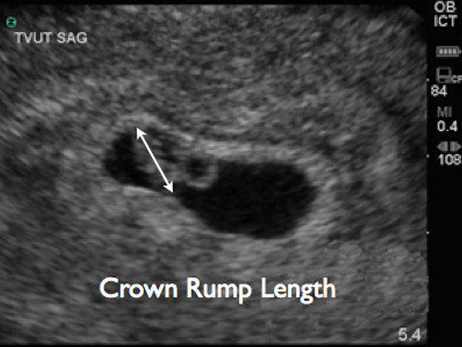
Footnote: Trans-vaginal ultrasound image demonstrating crown rump length measurement.
Crown rump length measurement
Crown rump length (CRL) is measured as the largest dimension of embryo, excluding the yolk sac and extremities. Crown rump length is used as a primary measure of gestational age between 6-13 weeks. After 13 weeks, head circumference, biparietal diameter, and femur length measurements become more useful measurements for assessing fetal growth.
The fetus floats in the amniotic fluid inside the uterus of the mother usually in a curved posture resembling the letter C. The crown rump length measurement can actually vary slightly if the fetus is temporarily stretching (straightening) its body. The crown rump length measurement needs to be in the natural state with an unstretched body which is actually C shaped. The measurement of CRL is useful in determining the gestational age (menstrual age starting from the first day of the last menstrual period) and thus the expected date of delivery. Different babies do grow at different rates and thus the gestational age is an approximation. Early in pregnancy it is accurate within +/- 4 days but later in pregnancy due to different growth rates, the accuracy is less. In that situation, other parameters can be used in addition to CRL.
Gestational age is not the same as fetal age. It takes about 14 days from the first day of the last menstrual period for conception to take place and thus for the conceptus to form. The age from this point in time (conception) is called the fetal age and is thus 2 weeks shorter than the gestational age. Thus a 6 week gestational age would be a 4 week fetal age. Some authorities however casually interchange these terms and the reader is advised to be cautious. An average gestational period (duration of pregnancy from the first day of the last menstrual period up to delivery) is 280 days. Thus if all months were of 31 days, it would be 9 months and 1 day.
Figure 2. Crown rump length (CRL) chart
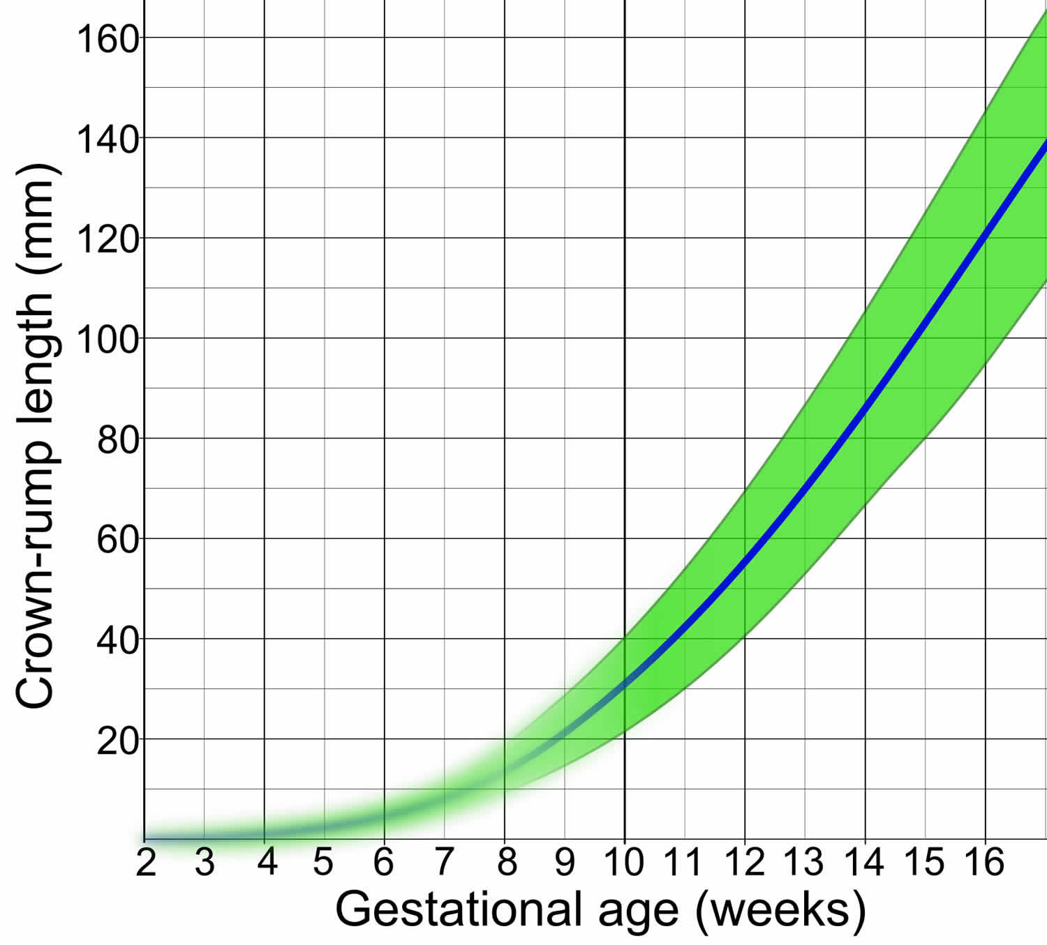
Table 1. Gestational age to crown rump length (CRL)
| Age | Length |
|---|---|
| 6.1 Weeks: | 0.4 cm |
| 7.2 Weeks: | 1.0 cm |
| 8.0 Weeks: | 1.6 cm |
| 9.2 Weeks: | 2.5 cm |
| 9.9 Weeks: | 3.0 cm |
| 10.9 Weeks: | 4.0 cm |
| 12.1 Weeks: | 5.5 cm |
| 13.2 Weeks: | 7.0 cm |
| 14.0 Weeks: | 8.0 cm |
Table 2. Crown rump length (CRL) in relation to gestational age
| CRL (mm) | Gestational age (weeks + days) | ||
|---|---|---|---|
| 5th centile | Median | 95th centile | |
| 5 | 5 + 6 | 6 + 2 | 6 + 6 |
| 10 | 7 + 0 | 7 + 4 | 8 + 1 |
| 15 | 7 + 5 | 8 + 2 | 9 + 0 |
| 20 | 8 + 2 | 9 + 0 | 9 + 5 |
| 25 | 8 + 6 | 9 + 4 | 10 + 2 |
| 30 | 9 + 2 | 10 + 0 | 10 + 6 |
| 35 | 9 + 5 | 10 + 3 | 11 + 2 |
| 40 | 10 + 1 | 10 + 6 | 11 + 5 |
| 45 | 10 + 3 | 11 + 2 | 12 + 1 |
| 50 | 10 + 6 | 11 + 5 | 12 + 4 |
| 55 | 11 + 1 | 12 + 0 | 13 + 0 |
| 60 | 11 + 3 | 12 + 3 | 13 + 3 |
| 65 | 11 + 6 | 12 + 5 | 13 + 5 |
| 70 | 12 + 1 | 13 + 0 | 14 + 0 |
| 75 | 12 + 3 | 13 + 3 | 14 + 3 |
| 80 | 12 + 5 | 13 + 5 | 14 + 5 |
| 85 | 13 + 0 | 14 + 0 | 15 + 1 |
| 90 | 13 + 2 | 14 + 2 | 15 + 3 |
| 95 | 13 + 4 | 14 + 4 | 15 + 5 |
| 100 | 13 + 6 | 15 + 0 | 16 + 1 |
- Doubilet PM. Ultrasound Evaluation of the First Trimester. Radiol. Clin. North Am. 2014;52 (6): 1191-1199. doi:10.1016/j.rcl.2014.07.004[↩]
- Doubilet PM, Benson CB, Bourne T et-al. Diagnostic criteria for nonviable pregnancy early in the first trimester. N. Engl. J. Med. 2013;369 (15): 1443-51. doi:10.1056/NEJMra1302417[↩]


