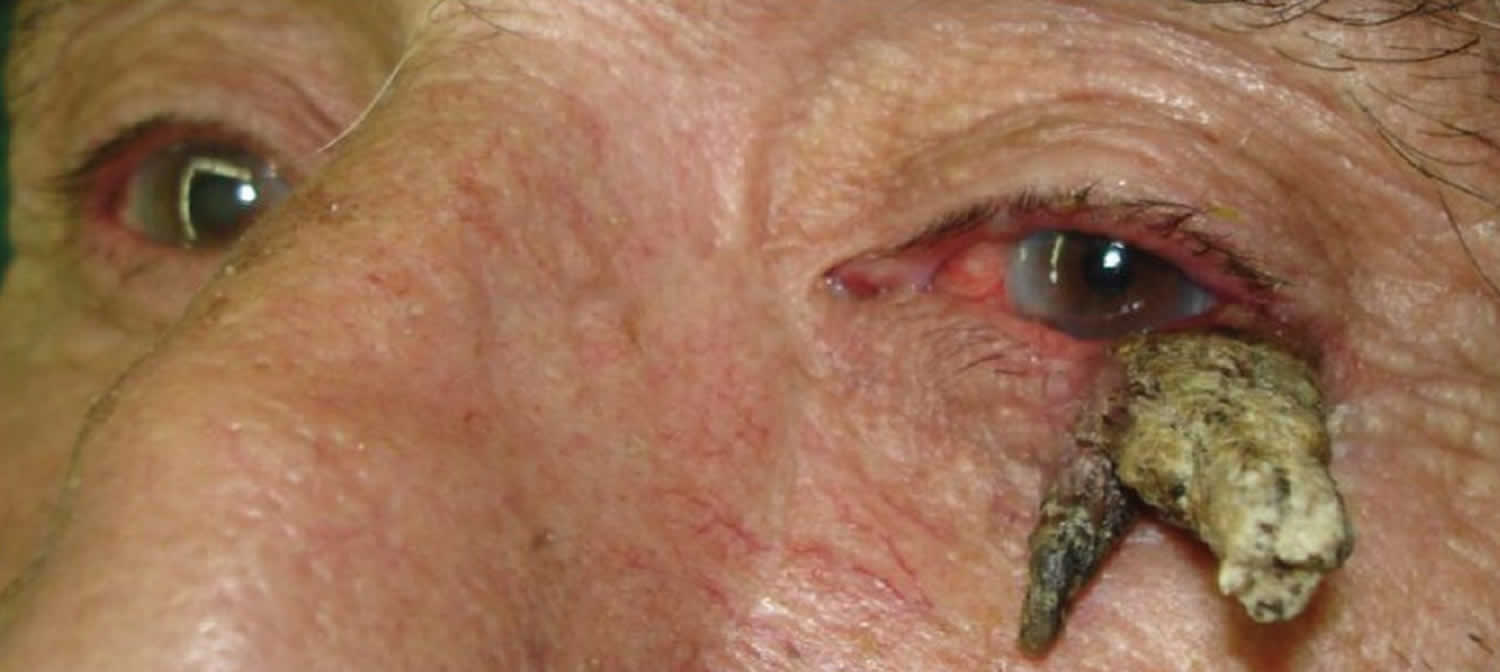
What is a cutaneous horn
A cutaneous horn also known as cornu cutaneum, refers to a specific appearance of a skin lesion in which a cone-shaped protuberance arises on your skin caused by overgrowth of the most superficial layer of skin (epidermis). Cutaneous horns are so named as they resemble an animal’s horn. Cutaneous horn arises from benign, premalignant or malignant skin lesions. A cutaneous horn is not a particular lesion but is a reaction pattern of your skin. Approximately 40% of cutaneous horns represent precancerous lesion called actinic keratosis. Cutaneous horns may also overlie skin cancer. When overlying cancerous skin, squamous cell carcinoma is typically at the base of the cutaneous horn, but basal cell carcinoma is also possible.
Around half of cutaneous horns have a benign base, and half are premalignant or malignant. The most common underlying lesions are seborrhoeic keratosis, viral warts (due to human papillomavirus), actinic keratosis, and well-differentiated squamous cell carcinoma associated with exposure to the sun and other sources of UV radiation.
See your doctor if a cutaneous horn is noted. A biopsy may be needed to assess whether the lesion is benign, precancerous, or cancerous.
Figure 1. Cutaneous horn
Who gets cutaneous horn?
- A cutaneous horn is more common in older patients, with the peak incidence in those between 60 and 70.
- Cutaneous horns are equally common in males and females, although there is a higher risk of the lesion being malignant in men.
- Cutaneous horns are more common in people with fairer skins (skin phototype 1 and 2).
Cutaneous horn causes
Cutaneous horns most often occur in adults, usually elderly, fair-skinned individuals with a history of significant sun exposure.
A common cause of a cutaneous horn is an actinic keratosis at the base of the horn; a cutaneous horn caused by an actinic keratosis must be distinguished from one arising from a squamous cell carcinoma (SCC), which may be present at the base of a cutaneous horn.
Cutaneous horns most frequently occur in elderly individuals, though they are occasionally seen in children (almost always as warts). In adults, the most common scenario is a cutaneous horn of the face, scalp, or ear presenting with a squamous cell carcinoma at the base. Warts and seborrheic keratoses are common benign lesions that may also be seen at the base of a cutaneous horn.
Skin conditions associated with cutaneous horns
| Benign | Premalignant or malignant |
|---|---|
|
|
What are the clinical features of a cutaneous horn?
Appearance
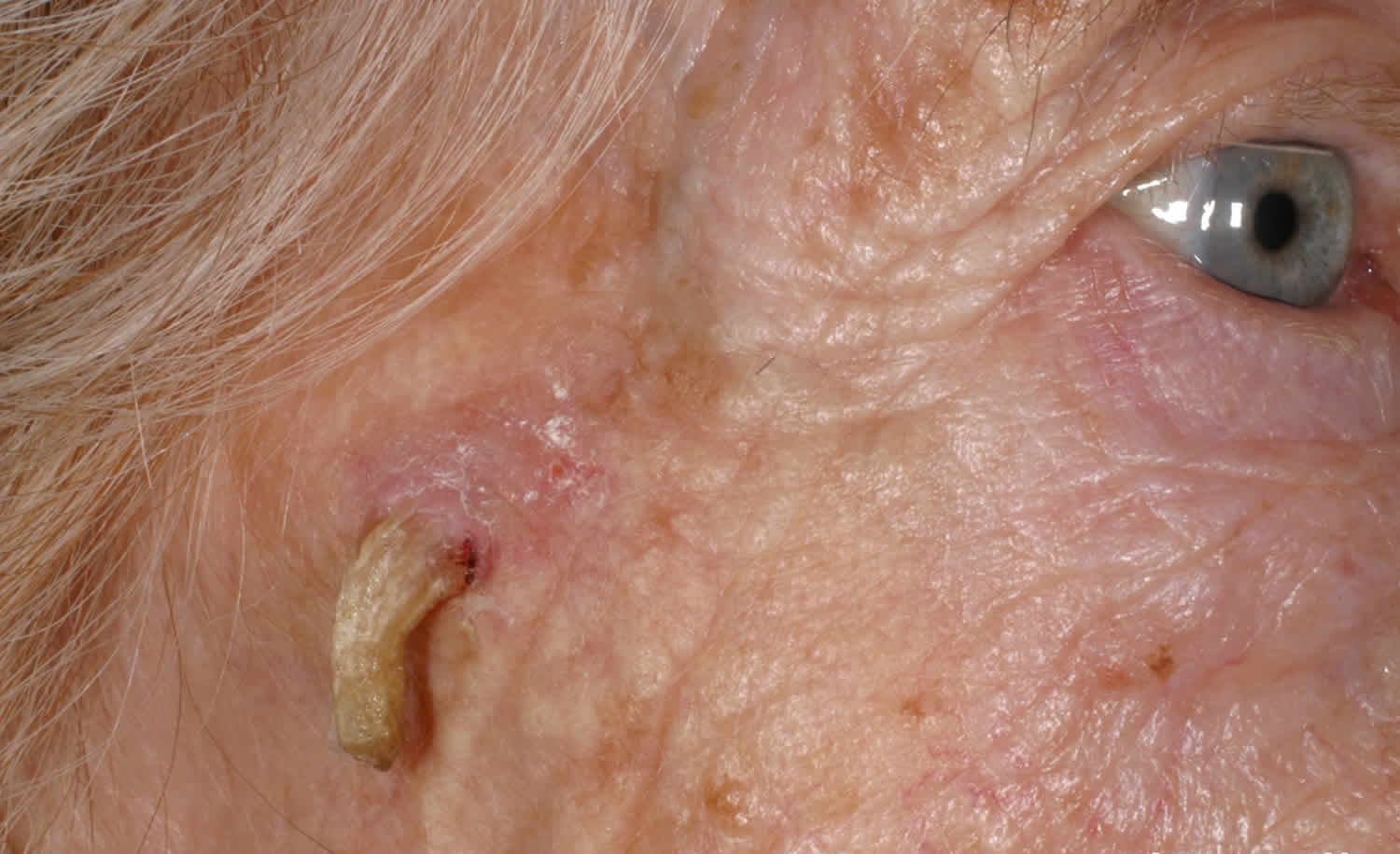
- A cutaneous horn generally presents as a straight or curved, hard, yellow-brown projection from the skin.
- It can be surrounded by normal skin or have a border of thickened skin.
- The side of the horn may be terrace-like or oyster shell-like with horizontal ridges.
- The base of the horn may be flat, protruding, or like a crater.
- Inflammation may be present, due to recurrent injury.
Size
- Typically, the horn is taller than twice the width at the base.
- It may vary from a few millimeters to several centimeters in size.
- Giant horns exist — the largest described is a 76-year-old Parisian woman named Madame Dimanche (Widow Sunday) in the 19th century, who grew a horn from her forehead that was 25-cm long.
Location
- Cutaneous horns are usually singular, but can be multiple.
- They can occur anywhere on the body, but are more common on sun-exposed areas especially the head and ears, back of hands and forearms.
- They may also occur on the chest, neck, shoulder and penis.
Cutaneous horn signs and symptoms
A cutaneous horn most often occurs on sun-exposed areas and appears as a cone-shaped protuberance arising from a skin-colored to red/pink bump or flat lesion. Cutaneous horns are usually asymptomatic. They can be injured causing pain and inflammation.
Worrying features suggestive of malignancy
Whilst no certain features can confidently confirm or exclude malignant lesions, malignant lesions are more common in older patients and in males compared to females. Squamous cell carcinoma is also likely if the horn has the following features:
- Painful
- Large size
- Induration at the base
- Anatomic site on the nose, ears, backs of hands, scalp, forearms, face and penis
- Wide base or low height to base ratio
- Redness at the base of the horn base
- Lack of terrace formation, due to rapid unorganized growth.
Cutaneous horn diagnosis
- A cutaneous horn is diagnosed by its clinical appearance.
- Histological examination of the horn base is crucial to rule out malignancy, as there are no certain clinical features that can definitively distinguish benign lesions from skin cancer.
- The lesion is usually completely excised. In some cases, a deep partial biopsy is taken to establish the diagnosis.
On histology, there is thickening of the stratum corneum or hyperkeratosis. Orderly horizontal parallel layers of keratin are associated more with benign lesions. Rapidly growing malignant lesions exhibit a more erratic growth. Acanthosis is often noted. The base of the lesion shows features of the underlying lesion.
Cutaneous horn treatment
Cutaneous horns are usually excised with appropriate margins, dependent of the nature of the lesion. If the lesion is benign, no further treatment may be needed.
If the lesion is precancerous, your physician may:
- Freeze the lesion with liquid nitrogen.
- Use a topical chemotherapy agent, such as 5-Fluorouracil or a topical medicine that stimulates the immune system, imiquimod.
- Scrape and burn (curettage and electrodesiccation) the lesion.
If the lesion is cancerous, your physician may:
- Perform surgery.
- Use a topical chemotherapy agent, such as 5-Fluorouracil or a topical medicine that stimulates the immune system, imiquimod.
- Scrape and burn (curettage and electrodesiccation) the lesion.
- Recommend radiation therapy.


