Eyelid dermatitis
Eyelid dermatitis also known as periocular dermatitis, periorbital dermatitis or eyelid eczema, is an umbrella term describing a group of inflammatory skin disorders that localize to the eyelids and resemble eczema. There are many causes of eyelid dermatitis, hence it is a vexing problem for patients and can represent a diagnostic and therapeutic dilemma. Knowledge of the common causes and their key features can focus the history and physical examination and alert the clinician to more serious conditions.
Time course, patient age, symptoms, presence or absence of scale or edema, distribution (isolated lesion vs multiple, discreet vs diffuse, bilateral vs unilateral, lid margin vs crease) help differentiate the different types of eyelid dermatitis.
Most common causes of eyelid dermatitis are:
- Contact dermatitis (allergic and irritant), 50% to 76% of cases
- Atopic dermatitis, 12% to 17%
- Seborrheic dermatitis 8% to 16%
- Rosacea, less than 5%
There are other conditions of the eyelids that mimic eyelid dermatitis and are not to be missed, such as dermatomyositis. The broader differential diagnosis includes other connective tissue diseases (discoid lupus erythematosus, Sjögren’s disease, etc), psoriasis, contact urticaria, infections (viral, bacterial or fungal), and drug reactions. Neoplasms benign or malignant can also mimic dermatitis and can localize to an eyelid.
Eyelid dermatitis causes
Most common causes of eyelid dermatitis are:
- Contact dermatitis (allergic and irritant), 50% to 76% of cases
- Atopic dermatitis, 12% to 17%
- Seborrheic dermatitis 8% to 16%
- Rosacea, less than 5%.
Eyelid dermatitis symptoms
As eyelid dermatitis can be an episodic occurrence, patients can present with relatively normal eyelid examination. In general, eyelid dermatitis appears as erythematous, often scaly, sometimes crusty and oozing plaques on either upper or lower eyelids or both, unilateral or bilateral. Edema is often present, but not without erythema and scaling.
The palpebral and bulbar conjunctiva are usually spared, but can appear slightly erythematous as a reaction to the surrounding inflammation. Upper lid involvement is more associated with airborne contact allergens, whereas lower lid dermatitis is more commonly associated with contact dermatitis induced by eye drops. In a photodistributed dermatitis again lower eyelids will be involved, while upper eyelids, especially creases, will be spared.
Allergic or irritant dermatitis more often presents as acute eyelid dermatitis with intense pruritus, weeping, brightly erythematous and edematous papules and plaques. Although vesicles are also a hallmark of acute contact dermatitis, they are less common periorbitally. Presence of vesicles should alert the clinician to consider a herpetic viral infection (herpes simplex or zoster; Figure 10). Yellow honey-color crusting can also indicate bacterial impetiginization.
Atopic eyelid dermatitis
Atopic eyelid dermatitis is a chronic relapsing skin condition with an age-dependent distribution. In the United States, it affects 10 to 20 percent of children and 1 to 3 percent of adults 1, with eyelid involvement in approximately 15 percent of cases 2.
Patients presenting with eyelid dermatitis caused by atopic dermatitis often have a history of chronic eyelid itching, redness and scaling; plus they often have eczema diagnosis since early childhood. Pruritus, sometimes unbearable, is often their main concern, as they become trapped in endless itch-scratch-itch cycles. Personal or family history of atopy (atopic dermatitis, or allergic rhinitis/hayfever or asthma) is typically present. Atopic dermatitis is diagnosed on the basis of major criteria (family history of eczema, severe pruritus, facial and extensor distribution in infants, flexural distribution in adults) and numerous minor criteria. As nipple dermatitis is fairly specific for atopic dermatitis, asking about that symptom can confirm diagnosis.
Atopic dermatitis is the most common cause of chronic eyelid dermatitis, which mimics changes seen in other chronic eczemas, with hyperpigmentation, lichenification, and diffuse scaling. Excessive chronic rubbing of the eyelids can cause eyelash and eyebrow hair loss. Dennie Morgan infraorbital folds, periorbital darkening (the “allergic shiner”), keratoconus, and anterior subcapsular cataracts are other clinical findings seen in chronic atopic eyelid dermatitis.
Full skin examination is important and helpful in identifying other typical areas of involvement by atopic dermatitis.
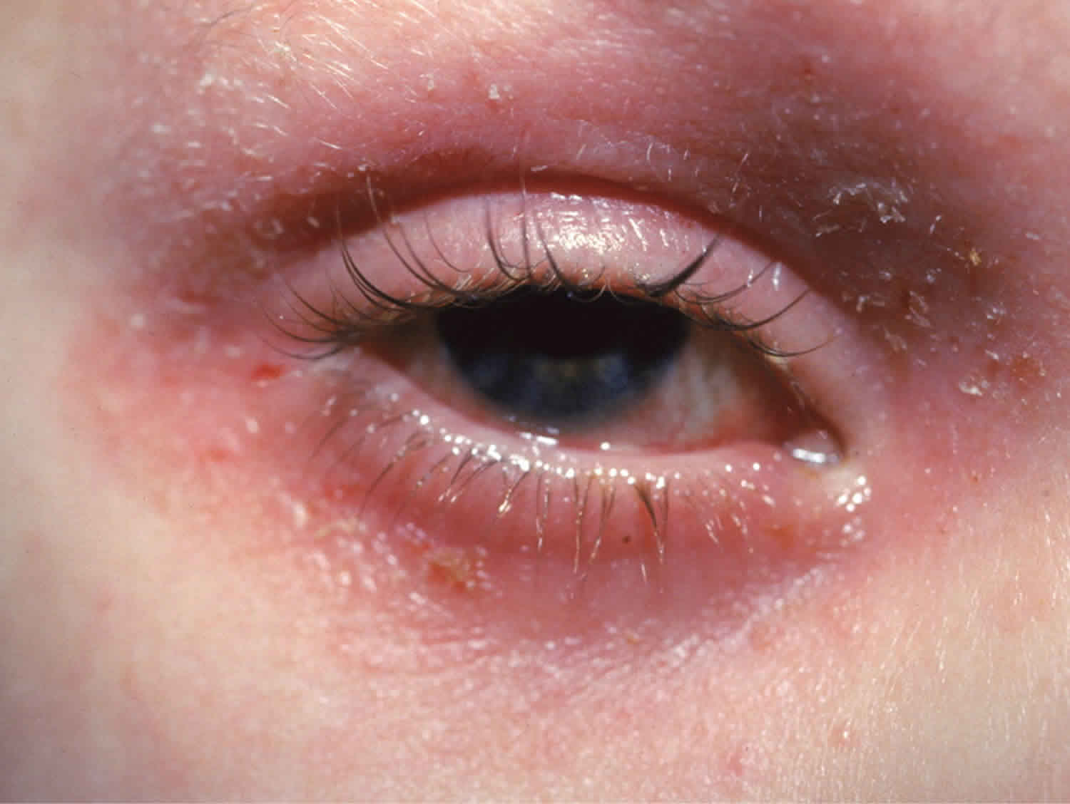
Figure 1. Atopic eyelid dermatitis
Atopic eyelid dermatitis causes
The cause of atopic dermatitis is thought to involve a combination of complex genetic, environmental, and immunologic interactions. There is often a strong familial pattern of inheritance. Altered T-cell function is present in the form of heightened T-helper 2 subtype activity. Inciting or exacerbating factors include aeroallergens, chemicals, foods, and emotional stress. Skin barrier function is also decreased in patients with atopic dermatitis, making them more sensitive to such allergens and irritants 1.
Atopic eyelid dermatitis diagnosis
Patients with atopic dermatitis involving the eyelid may present with pruritus, edema, erythema, lichenification, fissures, or fine scaling 3. Typically, edema and erythema of the eyelid are less prominent in atopic dermatitis than in contact dermatitis, and lichenification and fine scaling predominate. In some cases, however, the lesions may be difficult to distinguish from contact dermatitis 2. In these cases, the diagnosis may be made by the recognition of other features consistent with atopic dermatitis, such as a flexural distribution in older children and adults and a family history of asthma, rhinitis, and atopic dermatitis 4.
Atopic dermatitis may become complicated by infection or contact dermatitis, making the diagnosis more difficult. These complications should be suspected in patients who develop new or acute inflammation of the eyelid in the setting of well-controlled atopic dermatitis 3.
Atopic eyelid dermatitis treatment
Treatment involves oral antihistamines; moisturizers; and low-dose, short-term topical corticosteroids 5. The topical immunomodulators tacrolimus (Protopic) and pimecrolimus (Elidel) may be used in refractory cases. These agents are safe for use on the eyelids and face 6, but they should be reserved as second-line therapies because of their association with several types of cancer in animal studies and human case reports 7.
Eyelid contact dermatitis
Contact dermatitis is the most common cause of cutaneous eyelid inflammation 8. Eyelid skin is especially vulnerable to irritants and allergens because of its thinness and frequent exposure to chemicals via direct application or contamination from fingers and hands 9. As one of the most sensitive areas, eyelid skin may be the initial or only presenting area with signs of contact dermatitis, while other areas of the body remain unaffected by the same exposure 9.
Eyelid contact dermatitis also called eyelid eczema, is an inflammatory reaction involving the eyelid skin that is caused by contact with a trigger substance. Eyelid contact dermatitis may be due to allergy (allergic contact dermatitis) or irritation (irritant contact dermatitis). The presenting features of these types often are not readily distinguishable, but patients with irritant contact dermatitis often present with greater burning and stinging compared with the characteristic pruritus of allergic contact dermatitis 10.
Upper, lower or both eyelids on one or both sides can be affected by contact dermatitis. The patient may report itching, stinging or burning, and the eyelids are red and scaly. The eyelids may swell. With persistence of the dermatitis, the eyelids become thickened with increased skin markings (lichenification). The eyelid margins may become involved (blepharitis). The appearance is similar, whatever the cause.
Treatment of eyelid contact dermatitis involves avoidance of the offending agent 3. The patient should receive a list of common allergens or irritants and be instructed to carefully read all product labels. Acute allergic contact dermatitis of the eyelids can be treated with low-dose topical steroids twice daily for five to 10 days 3. Long-term use of these medications on the eyelid can cause skin atrophy and glaucoma 11 or cataracts 12; therefore, it is important to use the lowest potency preparation for the shortest period of time necessary to clear the eruption. Although delayed-type reactions of allergic contact dermatitis do not involve histamine release from mast cells, oral antihistamines may provide symptomatic relief as a result of their antipruritic and soporific effects 13.
Patients with irritant contact dermatitis may find it useful to apply a cool compress followed by an emollient 3. The use of topical steroids for irritant contact dermatitis was found to be ineffective in at least one study 14. However, in practice, steroids are often used because it can be difficult to differentiate between irritant and allergic contact dermatitis.
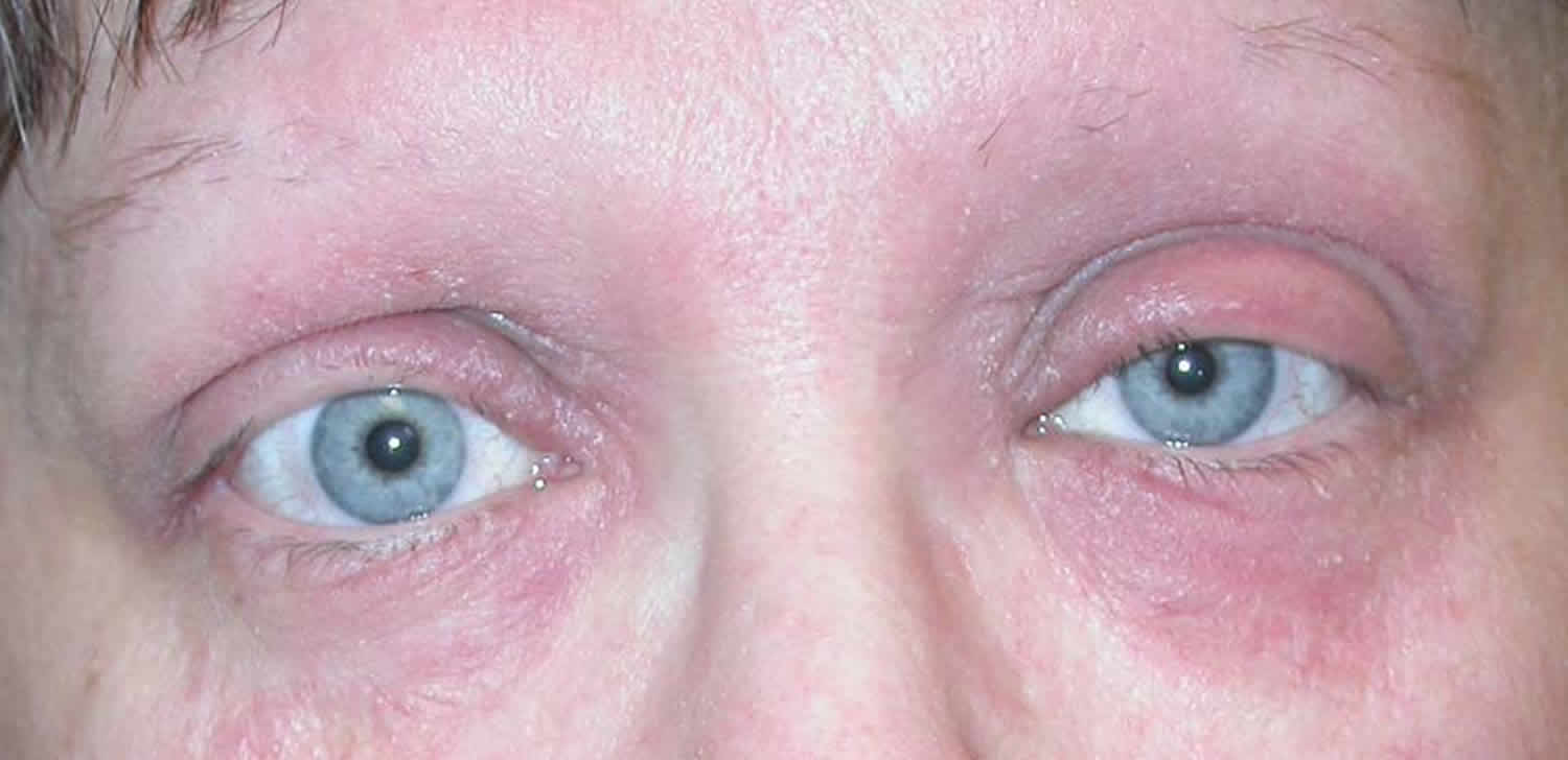
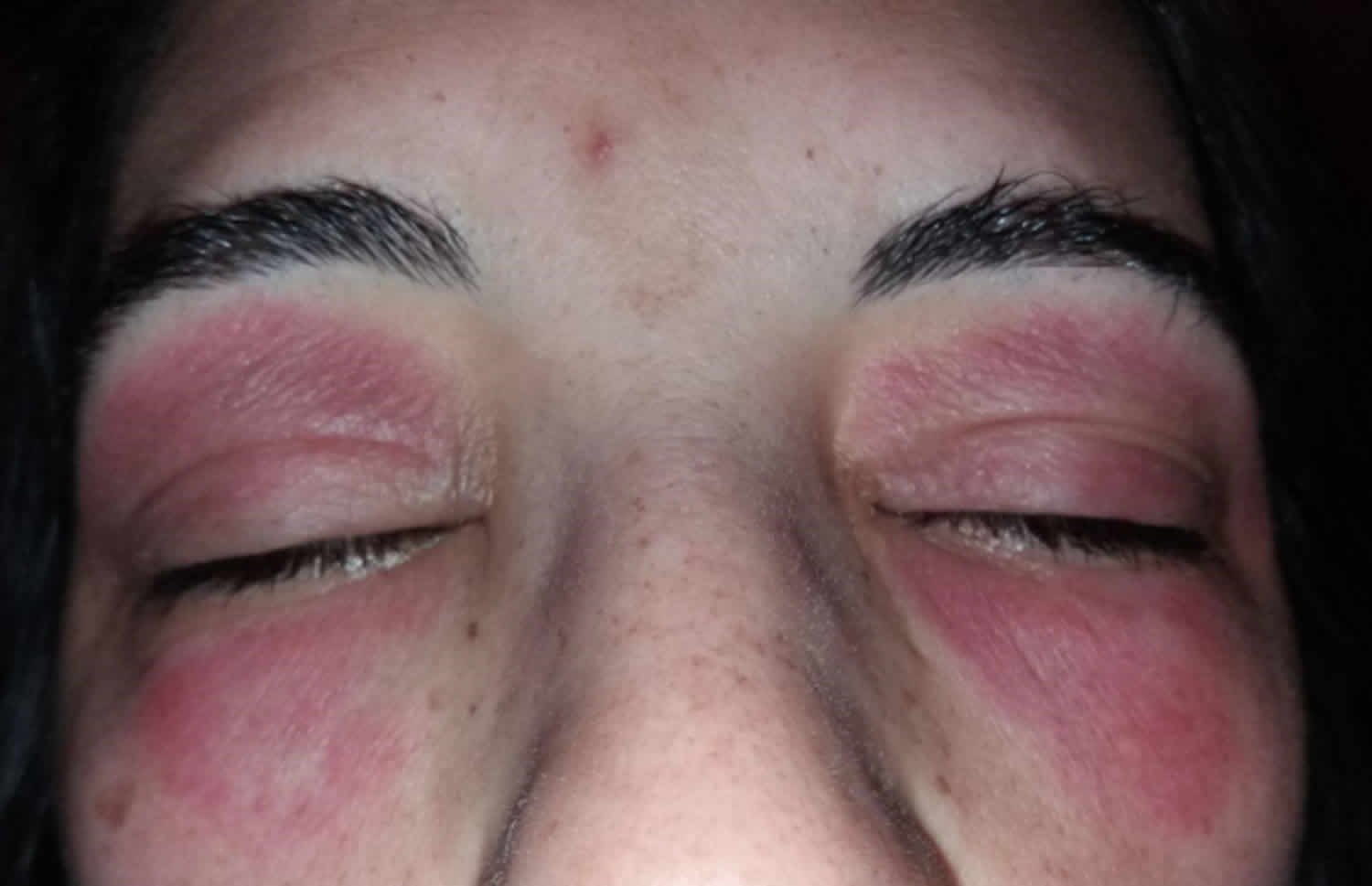
Figure 2. Eyelid contact dermatitis
Irritant eyelid contact dermatitis
Irritant contact dermatitis is an innate inflammatory reaction due to injury to the skin. Unlike allergic contact dermatitis, it does not involve specific antibodies (immunoglobulins) or specific immune cells (memory T cells ).
Irritant eyelid contact dermatitis can occur in anyone. However, it is more common in people with sensitive skin. People with a background of atopic eczema, asthma and hayfever (“atopy”) are more likely to suffer from irritant contact dermatitis than people without this history.
Any pre-existing inflammation of the skin can cause the skin’s waterproof “barrier” to be compromised and may make it more vulnerable to irritant contact dermatitis. Barrier function may also be defective for genetic reasons.
What triggers irritant eyelid contact dermatitis?
Irritant contact dermatitis may be triggered by contact with irritating substances and/or physical triggers.
Irritating substances that may cause irritant contact dermatitis of the eyelids include:
- Soaps and detergents
- Acids and alkalis
- Chemicals such as chlorine under goggles worn when swimming
- Dust particles
- Hydrophobic substances (comprised of molecules that repel water, i.e. drying agents)
- Cosmetics such as eyeliner, eye shadow, mascara and sunscreen
These substances may touch the eyelids directly or be transferred from the fingers (which may be unaffected by dermatitis).
Physical triggers that may cause irritant contact dermatitis include:
- Temperature extremes (heat or cold)
- Humidity extremes (dry or moist)
- Mechanical (rubbing or scratching)
Irritant eyelid contact dermatitis diagnosis
Diagnosis of eyelid irritant contact dermatitis is made by identifying its characteristic features.
- An itchy rash that affects one or both eyelids, which may occur intermittently or continuously
- Suspicion that it has been caused or aggravated by contact with an irritant (see the lists below).
- Often, pre-existing sensitive skin or atopic eczema
- Patch tests to suspected contact allergens are negative
It may be difficult to identify the precise cause.
Allergic eyelid contact dermatitis
Allergic contact dermatitis is caused by an immune reaction to the trigger; this is known as the allergen. This is often a substance that the patient has been exposed to many times previously without problem. The rash usually occurs one to several days after the contact with the allergen. This can make it difficult to identify the cause of the dermatitis. Allergic contact dermatitis involves a delayed-type hypersensitivity reaction (also called type IV hypersensitivity) and involves immune memory cells.
Allergic eyelid contact dermatitis can occur in anyone. It is no more likely in those with known sensitive skin or atopic eczema than it is in people that have previously had no skin problem.
Contact allergy usually develops after repeated previous contact with the allergen, so it is not necessarily a new substance that has triggered the allergy.
What triggers allergic eyelid contact dermatitis?
Eyelid contact dermatitis follows direct contact with an allergen.
Allergy to cosmetics may be due to contact with allergens in:
- Emollients and moisturizers
- Eye creams
- Sunscreens
- Makeups
- Cleansers
- Fragrances and essential oils
- Jewellery containing nickel and gold
Other potential allergens are found in:
- Eyelash curlers or tweezers (nickel)
- False eyelashes (components or more often, adhesives)
- Eye drops (preservatives and antiseptics)
- Contact lens solution
- Rubber goggles
- Spectacle frames
Eyelid contact dermatitis may also occur following indirect contact with an allergen, due to transfer of small amounts of substance from the hands (through rubbing or touching the eyelids). Such allergens may include:
- Nickel from metal coins or clothes fastenings
- Fragrances
- Nail cosmetics (varnishes and false nails)
- Hair dye, i.e., paraphenylenediamine allergy
Eyelid contact dermatitis is sometimes triggered by airborne allergens, such as dust mite and plant pollens. Compositae allergy is an example of an airborne allergen causing eyelid dermatitis.
Allergic eyelid contact dermatitis diagnosis
Diagnosis of eyelid allergic contact dermatitis is made by identifying its characteristic features.
- An itchy rash that affects one or both eyelids
- This is most likely to occur intermittently at intervals of days to years
- Suspicion that it has been caused or aggravated by contact with an allergen (see list above).
- Patch tests to one or more suspected contact allergens are positive
Knowing the details of all substances that the skin may have been exposed to in skin care, hobbies and work are an important part of the detective work required to find the cause of a eyelid dermatitis. Patch tests should be arranged to establish or confirm the triggering allergen.
Eyelid contact dermatitis causes
Contact dermatitis of the eyelid is mediated by a type IV hypersensitivity reaction in allergic contact dermatitis and by direct toxic effect in irritant contact dermatitis 3. It is more often caused by a product applied to the hair, nails, or face than by products applied directly to the eyelids 9. Below is a list of common exposures that can cause contact dermatitis 15.
Exposures that commonly cause eyelid contact dermatitis:
- Airborne pollen and dust 16
- Cosmetics
- Facial tissues
- Household cleaners and sprays
- Occupational exposures 20
- Ophthalmic solutions, medications, and ointments 21
- Poison ivy
Compounds that have most frequently caused allergic contact eyelid dermatitis in the last decade 22:
- Metals
- Gold sodium thiosulfate
- Nickel sulfate
- Cobalt
- Potassium dichromate
- Eye medications and antibiotics
- Aminoglycosides
- Gentamicin
- Neomycin
- Bacitracin
- Phenylephrine
- Aminoglycosides
- Personal care products
- Shampoos
- Fragrances
- Fragrance mix
- Balsam of Peru
- Cinnamic aldehyde
- Face creams
- Makeup (especially eye shadow)
- Makeup applicators
- Sponges (can contain thiuram mix)
- Brushes
- Botanicals and vitamin oils
- Ylang ylang oil
- d-α-Tocopherol acetate
- Nail products
- Tosylamide formaldehyde resin
- Cyanoacrylates
- Methacrylates
- Vehicles and additives
- Propylene glycol
- Lanolin alcohol
- Preservatives
- Methyldibromoglutaronitrile
- Quaternium-15
- Methylchloroisothiazolinone
- Dimethylol dimethyl hydantoin
- Benzalkonium chloride
- Formaldehyde
- Sodium disulfite
- Kathon CG
- Phenylmercuric acetate
- Protein contactants
- Dust mites
- Animal dander
- Cornstarch
- Latex
- Steroids
- Tixocortol
- Budesonide
- Desonide
- Airborne contactants
- Fragrance mix
- Resins
- Colophony
- Plants
- Monoterpenes
- Wood dust
- Surfactants and related chemicals
- 3-(dimethylamino)propylamine
- Amidoamine
- Cocamidopropyl betaine (in most cases, contaminants have not been evaluated)
- Oleamidopropyl dimethylamine
Eyelid contact dermatitis diagnosis
A careful history of exposure to agents known to cause eyelid contact dermatitis should be elicited. The patient should be asked about his or her occupation, hobbies, and cosmetic use including non-eye cosmetics, new products, and refills of a previously used product, because changes in cosmetic formulations are common 9. Patients with irritant contact dermatitis may have pruritus, burning, or stinging of the eyelids and periorbital area, with or without involvement of the face and hands 3. Examination may reveal a combination of erythema, edema, and vesiculation in patients with acute dermatitis, or scaling and desquamation if inflammation has been present for weeks 23. If the causative agent is not apparent after taking the history and performing the physical examination, referral to an allergist or dermatologist for patch testing may uncover an occult allergen.
Eyelid contact dermatitis treatment
It is important to avoid contact with irritants and known or potential allergens, to ensure that the skin is healthy and able to form a waterproof barrier.
- Avoid rubbing and scratching.
- Only touch eyelids with clean, rinsed hands.
- Wash eyelids with plain water or use a cream cleanser designed for sensitive skin.
- Avoid all contact with allergens detected by patch tests – this is necessary life-long.
- Avoid eyelid cosmetics while the dermatitis is active.
- Wear protective wrap-round spectacles if exposed to cold, wind, dust particles etc. Spectacles can also make it easier to stop scratching and rubbing the eyelids.
Short courses of mild topical corticosteroids (i.e., hydrocortisone 1% cream or ointment) or calcineurin inhibitors (ie pimecrolimus 1% cream) may be required to treat active inflammation. Severe contact dermatitis of the eyelids is usually treated with a short course of oral corticosteroids.
Eyelid dermatitis can be followed by postinflammatory pigmentation, one of the causes of dark circles under the eyes.
- Fitzpatrick TB, Freedberg IM. Fitzpatrick’s Dermatology in General Medicine. 6th ed. New York, N.Y.: McGraw-Hill, 2003.[↩][↩]
- Rich LF, Hanifin JM. Ocular complications of atopic dermatitis and other eczemas. Int Ophthalmol Clin. 1985;25:61–76.[↩][↩]
- Zug KA, Palay DA, Rock B. Dermatologic diagnosis and treatment of itchy red eyelids. Surv Ophthalmol. 1996;40:293–306.[↩][↩][↩][↩][↩][↩][↩]
- Hanifin JM, Lobitz WC Jr. Newer concepts of atopic dermatitis. Arch Dermatol. 1977;113:663–70.[↩]
- Donshik PC, Hoss DM, Ehlers WH. Inflammatory and papulosquamous disorders of the skin and eye. Dermatol Clin. 1992;10:533–47.[↩]
- Rikkers SM, Holland GN, Drayton GE, Michel FK, Torres MF, Takahashi S. Topical tacrolimus treatment of atopic eyelid disease. Am J Ophthalmol. 2003;135:297–302.[↩]
- FDA Approves Updated Labeling with Boxed Warning and Medication Guide for Two Eczema Drugs, Elidel and Protopic https://www.fda.gov/drugs/postmarket-drug-safety-information-patients-and-providers/fda-approves-updated-labeling-boxed-warning-and-medication-guide-two-eczema-drugs-elidel-and[↩]
- Guin JD. Eyelid dermatitis: experience in 203 cases. J Am Acad Dermatol. 2002;47:755–65.[↩]
- Rietschel RL, Fowler JF, Fisher AA. Fisher’s Contact Dermatitis. 5th ed. Philadelphia, Pa.: Lippincott Williams & Wilkins, 2001.[↩][↩][↩][↩]
- Altomare G, Capella GL, Frigerio E, Fracchiolla C. Recurrent oedematous irritant contact dermatitis of the eyelids from indirect application of glycolic acid. Contact Dermatitis. 1997;36:265.[↩][↩]
- Garrott HM, Walland MJ. Glaucoma from topical corticosteroids to the eyelids. Clin Experiment Ophthalmol. 2004;32:224–6.[↩]
- Renfro L, Snow JS. Ocular effects of topical and systemic steroids. Dermatol Clin. 1992;10:505–12.[↩]
- Mark BJ, Slavin RG. Allergic contact dermatitis. Med Clin North Am. 2006;90:169–85.[↩]
- Levin C, Zhai H, Bashir S, Chew AL, Anigbogu A, Stern R, et al. Efficacy of corticosteroids in acute experimental irritant contact dermatitis?. Skin Res Technol. 2001;7:214–8.[↩]
- Differential Diagnosis of the Swollen Red Eyelid. Am Fam Physician. 2007 Dec 15;76(12):1815-1824. https://www.aafp.org/afp/2007/1215/p1815.html[↩]
- Karlberg AT, Gafvert E, Meding B, Stenberg B. Airborne contact dermatitis from unexpected exposure to rosin (colophony). Rosin sources revealed with chemical analyses. Contact Dermatitis. 1996;35:272–8.[↩]
- Ross JS, White IR. Eyelid dermatitis due to cocamidopropyl betaine in an eye make-up remover. Contact Dermatitis. 1991;25:64.[↩][↩]
- Le Coz CJ, Leclere JM, Arnoult E, Raison-Peyron N, Pons-Guiraud A, Vigan M, for the Members of Revidal-Gerda. Allergic contact dermatitis from shellac in mascara. Contact Dermatitis. 2002;46:149–52.[↩]
- Guin JD. Eyelid dermatitis from methacrylates used for nail enhancement. Contact Dermatitis. 1998;39:312–3.[↩]
- Yesudian PD, King CM. Occupational allergic contact dermatitis from meropenem. Contact Dermatitis. 2001;45:53.[↩]
- Blondeau P, Rousseau JA. Allergic reactions to brimonidine in patients treated for glaucoma. Can J Ophthalmol. 2002;37:21–6.[↩]
- Knopp E, Watsky K. Eyelid dermatitis: contact allergy to 3-(dimethylamino)propylamine. Dermatitis. 2008;19(6):328–333. https://www.ncbi.nlm.nih.gov/pmc/articles/PMC4103018[↩]
- Kanski JJ, Nischal KK, Milewski SA. Ophthalmology: Clinical Signs and Differential Diagnosis. Philadelphia, Pa.: Mosby, 1999.[↩]


