What is keratoacanthoma
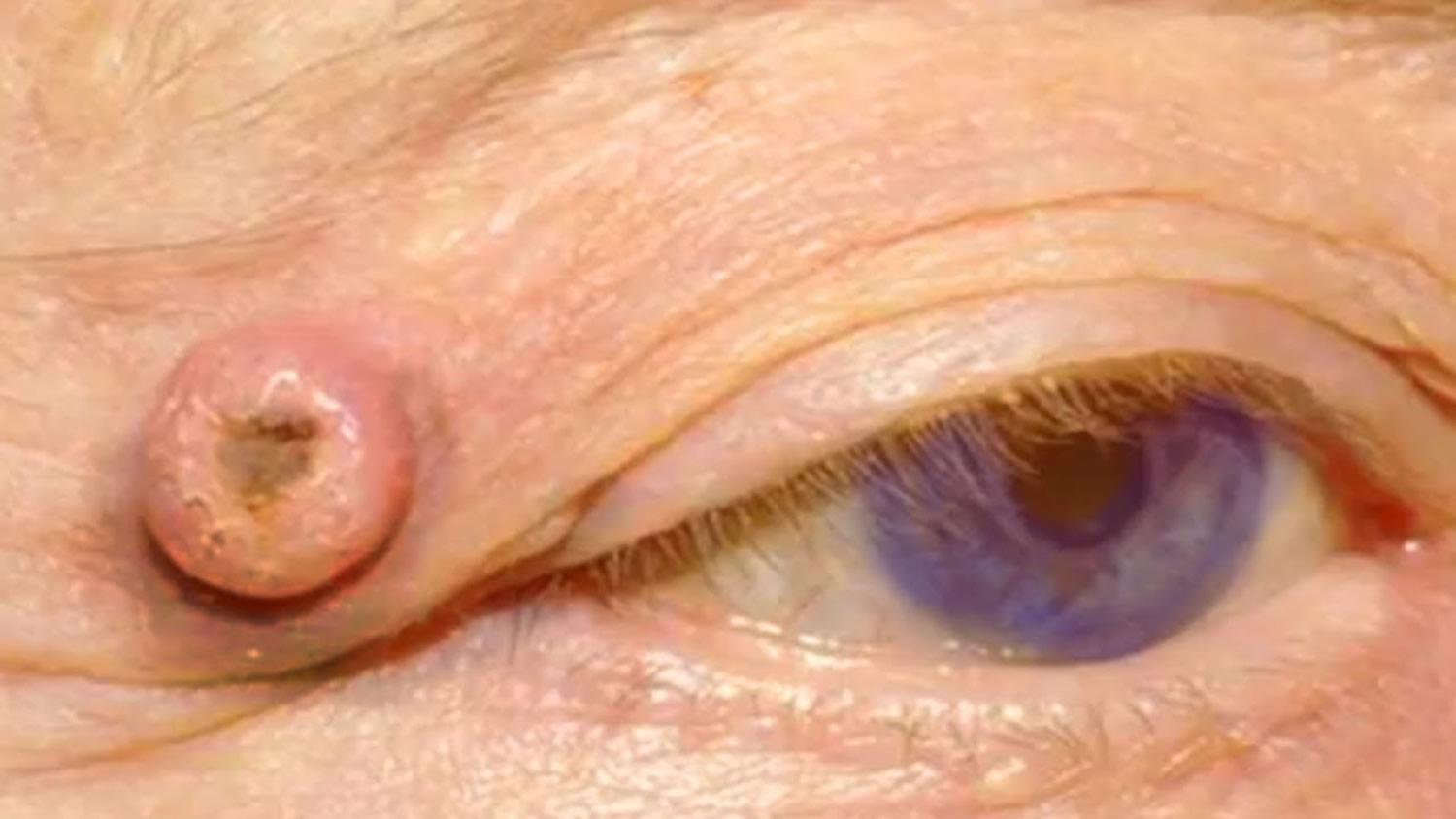
Keratoacanthoma is a rapidly growing skin cancer usually appearing as a volcano-like bump on the sun-damaged skin of middle-aged and elderly individuals. Many scientists consider keratoacanthoma to be a less serious form of squamous cell carcinoma (SCC). Most keratoacanthoma cause only minimal skin destruction, but a few behave more aggressively and can spread to lymph nodes.
Keratoacanthoma grows for a few months; then it may shrink and resolve by itself. Keratoacanthoma is considered to be a variant of the keratinocyte or non-melanoma skin cancer, squamous cell carcinoma. As it cannot be clinically reliably distinguished from more severe forms of skin cancer, keratoacanthomas are usually treated surgically.
Keratoacanthomas are reported in all age groups although they rarely appear before the age of 20. The peak incidence of solitary keratoacanthoma is between 50 and 69 years of age. Keratoacanthoma is most commonly seen in elderly, light-skinned people with a history of sun exposure or ultraviolet (UV) radiation. They occur more frequently in men with a male to female ratio of 2:1. The majority have been described in fair skin individuals with the highest rates found in those with Fitzpatrick I-III classification. Additionally, they occur more frequently in immunosuppressed individuals and are more locally invasive. Keratoacanthomas and squamous cell carcinoma share many epidemiological features.
Keratoacanthoma may start at the site of a minor injury to sun damaged and hair-bearing skin. At first, it may appear as a small pimple or boil and may be squeezed but is found to have a solid core filled with keratin (scale). It then proliferates, and it may be up to 2cm in diameter by the time it is brought to the attention of the doctor.
If left untreated, most keratoacanthoma spontaneously disappear (resolve) within 6 months, leaving a depressed scar. However, they may cause significant damage to the skin and underlying layers of tissue as well as psychological distress. Additionally, rare forms of keratoacanthoma may spread (invade) aggressively below the skin level and into the lymph glands, and your doctor has no way to tell this type from the more common form. Therefore, it is recommended to have the lesion removed to confirm that it is not a skin cancer and treatment are recommended.
One method of removal is to scrape the keratoacanthoma off with a sharp, spoon-like instrument (a curette) under a local anaesthetic and cauterize the raw area left behind. The specimen that has been scraped off is sent for microscopic examination. There is a chance that the keratoacanthoma may recur if the base is not removed completely.
The entire lesion of keratoacanthoma can be cut out (excised) and the area closed with stitches. Once the entire lesion is removed, it is unlikely to recur.
Occasionally, small keratoacanthomas can be treated by freezing with liquid nitrogen (cryotherapy).
There are no effective self-care treatments for keratoacanthoma. Preventing sun damage is crucial to avoiding the development of keratoacanthoma:
- Avoid ultraviolet (UV) light exposure from natural sunlight or from artificial tanning devices.
- Wear broad-spectrum sunscreens (blocking both UVA and UVB) with SPF 30 or higher, reapplying frequently.
- Wear wide-brimmed hats and long-sleeved shirts.
- Stay out of the sun in the middle of the day (between 10:00 AM and 3:00 PM).
Keratoacanthomas should be treated for several reasons.
- To obtain pathology: keratoacanthoma can be challenging to distinguish from invasive squamous cell carcinoma.
- To be rid of an unsightly, tender or worrisome lesion
- To minimise the scar, which can be more unsightly if the lesion resolves on its own.
Treatment requires the destruction of the lesion. Options include:
- Cryotherapy (used only for small lesions < 0.5 cm)
- Curettage and cautery or another form of electrosurgery
- Excision
- Radiotherapy
If you develop a new bump (lesion) on sun-exposed skin, or if you have a spot that bleeds easily or does not seem to be healing, then you should make an appointment with your primary care physician or with a dermatologist. You should also make an appointment if an existing spot changes size, shape, color, or texture, or if it starts to itch, bleed, or become sore to the touch.
Try to remember to tell your doctor when you first noticed the lesion and what symptoms, if any, it has. Also, young adults should ask adult family members whether or not they have ever had a skin cancer and relay this information to their physician.
Keratoacanthoma type
Keratoacanthoma is further divided into different subtypes with different presentations. Subtypes include solitary keratoacanthoma, subungual keratoacanthoma, mucosal keratoacanthoma, giant keratoacanthoma, keratoacanthoma centrifugum marginatum, generalized eruptive keratoacanthoma of Grzybowski, and multiple keratoacanthomas Ferguson-Smith syndrome 1.
Are keratoacanthomas hereditary?
Usually not. Rarely, keratoacanthoma can be due to genetic conditions in those who have multiple lesions and a history of other affected family members.
There are some rare conditions in which multiple keratoacanthomas appear. These are:
- Grzybowski eruptive keratoacanthomas
- Muir Torre syndrome
- Multiple self-healing squamous epitheliomas of Ferguson-Smith
- Keratoacanthoma centrifugum marginatum
- Drug-induced eruptive keratoacanthomas
- Responsible medications include BRAF inhibitors vemurafenib and dabrafenib, checkpoint inhibitors pembrolizumab and nivolumab, leflunomide, vismodegib, imiquimod.
Can a keratoacanthoma be cured?
Yes, once the lesion is removed, keratoacanthoma is considered cured.
What causes keratoacanthoma?
Keratoacanthoma arises from hair follicle skin cells for unknown reasons. Keratoacanthoma tends to occur on sun-exposed skin. It is more frequently seen in white skinned individuals and in those over 60 years of age.
Some keratoacanthomas appear to be related to infection with human papillomavirus (HPV), the cause of warts, but the majority of keratoacanthomas are not found to be due to HPV (human papillomavirus).
Multiple causes have also been suggested including exposure to chemical carcinogens, immunosuppression, use of BRAF inhibitors, genetic predisposition including mutations of p53 or H-Ras and recent trauma or surgery to the location 1. Keratoacanthoma can be commonly associated with syndromes such as autosomal dominant Muir-Torre syndrome, autosomal dominant Ferguson-Smith syndrome, autosomal recessive xeroderma pigmentosum, X-linked dominant incontinentia pigmenti, autosomal dominant Witten-Zak. The rare sporadic pruritic generalized eruption of keratoacanthoma is known as generalized eruptive keratoacanthoma of Gryzbowski 2.
Risk factors for developing keratoacanthoma
Risk factors for the development of keratoacanthoma include:
- Age over 50
- Fair skin, light hair, or light eyes
- Male
- Chronic exposure to sunlight or other ultraviolet light
- Exposure to certain chemicals, such as tar
- Exposure to radiation, such as X-ray treatment for internal cancers
- Long-term suppression of the immune system, such as organ transplant recipients
- Long-term presence of scars, such as from a gasoline burn
- Chronic ulcers
- Presence of particular strains of the wart virus (human papillomavirus)
- Previous skin cancer
Keratoacanthoma signs and symptoms
Keratoacanthomas occur more frequently on the face, and less often on the backs of the hands and forearms. Usually they are solitary and surrounded by normal skin.
The most common locations for keratoacanthoma include:
- Center of the face
- Backs of hands
- Forearms
- Ears
- Scalp
- Lower legs, especially in women
A keratoacanthoma appears and grows rapidly over the course of 2–6 weeks. Starting as a small, pimple-like lesion, a keratoacanthoma typically develops into a dome-shaped, skin-colored nodule with a central depression filled with keratin horn or scale (the major protein found in hair, skin, and nails). Sometimes, the center can erupt and form a crater. Keratoacanthoma usually range in size from 1–2.5 cm.
In rare cases, multiple keratoacanthomas may develop as part of a larger group of symptoms (syndrome).
Most keratoacanthoma are painless, though some may be itchy. Depending on the site of involvement, keratoacanthoma may interfere with normal function of the affected area.
Keratoacanthoma diagnosis
As keratoacanthoma can look similar to a skin cancer, it is important for the lesion to be removed at an early stage to have the diagnosis confirmed under the microscope. Your doctor is likely to refer you to a dermatologist who then performs the skin surgery to remove the lesion.
If your doctor suspects a keratoacanthoma, he or she will first want to establish the correct diagnosis by performing a biopsy. The procedure involves:
- Numbing the skin with an injectable anesthetic.
- Sampling a small piece of skin by using a flexible razor blade, a scalpel, or a tiny cookie cutter (called a “punch biopsy”). If a punch biopsy is taken, a stitch (suture) or 2 may be placed and will need to be removed 6–14 days later.
- Having the skin sample examined under the microscope by a specially trained physician (dermatopathologist).
Keratoacanthoma treatment
Once the diagnosis of keratoacanthoma is established, the treatment options usually include:
- Freezing with liquid nitrogen (cryosurgery), in which very cold liquid nitrogen is sprayed on the keratoacanthoma, freezing it and destroying it in the process.
- Electrodesiccation and curettage, also known as “scrape and burn.” After numbing the lesion, the doctor uses a sharp instrument (curette) to “scrape” the skin cancer cells away, followed by an electric needle to “burn” (cauterize) the tissue. The electrodesiccation helps to kill the cancer cells and also to stop any bleeding at the site.
- Removal (excision), in which the doctor uses a knife-like instrument (scalpel) to cut out the keratoacanthoma and then place stitches to bring the wound edges together.
- Mohs micrographic surgery, in which the physician takes tiny slivers of skin from the cancer site until it is completely removed. This technique is particularly useful for keratoacanthoma located on the nose, the ears, the lips, and the hands.
- Radiation treatment, where X-ray therapy is often useful for patients who might have difficulty with a surgical procedure because of other health issues.
Very rarely, keratoacanthoma are treated with medicine injected directly into the skin lesion (intralesional chemotherapy). In patients with more than one keratoacanthoma, the doctor may suggest taking a pill (isotretinoin) to reduce their size and number.
Patients with keratoacanthomas are at risk of further similar lesions and other skin cancers.
Multiple keratoacanthomas
There are some rare conditions in which multiple keratoacanthomas appear. These are:
- Grzybowski eruptive keratoacanthomas
- Muir Torre syndrome
- Multiple self-healing squamous epitheliomas of Ferguson-Smith
- Keratoacanthoma centrifugum marginatum
- Drug-induced eruptive keratoacanthomas
- Responsible medications include BRAF inhibitors vemurafenib and dabrafenib, checkpoint inhibitors pembrolizumab and nivolumab, leflunomide, vismodegib, imiquimod
Treatment requires surgery as well as oral drugs such as acitretin, methotrexate or cyclophosphamide. Drug-induced eruptive keratoacanthomas induced by checkpoint inhibitors have responded to topical steroids and intralesional steroid injections.
Keratoacanthoma prognosis
Keratoacanthoma has an excellent prognosis following surgical excision. Metastases are rare, but there are reports of this in addition to perineural spread in literature. Generally, patients with keratoacanthoma will have a history of sun exposure and will need to be followed for the development of new primary skin cancers.
It is important to remember that treatment of keratoacanthoma is not complete once the skin cancer has been removed. Frequent follow-up appointments with a dermatologist or with a physician trained to examine the skin are essential to ensure that the keratoacanthoma has not returned and that a new skin cancer has not developed somewhere else on your body. In addition, good sun protection habits are vital to preventing further damage from UV light.
If keratoacanthoma recurs, it should be treated again.
- Zito PM, Scharf R. Keratoacanthoma. [Updated 2019 Feb 21]. In: StatPearls [Internet]. Treasure Island (FL): StatPearls Publishing; 2019 Jan-. Available from: https://www.ncbi.nlm.nih.gov/books/NBK499931[↩][↩]
- Ranglani H, Pai VV, Shukla P. Keratoacanthoma-like cutaneous metastases in a case of squamous cell carcinoma of the tongue. Indian J Dermatol Venereol Leprol. 2018 Oct 29[↩]


