Pyelectasis
Pyelectasis is a dilation of the renal pelvis. The term is derived from the prefix pyelo- meaning “pelvis” and ectasia meaning “dilation or distension”. Pyelectasis is a relatively common finding on antenatal ultrasounds, often detected at the routine 2nd-trimester morphology scan and is three times more common in male fetuses. The estimated prevalence is at ~2% of routine second trimester scans 1. Fetal pyelectasis refers to a prominence of the renal pelvis in utero that is a relatively common finding, which in the majority of cases resolves spontaneously. In most cases fetal pyelectasis resolves normally, having no ill effects on the baby. The significance of pyelectasis in fetuses is not clear. It was thought to be a marker for obstruction, which in some cases it can be. In most cases it resolves spontaneously. In some studies it has been shown to appear and disappear several times throughtout the course of pregnancy. There is some discussion about what degree of pyelectasis is considered severe enough to warrant further investigation and most authorities use 6 mm as the cut-off point.
Pyelectasis is considered to be a “soft marker” for Down syndrome. This, along with other factors such as age and a Triple test, may be grounds for an amniocentesis to test for Down syndrome.
Babies with unresolved pyelectasis may experience urological problems requiring surgery.
Pyelectasis causes
Pyelectasis can result from a number of factors. In the majority of cases, it is physiological and resolves spontaneously. However, it may also herald the presence or evolution of renal tract pathology, such as:
- fetal pelviureteric junction obstruction
- fetal vesicoureteric junction obstruction
- urethral obstruction, e.g. posterior urethral valves
- vesicoureteric reflux
- duplex kidney
In previous publications, many authors found that the likelihood ratios of renal pyelectasis for Down syndrome is around 1.5 to 1.9 2, 3. However, just like other soft markers, isolated renal pyelectasis was found frequently among normal fetuses.
Renal pyelectasis diagnosis
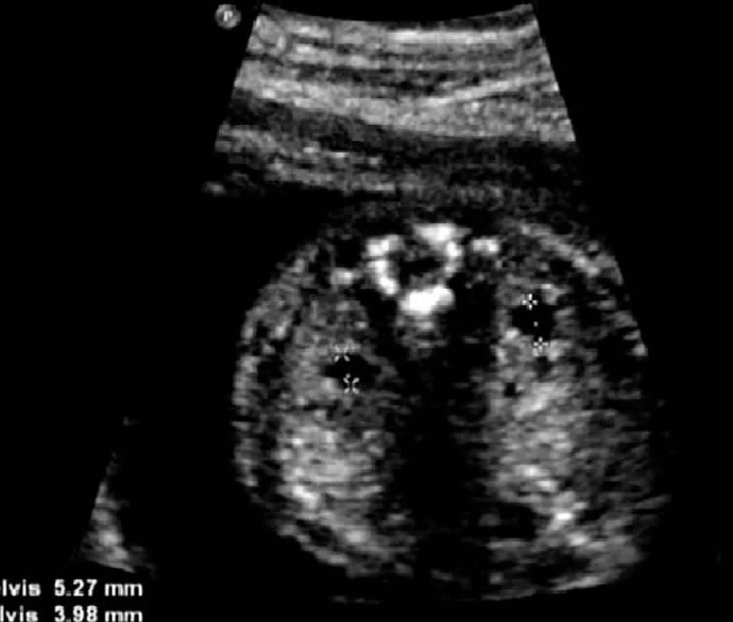
Renal pyelectasis was defined by Beacerraf et al 4 as an anteroposterior diameter of the fetal renal pelvis of ≥ 4 mm at 16–20 weeks, ≥ 5 mm at 20–30 weeks or ≥ 7 mm at 30–40 weeks gestation.
Fetal pyelectasis treatment
The vast majority of cases (~96%) with mild pyelectasis in the second trimester resolve, either during pregnancy or in the early postpartum period. The risk of postnatal renal pathology is increased with:
- increasing degree of pelvic dilatation
- in utero progression
- bilateral involvement
Antenatally detected renal pelvic dilatation, especially in isolation, is considered a weak predictor of vesicoureteric reflux 5 although postnatal sonographic evaluation is often recommended.
Some advocate a repeat prenatal scan at 30-40 weeks gestation for fetuses if ≥6 mm of renal pelvic dilatation is detected prior to 28 weeks as well as postnatal follow-up for persistent pyelectasis 6.
Fetal pyelectasis prognosis
Postnatally, most cases with pyelectasis resolve spontaneously in the first year of life, and invasive procedures are not required.
- Entezami M, Albig M, Knoll U et-al. Ultrasound Diagnosis of Fetal Anomalies. Thieme. (2003) ISBN:1588902129[↩]
- Smith-Bindman R, Hosmer W, Feldstein VA, et al. Second-trimester ultrasound to detect fetuses with Down syndrome: a meta-analysis. JAMA 2001;285: 1044–55.[↩]
- Nyberg DA, Souter VL, El-Bastawissi A, et al. Isolated sonographic markers for detection of fetal Down syndrome in the second trimester of pregnancy. J Ultrasound Med 2001;20:1053–63.[↩]
- Benacerraf BR, Mandell J, Estroff JA, et al. Fetal pyelectasis: a possible association with Down syndrome. Obstet Gynecol 1990;76:58–60.[↩]
- Antenatal renal pelvis dilatation: a predictor of vesicoureteral reflux? AJR Am J Roentgenol. 1996 Oct;167(4):897-900. https://www.ajronline.org/doi/pdf/10.2214/ajr.167.4.8819378[↩]
- Fetal pyelectasis: comparison of postnatal renal pathology with unilateral and bilateral pyelectasis. Prenat Diagn. 1997 May;17(5):451-5. https://doi.org/10.1002/(SICI)1097-0223(199705)17:5<451::AID-PD83>3.0.CO;2-4[↩]


