Thyroid biopsy procedure
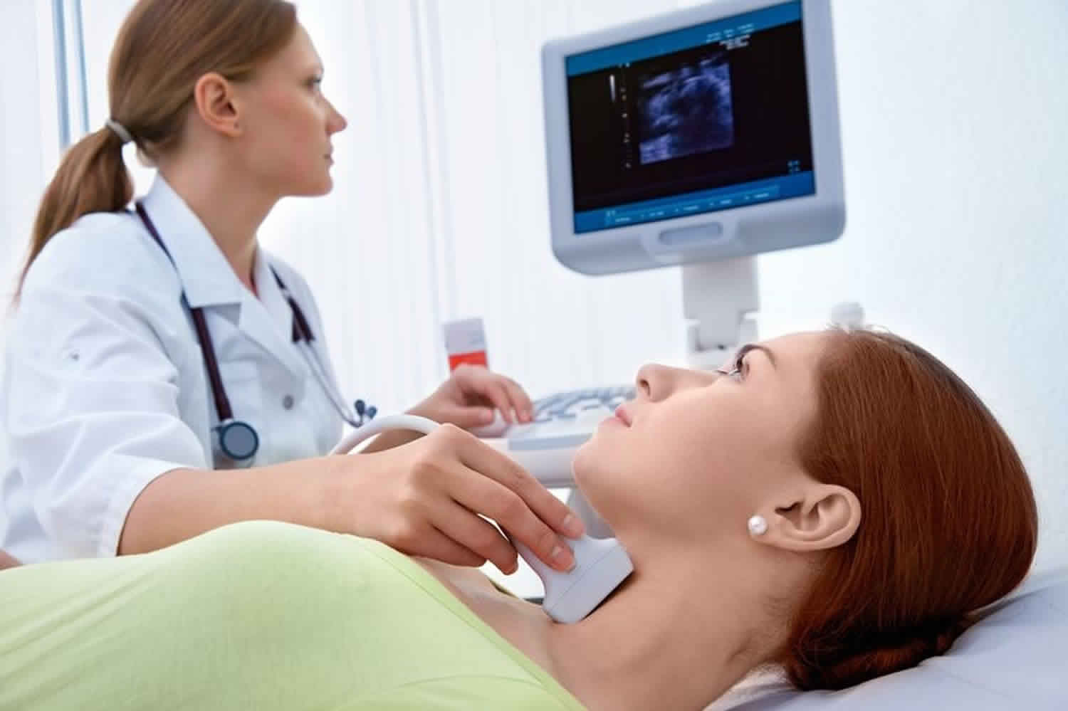
Ultrasound-guided fine needle aspiration biopsy (FNAB) of the thyroid refers to a minimally invasive procedure where tissue samples are collected from a thyroid nodule or other suspicious thyroid lesion using a thin hollow needle. The needle used is smaller in diameter than those used in most blood draws. The thyroid gland is located in front of the neck just above the neckline and is shaped like a butterfly, with two lobes on either side of the neck connected by a narrow band of tissue. Thyroid fine needle aspiration biopsy is usually done on a outpatient basis and generally complications are very minimal.
Fine needle biopsy of thyroid is usually completed in less than 30 minutes.
During a thyroid fine needle aspiration biopsy procedure, your doctor takes a small sample of tissue from your thyroid gland to diagnose your thyroid problem. This procedure is usually done in a treatment room by your doctor(s), with the help of a nurse. A technician with a microscope is also present in the room. Sometimes, this procedure is done by a radiologist in the diagnostic radiology department.
Thyroid biopsy is used to find the cause of a nodule in the thyroid gland. When a nodule is detected, imaging tests may be performed to help determine if it is benign (non-cancerous) or malignant (cancerous). However, it is not always possible to tell from these imaging tests whether a nodule is benign (non-cancerous) or cancerous.
Before the thyroid fine needle aspiration biopsy procedure, the doctor will explain it to you, including the risks and benefits. When you have a good understanding of what will be done, you will be asked to sign a consent form giving them permission to do the procedure. Please feel free to ask any questions you may have.
An ultrasound-guided fine needle aspiration biopsy uses sound waves to help locate a nodule or abnormality within the thyroid and remove a tissue sample for examination under a microscope. The procedure is less invasive than surgical biopsy, leaves little to no scarring and does not involve exposure to ionizing radiation.
An ultrasound-guided fine needle aspiration biopsy procedure requires little to no special preparation. Tell your doctor about any medications you’re taking, such as aspirin or blood thinners. For biopsies performed in children, sedation may be used. Specific instructions will be given at the time of scheduling.
Technique for thyroid fine needle aspiration biopsy varies widely from institution to institution, but there are elements common to all procedures.
Technique
- During the test, you will lie on your back with a pillow under your shoulders, your head tipped backward, and your neck extended. This position makes it easier for the radiologist to access the thyroid gland.
- You may feel some pressure on your neck from the ultrasound transducer and mild discomfort as the needle is moved to obtain the cells.
- You will be asked to remain still and not to cough, talk, swallow or make a sound during the procedure.
- The thyroid nodule is localized with the ultrasound transducer
- if using a guide, the guide path should not traverse important adjacent structures (e.g. the carotid)
- keeping the needle parallel to the transducer allows more of it to be seen and is often more useful in localizing the tip
- The skin is cleaned with the antiseptic agent
- A sterile probe cover is applied
- Many practitioners inject 1-2 mL of 1% lidocaine into the superficial tissue over the area of interest
- The patient should be instructed not to swallow or speak when the needle is below the skin surface
- The 25-27 gauge needle is guided into the lesion
- A to-and-fro action is used when the needle tip enters the nodule
- cells from the nodule enter the needle with capillary action
- some practitioners use suction with a 10 mL syringe, but this is controversial since other think it adds only blood to the sample
- The aspirated material is placed on a slide and often is checked by a pathologist or cytotechnologist for diagnostic quality
- Multiple passes (usually 3-6) are performed per nodule. This assures a better chance to find cancerous cells if they are present. If there is fluid in the nodule, a syringe may be used to drain it.
- New needles are used for additional samples.
- Depending on the situation, aspirated material may be reserved for cell block cytology or gene expression classifier tests
Once the biopsy is complete, pressure will be applied to the area to decrease the risk of bleeding. A bandage may be placed if necessary.
Aftercare instructions vary, but generally you can resume normal activities and any bandage can be removed within a few hours.
The biopsy site may be sore and tender for one to two days. You may take nonprescription pain medicine, such as acetaminophen, to relieve any discomfort.
A pathologist examines the removed specimen and makes a final diagnosis so that treatment planning can begin. Depending on the facility, the radiologist or your referring physician will discuss the results with you.
Does a thyroid biopsy hurt?
Generally, the thyroid biopsy procedure is not painful and the results are as accurate as when a tissue sample is removed surgically.
What are the limitations of thyroid needle biopsy?
In some cases, the specimens may be inadequate and the procedure may have to be repeated in order to obtain diagnostic results.
Thyroid biopsy results
Please note that different institutions and centers will have different rates of results depending on their specific populations:
- Benign – This accounts for up to 70% of biopsies when using the Bethesda System (one of the most common ways that cytopathologists classify nodule biopsy specimens). The risk of malignancy in this group is typically less than 3%. These nodules are generally monitored with a follow up ultrasound within 18 months and if needed, periodically after that.
- Malignant (cancer) – This accounts for 3-7% of all biopsy specimens. The most common type of thyroid cancer seen in these biopsies is papillary thyroid cancer. When a biopsy comes back as malignant, there is a 97- 99% chance that it is truly a cancerous lesion. Almost all of these nodules will go to surgery (thyroidectomy).
- Suspicious for malignancy – When a biopsy result returns as suspicious for malignancy, there is a 60-75% chance of cancer. The cytopathologist will see features that are worrisome, but not diagnostic of a malignant or cancerous lesion. The treatment is typically surgery.
- Atypia of Undetermined Significance (AUS) or Follicular Lesion of Undetermined Significance (FLUS) – These categories may be alternatively called “indeterminate.” These specimens have some features that are worrisome and some features that look more benign. This diagnosis carries a 5-15% risk of malignancy, although there is some variability among institutions. A repeat biopsy and/or genetic testing may be useful in these cases.
- Follicular Neoplasm or ‘Suspicious for follicular neoplasm’ – This category carries a 15-30% risk of malignancy. It is difficult to tell if the nodules are a benign condition called follicular adenoma or a malignant nodule unless it is taken out. Generally, at least half of the thyroid is removed (the side with the nodule) for diagnosis and treatment.
- Non-diagnostic – This means that there are not enough cells in the sample to make a diagnosis. Despite our best efforts and even when we can see that the needle was in the nodule during the biopsy, the specimen sometimes does not have enough thyroid follicular cells to make a proper diagnosis. Non-diagnostic samples can also occur when only cyst fluid is taken out, and for other reasons, such as the presence of too much blood. In these cases, the biopsy should be repeated, and if non-diagnostic a second time, consideration is given to a third biopsy, monitoring, or surgery.
Fine-needle aspiration biopsy of thyroid results are classified as diagnostic (satisfactory) or nondiagnostic (unsatisfactory) 1. Unsatisfactory smears (5-10%) result from hypocellular specimens usually caused by cystic fluid, bloody smears, or suboptimal preparation. Diagnostic smears are conventionally subclassified into benign, indeterminate, or malignant categories. Benign cytology (60-70%) is negative for malignancy, and includes cysts, colloid nodule, or Hashimoto thyroiditis 1. Malignant cytology (5%) is almost always positive for malignancy, and includes primary thyroid tumors or nonthyroid metastatic cancers. Papillary thyroid carcinoma is the most common malignancy, characterized by increased cellularity, sheets of cells, and typical nuclear abnormalities 1.
Indeterminate or suspicious specimens (10-20%) include atypical changes, Hurthle cells or follicular neoplasms, typically with absent or scanty colloid, hypercellularity sometimes with microfollicular arrangement. The new Bethesda Cytologic Classification has a 6-category classification, subdividing indeterminate further by risk factors. Overall, the indeterminate category has anywhere from 15% to 60% risk of malignancy, depending on the specifics of the report. Recent development of molecular markers are hoped to help further separate benign from malignant nodules with an indeterminate cytology. These tests are new and evolving, and no definite recommendations for use can be offered currently.
How long do thyroid biopsy results take?
Once your doctor has collected enough cells or tissue for analysis, your thyroid biopsy procedure is complete. The biopsy samples may be used to make slides immediately and/or collected in a solution to wash excess blood. Specially trained doctors, cytopathologists, then make slides from the material and examine them under a microscope to make a diagnosis. The results may be available in a few days, though more technical tests may require more time. Ask your doctor how long you can expect to wait. Generally, it can take anywhere from a few days to 2 weeks for the result to return.
Within the past few years, several molecular tests have become available to help determine whether some thyroid nodules are cancerous or benign. These tests look at many genes within the thyroid nodule’s genetic make-up (DNA). They are being used when a thyroid nodule biopsy comes back with a diagnosis of ‘indeterminate’. Sometimes, the person doing the thyroid biopsy will perform an additional pass of the needle to obtain material for such a test. This may be done on the first biopsy or at the time of a repeat biopsy.
Thyroid biopsy complications
Thyroid fine-needle aspiration biopsy, particularly under ultrasound-fine-needle aspiration, is very safe 1 and complications are minimal if the tip of the needle is visualized throughout the procedure. No serious complications such as tumor seeding, nerve damage, tissue trauma, or vascular injury have been reported 2. Needle puncture causes slight pain radiating to the ear and some skin discoloration at the aspiration site(s). However, even a minor localized hematoma is uncommon. The superficial location of the thyroid allows easy compression of bleeding. Patient use of anticoagulants or salicylates does not preclude fine-needle aspiration biopsy. Needle track implantation of thyroid carcinoma is extremely rare; it has been poorly documented and is not considered a real problem by most experts 3. Post-aspiration hemorrhage within a cystic lesion can occur, and the author has seen 1 patient who, within several hours after fine-needle aspiration biopsy, developed severe pain from bleeding into the nodule that warranted surgical excision. The specimen contained fresh blood consistent with hemorrhage caused by biopsy. However, this is the only example the authors have had among more than 40,000 biopsies performed at our institution during the past 4 decades 1.
Thyroid biopsy side effects
Thyroid biopsy carries a small risk of bleeding and infection at the site where the needle was inserted. Some mild pain can be expected after needle biopsy, though it is usually controlled with over-the-counter pain relievers or prescription medications.
Thyroid biopsy risks:
- Bleeding at the site of biopsy.
- Infection.
- Injury to structures adjacent to the thyroid.
Call your doctor if you experience:
- Fever
- Pain at the biopsy site that worsens or isn’t helped by medications
- Swelling at the biopsy site
- Drainage from the biopsy site
- Bleeding that doesn’t stop with pressure or a bandage
- Dean DS, Gharib H. Fine-Needle Aspiration Biopsy of the Thyroid Gland. [Updated 2015 Apr 26]. In: Feingold KR, Anawalt B, Boyce A, et al., editors. Endotext [Internet]. South Dartmouth (MA): MDText.com, Inc.; 2000-. Available from: https://www.ncbi.nlm.nih.gov/books/NBK285544[↩][↩][↩][↩][↩]
- Belfiore A, La Rosa GL. Fine-needle aspiration biopsy of the thyroid. Endocrinol Metab Clin North Am. 2001;30:361-400.[↩]
- Hales MS, Hsu FS. Needle tract implantation of papillary carcinoma of the thyroid following aspiration biopsy. Acta Cytol. 1990;34:801-4.[↩]


