Trichodynia
Trichodynia also called “hair pain”, scalp dysaesthesia or cutaneous dysesthesia syndrome, is defined as a painful sensation within scalp hair, which becomes more intense when hairs are touched 1. The term “trichodynia” has also been proposed for discomfort, pain or paresthesia (burning, or stinging) of the scalp related to diffuse hair loss (alopecia) 2. Sulzberger et al. 3 reported that trichodynia may also be circumscribed (“spotty”). The pain occurs with no pathological findings, and is usually considered a type of somatoform disorder 4. Trichodynia should be differentiated from scalp dysesthesia, which refers to situation when the symptoms reported by the patient are localized on the scalp skin, while in trichodynia patients indicate that the pain or burning is solely limited to the hair itself 1. Many patients with trichodynia present other psychiatric symptoms, including depression, obsessive-compulsive or anxiety disorders. The cause of trichodynia is not known, but it is suggested to be of multifactorial origin 5.
Trichodynia has been found that 34% of female patients with hair loss complained of this phenomenon 6. In a recent survey, Grimalt et al. 7 showed that 14% of their diffuse alopecia patients reported trichodynia. Complaints such as pain and burning of the scalp in patients with diffuse hair loss (alopecia) were described in the earlier dermatology literature 8. Both such studies and clinical observations have led to the idea that the diffuse alopecia or telogen effluvium and trichodynia are related. By definition, telogen effluvium is a nonscarring and diffuse hair loss from the scalp that occurs a few months after a triggering event 2.
Although dealing with trichodynia can be distressing and literature support is weak, there are a number of treatments available.
Trichodynia causes
The cause of trichodynia is not known, but it is suggested to be of multifactorial origin 9. It is commonly believed that trichodynia correlates with emotional upset 10. Trichodynia is almost exclusive of patients with active telogen effluvium, was described in the alopecia areata (14% of cases), folliculitis decalvans, or frontal fibrosing alopecia (hair loss) 11.
The underlying mechanisms creating the pain are not clear, though it has been proposed that it is probably multi-etiological. The most accepted hypotheses are increased expression of the neuropeptide substance P, underlying psychiatric disorders, nutritional deficiencies, and perifollicular inflammation 12. Substance P is involved in pain perception by the nerve endings, and changes in the production and activity of substance P around the hair follicles may be responsible for the pain and burning sensation 13. Hair follicles are innervated by unmyelinated neural plexuses located around the hair follicle stem cells. These nerve fibers contain neuropeptides including substance P and calcitonin gene-related peptide (CGRP). These neuropeptides play an important role in the regulation of hair growth and are associated with the neurogenic inflammatory response. Perifollicular substance P is also involved in the regulation of hair growth 14. An imbalance in the tonic release of neuropeptides may result in inhibition of hair growth. Cutrer et al. 15 hypothesized that chronic activation of the c-fibers, in addition to mediating inflammatory pain and follicular injury, might reduce substance P and CGRP concentrations resulting in altered peribulbar antigen presentation and inhibition of further hair growth.
Another explanation may be an underlying psychiatric disorder. It has been found that 76% of the people who had trichodynia had psychopathic signs versus 20% in the control group, supporting this idea. Researchers have observed and speculated that there is a connection between psychopathologic findings (such as anxiety) and trichodynia 16. In 2006 Gupta and Gupta 17 found that numbness and pain are common symptoms of somatoform dissociation or conversion reaction. Kivanç et al. 18 found that trichodynia was associated with depression in the telogen alopecia group and with obsessive-compulsive personality disorder in the androgenic alopecia group. However, this idea is controversial. Although increased rates of psychiatric problems have been reported in patients with trichodynia, Ozturk et al. 19 found no association between trichodynia and depression or anxiety. In this study the patients with telogen alopecia were consisting the control group, and they could have the opportunity to evaluate only the trichodynia patients 19.
Neuropathic pain can also be associated with nutritional deficiencies (Iron [Fe], vitamin B12, ferritin, zinc, vitamin D, vitamin E). Nutritional factors affect the hair directly, and dietary supplements containing B complex vitamins can influence hair growth 19. Nutritional deficiencies have been reported in other cutaneous dysesthesia syndromes. For example, glossodynia is characterized by a burning sensation of the tongue and oral mucosa. Menopause, psychogenic disorders, and nutritional factors have also been suggested to cause this phenomenon 20. However, evidence level is very low to confirm this nutritional hypothesis for trichodynia patients 21.
Other dermatological conditions causing scalp pain
Scalp pain can occur with cicatricial alopecia that can be caused by a fungus infection or autoimmune conditions such as cutaneous lupus and lichen planopilaris. Folliculitis decalvans and dissecting cellulitis are forms of primary neutrophilic scarring alopecia that are characterized clinically by chronic suppurative folliculitis and often associated with pruritus or even pain. The inflammatory cells may irritate nerve endings leading to a burning or painful sensation. Hair dye-related dermatitis may also cause burning sensations.
There are also painful tumoral lesions of the skin and subcutaneous tissue. These lesions can be found anywhere in the peripheral nerve tissue. They have a propensity for developing on the skin and subcutaneous tissue, as well as in oral and pharyngeal locations. An old acronym may help us to remember them. LEND AN EGG tumors (leiomyoma, eccrine spiradenoma, neuroma, dermatofibroma, angiolipoma, neurilemmoma, endometrioma, glomus tumor, and granular cell tumors) must always be considered when there is a tumoral lesion associated with pain 22.
Trichodynia symptoms
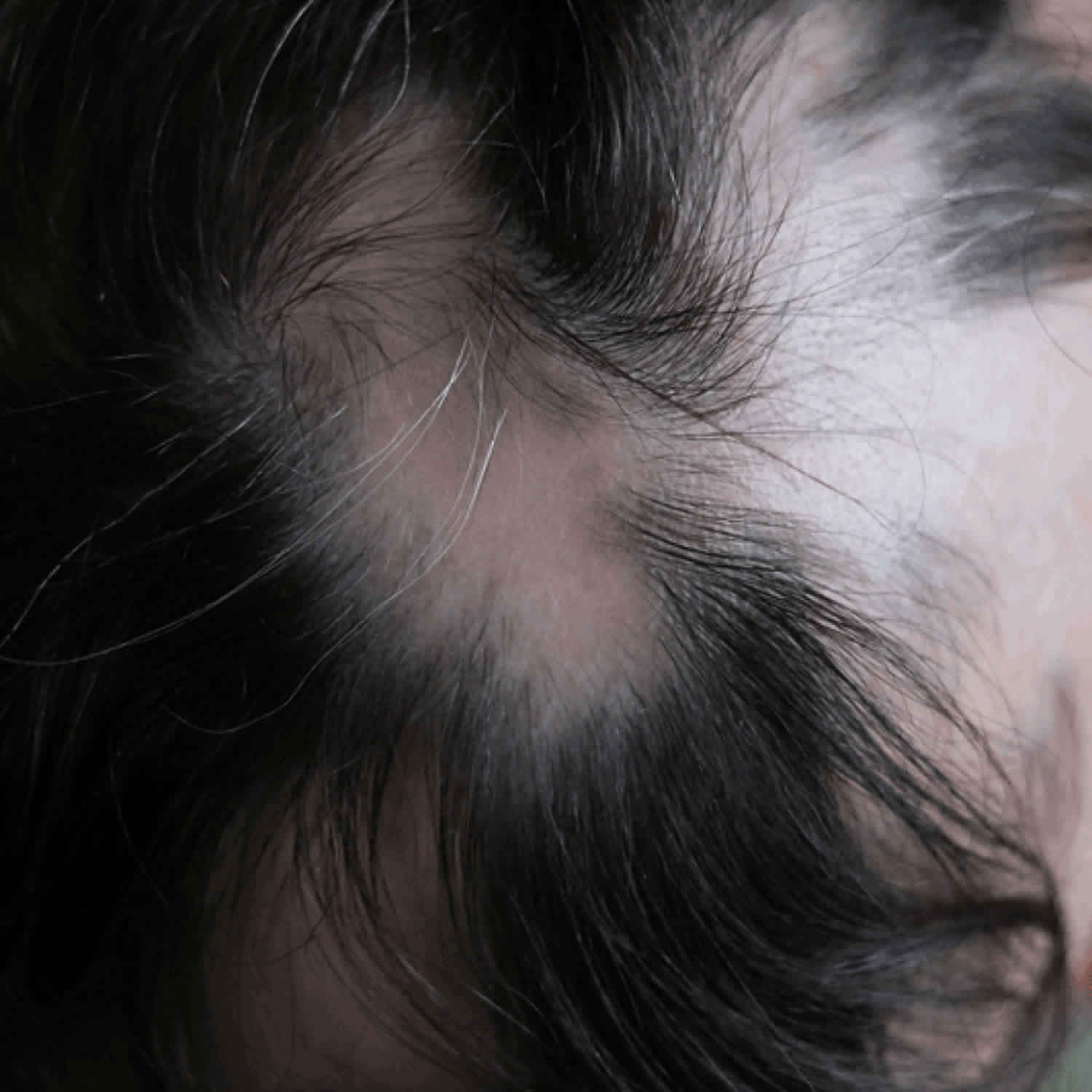
Trichodynia is defined as a painful sensation in the skin of the scalp or the hair itself and becomes more intense when hairs are touched. Trichodynia presents with complaints such as tenderness, pain, burning, itching, stinging, and diffuse hair loss 7.
Trichodynia diagnosis
Trichodynia diagnosis is based on clinical suspicion. A comprehensive history and examination are needed to identify any underlying cause and excluded a primary dermatological disease 23. If the patient reports headache or temporal pain by palpation, tension headaches or temporal arteritis should be evaluated first.
Diagnostic tests may include:
- Trichoscopy. Trichoscopy is a non-invasive technique based on dermoscopic evaluation of the scalp hair, which allows a 10× magnification. Recently, besides manual trichoscopy, videodermoscopy has gained importance. It is a non-invasive diagnostic tool, initially used to evaluate and monitor pigmented skin lesions, that has proven to be useful in the scalp diseases. A magnification ranging from 20× to 70× allows the in vivo visualization of scalp epidermis, follicles, hair shafts and vascular patterns. Moreover, images can be stored to get a global photographic assessment that can be used in further controls to evaluate the hair growth 24.
- Phototrichogram. This technique involves trimming the hair of a 2 square cm area of scalp, pictures of the same area taken on different days, and then compared in hair density, hair growth, and rate of shedding. Since only anagen hair would elongate it helps in the assessment of the ratio of anagen:telogen hair. A TrichoScan is a fully computerized phototrichogram 25. A TrichoScan is a simpler, noninvasive, reproducible, and more sensitive than a classical trichogram and very useful in the diagnosis of hair loss 25.
- Trichogram is a plucking of hair in a defined area (40-60 hair). Cases of telogen effluvium show a significant reduction of the anagen:telogen ratio. More than 25% of hair are found to be in the telogen phase in the case of telogen effluvium 26.
- Scalp biopsy. Scalp biopsy can be useful when the clinical evaluation does not provide a definitive diagnosis, and cicatricial alopecias or alopecia areata are suspected.
- Blood tests.
- A hyperandrogenic state should be considered in women with female androgenetic alopecia associated with signs of androgen excess (e.g. hirsutism, irregular menses, acne, hyperprolactinemia, acanthosis nigricans); while it is not indicated in all women with female androgenetic alopecia 27. Free androgen index test (FAI = total testosterone [nmol L−1] × 100/sex hormone binding globulin [SHBG]) and prolactin as screening parameters for ovarian hyperandrogenism and dehydroepiandrosterone sulfate (DHEA) and 17-hydroxyprogesterone (17-OH-P) as screening parameters for androgen-producing tumors and adrenal congenital hyperplasia are recommended 28. Note that testing of androgen levels should be done during the follicular phase, between the fourth and the seventh day of the cycle, and that oral contraceptives should be discontinued for at least 2 months prior to this testing 29.
- Measurements of blood iron, ferritin, vitamin D, zinc and thyroid profile may be useful to assess and treat other conditions that may impact on hair regrowth in female androgenetic alopecia 27.
- Serology: antinuclear antibody (ANA), antineutrophil cytoplasmic antibodies (ANCA), C-reactive protein (CRP)
- Ovarian and adrenal ultrasound could be prescribed to rule out the presence of ovarian cysts and androgen-producing tumours or adrenal congenital hyperplasia 28. Depending on the results, further investigations may be needed and an interdisciplinary approach involving gynaecologists, endocrinologists and dermatologists may be required 28.
Trichodynia treatment
Although dealing with trichodynia can be distressing and literature support is weak, there are a number of treatments available. L-Cystine-containing oral preparations, topical corticosteroids (both high potency and low), and anti-inflammatory drugs have been advocated 2. Inhibitors of substance P can also be tried. Cannabinoids, for example, have been demonstrated to inhibit substance P 30. Capsaicin cream has been used because it blocks substance P when applied to the hair follicles. On the basis of psychiatric origin, the physician also may use low-dose antidepressants (venlafaxine, amitriptyline, and doxepin) and also pregabalin 31.
In 2009, Cutrer et al. 15 have investigated the efficacy of botulinum toxin treatment in cephalalgia alopecia patients and obtained improved pain control and hair regrowth following botulinum toxin A injections. They also observed that botulinum increases substance P and calcitonin gene-related peptide-containing cutaneous nerves in the scalp. Botulinum toxin A injections does not block low-level trophic release of neuropeptides such as calcitonin gene-related peptide and allows resumption of substance P and calcitonin gene-related peptide baseline regulation of the hair follicle and hair regrowth 32. However, it should be kept in mind that botulinum toxin A treatment is temporary. The process of painful inflammatory activation, hair follicle regression, and hair loss is repeated after a few months.
Sensory tests revealed that trichodynia patients were significantly more sensitive to touch and to pressure pain and exhibited cranial mechanical hyperesthesia and cranial hyperalgesia 33. So, gentle scalp maintenance may provide some relief. To support the treatment, it is important to inform the patients about not to use over hot water and harsh shampoos or wear tight pony tail. Other relaxation techniques such as gentle scalp massage may also help in reducing symptoms.
- Itchy Hair – Trichoknesis: A Variant of Trichodynia or a New Entity? Acta Derm Venereol 2013; 93: 591 DOI: 10.2340/00015555-1543 https://www.medicaljournals.se/acta/content_files/files/pdf/93/5/3865.pdf[↩][↩]
- Müge Güler Özden (April 12th 2017). Trichodynia (Scalp Dysesthesia), Current Perspectives on Less-known Aspects of Headache, Hande Turker, IntechOpen, DOI: 10.5772/67792 https://www.intechopen.com/books/current-perspectives-on-less-known-aspects-of-headache/trichodynia-scalp-dysesthesia-[↩][↩][↩]
- Diffuse alopecia in women: its unexplained apparent increase in incidence. Sulzberger MB, Witten VH, Kopf AW. AMA Arch Derm. 1960;81:556–560.[↩]
- Kivanç-Altunay I, Savaş C, Gökdemir G, Köşlü A, Ayaydin EB. The presence of trichodynia in patients with telogen effluvium and androgenetic alopecia. Int J Dermatol 2003; 42: 691–693.[↩]
- Harth W, Gieler U, Kusnir D, Tausk FA. Persistent somatoform pain disorders. In: Harth W, Gieler U, Kusnir D, Tausk FA. Clinical management in psychodermatology. Berlin, Heidelberg: Springer Verlag, 2009: p. 62–63.[↩]
- Rebora A, Semino MT, Guarrera M. Trichodynia. Dermatology. 1996;192:292–3.[↩]
- Grimalt R, Ferrando J, Grimalt F. Trichodynia. Dermatology. 1998;196(3):374.[↩][↩]
- Hoss D, Segal S. Scalp dysesthesia. Arch Dermatol. 1998;134:327–30.[↩]
- Rebora A. Trichodynia: A review of the literature. Int J Dermatol. 2016;55:382–4.[↩]
- Brzezinski P, Zawar V, Chiriac A. Trichodynia Silenced Effectively with Propranolol. Int J Trichology. 2019;11(1):41-42. doi:10.4103/ijt.ijt_8_19 https://www.ncbi.nlm.nih.gov/pmc/articles/PMC6385513[↩]
- Heppt MV, Letulé V, Laniauskaite I, Reinholz M, Tietze JK, Wolff H, et al. Frontal fibrosing alopecia: A Retrospective analysis of 72 patients from a German academic center. Facial Plast Surg. 2018;34:88–94.[↩]
- Baldari M, Montinari M, Guarrera M, Rebora A. Trichodynia is a distinguishing symptom of telogen effluvium. J Eur Acad Dermatol Venereol. 2009;23:733–4.[↩]
- Ericson M, Gabrielson A, Worel S, Lee WS, Hordinsky MK. Substance P (SP) in innervated and non-innervated blood vessels in the skin of patients with symptomatic scalp. Exp Dermatol. 1999;8:344–5.[↩]
- Paus R, Heinzelmann T, Schultz KD, Furkert J, Fechner K, Czarnetzki BM. Hair growth induction by substance P. Lab Investig. 1994;71:134–40.[↩]
- Cutrer FM, Sandroni P and Wendelschafer-Crabb G. Botulinum toxin treatment of cephalalgia alopecia increases substance P and calcitonin gene-related peptide-containing cutaneous nerves in scalp. Cephalalgia. 2010;30(8):1000–6.[↩][↩]
- Durusoy C, Ozenli Y, Adiguzel A, Budakoglu IY, Tugal O, Arikan S, et al. The role of psychological factors and serum zinc, folate and vitamin B12 levels in the aetiology of trichodynia: A case-control study. Clin Exp Dermatol. 2009;34:789–92.[↩]
- Gupta MA, Gupta AK. Stressful major life events are associated with a higher frequency of cutaneous sensory symptoms: An empirical study of non-clinical subjects. J Eur Acad Dermatol Venereol. 2004;18:560–5.[↩]
- Kivanç-Altunay I, Savaş C, Gökdemir G, Köşlü A, Ayaydin EB. The presence of trichodynia in patients with telogen effluvium and androgenetic alopecia. Int J Dermatol. 2003;42:691–3.[↩]
- Ozturk P, Orhan FO, Ozer A, Akman Y, Kurutas E. Evaluation of anxiety and levels of serum B12, folate, TSH, ferritin, and zinc in telogen alopecia patients with trichodynia. Int J Trichol. 2012;4(4):251–4.[↩][↩][↩]
- Moore PA, Guggenheimer J, Orchard T. Burning mouth syndrome and peripheral neuropathy in patients with type 1 diabetes mellitus. J Diabet Complicat. 2007;21:397–402.[↩]
- Durusoy, Ozturk P, Orhan FO, Ozer A, Akman Y, Kurutas E. Evaluation of anxiety and levels of serum B12, folate, TSH, ferritin, and zinc in telogen alopecia patients with trichodynia. Int J Trichol. 2012;4(4):251–4.[↩]
- Naversen DN, Trask DM, Watson FH, Burket JM. Painful tumors of the skin: “LEND AN EGG”. J Am Acad Dermatol. 1993;28(2):298–300.[↩]
- Willimann B, Trüeb RM. Hair pain (trichodynia): Frequency and relationship to hair loss and patient gender. Dermatology. 2002;205:374–7.[↩]
- Kanti V, Messenger A, Dobos G, Reygagne P, Finner A, Blumeyer A, et al. Evidence-based (S3) guideline for the treatment of androgenetic alopecia in women and in men. Eur Acad Dermatol Venereol. 2018;32(1):11–22.[↩]
- Phototrichogram. Dhurat R. Indian J Dermatol Venereol Leprol. 2006;72:242–244.[↩][↩]
- The hair pull test and the hair pluck for analysis of hair abnormalities. Chong A, Wade M, Sinclair R. Modern Med Australia. 1999;42:105–110.[↩]
- Carmina E, Azziz R, Bergfeld W, Escobar Morreale HF, Futterweit W, Huddleston H, et al. Female pattern hair loss and androgen excess: a report from the Multidisciplinary Androgen Excess and PCOS Committee. J Clin Endocrinol Metab. 2019. https://doi.org/10.1210/jc.2018-02548 (Epub Ahead of Print).[↩][↩]
- Bienenfeld A, Azarchi S, Lo Sicco K, Marchbein S, Shapiro J, Nagler AR. Androgens in women: androgen mediated skin disease and patient evaluation (part I). J Am Acad Dermatol. 2018. https://doi.org/10.1016/j.jaad.2018.08.062[↩][↩][↩]
- Sánchez LA, Pérez M, Centeno I, David M, Kahi D, Gutierrez E. Determining the time androgens and sex hormonebinding globulin take to return to baseline after discontinuation of oral contraceptives in women with polycystic ovary syndrome: a prospective study. Fertil Steril. 2007;873(3):712–4.[↩]
- OConnor TM, OConnell J, Obrien DJ, et al. The role of substance P in inflammatory disease. J Cell Physiol. 2004;201:167–180.[↩]
- Thorsberry L, English J. Scalp dysesthesia related to cervical spine disease. JAMA Dermatol. 2013;149:200–3.[↩]
- Durham PL, Cady R, Cady R. Regulation of calcitonin gene-related peptide secretion from trigeminal nerve cells by botulinum toxin type A: Implications for migraine therapy. Headache. 2004;44:35–43.[↩]
- Defrin R, Lurie R. Indications for peripheral and central sensitization in patients with chronic scalp pain (trichodynia). Clin J Pain. 2001;29(5):417–424.[↩]


