Tumor suppressor genes
Tumor suppressor genes are normal genes that slow down cell division, repair DNA mistakes or tell cells when to die (a process known as apoptosis or programmed cell death). When tumor suppressor genes don’t work properly, cells can grow out of control, which can lead to cancer. Loss of function mutations in tumor suppressor genes has been identified in many types of cancers, including ovarian, lung, colorectal, head and neck, pancreatic, uterine, breast, and bladder cancer 1. Examples of tumor suppressor genes include BRCA1, BRCA2, and p53 or TP53.
A tumor suppressor gene is like the brake pedal on a car. Tumor suppressor gene normally keeps the cell from dividing too quickly, just as a brake keeps a car from going too fast. When something goes wrong with the tumor suppressor gene, such as a mutation, cell division can get out of control.
Tumor suppressor genes function to either repress or inhibit the cell cycle or promote apoptosis. The more specific functions of tumor suppressor proteins fall into several categories, including 2, 3:
- Inhibition of mitogenic signaling pathways
- Inhibition of cell cycle progression
- Inhibition of “pro-growth” programs of metabolism and angiogenesis
- Inhibition of invasion and metastasis
- Stabilization of the genome
- DNA repair factors
- Induction of apoptosis
Inherited abnormalities of tumor suppressor genes have been found in some family cancer syndromes. They cause certain types of cancer to run in families. But most tumor suppressor gene mutations are acquired, not inherited.
Germline mutations in BRCA1 or BRCA2 genes increase a woman’s risk of developing hereditary breast or ovarian cancers and a man’s risk of developing hereditary prostate or breast cancers. They also increase the risk of pancreatic cancer and melanoma in women and men.
Abnormalities of the TP53 gene (which codes for the p53 protein) have been found in more than half of human cancers e.g., Li-Fraumeni syndrome 4. Acquired mutations of TP53 gene appear in a wide range of cancers.
An important difference between oncogenes and tumor suppressor genes is that oncogenes result from the activation (turning on) of proto-oncogenes, but tumor suppressor genes cause cancer when they are inactivated (turned off).
Proto-oncogenes are genes that normally help cells grow. When a proto-oncogene mutates (changes) or there are too many copies of it, it becomes a “bad” gene that can become permanently turned on or activated when it is not supposed to be. When this happens, the cell grows out of control, which can lead to cancer. This bad gene is called an oncogene. A few cancer syndromes are caused by inherited mutations of proto-oncogenes that cause the oncogene to be turned on (activated). But most cancer-causing mutations involving oncogenes are acquired, not inherited. They generally activate oncogenes by:
- Chromosome rearrangements: Changes in chromosomes that put one gene next to another, which allows one gene to activate the other
- Gene duplication: Having extra copies of a gene, which can lead to it making too much of a certain protein
Two common oncogenes are:
- HER2, a specialized protein that controls cancer growth and spread. It is found in some cancer cells. For example, breast and ovarian cancer cells.
- The RAS family of genes, which makes proteins involved in cell communication pathways, cell growth, and cell death.
Tumor suppressor genes function
Tumor suppressor genes function to either repress or inhibit the cell cycle or promote apoptosis. The more specific functions of tumor suppressor genes can be broadly divided into five types functionally 5:
- Genes that encode intracellular proteins that are crucial in controlling the progression of cell cycle stages – (e.g., pRB and p16) 6
- Genes that encode receptors or signal transducers which orchestrate signals that inhibit cell proliferation [e.g., adenomatous polyposis coli (APC) and transforming growth factor (TGF)-β] 7
- Genes that encode the checkpoint-control proteins, which are useful in triggering cell cycle arrest in case of DNA damage or chromosomal defects [e.g., p16, p14, and breast cancer type 1 susceptibility protein (BRCA1)] 8
- Genes that encode proteins useful for the induction of apoptosis (e.g., p53) 9
- Genes that encode proteins involved in the repair of DNA [e.g., DNA mismatch repair protein 2 (MSH2) and p53] 10
Many tumor suppressor genes have been the object of studies, and there are likely many more that have yet to be discovered. The mechanisms of each tumor suppressor gene and its protein products are complex and interrelated to other cell signaling pathways, but some better-known mechanisms appear below.
Most of our current understanding of tumor suppressor genes comes from the initial studies of the retinoblastoma (RB) gene. This was the first tumor suppressor gene discovered and mutations in RB gene cause childhood retinoblastoma. This is an inherited condition caused by an inactivating mutation in the RB1 gene that causes a 10,000-fold increased risk of developing retinoblastoma (often in both eyes) as compared to the general population. These patients are also at increased risk of developing osteosarcoma and other sarcomas. Interestingly, about 60% of retinoblastomas occur sporadically (almost always in one eye), and these patients are not at increased risk for other forms of cancer 11. To explain this dichotomy, Knudson proposed a “two-hit” hypothesis:
- In the inherited form of retinoblastoma, children inherit one mutated RB allele (germ-line mutation) and the other copy is normal. Retinoblastoma develops when the normal retinoblastoma (RB) allele undergoes a spontaneous somatic mutation. This is the “second hit.”
- In sporadic cases of retinoblastoma, both normal RB alleles must undergo a somatic mutation in the same cell. The probability of this is low which explains why retinoblastoma is uncommon in the general population. In both cases, the resulting two hits lead to the development of retinoblastoma 12.
Based on these original observations, there are three important properties of classic tumor suppressor genes:
- Classic tumor suppressor genes are recessive at the cellular level, with inactivation of both alleles typically found in tumors
- Inheritance of a single mutant allele increases tumor susceptibility because only one additional inactivating event is required for complete loss of gene function
- The same gene is frequently inactivated in sporadic cancers 13.
Tumor suppressor genes examples
There are many tumor suppressor genes that have been studied, and there are likely many more that have yet to be discovered. The mechanisms of each tumor suppressor gene and its protein products are complex and interrelated to other cell signaling pathways, but some better-known mechanisms are described below.
Retinoblastoma (RB)
The retinoblastoma (RB) gene also called “Governor of the Cell Cycle”, encodes the RB protein that when hypophosphorylated, binds and inhibits E2F transcriptions factors 14. The E2F transcriptions factors regulate genes that are essential for cells to pass from G1 to the S phase of the cell cycle. Typical growth factor signaling causes RB hyperphosphorylation and inactivation, thus causing cell cycle progression 15. A variety of mechanisms like loss-of-function mutations affecting RB, CDK4, and cyclin D gene amplification, cyclin-dependent kinase inhibitors loss (p16/INK4a), and inhibition of RB protein by the binding of viral oncoproteins (E7 protein of HPV) can revoke the antiproliferative effect of RB protein in cancers 16.
TP53 Tumor Suppressor Gene
TP53 Tumor Suppressor Gene also known as the “guardian of the genome” as it serves to monitor for cellular stress like anoxia, identify DNA damage, or inappropriate signaling by mutated oncoproteins 17, 18. The TP53 gene encodes for the p53 protein, which controls the expression of proteins and their activity in cell cycle arrest, cellular senescence, DNA repair, and apoptosis. Loss of p53 protein can cause continued cell replication despite DNA damage and failure to activate programmed cell death 19. The DNA damage is perceived by complexes comprising kinases of the ATM/ATR family 3. These kinases phosphorylate p53, releasing it from inhibitors such as MDM2. The active p53 upregulated the expressions of important proteins like cyclin-dependent kinase inhibitor p21, which causes G1-S checkpoint arrest of the cell cycle. In instances where the DNA damage is not repairable, p53 induces events like activating the BAX gene, which encodes a pro-apoptotic protein that finally lead to cellular apoptosis or senescence. It also works to inhibit the BCL2 anti-apoptotic gene and stimulates the release of cytochrome c from the mitochondria. Cytochrome c activates caspases within the cell responsible for its eventual degradation. Similar to RB, p53 can be inactivated by viral oncoproteins like the E6 protein of HPV, thus revoking the antiproliferative and other important cellular effects. Most of the cancers demonstrate a biallelic loss-of-function mutation in TP53 tumor suppressor gene. Uncommon patients with Li-Fraumeni syndrome have a very high incidence of a wide variety of cancers like breast cancer, soft-tissue, and bone sarcomas, and brain tumors since they inherit one defective copy of TP53 tumor suppressor gene 20, 21, 22.
Phosphatase and Tensin Homolog (PTEN) Gene
The phosphatase and tensin homolog (PTEN) gene negatively regulates the phosphoinositide-3-kinase (PI3K)-AKT and the target of mTOR signaling pathways, which are vital for cell proliferation, cell cycle progression, and apoptosis 23. The PTEN protein also functions to keep migration, adhesion, and angiogenesis in check. It also plays a role in the overall stabilization of the genome. A biallelic loss-of-function is common in diverse cancers. Cowden syndrome is an autosomal dominant disorder resulting from germline loss-of-function mutations of this gene and correlates with a higher risk of breast and endometrial cancer 24.
E-cadherin (CDH1)
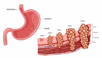
Normal cells stop proliferating once they come into contact with neighboring cells. This helps to maintain the structure and architecture of the tissue and is referred to as contact inhibition. Mediation of cell-to-cell contact in many tissues is the function of a group of proteins called cadherins 25. E-cadherin (epithelial cadherin) regulates contact inhibition by binding to a key component of the WNT signaling pathway, ß-catenin. This binding prevents E-catenin from translocating to the nucleus of the cell, stopping it from activating transcription of pro-growth target genes 26. Overall, this interaction regulates the morphology and organization of epithelial cell linings 27. Autosomal dominant familial gastric carcinoma is associated with a germline loss-of-function in E-cadherin (CDH1) gene 27.
NF1 and NF2
NF1 gene encodes for Neurofibromin-1, which is a GTPase that functions as a negative regulator of RAS. A germline loss of function mutation of this gene causes Neurofibromatosis type 1, an autosomal dominant disorder associated with the formation of neurofibromas, brain tumors like optic gliomas, and malignant nerve sheath tumors peripherally 28, 29.
NF2 gene encodes Neurofibromin-2 also known as Merlin, which is a cytoskeletal protein associated with contact inhibition. Loss of function mutations of NF2 gene leads to neurofibromatosis type 2, which is also an autosomal dominant disorder associated with an increased risk of bilateral schwannomas among other tumors 30.
BRCA 1, BRCA 2 and PARP-1
BRCA1 and BRCA2 are tumor suppressor genes that encode proteins involved in the repair of DNA double-strand breaks through the homologous recombination repair pathway 31.
PARP-1 encodes a protein that assists with the repair of single-stranded breaks in the DNA. Without functional proteins that repair DNA, the cell cycle continues to pass along defective and mutated genetic material that leads to aberrant daughter cells.
APC
The adenomatous polyposis coli (APC) gene encodes a tumor suppressor protein that negatively regulates the WNT signaling pathway. This regulation leads to enhancement of the formation of a complex that degrades β-catenin that is involved in the regulation and co-ordination of cell-cell adhesion and gene transcription 32, 33. The APC gene mutation is present in familial adenomatous polyposis (FAP), an autosomal dominant disorder where thousands of colonic polyps develop with early onset of colon carcinoma. The tumor development is associated with a loss of a single normal APC allele 33.
CDKN2A
Cyclin-dependent kinase inhibitor 2A (CDKN2A) gene encodes two tumor suppressor proteins, p16/INK4a, and ARF, which augments RB function and stabilizes p53, respectively 34. Loss-of-function germline mutations in CDKN2A gene occur in autosomal dominant familial melanoma. A biallelic loss-of-function presents in multiple cancers including melanomas, leukemias, and carcinomas.
WT1
Wilms’ tumor 1 (WT1) gene encodes for transcription factors required for normal genitourinary tissue development 35. Wilms tumor, a kidney cancer in chidlren, is associated with a germline loss-of-function mutation in WT1 gene. Sporadic Wilms tumor also correlates with similar WT1 mutations 35.
PTCH1
PTCH1 tumor suppressor gene encodes protein patched homolog 1 that negatively regulated the hedgehog signaling pathway 36, 37. Gorlin syndrome also known as nevoid basal cell carcinoma syndrome is an autosomal dominant disorder that correlates with a germline loss-of-function mutation in PTCH1 gene and has a high risk of developing basal cell carcinoma and medulloblastoma. Sporadic cases of basal cell carcinoma and medulloblastomas are frequently associated with acquired biallelic loss-of-function PTCH1 mutations 36.
VHL
Von Hippel-Lindau tumor suppressor gene or VHL gene provides instructions for making a protein that functions as part of a complex (a group of proteins that work together) called the VCB-CUL2 complex 38. The VCB-CUL2 complex targets other proteins to be broken down (degraded) by the cell when they are no longer needed. Protein degradation is a normal process that removes damaged or unnecessary proteins and helps maintain the normal functions of cells.
One of the targets of the VCB-CUL2 complex is a protein called hypoxia-inducible factor 2-alpha (HIF-2α). HIF-2α is one part (subunit) of a larger protein complex called HIF, which plays a critical role in the body’s ability to adapt to changing oxygen levels. HIF controls several genes involved in cell division, the formation of new blood vessels, and the production of red blood cells. It is the major regulator of a hormone called erythropoietin, which controls red blood cell production. HIF’s function is particularly important when oxygen levels are lower than normal (hypoxia). However, when adequate oxygen is available, the VCB-CUL2 complex keeps hypoxia-induced factor (HIF) from building up inappropriately in cells.
The VHL protein likely plays a role in other cellular functions, including the regulation of other genes and control of cell division. Based on this function, the VHL protein is classified as a tumor suppressor, which means it prevents cells from growing and dividing too rapidly or in an uncontrolled way. The VHL protein is also involved in the formation of the extracellular matrix, which is an intricate lattice that forms in the spaces between cells and provides structural support to tissues.
Von Hippel-Lindau syndrome, an autosomal dominant disorder, is associated with loss-of-function germline mutations of VHL gene and poses a high risk of developing renal cell carcinoma and pheochromocytoma 39, 40.
- Joyce C, Kasi A. Cancer, Tumor-Suppressor Genes. [Updated 2018 Nov 15]. In: StatPearls [Internet]. Treasure Island (FL): StatPearls Publishing; 2019 Jan-. Available from: https://www.ncbi.nlm.nih.gov/books/NBK532243[↩]
- Sherr CJ. Principles of tumor suppression. Cell. 2004 Jan 23;116(2):235-46. doi: 10.1016/s0092-8674(03)01075-4[↩]
- Kaseb H, Rayi A, Hozayen S. Chromosome Instability Syndromes. [Updated 2022 Sep 19]. In: StatPearls [Internet]. Treasure Island (FL): StatPearls Publishing; 2023 Jan-. Available from: https://www.ncbi.nlm.nih.gov/books/NBK537198[↩][↩]
- Varley JM, Evans DG, Birch JM. Li-Fraumeni syndrome–a molecular and clinical review. Br. J. Cancer. 1997;76(1):1-14.[↩]
- Wang LH, Wu CF, Rajasekaran N, Shin YK. Loss of Tumor Suppressor Gene Function in Human Cancer: An Overview. Cell Physiol Biochem. 2018;51(6):2647-2693. doi: 10.1159/000495956[↩]
- Leiderman YI, Kiss S, Mukai S. Molecular genetics of RB1–the retinoblastoma gene. Semin Ophthalmol. 2007 Oct-Dec;22(4):247-54. doi: 10.1080/08820530701745165[↩]
- Smith AL, Robin TP, Ford HL. Molecular pathways: targeting the TGF-β pathway for cancer therapy. Clin Cancer Res. 2012 Sep 1;18(17):4514-21. doi: 10.1158/1078-0432.CCR-11-3224[↩]
- Savage, K.I. and Harkin, D.P. (2015), BRCA1, a ‘complex’ protein involved in the maintenance of genomic stability. FEBS J, 282: 630-646. https://doi.org/10.1111/febs.13150[↩]
- Nayak SK, Panesar PS, Kumar H. p53-Induced apoptosis and inhibitors of p53. Curr Med Chem. 2009;16(21):2627-40. doi: 10.2174/092986709788681976[↩]
- Nazneen Rahman, Richard H. Scott, Cancer genes associated with phenotypes in monoallelic and biallelic mutation carriers: new lessons from old players, Human Molecular Genetics, Volume 16, Issue R1, 15 April 2007, Pages R60–R66, https://doi.org/10.1093/hmg/ddm026[↩]
- Manning AL, Dyson NJ. RB: mitotic implications of a tumour suppressor. Nat. Rev. Cancer. 2012 Feb 09;12(3):220-6[↩]
- Knudson AG. Hereditary cancer, oncogenes, and antioncogenes. Cancer Res. 1985 Apr;45(4):1437-43[↩]
- Sherr CJ. Principles of tumor suppression. Cell. 2004 Jan 23;116(2):235-46[↩]
- Huang S, Ingber DE. The structural and mechanical complexity of cell-growth control. Nat Cell Biol. 1999 Sep;1(5):E131-8. doi: 10.1038/13043[↩]
- Manning AL, Dyson NJ. RB: mitotic implications of a tumour suppressor. Nat Rev Cancer. 2012 Feb 9;12(3):220-6. doi: 10.1038/nrc3216[↩]
- Javanmard D, Moein M, Esghaei M, Naseripour M, Monavari SH, Bokharaei-Salim F, Sadeghipour A. Molecular evidence of human papillomaviruses in the retinoblastoma tumor. Virusdisease. 2019 Sep;30(3):360-366. doi: 10.1007/s13337-019-00540-7[↩]
- Toufektchan E, Toledo F. The Guardian of the Genome Revisited: p53 Downregulates Genes Required for Telomere Maintenance, DNA Repair, and Centromere Structure. Cancers (Basel). 2018 May 6;10(5):135. doi: 10.3390/cancers10050135[↩]
- Hofseth LJ, Hussain SP, Harris CC. p53: 25 years after its discovery. Trends Pharmacol Sci. 2004 Apr;25(4):177-81. doi: 10.1016/j.tips.2004.02.009[↩]
- Goh, A.M., Coffill, C.R. and Lane, D.P. (2011), The role of mutant p53 in human cancer. J. Pathol., 223: 116-126. https://doi.org/10.1002/path.2784[↩]
- Aiman W, Gasalberti DP, Rayi A. Low Grade Gliomas. [Updated 2022 Nov 4]. In: StatPearls [Internet]. Treasure Island (FL): StatPearls Publishing; 2023 Jan-. Available from: https://www.ncbi.nlm.nih.gov/books/NBK560668[↩]
- Malkin D. Li-fraumeni syndrome. Genes Cancer. 2011 Apr;2(4):475-84. doi: 10.1177/1947601911413466[↩]
- Kratz CP, Achatz MI, Brugières L, Frebourg T, Garber JE, Greer MC, Hansford JR, Janeway KA, Kohlmann WK, McGee R, Mullighan CG, Onel K, Pajtler KW, Pfister SM, Savage SA, Schiffman JD, Schneider KA, Strong LC, Evans DGR, Wasserman JD, Villani A, Malkin D. Cancer Screening Recommendations for Individuals with Li-Fraumeni Syndrome. Clin Cancer Res. 2017 Jun 1;23(11):e38-e45. doi: 10.1158/1078-0432.CCR-17-0408[↩]
- Stambolic V, Suzuki A, de la Pompa JL, Brothers GM, Mirtsos C, Sasaki T, Ruland J, Penninger JM, Siderovski DP, Mak TW. Negative regulation of PKB/Akt-dependent cell survival by the tumor suppressor PTEN. Cell. 1998 Oct 2;95(1):29-39. doi: 10.1016/s0092-8674(00)81780-8[↩]
- Joyce C, Rayi A, Kasi A. Tumor-Suppressor Genes. [Updated 2022 Aug 29]. In: StatPearls [Internet]. Treasure Island (FL): StatPearls Publishing; 2023 Jan-. Available from: https://www.ncbi.nlm.nih.gov/books/NBK532243[↩]
- van Roy F, Berx G. The cell-cell adhesion molecule E-cadherin. Cell Mol Life Sci. 2008 Nov;65(23):3756-88. doi: 10.1007/s00018-008-8281-1[↩]
- Liu X, Chu KM. E-cadherin and gastric cancer: cause, consequence, and applications. Biomed Res Int. 2014;2014:637308. doi: 10.1155/2014/637308[↩]
- Pharoah PD, Guilford P, Caldas C; International Gastric Cancer Linkage Consortium. Incidence of gastric cancer and breast cancer in CDH1 (E-cadherin) mutation carriers from hereditary diffuse gastric cancer families. Gastroenterology. 2001 Dec;121(6):1348-53. doi: 10.1053/gast.2001.29611[↩][↩]
- Shen MH, Harper PS, Upadhyaya M. Molecular genetics of neurofibromatosis type 1 (NF1). J Med Genet. 1996 Jan;33(1):2-17. doi: 10.1136/jmg.33.1.2[↩]
- Campian J, Gutmann DH. CNS Tumors in Neurofibromatosis. J Clin Oncol. 2017 Jul 20;35(21):2378-2385. doi: 10.1200/JCO.2016.71.7199[↩]
- Morrison H, Sherman LS, Legg J, Banine F, Isacke C, Haipek CA, Gutmann DH, Ponta H, Herrlich P. The NF2 tumor suppressor gene product, merlin, mediates contact inhibition of growth through interactions with CD44. Genes Dev. 2001 Apr 15;15(8):968-80. doi: 10.1101/gad.189601[↩]
- Roy R, Chun J, Powell SN. BRCA1 and BRCA2: different roles in a common pathway of genome protection. Nat Rev Cancer. 2011 Dec 23;12(1):68-78. doi: 10.1038/nrc3181[↩]
- Aoki K, Taketo MM. Adenomatous polyposis coli (APC): a multi-functional tumor suppressor gene. J Cell Sci. 2007 Oct 1;120(Pt 19):3327-35. doi: 10.1242/jcs.03485[↩]
- Nicola S. Fearnhead, Michael P. Britton, Walter F. Bodmer, The ABC of APC, Human Molecular Genetics, Volume 10, Issue 7, 1 April 2001, Pages 721–733, https://doi.org/10.1093/hmg/10.7.721[↩][↩]
- Zhao R, Choi BY, Lee MH, Bode AM, Dong Z. Implications of Genetic and Epigenetic Alterations of CDKN2A (p16(INK4a) in Cancer. EBioMedicine. 2016 Jun;8:30-39. doi: 10.1016/j.ebiom.2016.04.017[↩]
- Yang L, Han Y, Suarez Saiz F, Minden MD. A tumor suppressor and oncogene: the WT1 story. Leukemia. 2007 May;21(5):868-76. doi: 10.1038/sj.leu.2404624. Epub 2007 Mar 15. Erratum in: Leukemia. 2007 Jul;21(7):1603. Saurez Saiz, F [corrected to Suarez Saiz, F].[↩][↩]
- Lo Muzio L. Nevoid basal cell carcinoma syndrome (Gorlin syndrome). Orphanet J Rare Dis. 2008 Nov 25;3:32. doi: 10.1186/1750-1172-3-32[↩][↩]
- Pan S, Dong Q, Sun LS, Li TJ. Mechanisms of inactivation of PTCH1 gene in nevoid basal cell carcinoma syndrome: modification of the two-hit hypothesis. Clin Cancer Res. 2010 Jan 15;16(2):442-50. doi: 10.1158/1078-0432.CCR-09-2574[↩]
- VHL gene. https://medlineplus.gov/genetics/gene/vhl[↩]
- Neumann HP, Wiestler OD. Clustering of features of von Hippel-Lindau syndrome: evidence for a complex genetic locus. Lancet. 1991 May 4;337(8749):1052-4. doi: 10.1016/0140-6736(91)91705-y[↩]
- Ganeshan D, Menias CO, Pickhardt PJ, Sandrasegaran K, Lubner MG, Ramalingam P, Bhalla S. Tumors in von Hippel-Lindau Syndrome: From Head to Toe-Comprehensive State-of-the-Art Review. Radiographics. 2018 May-Jun;38(3):982. doi: 10.1148/rg.2018184005. Erratum for: Radiographics. 2018 May-Jun;38(3):849-866.[↩]


