Warfarin induced skin necrosis
Warfarin or Coumadin is an oral anticoagulant medicine (blood thinner) used to treat and prevent blood clots. Warfarin-induced skin necrosis or Coumadin-induced skin necrosis refers to a relatively uncommon, adverse reaction to warfarin or coumadin in which there is paradoxical blood clotting. Skin necrosis occurs secondary to the development of microthrombi (blood clots) and endothelial cell damage in the blood vessels of dermal and subcutaneous tissues. Skin necrosis affects areas of the body with a high fat content. Warfarin-induced skin necrosis usually observed between the second and tenth days of warfarin therapy when the blood tends to clot more than is normal, with the highest incidence occurring between days three to six 1. However, there are documented cases of warfarin-induced skin necrosis occurring months to years after the initiation of warfarin 2, 3, 4, 5. Many of these late-onset cases involved patients who were predisposed to prothrombotic states 6. Furthermore, there have been three reported cases of warfarin-induced skin necrosis upon warfarin recommencement in warfarin non-naïve patients with no prior complications 6.
O’Dempsey et al. 6 reported the case of a male with a history of thrombophilia, factor V Leiden and prothrombin mutations who had been taking warfarin for 20 years without complication for thrombophilia. The warfarin was stopped for one day while he underwent repair of a ruptured aortic aneurysm. The patient developed warfarin-induced skin necrosis eight days after restarting warfarin, which was being administered with enoxaparin. Stewart et al. 7 reported two patients who had been anticoagulated with warfarin without complication who then developed skin necrosis upon recommencement. The first patient was anticoagulated with warfarin for six months after developing a deep vein thrombosis. She developed another thrombus eight years later at which time warfarin was restarted; she developed warfarin-induced skin necrosis three days later. Laboratory investigation revealed lupus anticoagulant. The second patient reported by Stewart et al. 7 was on warfarin for a history of pulmonary embolism. Upon becoming pregnant, she was anticoagulated with heparin alone and the warfarin was restarted after delivery. She developed warfarin-induced skin necrosis seven days later.
Warfarin-induced skin necrosis affects one in every 10,000 patients prescribed warfarin 8. Warfarin-induced skin necrosis is more common in obese middle-aged women than men and in patients with preexisting protein C deficiency or protein S deficiency. It usually occurs between the age of 50 and 70 years. It is more common in obese patients and perimenopausal women. Patients initially become hypercoagulable because warfarin depresses levels of the anticoagulant proteins C and S more quickly than it does coagulant proteins II, VII, IX, and X. Hypercoagulable states such as protein C or S deficiency, antithrombin III deficiency, factor V Leiden mutation, antiphospholipid antibody syndrome and infectious states have all been described in association with warfarin-induced skin necrosis 9.
Extensive thrombosis of the venules and capillaries occurs within the subcutaneous fat. Favored areas of involvement include those high in subcutaneous fat such as the abdomen, thighs, breasts, and buttocks. Women note an intense, painful burning in areas such as the thigh, buttocks, waist, and/or breast several days after beginning warfarin; skin necrosis and permanent scarring may follow 1.
Warfarin-induced skin necrosis is more likely if warfarin is given without heparin or if a higher loading dose of warfarin is given in the first day or two of treatment.
Very rarely, warfarin-induced skin necrosis occurs weeks or months after starting warfarin therapy. This may occur in the following circumstances:
- the dose of warfarin has been increased
- prescribed doses have not been taken
- liver disease
- drug interactions.
Warfarin can also give rise to calciphylaxis, a form of cutaneous necrosis due to occlusion of blood vessels with calcium. Calciphylaxis also known as calcific uremic arteriolopathy, is more often seen in the setting of chronic renal failure (end-stage renal disease) but may be rarely caused by warfarin when kidney function is normal 10.
When skin necrosis occurs, it can be extremely severe and disfiguring and may require treatment through debridement or amputation of the affected tissue, limb, breast, or penis.
Immediate withdrawal of warfarin therapy is indicated. Heparin can be substituted safely for warfarin; however, treatment of patients who require long-term anticoagulant therapy remains problematic.
Restarting warfarin therapy at a low dose (eg, 2 mg) while continuing heparin treatment for 2-3 days may be reasonable. The dosage of warfarin can be increased gradually over several weeks.
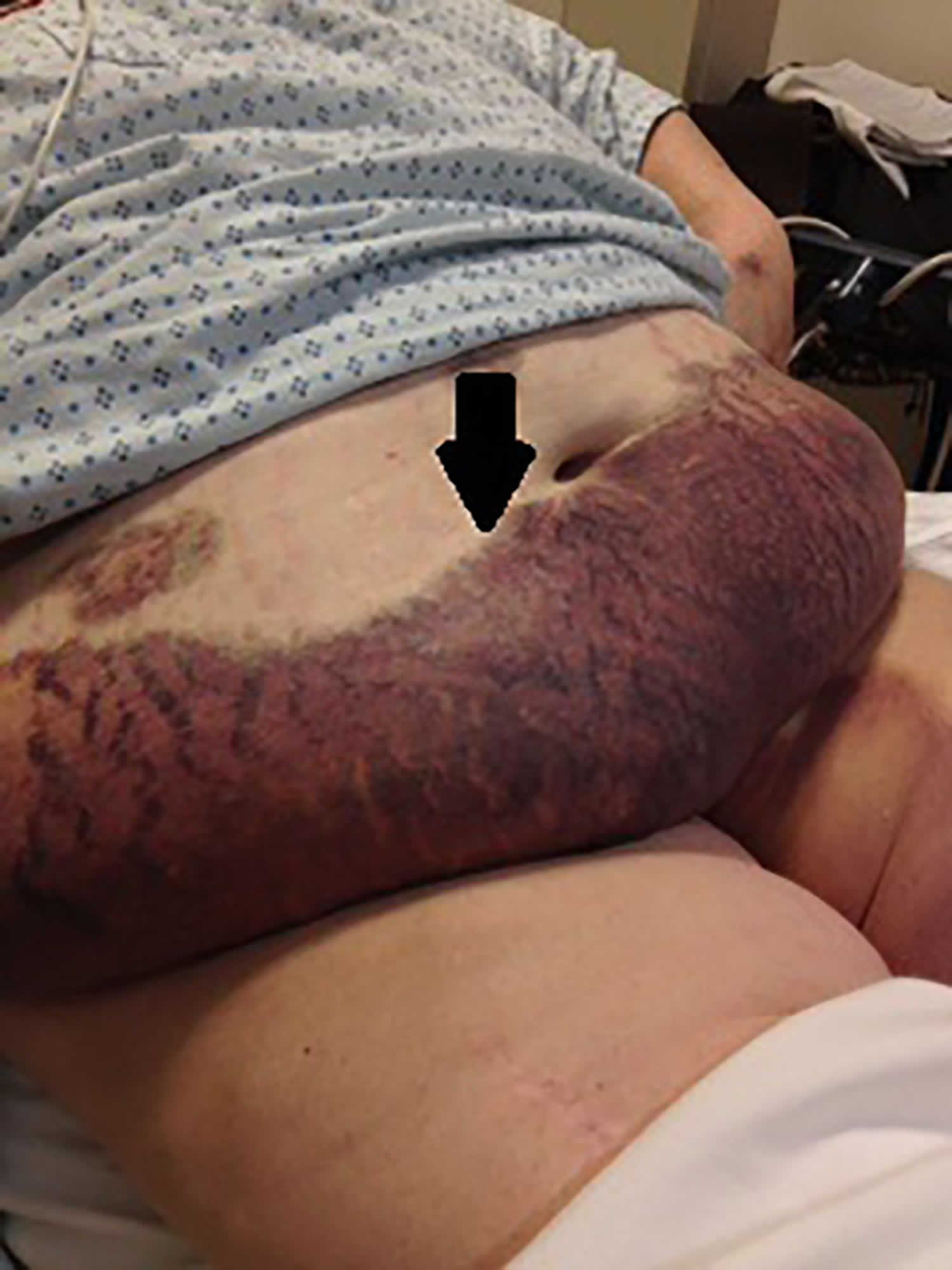
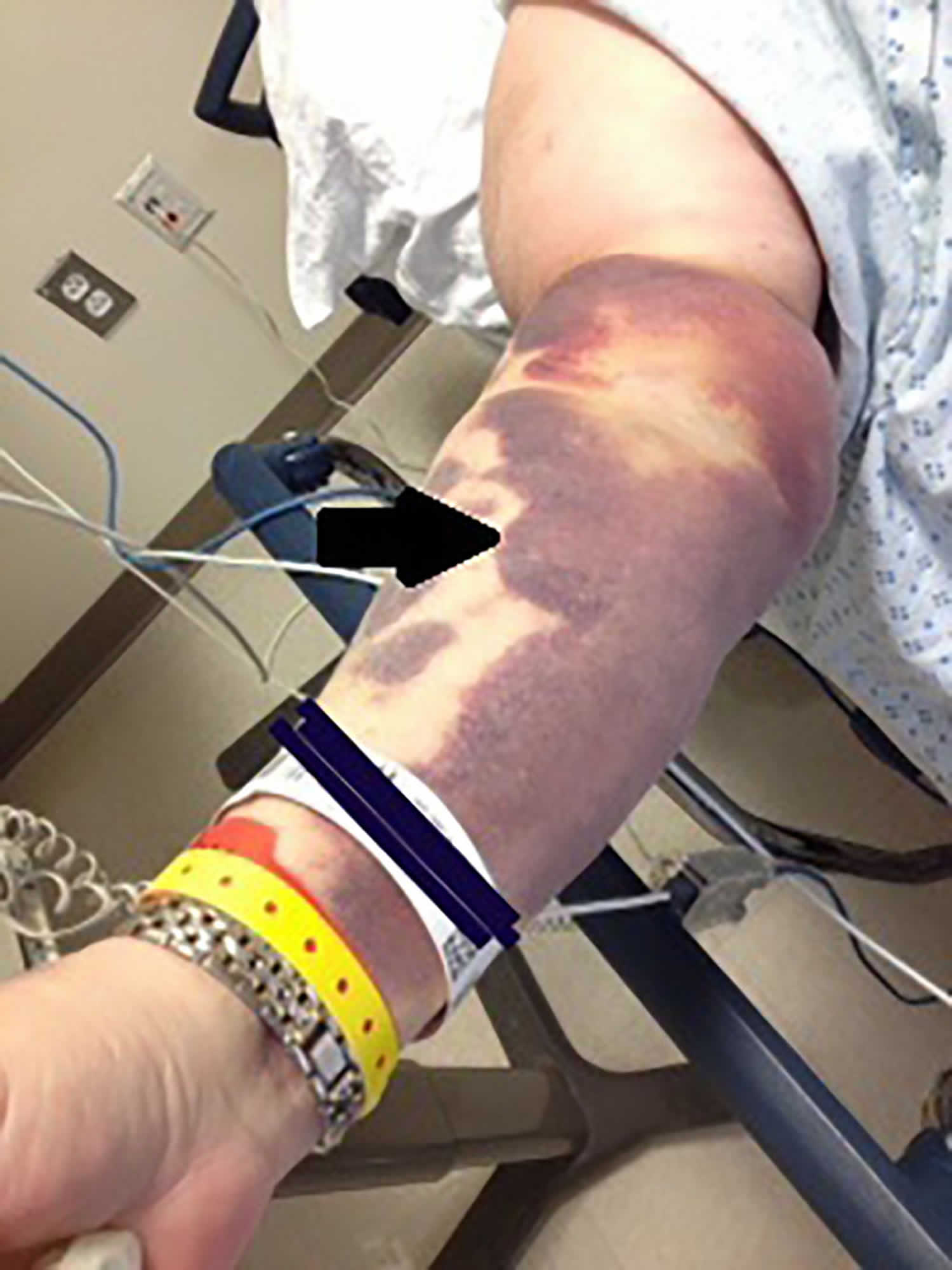
Figure 1. Warfarin induced skin necrosis
Warfarin induced skin necrosis causes
While the precise cause of warfarin-induced skin necrosis is heavily debated, it is agreed that the rapid decline of vitamin K-dependent coagulation factors with short half-lives, such as protein C or S, predisposes one to a temporary hypercoagulable state 11. Warfarin or Coumadin is a widely used anticoagulant or blood-thinner. Warfarin competitively inhibits the vitamin K epoxide reductase complex 1 (VKORC1), which is an essential enzyme for activating the vitamin K available in the body. Through this mechanism, warfarin can deplete functional vitamin K reserves and therefore reduce the synthesis of active clotting factors. The hepatic synthesis of coagulation factors II, VII, IX, and X, as well as coagulation regulatory factors protein C and protein S (natural anticoagulants), require the presence of vitamin K. Vitamin K is an essential cofactor for the synthesis of all of these vitamin K-dependent clotting factors II, VII, IX and X.
Half the activated Protein C disappears within 6 hours (its half-life). So, Protein C runs out during the first few days of warfarin therapy, before Factor X and II disappear, which have half-lives of 2-5 days. In some circumstances, this leads to excessive clotting.
Why the skin necrosis happens in the areas of fat abundance is unclear; possibly these areas are more susceptible because of reduced blood supply.
Warfarin-induced calciphylaxis may be due to inhibition of the matrix protein Gla, which normally prevents calcium deposition in the blood vessels.
Risk factors for warfarin-induced skin necrosis
Risk factors for warfarin-induced skin necrosis include:
- Inherited deficiency of Protein C, Protein S or Factor V Leiden
- Mutations in methylene tetrahydrofolate reductase gene causing hyperhomocysteinaemia
- Antithrombin III deficiency
- Antiphospholipid antibodies.
Warfarin induced skin necrosis signs and symptoms
The first sign of warfarin induced skin necrosis typically manifests as abrupt, painful purpura (a purplish bruise-like rash), which over a few days becomes bluish-black with a red rim. Blood blisters and full thickness skin necrosis (skin death) follows within 24 hours. There may be a red netlike rash around the necrotic area (retiform purpura). An eschar then forms, which eventually sloughs, revealing necrosis that may extend to the subcutaneous tissue. Affected areas are most often the breasts, thighs, buttocks, hips and abdomen, but early warfarin-induced skin necrosis can also cause blue toe syndrome or purple toe syndrome. Purple toe syndrome is a complication characterized by cholesterol microembolization that causes purple lesions to develop on the toes and sides of the feet. Purple toe syndrome usually develops 3 to 8 weeks after the initiation of warfarin therapy.
Warfarin induced skin necrosis diagnosis
The diagnosis of warfarin-induced skin necrosis is made clinically.
A skin biopsy can aid in diagnosis. Histopathology of warfarin necrosis usually reveals clotting within blood vessels in the skin without any inflammation. Warfarin can also precipitate calciphylaxis (calcium uremic arteriolopathy), recognized on biopsy by calcium deposition in the affected skin.
Blood tests for protein C and protein S levels are important to assess the likely predisposing causes.
Warfarin induced skin necrosis treatment
The mainstay of treatment of warfarin-induced skin necrosis is to stop warfarin. If anticoagulation is required, unfractionated heparin or low molecular weight heparin can be used. Sometimes vitamin K is used to hasten the reversal of warfarin effects. If there is life-threatening coagulation then protein C concentrates can be used. Prostacyclin is also effective 12.
Once warfarin is stopped, small areas of skin necrosis can be left to heal, but larger areas of skin necrosis may require surgery and skin grafting.
Warfarin has been cautiously restarted in lower doses in some patients when needed for long-term anticoagulation. This is best done with advice from a hematologist.
- What causes skin necrosis in warfarin and superwarfarin toxicity? https://www.medscape.com/answers/821038-119730/what-causes-skin-necrosis-in-warfarin-and-superwarfarin-toxicity[↩][↩]
- Ward CT, Chavalitanonda N. Atypical warfarin-induced skin necrosis. Pharmacotherapy. 2006;26(8):1175-9.[↩]
- Goldberg SL, Orthner CL, Yalisove BL, et al. Skin necrosis following prolonged administration of coumarin in a patient with inherited protein S deficiency. Am J Hematol. 1991;38(1):64-6.[↩]
- Scarff CE, Maker C, Hill P, et al. Late-onset warfarin necrosis. Australas J Dermatol. 2002;45(3):202-6.[↩]
- Sternberg ML, Pettyjohn FS. Warfarin sodium-induced skin necrosis. Ann Emerg Med. 1995;26(1):94-7.[↩]
- O’Dempsey RM, Choong AM, Patel A, et al. Warfarin-induced skin necrosis following recommencement of warfarin after perioperative Prothrombinex-VF. Med J Aust. 2015;202(9):499-500.[↩][↩][↩]
- Stewart AJ, Penman ID, Cook MK, et al. Warfarin-induced skin necrosis. Postgrad Med J. 1999;75(882):233-5.[↩][↩]
- Chan YC, Valenti D, Mansfield AO, Stansby G. Warfarin-induced skin necrosis. Br J Surg. 2000;87(3):266-72.[↩]
- Choudhary SJ, Madnani A, Singh AL. Late onset warfarin-induced skin necrosis in human immunodeficiency virus infected patient with pulmonary tuberculosis. Indian J Sex Transm Dis. 2013;34(1):47-9.[↩]
- Westphal SG, Plumb T. Calciphylaxis. [Updated 2020 Aug 11]. In: StatPearls [Internet]. Treasure Island (FL): StatPearls Publishing; 2020 Jan-. Available from: https://www.ncbi.nlm.nih.gov/books/NBK519020[↩]
- An Atypical Case of Warfarin-Induced Skin Necrosis. Case Report , Clinical Cases and Reports in EM , CPC-EM: Volume 1 Issue 4. https://westjem.com/case-report/an-atypical-case-of-warfarin-induced-skin-necrosis.html[↩]
- Nazarian RM, Van Cott EM, Zembowicz A, Duncan LM. Warfarin-induced skin necrosis. J Am Acad Dermatol. 2009 Aug;61(2):325-32. doi: 10.1016/j.jaad.2008.12.039[↩]


