Transillumination
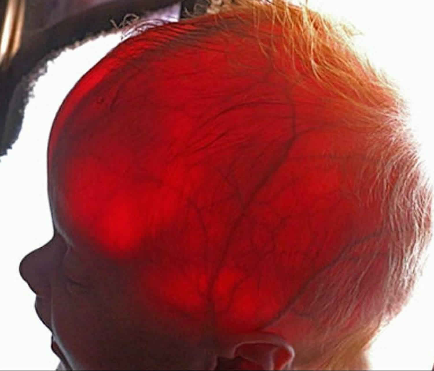
Transillumination is the shining of a light through a body area, tissues of the body or organ to check for abnormalities. A common example is the transmission of a flash of light through fingers, producing a red glow. This is due to the fact that red blood cells absorbed other colors of the beam and transmitted only the red component.
Transillumination is a major application of visible light in medicine. Transillumination is mainly used by pediatricians to shine light in bodies of infants and observe the amount of scattered light. Since their skeleton is not fully calcified, light can easily penetrate tissues. In newborns, a bright halogen light may be used to transilluminate the chest cavity if there are signs of a collapsed lung or air around the heart. Transillumination through the chest is only possible on small newborns.
Common examples are diagnosis of:
- Hydrocephalus (water head): Light penetrates to the inside of the skull of the infant. If there is an excess of cerebrospinal fluid (CSF), light is scattered to different parts of the skull, producing patterns characteristic to hydrocephalus. The device used in this operation is a Chun gun that uses a 150 watt projection bulb as a light source.
- Pneumothorax (collapsed lungs): Bright light penetrates the thin front chest wall and reflects off the back chest wall to indicate the degree of pneumothorax. To treat it, a physician insert a needle attached to a syringe into the area of collapse to remove the air between lungs and chest wall, causing the lung to reinflate.
- When done on the breast:
- Internal areas will be dark to black if there is a lesion and bleeding has occurred (because blood does not transilluminate).
- Benign tumors tend to appear red.
- Malignant tumors are brown to black.
Other studies include the sinuses, the gums, teeth (cracked tooth), the breasts (for breast lesions or cysts in women) and the testes (for fluid-filled sac in the scrotum [hydrocele] or a tumor in the testicle).
Transillumination is also sometimes used to find blood vessels.
In some locations in the stomach and intestine, the light can be seen through the skin and tissues at the time of upper endoscopy and colonoscopy.
In general, transillumination is not an accurate enough test to rely on. Further tests, such as an x-ray, CT, or ultrasound, are needed to confirm the diagnosis.
How the transillumination test is performed
The room lights are dimmed or turned off so that the area of the body may be seen more easily. A bright light is then pointed at that area. Areas where transillumination test is used include the:
- Head
- Scrotum
- Chest of a premature or newborn infant
- Breast of an adult female
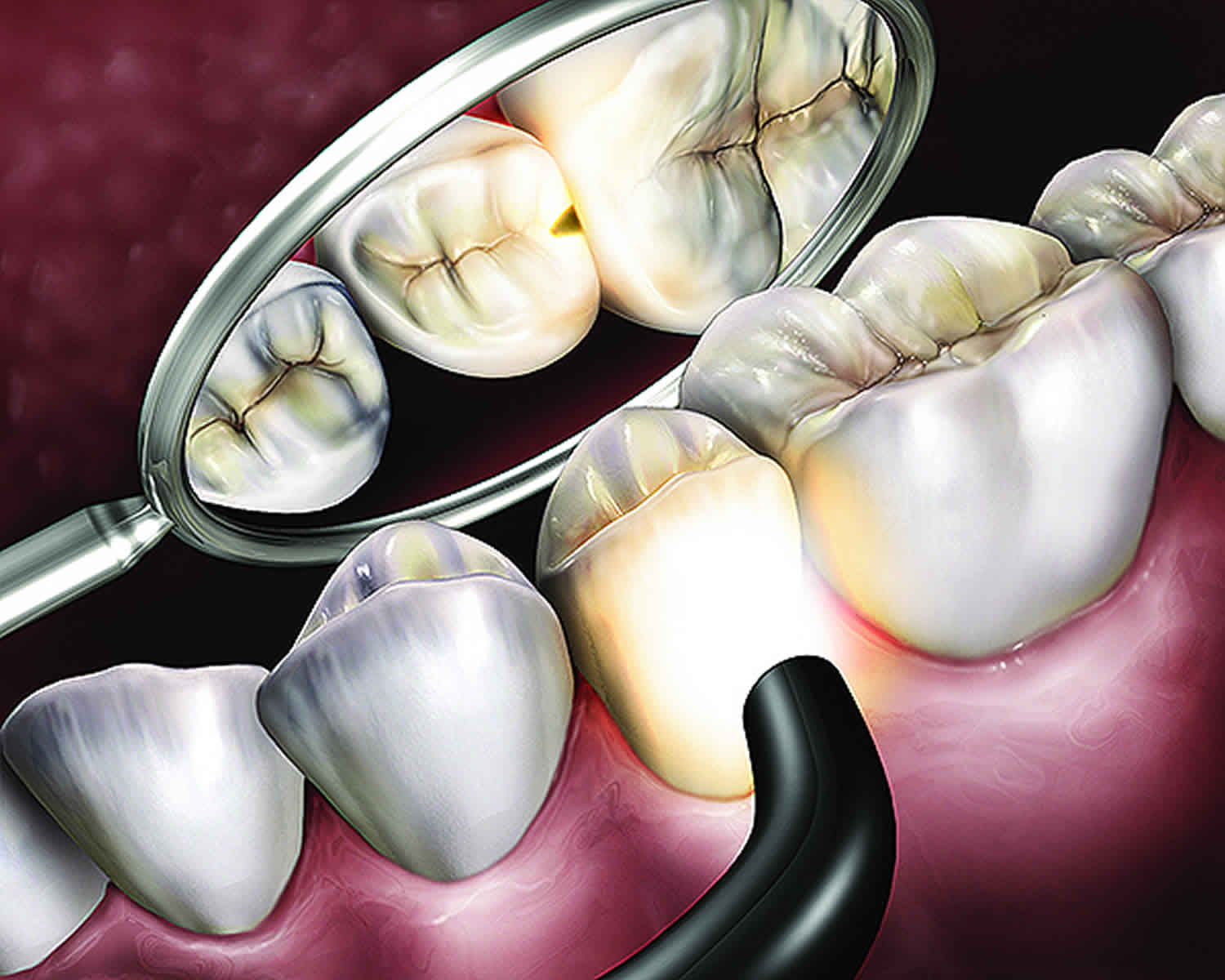
Transillumination teeth
The detection of a cracked tooth is often difficult. Normal diagnostic tests that are useful in other situations, such as sensitivity testing and radiographic evaluation, may not be as helpful for detecting a tooth with a crack. This is because the crack tooth with its potential to harbor bacteria and bacterial byproducts may have not completely extended to the pulp. The pulpal diagnosis for a tooth with a crack could be normal, reversible pulpitis, irreversible pulpitis or necrosis. This can be very confusing from the clinical and research perspectives.
Transillumination of teeth provides the most information, and easily and graphically represents whether a crack is present in a tooth. It is based on a law of physics, namely that a beam of light will continue to penetrate through a substance until is meets a space, after which the light beam is reflected. This results in a light and a dark area of the tooth separated by the fracture line. Sources of light other than from the transilluminator (i.e., overhead lighting and operatory lighting) should not be used. A dental mirror must be used to evaluate whether a fracture is present. A dental (surgical) operating microscope is useful, particularly by turning off the light source and using only magnification along with the dental mirror and transilluminator. Various transilluminators are commercially available, but dentist could use a fiberoptic handpiece with the bur removed (do not forget to turn the water off), composite curing lights (protect your eyes if using ultraviolet light) and other tools as transilluminators.
Figure 1. Transillumination dental
Transillumination of teeth to detect the presence of cracks should be performed in all of the following instances:
- On the marginal ridges,
- Floor of the cavity preparation
- The pulpal floor after endodontic access preparation
- Accessible proximal surfaces
- During surgical flap reflection procedures
Transillumination is particularly beneficial when performed after restorations are removed. Many fractures are not visualized without transillumination. The more transillumination is performed, the more fractures will be identified.


