close
Health topics

Necrotizing fasciitis

necrotizing fasciitis

What is necrotizing fasciitis

Necrotizing fasciitis represents a group of rare but highly lethal bacterial infections characterized by rapidly progressing inflammation and necrosis of skin and soft tissue 1. The bacteria most commonly get into the body through a break in the skin. Once in the body, the bacteria spread quickly and destroy the tissue they infect. The word “necrotizing” refers to something that causes body tissue to die. “Fasciitis” means inflammation of the fascia (the tissue under the skin that surrounds muscles, nerves, fat, and blood vessels). The spectrum of necrotizing fasciitis disease ranges from necrosis (death) of the skin to life-threatening infections involving the subcutaneous tissue, fascia, and muscle 2. The first clear description of necrotizing fasciitis was given by Joseph Jones 2, a surgeon in the Confederate Army of the United States in 1871. He described it as “hospital gangrene” in 2,642 soldiers with a mortality of 46 % during the Civil War. Necrotizing fasciitis itself was described in 1952 by Wilson when he observed edema and necrosis of subcutaneous fat and fascia with sparing of the underlying muscle in a series of 22 patients 3. Unfortunately, necrotizing fasciitis can result in a loss of limbs and even death. Accurate diagnosis, rapid antibiotic treatment, and prompt surgery are important to stopping this infection.

Necrotizing fasciitis has been classified according to the microbial infestation of the necrotic wound.

  • Type I necrotizing fasciitis is caused by polymicrobials (i.e. more than one bacteria involved) including the anaerobes. Type I is the most common (70-80%) necrotizing fasciitis 4. Bacteria causing type 1 necrotizing fasciitis include Staphylococcus aureus, Haemophilus, Vibrio and several other aerobic and anaerobic strains (Escherichia coli, Bacteroides fragillis). It is usually seen in the elderly or in patients affected with diabetes or other conditions.
  • Type II necrotizing fasciitis is caused by monomicrobials (only one bacteria involved) commonly by hemolytic group A streptococcus, staphylococci including methicillin resistant strains (MRSA) 4. Type II necrotizing fasciitis has been sensationalized in the media and is commonly referred to as flesh-eating disease. It affects all age groups. Healthy people are also prone to infection with this group.
  • Type III necrotizing fasciitis is caused by Clostridia perfringens or less commonly Clostridia septicum. It usually follows significant injury or surgery and results in gas under the skin: this makes a crackling sound called crepitus. IV drug users injecting “black tar” heroin subcutaneously can also be infected with clostridia and develop necrotizing fasciitis. Necrotizing fasciitis due to marine organisms is usually due to contamination of wounds by sea water, cuts by fish fins or stingers, or consumption of raw seafood. It occurs more commonly in patients with liver disorders. These infections can be very serious and can be fatal if not attended within 48 hours.
  • Type IV necrotizing fasciitis is a fungal infections caused by candida and zygomecetes, especially in cases of trauma and immunocompromised individuals.
  • Necrotizing fasciitis affecting perineal, genital and perianal regions is known as Fournier’s gangrene. This has a particularly high death rate ranging from 15% to 50% 5.

Necrotising fasciitis can start from a relatively minor injury, such as a small cut, but gets worse very quickly and can be life threatening if it’s not recognized and treated early on. See a doctor right away if you have a fever, dizziness, or nausea soon after an injury or surgery. Necrotizing fasciitis usually involves the muscular fascia and subcutaneous tissue rapidly and extensively but can also affect the skin and muscle 6. These infections can have a fulminant presentation, and their clinical course is unpredictable 3. They may manifest as a low-grade cellulitis that quickly deteriorates to a limb or life-threatening infection 7. Necrotizing fasciitis is usually rapidly fatal unless there is a prompt recognition and aggressive surgical treatment 8. Necrotizing fasciitis must be treated as an emergency with repeated surgical interventions and high doses of broad-spectrum antibiotics through intravenous route 9.

Your chances of getting necrotizing fasciitis are extremely low if you have a strong immune system and practice good hygiene and proper wound care.

Most people who get necrotizing fasciitis have other health problems that may lower their body’s ability to fight infection. Some of these conditions include:

  • Diabetes
  • Kidney disease
  • Cancer

Symptoms of necrotizing fasciitis can often be confusing and develop quickly. This fulminant tissue necrosis is usually accompanied by generalized toxicity, which may progress to shock and multiple organ failure. Without prompt recognition and immediate aggressive management, necrotizing fasciitis is often rapidly fatal. In the United States from 2003 to 2013, the overall mortality rate for necrotizing fasciitis-related death was 4.8 per 1,000,000 person-years 10.

Early symptoms of necrotizing fasciitis include:

  • A red or swollen area of skin that spreads quickly
  • Severe pain, including pain beyond the area of the skin that is red or swollen
  • Fever

See a doctor right away if you have these symptoms after an injury, even if the injury doesn’t break the skin. Even though minor illnesses can cause symptoms like these, people should not delay getting medical care.

Later symptoms of necrotizing fasciitis can include:

  • Ulcers, blisters, or black spots on the skin
  • Changes in the color of the skin
  • Pus or oozing from the infected area
  • Dizziness
  • Fatigue (tiredness)
  • Diarrhea or nausea

Doctors often need to perform surgery in order to:

  • Diagnose necrotizing fasciitis
  • See how deep the infection has spread
  • Quickly remove diseased tissue

Since necrotizing fasciitis can spread so rapidly, patients often must get surgery done very quickly. Doctors also give strong antibiotics through a needle into a vein (IV antibiotics) to try to stop the infection. Sometimes, however, antibiotics cannot reach all of the infected areas because the bacteria have killed too much tissue and reduced blood flow. When this happens, doctors have to surgically remove the dead tissue. Sometimes doctors have to remove the infected limb (e.g., leg, foot, arm) to stop the infection from spreading further. It is not unusual for someone with necrotizing fasciitis to end up needing multiple surgeries.

When to get medical help

Necrotizing fasciitis is a medical emergency that requires immediate treatment.

Go to your nearest accident and emergency department as soon as possible if you think you have it. Call your local emergency services number for an ambulance if you’re too unwell to get yourself to emergency department.

Blood tests and scans may be carried out to find out what’s causing your symptoms, although a diagnosis of necrotizing fasciitis can usually only be confirmed by having an operation to examine the affected tissue.

Necrotizing fasciitis bacteria

Many types of bacteria can cause necrotizing fasciitis, including 11:

  • Group A Streptococcus or group A strep (Streptococcus pyogenes)
  • Bacterioides
  • Klebsiella
  • Clostridium
  • Escherichia coli or E. coli
  • Staphylococcus aureus
  • Aeromonas hydrophila
  • Pseudomonas
  • Proteus

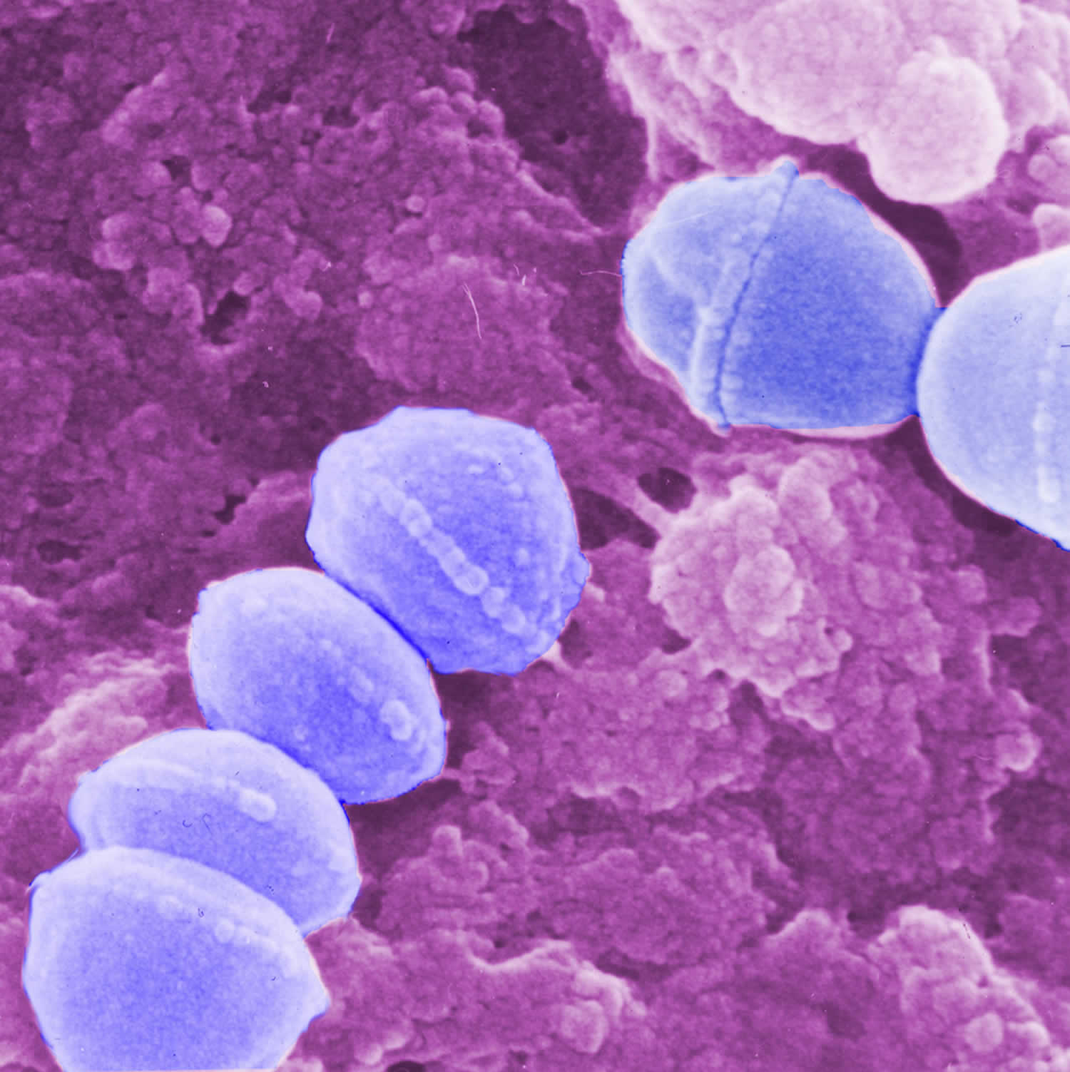
Media reports often call them “flesh eating bacteria” and necrotizing fasciitis is sometimes called the “flesh-eating disease”, although the bacteria that cause it don’t “eat” flesh – they release toxins that damage nearby tissue. Public health experts believe group A Streptococcus (group A strep) is the most common cause of necrotizing fasciitis 12. Group A strep (Streptococcus pyogenes) are the same bacteria that cause strep throat, scarlet fever, impetigo, toxic shock syndrome, cellulitis and necrotizing fasciitis. Group A strep bacteria live in the nose and throat. When someone who is infected coughs or sneezes, the bacteria travel in small droplets of water called respiratory droplets. One of the ways you can get sick is if you breathe in those droplets or if you touch something that has the droplets on it and then touch your mouth, nose, or eyes.

Figure 1. Group A Streptococcus (Streptococcus pyogenes) bacteria

Group A Streptococcus

Is necrotizing fasciitis contagious?

People rarely spread necrotizing fasciitis to other people. If you’re in close contact with someone who has necrotizing fasciitis, you may be given a course of antibiotics to reduce your risk of infection.

In general, someone with necrotizing fasciitis does not spread the infection to others. Most cases of necrotizing fasciitis occur randomly.

How do you get necrotizing fasciitis

Necrotizing fasciitis may occur in anyone, in fact, almost half of all known cases of streptococcal necrotizing fasciitis have occurred in young and previously healthy individuals.

The most common way of getting necrotizing fasciitis is when the bacteria enter the body through a break in the skin, including:

  • Cuts and scrapes
  • Burns
  • Insect bites
  • Puncture wounds (including those due to IV drug use)
  • Surgical wounds

Necrotizing fasciitis infection may occur if the right set of conditions is present, these include:

  • An opening in the skin that allows bacteria to enter the body. This may occur following minor injury (eg small cut, graze, pinprick, injection), or a large wound due to trauma or surgery (e.g., laparoscopy, sclerotherapy, endoscopic gastrostomy, thoracostomy, caesarean section, hysterectomy). Sometimes no point of entry can be found.
  • Cervicofacial necrotizing fasciitis can follow mandibular fracture or dental infection.
  • Direct contact with a person who is carrying the bacteria or the bacteria is already present elsewhere on the person.
  • Particularly invasive strains of bacteria, eg streptococci that evade the immune system and produce a toxin called cysteine protease SpeB, which dissolves tissue.
  • In children, type II necrotizing fasciitis may complicate chickenpox. Other causes of necrotizing fasciitis in chidren include omphalitis, necrotizing enterocolitis and urachal anomalies.

Risks for necrotizing fasciitis include:

  • Aspirin and non-steroidal anti-inflammatory drugs
  • Advanced age
  • Diabetes
  • Immune suppression
  • Obesity
  • Drug abuse
  • Severe chronic illness
  • Malignancy.

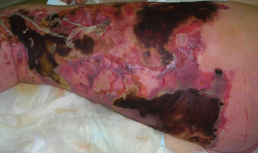
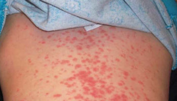
Figure 2. Necrotizing fasciitis

necrotizing fasciitis

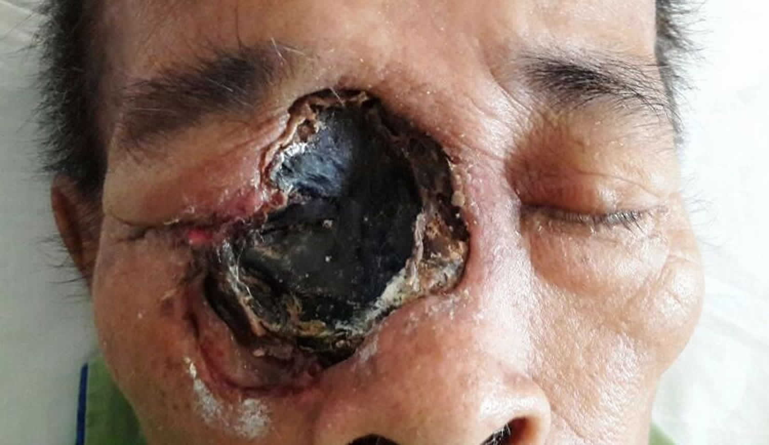
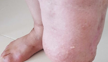
Figure 3. Necrotizing fasciitis upper limb

necrotizing fasciitis

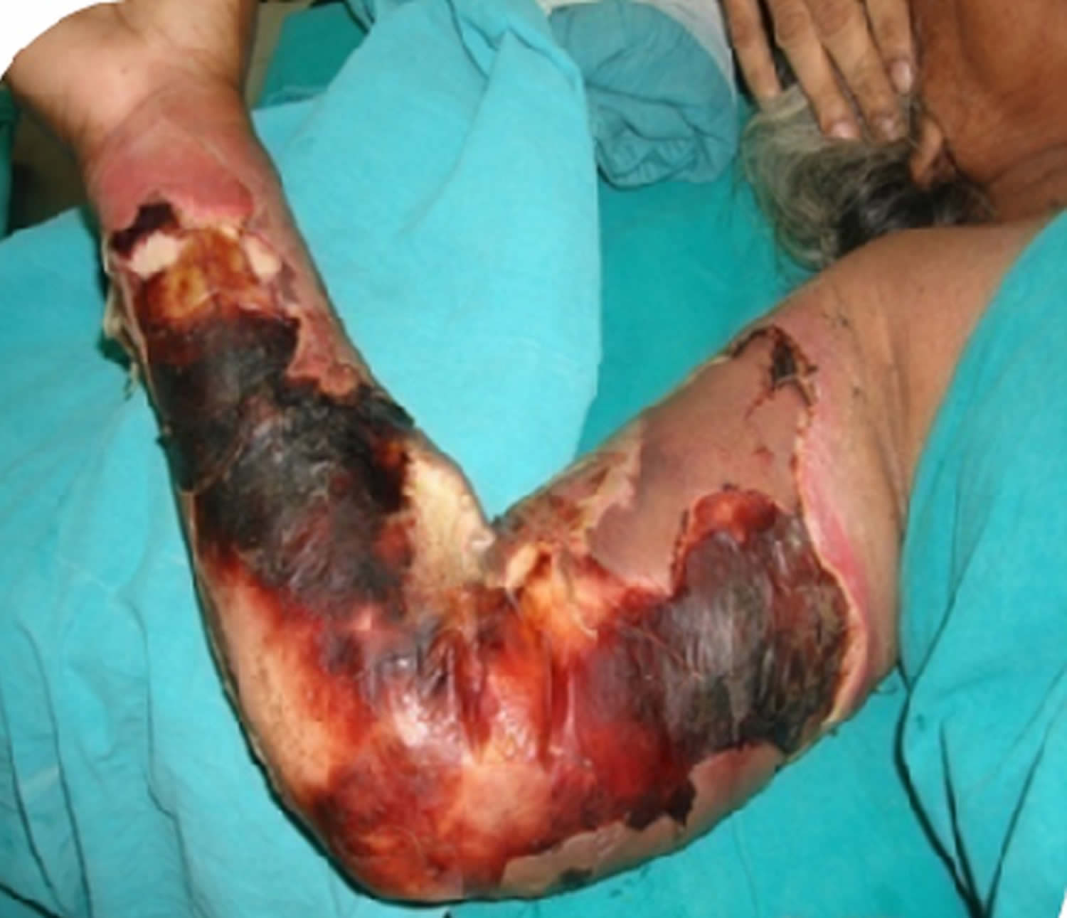
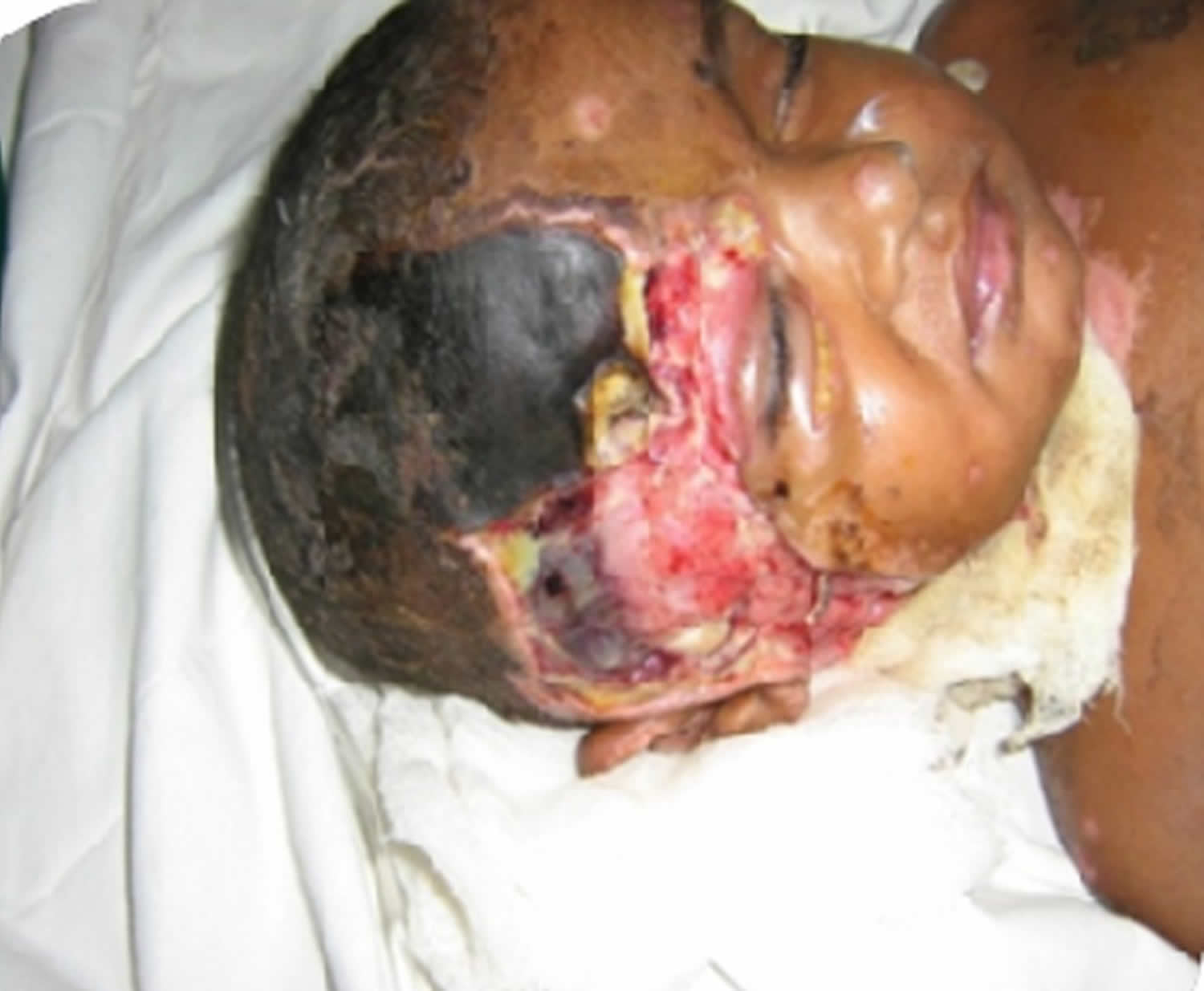
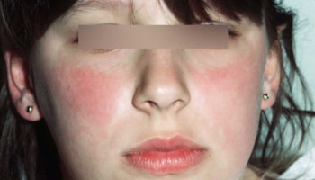
Figure 4. Necrotizing fasciitis face

Necrotizing fasciitis face

Necrotizing fasciitis prognosis

Necrotizing fasciitis can progress very quickly and lead to serious problems such as blood poisoning (sepsis) and organ failure.

Necrotizing fasciitis carries a mortality rate between 8.7-76% 13. Mortality is high when this infection is associated with delayed treatment and immune compromised host 14. Early diagnosis and prompt treatment offers the only chance of cure. Even with treatment, it’s estimated that 1 or 2 in every 5 cases are fatal, due to complications such as renal failure and septicemia (blood poisoning) and multiorgan failure 15.

People who survive the infection are sometimes left with long-term disability as a result of amputation or the removal of a lot of infected tissue.

They may need further surgery to improve the appearance of the affected area and may need ongoing rehabilitation support to help them adapt to their disability. Thus necrotizing fasciitis is a condition that incurs significant morbidity and both early and late mortality.

Necrotizing fasciitis symptoms

The symptoms of necrotizing fasciitis develop quickly over hours or days. They may not be obvious at first and can be similar to less serious conditions such as flu, gastroenteritis or cellulitis.

Early symptoms can include:

  • A small but painful cut or scratch on the skin. The most common site of infection is the lower leg. necrotizing fasciitis may also affect upper limb, perineum, buttocks, trunk, head and neck.
  • Intense pain that’s out of proportion to any damage to the skin. Pain is often very severe at presentation and worsens over time.
  • A high temperature (fever) and other flu-like symptoms, such as nausea, fever, diarrhea, dizziness and general malaise.
  • Intense thirst develops as body becomes dehydrated.
  • Symptoms appear usually within 24 hours of a minor injury.

After a few hours to 3 to 4 days, you may develop:

As necrotizing fasciitis takes hold:

  • The affected area starts to swell and may show a purplish rash and redness in the painful area – the swelling will usually feel firm to the touch
  • Diarrhea and vomiting
  • Large dark blotches on the skin that turn into fluid-filled blisters with dark fluid
  • Wound starts to die and area becomes blackened (necrosis)
  • Edema is common
  • A fine crackling sensation under the skin (crepitus) is due to gas in the tissues
  • Severe pain continues until necrosis/gangrene destroys peripheral nerves when the pain subsides
  • The infection may not improve when antibiotics are given.
  • If left untreated, the infection can spread through the body quickly and cause symptoms such as dizziness, weakness and confusion.

By about days 4–5, the patient is very ill with dangerously low blood pressure and high temperature. The infection has spread into the bloodstream and the body goes into toxic shock. The patient may have altered levels of consciousness or become totally unconscious.

Metastatic abscesses can develop in liver, lung, spleen, brain, pericardium, and rarely, in the skin.

What causes necrotizing fasciitis

Necrotizing fasciitis can be caused by several different types of bacteria.

The bacteria that cause the infection live in the gut, in the throat, or on the skin of some people, where they don’t usually cause any serious problems.

But in rare cases, they can cause necrotizing fasciitis if they get into deep tissue – either through the bloodstream, or an injury or wound, such as:

  • cuts and scratches
  • insect bites
  • puncture wounds caused by injecting drugs
  • surgical wounds

Necrotizing fasciitis typically occurs when bacteria already on the skin or in the body get into deep tissue. The infection can also be spread from person to person, but this is very rare.

Anyone can get necrotizing fasciitis – including young and otherwise healthy people – but it tends to affect older people and those in poor general health.

Pathophysiology of necrotizing fasciitis

Infection starts in the superficial fascia. Enzymes and proteins released by the responsible organisms cause necrosis of fascial layers. Horizontal spread of infection may not be clinically apparent on the skin surface and hence diagnosis may be delayed. Infection then spreads vertically up into the skin and down into deeper structures. Thrombosis occludes the arteries and veins leading to ischemia and necrosis of the tissues.

Streptococci produce:

  • M proteins, which initiate an inflammatory response with release of numerous cytokines (L-1, IL-6, TNFα)
  • Exotoxins, which destroy neutrophils allowing bacterial growth and destroying tissues.

Aerobic and anaerobic bacteria produce hydrogen, nitrogen, hydrogen sulfide, and methane gases that destroy hyaluronic acid enabling spread of infection.

Risk factors for developing necrotizing fasciitis

  • Chronic alcoholism
  • Diabetes
  • Vascular disease
  • Peripheral vascular deficiency
  • Acquired immune-deficiency syndrome
  • Age >50 years
  • Poor personal hygiene
  • Malnutrition 16
  • Kidney failure 16
  • Obesity 17

Non-steroidal anti-inflammatory drugs (NSAIDs) have been suggested as possible risk factors for necrotizing fasciitis 18.

Necrotizing fasciitis prevention

There’s no vaccine for necrotizing fasciitis and it’s not always possible to prevent it. If you’re in close contact with someone who has necrotizing fasciitis, you may be given a course of antibiotics to reduce your risk of infection.

Common sense and good wound care are the best ways to prevent bacterial skin infections.

  • Clean all minor cuts and injuries that break the skin (like blisters and scrapes) with soap and water.
  • Clean and cover draining or open wounds with clean, dry bandages until they heal.
  • Keep wounds clean and dry – after a wound has been cleaned, cover it with a sterile dressing (such as a plaster); change the dressing if it gets wet or dirty.
  • See a doctor for puncture and other deep or serious wounds.
  • Wash hands often with soap and water or use an alcohol-based hand rub if washing is not possible.
  • Care for fungal infections like athlete’s foot.

If you have an open wound or skin infection, avoid spending time in:

  • Hot tubs
  • Swimming pools
  • Natural bodies of water (e.g., lakes, rivers, oceans)

Necrotizing fasciitis treatment

Necrotizing fasciitis needs to be treated in hospital.

The main treatments are:

  • surgery to remove infected tissue – this may be repeated several times to ensure all the infected tissue is removed, and occasionally it may be necessary to amputate affected limbs
  • antibiotics – usually several different types are given directly into a vein
  • supportive treatment – including treatment to control your blood pressure, fluid levels and organ functions
  • hyperbaric oxygen and intravenous immunoglobulin may also be considered.

People with necrotizing fasciitis will often need to be looked after in an intensive care unit and may need to stay in hospital for several weeks.

While in hospital, they may be isolated from other patients to reduce the risk of spreading the infection.

  1. Singh G, Bharpoda P, Reddy R. Necrotizing Fasciitis: A Study of 48 Cases. The Indian Journal of Surgery. 2015;77(Suppl 2):345-350. doi:10.1007/s12262-013-0835-2. https://www.ncbi.nlm.nih.gov/pmc/articles/PMC4692953/[]
  2. Bosshardt TL, Henderson VJ, Organ CH. Necrotizing soft tissue infections. Arch Surg. 1996;131:846–54. doi: 10.1001/archsurg.1996.01430200056011[][]
  3. Wilson B. Necrotizing fasciitis. Am J Surg. 1952;18:416–31.[][]
  4. Diagnosis and management of necrotising fasciitis: a multiparametric approach. Morgan MS. J Hosp Infect. 2010 Aug; 75(4):249-57. https://www.ncbi.nlm.nih.gov/pubmed/20542593/[][]
  5. Kuzaka B, Wróblewska MM, Borkowski T, et al. Fournier’s Gangrene: Clinical Presentation of 13 Cases. Medical Science Monitor : International Medical Journal of Experimental and Clinical Research. 2018;24:548-555. doi:10.12659/MSM.905836. https://www.ncbi.nlm.nih.gov/pmc/articles/PMC5798415/[]
  6. Kotrappa KS, Bansal RS, Amin NM. Necrotizing fasciitis. Am Fam Physician. 1996;53(5):1691–7.[]
  7. Ardire L. Necrotizing fasciitis: case study of a nursing dilemma. Ostomy Wound Manage. 1997;43(5):30–4.[]
  8. Buchholz J, Jeske M, Kuhnen C, et al. (1997) Necrotizing fasciitis of extremities: clinical and histological examinations. Br J Surg 84–7[]
  9. Harmonson JK, Lobar MY, Harkless LB. Necrotizing fasciitis. Clin Podiatr Med Surg. 1996;13(4):635–46.[]
  10. Arif N, Yousfi S, Vinnard C. Deaths from Necrotizing Fasciitis in the United States, 2003–2013. Epidemiology and infection. 2016;144(6):1338-1344. doi:10.1017/S0950268815002745. https://www.ncbi.nlm.nih.gov/pmc/articles/PMC5725950/[]
  11. Acting Fast Is Key with Necrotizing Fasciitis. https://www.cdc.gov/Features/NecrotizingFasciitis/[]
  12. Lancerotto L, Tocco I, Salmaso R, Vindigni V, Bassetto F. Necrotizing fasciitis: classification, diagnosis, and management. Journal of Trauma and Acute Care Surgery 2012;72(3):560‐6.[]
  13. Current concepts in the management of necrotizing fasciitis. Misiakos EP, Bagias G, Patapis P, Sotiropoulos D, Kanavidis P, Machairas A. Front Surg. 2014; 1():36.[]
  14. Necrotizing fasciitis of the vulva. Stephenson H, Dotters DJ, Katz V, Droegemueller W. Am J Obstet Gynecol. 1992 May; 166(5):1324-7.[]
  15. Clark LA, Moon RE. Hyperbaric oxygen in the treatment of life‐threatening soft‐tissue infections. Respiratory Care Clinics of North America 1999;5(2):203‐19.[]
  16. Navinan MR, Yudhishdran J, Kandeepan T, Kulatunga A. Necrotizing fasciitis – a diagnostic dilemma: two case reports. Journal of Medical Case Reports. 2014;8:229. doi:10.1186/1752-1947-8-229. https://www.ncbi.nlm.nih.gov/pmc/articles/PMC4086700/[][]
  17. [Necrotizing fasciitis. Clinical criteria and risk factors]. Roujeau JC. Ann Dermatol Venereol. 2001 Mar; 128(3 Pt 2):376-81.[]
  18. Necrotizing fasciitis. Puvanendran R, Huey JC, Pasupathy S. Can Fam Physician. 2009 Oct; 55(10):981-7.[]
read more
Health topics

Fainting

fainting

What is syncope

Fainting also known as syncope or “passing out”, is a period of temporary loss of consciousness that happens when the blood flow to the brain is reduced and your brain temporarily doesn’t receive enough oxygen, causing you to lose consciousness. Fainting might have no medical significance. Fainting can occur when you’re sitting, standing, or when you get up too quickly. Or the cause can be a serious disorder, often involving the heart. Therefore, treat loss of consciousness as a medical emergency until the signs and symptoms are relieved, and the cause is known. Talk to your doctor if you faint more than once.

Typically, a fainting spell lasts only a few seconds or minutes, and then the person regains consciousness. Fainting is common and a single spell usually is not serious. Fainting (syncope) may be explained by factors such as stress, grief, overheating, dehydration, painful stimuli, exhaustion, anxiety or illness. Profound blood loss or fluid loss (severe diarrhea or vomiting) may also cause syncope.

Many causes of syncope, however, are not easy to explain and are not serious. However, some are life threatening, especially fainting during exertion. The most serious causes of syncope are related to heart damage or abnormal electrical system disorders that affect the heart’s ability to pump blood efficiently. In some cases, fainting is the only warning sign of an abnormal heart rhythm (arrhythmia) that could lead to sudden cardiac arrest and death.

There are a number of things that can cause you to faint, including:

  • changes to your blood pressure, especially when you stand up
  • dehydration
  • anemia
  • some medicines
  • diabetes
  • drop in blood sugar
  • a nervous system problem
  • a heart problem
  • a seizure.

In some people, fainting is caused by a temporary glitch in the autonomic nervous system that regulates your heart rate and blood pressure. This can be triggered by:

  • experiencing high levels of pain
  • exposure to sights you find unpleasant, such as the sight of blood
  • high levels of anxiety
  • standing up for long periods of time
  • coughing, sneezing or laughing
  • straining on the toilet
  • heat exposure.

People who faint might fall and injure themselves if they are not sitting or lying down at the time that they lose consciousness. Sometimes when people faint, their muscles twitch and their bodies make jerking movements; this can sometimes be confused with seizures but are not actual seizures.

Fainting itself is generally not serious, but harm from related falls or other accidents can cause injury. The main concern is head injury.

Although fainting itself might or might not be preventable, it is important to prevent injuries when people do faint.

If you feel faint:

  • Lie down or sit down. To reduce the chance of fainting again, don’t get up too quickly.
  • Place your head between your knees if you sit down.

If you are looking after someone who has fainted you should:

  • Place the person on their back. If there are no injuries and the person is breathing, raise the person’s legs above heart level — about 12 inches (30 centimeters) — so that blood flows back to the brain. Loosen belts, collars or other constrictive clothing. You can kneel down and rest their legs on your shoulders.
  • Make sure they have plenty of fresh air.
  • If they don’t recover quickly, make sure their airway is open and clear and check their breathing.
  • If they are unresponsive, call your local emergency services number and start cardiopulmonary resuscitation (CPR). Continue CPR until help arrives or the person begins to breathe.
  • If the person was injured in a fall associated with a faint, treat bumps, bruises or cuts appropriately. Control bleeding with direct pressure.

Most cases of fainting aren’t a cause for concern and don’t require treatment, but you should see your doctor if you’re at all concerned.

You should also see your doctor after fainting if you:

  • have no previous history of fainting
  • experience repeated episodes of fainting
  • injure yourself during a faint
  • have diabetes – a lifelong condition that causes your blood glucose level to become too high
  • are pregnant
  • have a history of heart disease – where your heart’s blood supply is blocked or interrupted
  • experienced chest pains, an irregular heartbeat or a pounding heartbeat before you lost consciousness
  • experienced a loss of bladder or bowel control
  • took longer than a few minutes to regain consciousness
Fainting or Stroke or Sudden Cardiac Arrest?

Sudden Cardiac Arrest (sudden cardiac death)

Sudden cardiac arrest occurs when the heart suddenly stops beating, which stops oxygen-rich blood from reaching the brain and other organs. Sudden cardiac arrest occurs suddenly and often without warning. For most people, the first sign of sudden cardiac arrest is fainting or a loss of consciousness, which happens when the heart stops beating. Breathing may also stop at this time. Some people may experience dizziness or lightheadedness just before they faint.

Sudden cardiac arrest is triggered by an electrical malfunction in the heart that causes an irregular heartbeat (arrhythmia). With its pumping action disrupted, the heart cannot pump blood to the brain, lungs and other organs. Seconds later, a person loses consciousness and has no pulse. Death occurs within minutes if the victim does not receive proper treatment.

Sudden cardiac arrest can occur after a heart attack, or during recovery. Sudden cardiac arrest is not a heart attack. Heart attacks increase the risk for sudden cardiac arrest, but the two terms do not mean the same thing.

Sudden cardiac arrest is a leading cause of death – over 320,000 out-of-hospital cardiac arrests occur annually in the United States.

What to do in sudden cardiac arrest

Cardiac arrest is reversible in most victims if it’s treated within a few minutes with a defibrillator (a device that sends an electrical shock to the heart to restore its normal rhythm).

  • First, call your local emergency services number for emergency medical services and ask for an ambulance.
  • Then get an automated external defibrillator if one is available and use it as soon as it arrives.
  • If there is no defibrillator available, begin cardiopulmonary resuscitation (CPR) immediately and continue until professional emergency medical services arrive. If two people are available to help, one should begin CPR immediately while the other calls your local emergency services number and finds an automated external defibrillator.

Stroke

Fainting can sometimes be mistaken for a serious medical condition, such as a stroke. A stroke is a medical emergency that occurs when blood supply to the brain is interrupted.

You should dial your local emergency services number immediately and ask for an ambulance if you think you or someone else is having a stroke.

The main symptoms of stroke can be remembered with the word FAST, which stands for Face-Arms-Speech-Time:

  • Face – the face may have fallen on one side, the person may not be able to smile, or their mouth or eye may have drooped
  • Arms – the person may not be able to raise both arms and keep them there because of weakness or numbness
  • Speech – the person may have slurred speech or garbled, or the person may not be able to talk at all despite appearing to be awake.
  • Time – it’s time to dial your local emergency services number immediately if you see any of these signs or symptoms

It’s important for everyone to be aware of these signs and symptoms, particularly if you live with or care for somebody in a high-risk group, such as someone who is elderly or has diabetes or high blood pressure.

Figure 1. Recovery position

seizure recovery position

How to perform a Cardiopulmonary Resuscitation (CPR)

If the person’s breathing or heart stops, cardiopulmonary resuscitation (CPR) should be performed immediately.

Hands-only CPR

  • Ensure the area is safe
  • Check for hazards, such as electrical equipment or traffic.

To carry out a chest compression:

  1. Place the heel of your hand on the breastbone at the center of the person’s chest. Place your other hand on top of your first hand and interlock your fingers.
  2. Position yourself with your shoulders above your hands.
  3. Using your body weight (not just your arms), press straight down by 5-6cm (2-2.5 inches) on their chest.
  4. Keeping your hands on their chest, release the compression and allow the chest to return to its original position.
  5. Repeat these compressions at a rate of 100 to 120 times per minute until an ambulance arrives or you become exhausted.

When you call for an ambulance, telephone systems now exist that can give basic life-saving instructions, including advice about CPR. These are now common and are easily accessible with mobile phones.

Cardiopulmonary Resuscitation (CPR) with rescue breaths

If you’ve been trained in CPR, including rescue breaths, and feel confident using your skills, you should give chest compressions with rescue breaths. If you’re not completely confident, attempt hands-only CPR instead (see above).

Adults

  1. Place the heel of your hand on the centre of the person’s chest, then place the other hand on top and press down by 5-6cm (2-2.5 inches) at a steady rate of 100 to 120 compressions per minute.
  2. After every 30 chest compressions, give two rescue breaths.
  3. Tilt the casualty’s head gently and lift the chin up with two fingers. Pinch the person’s nose. Seal your mouth over their mouth and blow steadily and firmly into their mouth for about one second. Check that their chest rises. Give two rescue breaths.
  4. Continue with cycles of 30 chest compressions and two rescue breaths until they begin to recover or emergency help arrives.

Children over one year old

  1. Open the child’s airway by placing one hand on the child’s forehead and gently tilting their head back and lifting the chin. Remove any visible obstructions from the mouth and nose.
  2. Pinch their nose. Seal your mouth over their mouth and blow steadily and firmly into their mouth, checking that their chest rises. Give five initial rescue breaths.
  3. Place the heel of one hand on the center of their chest and push down by 5cm (about two inches), which is approximately one-third of the chest diameter.
  4. The quality (depth) of chest compressions is very important. Use two hands if you can’t achieve a depth of 5cm using one hand.
  5. After every 30 chest compressions at a rate of 100 to 120 per minute, give two breaths.
  6. Continue with cycles of 30 chest compressions and two rescue breaths until they begin to recover or emergency help arrives.

Infants under one year old

  1. Open the infant’s airway by placing one hand on their forehead and gently tilting the head back and lifting the chin. Remove any visible obstructions from the mouth and nose.
  2. Place your mouth over the mouth and nose of the infant and blow steadily and firmly into their mouth, checking that their chest rises. Give five initial rescue breaths.
  3. Place two fingers in the middle of the chest and push down by 4cm (about 1.5 inches), which is approximately one-third of the chest diameter. The quality (depth) of chest compressions is very important. Use the heel of one hand if you can’t achieve a depth of 4cm using the tips of two fingers.
  4. After 30 chest compressions at a rate of 100 to 120 per minute, give two rescue breaths.
  5. Continue with cycles of 30 chest compressions and two rescue breaths until they begin to recover or emergency help arrives.

Types of syncope

  • Cardiovascular syncope – The most dangerous but rare type of fainting is caused by an abnormal heart rhythm (arrhythmia) or by structural damage to the heart.
  • Non-cardiovascular syncope – The most common type (also called vasovagal type). There are a variety of causes not necessarily related to the heart. This type of syncope is often related to problems with the autonomic nervous system, which controls functions such as heart rate and blood pressure. They may occur even in young, otherwise healthy people with normal heart function.

People with underlying heart disease are at higher risk for cardiovascular syncope, although this may be the initial symptom suggesting such a problem.

Risk Factors for Cardiovascular Syncope

The risk of cardiovascular syncope increases with age. Those at greatest risk are people who have any of the following conditions:

  • Coronary artery disease (clogged blood vessels to the heart)
  • Chest pain caused by angina (lower blood supply to the heart)
  • Prior heart attack (myocardial infarction)
  • Ventricular dysfunction (weakness in the heart’s pumping chambers)
  • Cardiomyopathy (structural problems with the heart’s muscles)
  • An abnormal electrocardiogram (a common test to check for abnormal heart rhythms)
  • Recurrent episodes of Fainting that come on suddenly and without warning
  • Fainting during exertional activity or exercise
  • Some congenital heart defects or syndromes (like William’s syndrome)
  • Channellopathies (defects of the electrical system of the heart) like Long QT syndrome (LQTS) and Brugada Syndrome

Signs of Cardiovascular Syncope

Cardiovascular syncope usually is sudden. There may be no warning signs that an individual is about to faint. Some people do feel the following:

  • Shortness of breath
  • Heart palpitations (feeling like your heart is racing, pounding, or fluttering)
  • Pain, pressure, tightness, or discomfort in the chest

Cardiovascular Syncope and Long QT Syndrome

Fainting is the primary symptom – and may be the only warning sign – of Long QT Syndrome (LQTS), an inherited electrical disorder of the heart. Long QT Syndrome is believed to be a common cause of sudden and unexplained death in children and young adults. It may occur in as many as 1 in 2,000 individuals and causes up to 4,000 deaths in children and young adults each year in the United States.

Fainting symptoms

Fainting (syncope) is a sudden temporary loss of consciousness that usually results in a fall. When you faint, you’ll feel weak and unsteady before passing out for a short period of time, usually only a few seconds.

There may not be any warning symptoms, but some people experience:

  • yawning
  • a sudden, clammy sweat
  • feeling sick (nausea)
  • changes to your breathing, such as breathing faster and deeply
  • sweating
  • confusion
  • dizziness
  • lightheadedness
  • blurred vision or spots or lights in front of your eyes
  • ringing in your ears

This will usually be followed by a loss of strength and consciousness.

When you collapse to the ground, your head and heart are on the same level. This means your heart doesn’t have to work as hard to push blood up to your brain.

You should return to consciousness after about 20 seconds. Recovery from a faint usually happens quite quickly as the blood flow back to the brain returns to normal.

Dial your local emergency services number and ask for an ambulance if someone faints and doesn’t regain consciousness within two minutes.

After fainting

After fainting, you may feel confused and weak for about 20-30 minutes. You may also feel tired and not be able to remember what you were doing just before you fainted.

Causes of fainting

Syncope can be due to a number of different causes. The most common cause is vasovagal syncope (also known as neurally mediated syncope, a temporary glitch in the autonomic nervous system), where in response to a certain stimulus there is a simultaneous drop in heart rate or or your heartbeat pause for a few seconds and dilatation (widening) of blood vessels. This causes pooling of blood in the legs, leading to a temporary interruption of blood flow to the brain.

There are a number of common triggers for vasovagal syncope, including prolonged standing (especially in a hot environment), sitting or standing up suddenly, severe pain, emotional distress, the sight or smell of something unpleasant, severe coughing, urinating or opening the bowels.

Other less common causes of syncope include:

  • cardiac arrhythmias (abnormal heart rhythms);
  • orthostatic hypotension (a drop in blood pressure when standing), which may in turn be due to a number of causes;
  • structural heart disease;
  • heart attack, cardiac tumor, or blood clot in the arteries supplying the lungs and
  • cerebrovascular diseases (conditions caused by problems with the blood vessels in the brain).

Conditions that are sometimes misdiagnosed as syncope include epilepsy, intoxication and certain psychiatric disorders.

Vasovagal syncope

One of the most common types of syncope is called vasovagal syncope or neurocardiogenic syncope. A variety of conditions can trigger vasovagal syncope, including physical or psychological stress, dehydration, bleeding, or pain. The heart rate slows dramatically and the blood vessels in the body expand, causing blood to pool in the legs, resulting in low blood pressure (hypotension). This causes a decrease in blood flow to the brain.

In some cases, vasovagal syncope is triggered by an emotional response to a stimulus, such as fear of injury, heat exposure, the sight of blood, or extreme pain. In other cases, it is caused by abnormal nervous system responses to activities such as urinating, having a bowel movement, coughing, or swallowing. In still other cases, no trigger can be identified.

In most cases of vasovagal syncope, you have some warning that you are near fainting. These signs include dizziness, feeling hot or cold, nausea, pale skin, “tunnel-like” vision, and profuse sweating. After the episode, symptoms may continue because of continued low blood pressure. Some people feel extremely tired.

Orthostatic hypotension

Fainting can also be caused by a fall in blood pressure when you stand up. This is called orthostatic hypotension, and tends to affect older people, particularly those aged over 65. It’s a common cause of falls in older people.

When you stand up after sitting or lying down, gravity pulls blood down into your legs, which reduces your blood pressure.

The nervous system usually counteracts this by making your heart beat faster and narrowing your blood vessels. This stabilises your blood pressure.

However, in cases of orthostatic hypotension, this doesn’t happen, leading to the brain’s blood supply being interrupted and causing you to faint.

Possible triggers of orthostatic hypotension include:

  • Dehydration or blood loss – if you’re dehydrated, the amount of fluid in your blood will be reduced and your blood pressure will decrease; this makes it harder for your nervous system to stabilize your blood pressure and increases your risk of fainting
  • Diabetes – uncontrolled diabetes makes you urinate frequently, which can lead to dehydration; excess blood sugar levels can also damage the nerves that help regulate blood pressure
  • Medication – certain medications can interfere with the normal mechanisms that maintain blood pressure. Examples include some antidepressants, certain blood pressure or heart medicines, or medicines containing opiates, such as morphine.
  • Neurological conditions – conditions that affect the nervous system, such as Parkinson’s disease, can trigger orthostatic hypotension in some people
  • Illnesses that affect the nervous system – A number of illnesses can affect the specialized branch of the nervous system that helps maintain blood pressure (the autonomic nervous system). Examples are Parkinson disease, diabetes mellitus, the Shy-Drager syndrome, and amyloidosis.
  • Alcohol – Drinking alcohol can cause blood vessels to expand, causing blood pressure to fall and syncope to occur.
  • Carotid sinus hypersensitivity – Carotid sinus hypersensitivity is a condition in which reflexes lead to a slow heart rate and/or enlargement of blood vessels. This may be triggered by pressure on the carotid arteries (the main artery in the neck), and can lead to low blood pressure and syncope.

Heart problems

Heart problems can also interrupt the brain’s blood supply and cause fainting. This type of fainting is called cardiac syncope.

The risk of developing cardiac syncope increases with age. You’re also at increased risk if you have:

  • narrowed or blocked blood vessels to the heart (coronary heart disease)
  • chest pain (angina)
  • had a heart attack in the past
  • weakened heart chambers (ventricular dysfunction)
  • structural problems with the muscles of the heart (cardiomyopathy)
  • an abnormal electrocardiogram – a test that checks for abnormal heart rhythms
  • repeated episodes of fainting that come on suddenly without warning

See your doctor as soon as possible if you think your fainting is related to a heart problem.

Heart rhythm problems

A number of disturbances in the rate and/or rhythm of the heart can cause syncope. These disturbances are called arrhythmias.

The heart includes an area of specialized cells in the upper right chamber of the heart (right atrium) called the sinus node (figure 1). These cells send a series of regular electrical impulses to the atria that regulate the heart’s rhythm and pace. These impulses travel in an organized way along conduction tissues within the heart muscle and then spread along smaller fibers that go to each muscle cell of the heart. The impulses cause the heart muscle cells to contract in an organized and regular way, generating an effective pumping of blood to all areas of the body.

Syncope can occur because of problems at several places in this system. The problems may be due to primary heart rhythm problems, underlying heart disease, use of a medication, or a transient abnormal communication between the heart and the nervous system The following are common rhythm problems that cause syncope.

Sinus bradycardia — Bradycardia means a slow heart rate. In sinus bradycardia, the heart rate is slower than normal. A dramatically slowed heart rate can decrease the blood supply to the brain by reducing the amount of blood that the heart can pump per minute.

Sometimes, sinus bradycardia occurs because of an abnormality in the sinus node itself. This is called sick sinus syndrome. In other cases, the slowed firing of the sinus node is due to medications. In still others, problems with the nerves that lead to the heart muscle and regulate the sinus node rate are to blame. For syncope to occur due to this problem, the heart usually stops for six or more seconds. This is known as asystole.

Heart block — Sometimes, part of the conduction system between the sinus node and the rest of the heart becomes disrupted due to heart disease. Most often, this occurs in the middle of the heart where a special set of fibers conduct the electrical impulse to the pumping chambers (the ventricles), preventing the normal flow of electrical impulses. If the electrical signal from the sinus node fails to get through the entire conduction pathway, the heartbeat can be interrupted. If the interruption is significant and the heart rate is too slow, it can impair blood flow to the brain.

Ventricular tachycardia — Tachycardia is a fast heart rate. The ventricles are the heart’s main pumping chambers. Ventricular tachycardia (VT) occurs when muscle in the ventricles send out their own rapid electrical impulses, taking over the rhythm normally controlled by the sinus node. The heartbeat that results from these abnormal impulses is also abnormal, and often fails to pump blood in an adequate way. The heart is racing but does not pump effectively, so blood flow to the brain may be decreased.

Most people with syncope due to ventricular tachycardia have underlying heart disease, most commonly coronary heart disease. Treatment of people with syncope caused by ventricular tachycardia usually includes an implantable cardioverter-defibrillator.

Supraventricular tachycardia (SVT) — Rapid heartbeats can originate above the ventricles (supraventricular tachyarrhythmias). This is not commonly associated with syncope, unless the heart rate is very rapid.

Blockage of blood flow from the heart

Any problem with the structure of the heart that interferes with the flow of blood can cause syncope. The two most common causes of outflow obstruction are hypertrophic cardiomyopathy and aortic stenosis.

Hypertrophic cardiomyopathy – Hypertrophic cardiomyopathy is an inherited condition in which the areas of the left ventricular muscle walls are thickened. In some cases, the condition can interfere with blood flow out from the left ventricle and can cause syncope. More information about hypertrophic cardiomyopathy, including treatment recommendations, is available separately. )

Mitral stenosis or aortic stenosis – In the normal circulation, blood flows through the mitral and aortic valves and into the body’s largest artery, the aorta, to supply blood to the body. Disease causing narrowing or obstruction of these valves will reduce the amount of blood being pumped and may predispose to syncope.

When severe, mitral or aortic stenosis can reduce blood flow to the brain and the rest of the body. Treatment of syncope caused by mitral or aortic stenosis often includes valve replacement surgery.

Reflex anoxic seizures

A reflex anoxic seizure is a type of fainting that mainly occurs in young children. Reflex anoxic seizures is caused by an involuntary slowing of the heart rate, to the extent that the heart actually stops beating for 5-30 seconds. After 5 seconds of the heart beat stopping, the child will start to lose consciousness, and after 10 seconds they’ll be deeply unconscious. Usually, by 30 seconds the vagus nerve starts to lose its signal strength and the heart starts beating again on its own. The heart will always automatically start to beat on its own as the strength of the vagus nerve signal fades. The heart is normal and the slowing is caused by an excessively strong reflex signal from the brain via the vagus nerve. The vagus nerve is one of 12 nerves in the head. It runs down the inside of the head, passes through the neck, and into the chest and abdomen.

Reflex anoxic seizures are involuntary, aren’t dangerous and don’t cause brain damage or death. They’re not a sign of a difficult child or poor parenting. They’re not epileptic seizures.

The seizures will improve with age, but may get more frequent before becoming less frequent or disappearing altogether.

Reflex anoxic seizures are often triggered by a sudden unexpected fright or pain, such as a fall with a minor head injury. The seizure isn’t caused by the injury itself, but by the sudden fright or pain.

The child will often open their mouth as if they’re going to cry, but make no sound before turning pale grey and losing consciousness.

They’ll either become limp – or, more often, stiff – with their eyes rolling upwards and their fingers clawed. Their body may also jerk a few times.

The seizure usually lasts less than a minute. Afterwards, the child will regain consciousness, but may appear sleepy and confused for a few hours.

Reflex anoxic seizures can be frightening to witness, but they aren’t dangerous and don’t harm the child.

In the pre-school years, some children may have several reflex anoxic seizures a month, but most will grow out of them by the time they start school. The seizures will become less frequent as the child gets older and usually disappear by the time they’re four or five years of age. Some children will continue to occasionally have them into adult life.

If the child is iron deficient, treatment with iron supplements can help. A simple blood test can be used to determine whether the child is anemic or iron deficient.

Syncope diagnosis

Your doctor will take a detailed history from you and perform a physical examination to try to determine the cause of the syncope.

An electrocardiogram (ECG), which records the electrical activity of your heart, will usually be performed.

If the cause of the fainting is still not apparent after the initial assessment, further tests may be performed including blood tests, an echocardiogram (ultrasound of the heart) and prolonged ECG monitoring. Sometimes additional specialized testing is required.

Electrocardiogram (ECG)

If your doctor thinks your fainting episode may have been caused by a heart problem, they may suggest that you have an electrocardiogram (ECG).

An ECG records your heart’s rhythm and electrical activity. A number of small, sticky patches called electrodes are placed on your arms, legs and chest. Wires connect the electrodes to an ECG machine.

Every time your heart beats, it produces tiny electrical signals. The ECG machine traces these signals on paper, recording any abnormalities in your heartbeat.

An ECG is usually carried out at a hospital or doctor surgery. The procedure is painless and takes about five minutes.

Heart rhythm monitoring

Heart rhythm monitoring may be recommended to diagnose rhythm problems that come and go and have not been detected with a routine ECG. This monitoring may be done at home or in the hospital.

Holter monitor – You may be asked to wear a monitoring device, called a Holter monitor, for 24 or 48 hours while performing normal daily activities at home. The device is connected to several long thin cables that are attached to your chest with sticky pads (similar to an ECG). The cables connect to a small, portable machine that can be attached to a belt or strap that is carried over the shoulder.

However, this type of monitoring has limited use and provides a diagnosis in only about 2 to 3 percent of people with syncope. If you do not experience a syncopal episode while wearing the Holter monitor, the test may need to be repeated, or an alternate form of long-term monitoring may be recommended.

Event recorder – An event recorder may be recommended to capture rhythm problems associated with a syncopal episode. The advantages of an event recorder compared with a Holter monitor are its small size and the ability to monitor for abnormal rhythms for longer periods of time (usually one to two months).

Some devices require you to activate the recorder when you feel symptoms of a syncopal episode. However, if you lose consciousness and another person is not available to assist with the recording, the opportunity to “capture” the event on the monitor may be lost.

Intermittent loop recorders – Intermittent loop recorders were developed to capture rhythm problems that occur before the device is activated. When you activate the monitoring device after regaining consciousness, the ECG recordings from the previous few minutes are retrieved and stored for analysis at a later time.

An implantable loop recorder provides a way to monitor rhythms over an extended period of time (eg, 18 to 24 months). The implantable loop recorder is implanted under the skin on the upper left chest area. It stores events automatically according to programmed criteria, or can be activated by the patient. The implantable loop recorder may be most useful if your symptoms are infrequent and an arrhythmia is suspected, but other forms of testing are negative or inconclusive.

Echocardiogram

An echocardiogram is useful for identifying underlying structural heart disease such as hypertrophic cardiomyopathy or significant aortic stenosis. These findings alone do not conclusively establish the specific cause for syncope.

An echocardiogram uses ultrasound (sound waves) to obtain detailed pictures of your heart as it beats. A technician presses a transducer (wand) against your chest and abdomen. The transducer is attached to a recording device and monitor. You are awake during the procedure. An echocardiogram does not use radiation.

Upright tilt table test

This test is often done in healthy patients who have syncope. You lie on a flat table and are tilted at various angles while your heart rate and blood pressure are monitored closely (figure 5). Your response to the change in position can sometimes give clues about the cause of syncope.

Electrophysiology study

An electrophysiology study may be performed if you have heart disease or if a rhythm problem is suspected.

Most people undergo electrophysiology study in a hospital setting. You will be given a sedative before the procedure but may be awake during testing. The physician uses a local anesthetic to numb a small area over a blood vessel, usually in the groin, and then threads small wires through the blood vessels into the heart using x-ray (fluoroscopic) guidance. Once in the heart, precise measurements of the heart’s electrical function can be obtained.

Exercise testing

In some people, especially those with a history of syncope during exertion, an exercise test is useful. Your blood pressure, heart rate, and rhythm are monitored while exercising on a treadmill or bicycle.

Carotid sinus test

If your doctor thinks your fainting episode was associated with carotid sinus syndrome, they may massage your carotid sinus to see whether it makes you feel faint or lightheaded.

The carotid sinus is a collection of sensors in the carotid artery, which is the main artery in your neck that supplies blood to your brain.

If the carotid sinus massage causes symptoms, it may indicate that you have carotid sinus syndrome.

Blood tests

Blood tests may be carried out to rule out conditions such as diabetes or anemia, a condition where the body doesn’t produce enough oxygen-rich red blood cells.

Your doctor may measure your blood pressure while you’re lying down and again after you stand up. You may have orthostatic hypotension if your blood pressure falls after you stand up.

If you have orthostatic hypotension, you may be asked further questions to help determine the cause. For example, it can sometimes occur as a side effect of taking some medications.

Electroencephalogram

An electroencephalogram (EEG) is used to diagnose seizures, but may be part of the evaluation of unexplained “collapse” events. It involves the measurement of electrical activity in the brain. It can be performed in a provider’s office or in a hospital, and generally takes approximately one hour. Multiple electrodes (small, flat metal discs) will be attached to your head and face with a sticky paste. The electrodes are connected to a recording device with long, thin wires. You must lie still and avoid speaking during the test.

An EEG is frequently obtained in people with syncope, but is rarely useful. It can be helpful if you have syncope and seizure-like activity.

Syncope treatment

Syncope is a symptom that can be caused by many different factors. When it comes to treatment, there is no “one size fits all” approach, making an accurate diagnosis very important. Treatment should address the underlying cause of syncope.

People with vasovagal syncope should watch for warning signs of fainting — such as light-headedness, sweating, nausea, ringing in the ears — and try to lie down (preferably with legs elevated), or if this is not possible, to sit down at the earliest warning signs.

Other measures that can help to temporarily increase blood pressure and avoid collapse include squeezing the calf muscles or buttocks together.

They should also avoid becoming dehydrated and generally try to avoid situations likely to trigger fainting. Occasionally, other treatments such as medication may be required for vasovagal syncope.

If the syncope is due to another condition — such as underlying heart disease — specific treatment for that condition will be arranged. Your doctor will advise you as to the appropriate treatment in your case.

Cardiovascular syncope treatment

Depending on the diagnosis, cardiovascular syncope may be stopped or controlled with one or more of the following therapies:

  • Simple reassurance, proper hydration, anticipatory guidance, safety precautions, and increased salt intake are helpful for common type fainting (vasovagal syncope) especially in children and young adults.
  • Insertion of a pacemaker is the standard treatment for syncope caused by a slow heartbeat (bradycardia). The pacemaker continuously monitors the heart’s natural rhythm. It delivers an electrical impulse to stimulate (pace) the heart’s muscle contractions if the heart rate drops below a certain number of beats per minute.
  • Treatment for a rapid heartbeat (tachycardia) depends on whether it occurs in the upper chambers (atria) or lower chambers (ventricles) of the heart. These treatments may include:
    • Medications to control irregular heart rhythms or underlying disease.
    • Catheter ablation, which is a procedure in which one or more flexible, thin wires (catheters) are inserted using x-ray or three dimensional computerized navigation system guidance into the blood vessels and directed to the heart muscle. Most commonly, a burst of energy heats and destroys very small areas of tissue that cause abnormal electrical signals.
    • Cardioversion is a controlled, electric shock that is delivered to restore the heart’s normal rhythm. It is most often used to treat abnormal rhythms that come from the heart’s upper chambers or atria.
    • An implantable cardioverter defibrillator (ICD) is a pacemaker-like device that continually monitors the heart, and delivers a life-saving shock if a dangerous heart rhythm is detected. The device significantly improves survival in certain groups of patients who are at high risk for a deadly heart rhythm disorder called ventricular fibrillation (VF). Ventricular fibrillation is the primary cause of sudden cardiac arrest, also known as sudden cardiac death.

Treating fainting associated with the nervous system

Most fainting episodes are associated with a temporary malfunction of the autonomic nervous system, which regulates the body’s automatic functions, such as heartbeat and blood pressure.

This type of fainting is called neurally mediated syncope. Treatment for neurally mediated syncope involves avoiding any possible triggers.

If you’re not sure what caused your fainting episode, your doctor may suggest keeping a diary of any symptoms you experience.

It may help to identify possible causes by making a note of what you were doing at the time you fainted.

There are also steps you can take to avoid losing consciousness if you think you may be about to faint.

Fainting associated with an external trigger

Fainting can occur when an external trigger, such as a stressful situation, causes a temporary malfunction in your autonomic nervous system. This is called vasovagal syncope.

In most cases of vasovagal syncope, further treatment isn’t required. However, you may find it useful to avoid potential triggers, such as stress or excitement, hot and stuffy environments, and long periods spent standing.

If you know injections or medical procedures like blood tests make you feel faint, you should tell the doctor or nurse beforehand. They’ll make sure you’re lying down during the procedure.

Fainting associated with bodily functions

Fainting can occur when a bodily function or activity – such as coughing – places a sudden strain on the autonomic nervous system. This is called situational syncope.

There’s no specific treatment for situational syncope, but avoiding the triggers may help. For example, if coughing caused you to faint, you may be able to suppress your urge to cough and avoid fainting.

Carotid sinus syndrome

Carotid sinus syndrome is where pressure on your carotid sinus causes you to faint. It’s more common in older men.

Your carotid sinus is a collection of sensors in the carotid artery, which is the main artery in your neck that supplies blood to your brain.

You can avoid fainting by not putting any pressure on your carotid sinus – for example, by not wearing shirts with tight collars.

In some people, carotid sinus syndrome can be treated by having a pacemaker fitted. A pacemaker is a small electrical device that’s implanted in your chest to help keep your heart beating regularly.

Treating fainting associated with low blood pressure

Fainting can occur when your blood pressure drops as you stand up. This drop in blood pressure is called orthostatic hypotension.

Avoiding anything that lowers your blood pressure should help prevent fainting. For example, avoid becoming dehydrated by increasing your fluid intake.

Your doctor may also advise you to eat small, frequent meals rather than large ones, and increase your salt intake.

Taking certain medications can also decrease blood pressure. However, don’t stop taking a prescribed medication unless your doctor or another qualified healthcare professional in charge of your care advises you to do so.

Physical counterpressure manoeuvers

Physical counterpressure manoeuvres are movements designed to raise your blood pressure and prevent you losing consciousness.

One study found training in physical counterpressure manoeuvres can reduce fainting in some people.

Physical counterpressure manoeuvres include:

  • crossing your legs
  • clenching the muscles in your lower body
  • squeezing your hands into a fist
  • tensing your arm muscles

You need to be trained to carry out these movements correctly. You can then do them if you experience any symptoms that suggest you’re about to faint, such as feeling lightheaded.

Safety at work

If you’ve fainted, it may affect your safety at work or the safety of others. For example, continuing to operate machinery may be dangerous if it’s likely you’ll faint again.

The healthcare professionals who diagnose and treat your condition can tell you whether it’s likely to affect your work. If it is, speak to your health and safety representative.

read more
Health topics

Fibromyalgia

fibromyalgia
Fibromyalgia Fibromyalgia also called fibromyalgia syndrome (a set of symptoms), fibro or FMS, is a chronic musculoskeletal pain syndrome characterized widespread body pain, stiffness, and tenderness
read more
1 374 375 376 377 378 544
Page 376 of 544
Health Jade