Auditory cortex
The auditory cortex is located on the lateral surface in the temporal lobe of the brain. The primary auditory cortex is about the same as Brodmann areas 41 and 42. It lies in the posterior half of the superior temporal gyrus and also dives into the lateral sulcus as the transverse temporal gyri also called Heschl’s gyri. The primary auditory cortex is the region of the brain that is responsible for processing of auditory (sound) information. Although the auditory cortex has a number of subdivisions, a broad distinction can be made between a primary auditory cortex, secondary auditory cortex (peripheral area) and tertiary auditory cortex (belt area) (Figure 1). These structures are formed concentrically around one another, with the primary auditory cortex in the middle and the tertiary auditory cortex on the outside. The primary auditory cortex (A1) is located on the superior temporal gyrus called Heschl’s gyrus, a ridge in the temporal lobe, on the lower lip of the deep cleft between the temporal and parietal lobes, known as the lateral sulcus (Sylvian fissure) 1. The primary auditory cortex (A1) receives point-to-point input from the ventral division of the medial geniculate complex; thus, it contains a precise tonotopic map 2. The belt areas of the auditory cortex receive more diffuse input from the belt areas of the medial geniculate complex and therefore are less precise in their tonotopic organization.
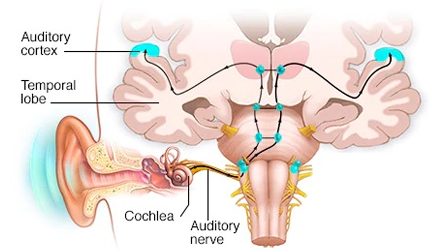
Information from the peripheral auditory system reaches central auditory nuclei via the auditory nerve. The auditory nerve transmits auditory information up a series of nuclei to cortex where perception occurs. These nuclei include: 1) cochlear nucleus, 2) superior olivary nuclei, 3) lateral lemniscus, 4) inferior colliculus, and 5) medial geniculate nuclei 3. Auditory information ascending through the auditory pathways start at the auditory nerve. These nerves synapse within the cochlear nucleus. A majority of auditory information is then transmitted through crossing fibers into the superior olivary complex. From there, the information ascends through the contralateral side of the brainstem and brain to cortex. It is of note that a significant number of neurons within the auditory system have crossing fibers at every level of the auditory system. This is likely due to the need for both ipsilateral and contralateral information for many aspects of auditory processing. Therefore, all levels of the central auditory system receive and process information from both the ipsilateral and contralateral sides.
The primary auditory cortex (A1) is tonotopically organized and has a topographical map of the cochlea (Figure 1), which means that certain cells in the auditory cortex are sensitive to specific frequencies, just as the primary visual cortex (V1) and the primary somatic sensory cortex (S1) have topographical maps of their respective sensory epithelia. Unlike the visual and somatic sensory systems, however, the cochlea has already decomposed the acoustical stimulus so that it is arrayed tonotopically along the length of the basilar membrane. Thus, primary auditory cortex (A1) is said to comprise a tonotopic map, as do most of the ascending auditory structures between the cochlea and the cortex. Orthogonal to the frequency axis of the tonotopic map is a striped arrangement of binaural properties. The neurons in one stripe are excited by both ears and are therefore called EE cells, while the neurons in the next stripe are excited by one ear and inhibited by the other ear (EI cells). The EE and EI stripes alternate. The sorts of sensory processing that occur in the other divisions of the auditory cortex are not well understood, but they are likely to be important to higher-order processing of natural sounds, including those used for communication. It appears that some areas are specialized for processing combinations of frequencies, while others are specialized for processing modulations of amplitude or frequency. This is a fascinating function which has been preserved throughout most of the audition circuit. This area of the brain “is thought to identify the fundamental elements of music, such as pitch and loudness. This makes sense as this is the area which receives direct input from the medial geniculate nucleus of the thalamus.
The secondary auditory cortex has been indicated in the processing of “harmonic, melodic and rhythmic patterns.” The tertiary auditory cortex supposedly integrates everything into the overall experience of music 4.
Sounds that are especially important for intraspecific communication often have a highly ordered temporal structure. In humans, the best example of such time-varying signals is speech, where different phonetic sequences are perceived as distinct syllables and words. Behavioral studies in cats and monkeys show that the auditory cortex is especially important for processing temporal sequences of sound. If the auditory cortex is ablated in these animals, they lose the ability to discriminate between two complex sounds that have the same frequency components but which differ in temporal sequence. Thus, without the auditory cortex, monkeys cannot discriminate one conspecific communication sound from another. Studies of human patients with bilateral damage to the auditory cortex also reveal severe problems in processing the temporal order of sounds. It seems likely, therefore, that specific regions of the human auditory cortex are specialized for processing elementary speech sounds, as well as other temporally complex acoustical signals, such as music. Wernicke’s area, which is critical to the comprehension of human language, lies within the secondary auditory cortex (Figure 2).
There are additional areas of the human cerebral cortex that are involved in processing sound, in the frontal and parietal lobes. Animal studies indicate that auditory fields of the cerebral cortex receive ascending input from the auditory thalamus, and that they are interconnected on the same and on the opposite cerebral hemispheres.The auditory cortex is composed of fields, which differ from each other in both structure and function 5.
Figure 1. Auditory cortex
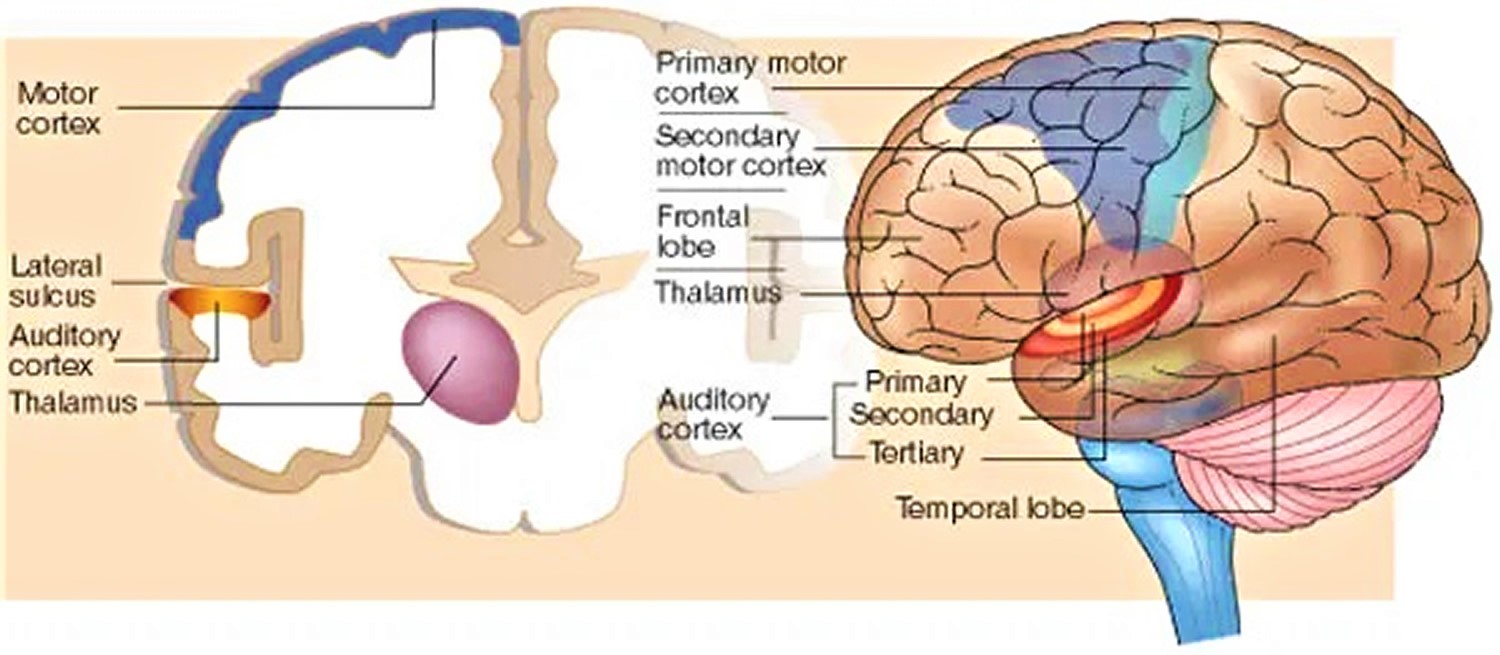
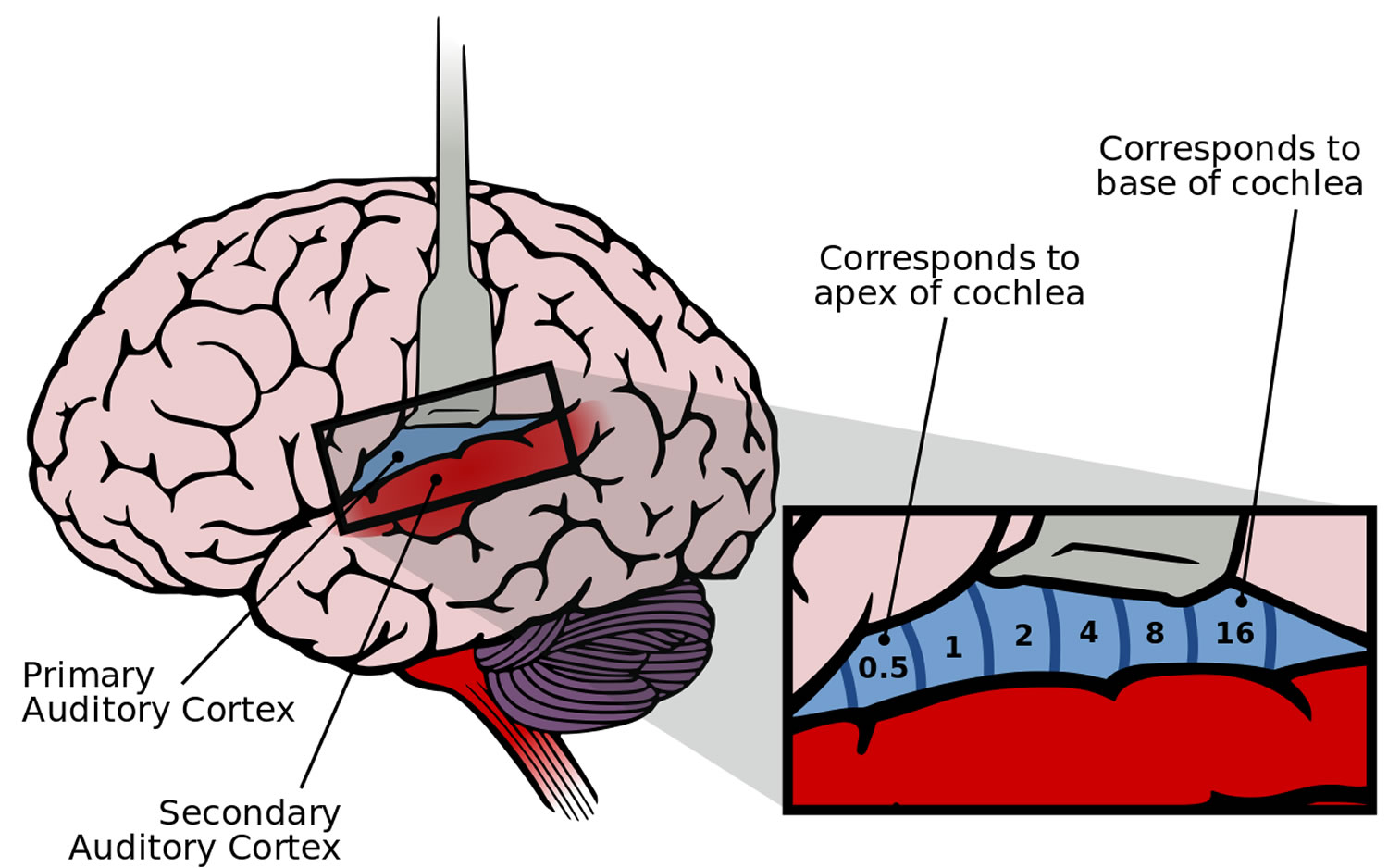
Footnote: The human auditory cortex. (A) Diagram showing the brain in left lateral view, including the depths of the lateral sulcus, where part of the auditory cortex occupying the superior temporal gyrus normally lies hidden. The primary auditory cortex (A1) is shown in blue; the surrounding belt areas of the auditory cortex are in red. (B) The primary auditory cortex has a tonotopic organization, as shown in this blowup diagram of a segment of A1.
[Source 2 ]Figure 2. Secondary auditory cortex
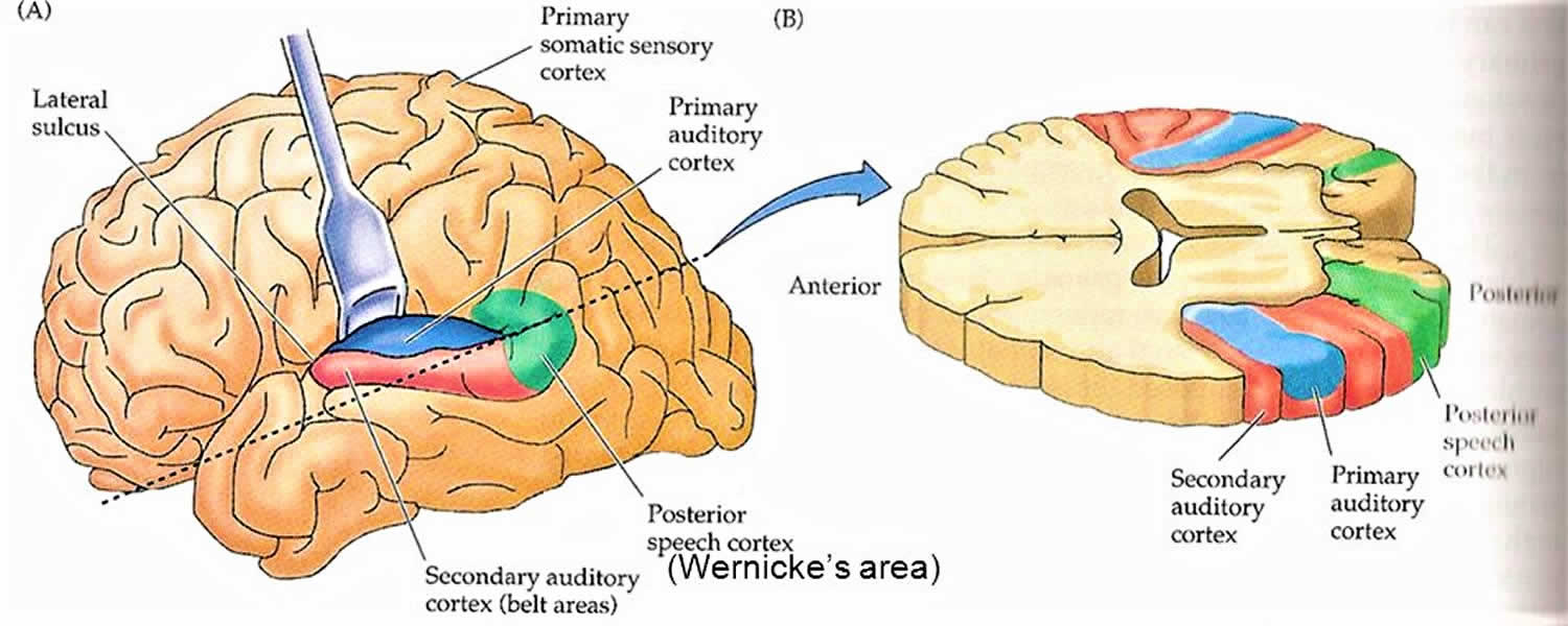
Footnote: The human auditory cortical areas related to processing speech sounds. (A) Diagram of the brain in left lateral view, showing locations in the intact hemisphere. (B) An oblique section (plane of dashed line in A) shows the cortical areas on the superior surface of the temporal lobe. Note that Wernicke’s area, a region important in comprehending speech, is just posterior to the primary auditory cortex.
Figure 3. Auditory circuits
Primary auditory cortex
The primary auditory cortex is the first region of the cerebral cortex to receive auditory input. In humans and other primates, the primary acoustic area in the cerebral cortex is the superior transverse temporal gyri also called Heschl’s gyri, a ridge in the temporal lobe, on the lower lip of the deep cleft between the temporal and parietal lobes, known as the lateral sulcus (Sylvian fissure), which contains the primary auditory cortex. Primary auditory cortex is responsible for translating and processing all sounds and tones, and it is minimally affected by task requirement. Task requirement: a test where the examiner pronounces some words and ask the participant to categorize them acoustically, or phonemically, or semantically 6. The superior temporal plane has another important area next to the Heschl gyrus called Wernicke’s area. In the past, this area was thought to have a significant role in speech perception and comprehension, but recent evidence shows that this area is not involved in this process. Researchers found that this process is not a simple task, but moreover, it is a complex task that is distributed all over the brain. The primary function of this area is the phonological representation, a process where the pronounced word is interpreted based on their tones and sound and trying to link it to a previously learned sound 7.
The lateral surface of the superior temporal gyrus is thought to be the secondary auditory cortex that also functions in interpreting sounds, but mostly in the activities that involve task requirement 6.
Because about half of the fibers of the auditory pathways cross the midline whereas others ascend on the same side of the brain, each ear is represented in both the right and left primary auditory cortex. For this reason, even when the auditory cortical area of one side is injured by trauma or stroke, binaural hearing may be minimally affected.
Perception of sound is associated with the right posterior superior temporal gyrus. The superior temporal gyrus contains several important structures of the brain, including Brodmann 41 and 42, marking the location of the primary auditory cortex, the cortical region responsible for the sensation of basic characteristics of sound such as pitch and rhythm.
The auditory association area is located within the temporal lobe of the brain, in an area called the Wernicke area or area 22. This area, near the lateral cerebral sulcus, is an important region for the processing of acoustic signals so that they can be distinguished as speech, music, or noise.
As is common for thalamocortical connections, nuclei within the medial geniculate body that send fibers to the auditory cortex also receive fibers from the same area of the cortex. Impaired hearing due to bilateral cortical injury involving both auditory areas has been reported, but it is extremely rare. However, bilateral lesions of the temporal lube have been shown to produce wide-ranging effects (cortical deafness, in which several behaviors are affected, including speech discrimination, localization of sound, and the detection of faint, short-duration signals).
Secondary auditory cortex
The lateral surface of the superior temporal gyrus is thought to be the secondary auditory cortex that also functions in interpreting sounds, but mostly in the activities that involve task requirement 6.
Auditory cortex function
Classically, two main functional regions have been described in auditory cortex:
- Primary auditory cortex (AI), composed of neurons involved in decoding the cochleotopic and tonotopic spatial representation of a stimulus.
- Secondary auditory cortex (AII), which doesn’t have clear tonotopic organisation but has an important role in sound localisation and analysis of complex sounds: in particular for specific animal vocalisations and human language. It also has a role in auditory memory.
- The belt region, surrounding AI and AII, which helps to integrate hearing with other sensory systems.
When awake, humans, like other animals, are able to perceive the small temporal variations of complex sounds. These variations are essential to the comprehension of human speech. A number of studies investigating AI have identified that in awake primates, two distinct populations of synchronous and asynchronous neurons (respectively) encode sequential stimuli differently.
Synchronous neurons analyse slow temporal changes. They respond precisely to low-rate stimulation (A1), but are unable to maintain their activity if the number of stimuli increases. The fast changes in rate are perceived by these neurons as a continuous tone. They are involved in both frequency and intensity analysis.
Asynchronous neurons analyse fast temporal changes (of many stimuli). They can determine short-duration variations and can accurately distinguish one stimulus from the next.
The functional division of the auditory cortex enables temporal variations of a stimulus to be decoded extremely accurately compared to other centers of the auditory pathway. It allows more information to be obtained about complex sounds, as well as the location of a sound source and its motion.
Synchronous and asynchronous neurons
- Synchronous neurons always respond to each stimulus (click) when the stimulus trains have intervals greater than 20 ms (A1). As the intertrain interval decreases (i.e. the repetition rate gets faster), these neurons start to desychronise their firing rate. When the interstimulus interval falls below 10 ms (B1), these neurons only fire at the beginning and the end of the stimulus (onset and offset responses, respectively).
- Asynchronous neurons do not respond synchronously to stimuli (A2 and B2), but their activity increases progressively to a very high discharge rate (B2).
Types of processing
Different aspects of environmental sounds (e.g., attenuation: how loud the sound is; location in space; frequency, and combination sensitivity) are processed in each of the central auditory areas. Most of the auditory nuclei throughout the brain are tonotopically arranged. In this way, auditory signals ascending to cortex can preserve the frequency information from the environment 3.
Attenuation (the intensity of a sound), is processed within the auditory system by neurons that fire action potentials at different rates based on the sound intensity. Most neurons respond by increasing their firing rate in response to increased attenuation. More specialized neurons respond maximally to environmental sounds within specific intensity ranges 3.
The brain processes the location of a sound in space by comparing differences in attenuation and timing of inputs from both ears within the superior olivary complex. If a sound is directly midline (i.e., front or back of the head), it would reach both ears at the same time. If it is to the right or left of midline, a temporal delay occurs between the inputs for the two ears. Within the superior olivary complex, specialized neurons receive input from both ears and can code for this temporal delay (i.e., binaural processing) 3.
Combination-sensitive neurons are another subset of neurons within the auditory system that have either enhanced or inhibited responses specifically to 2 or more sounds with a specific temporal delay. Combination-sensitive neurons are located within the inferior colliculus, lateral lemniscus, medial geniculate, and auditory cortex 8. Because most sounds in the environment are not pure tones, these types of combination-sensitive neurons are thought to facilitate enhancement of processing for combinations of sounds that may be important to the individual (e.g., speech, communication sounds) 9.
Descending circuits
It was once thought that auditory processing was a simple relay from the environmental signals up to cortex. Scientists now know that there is a significant descending system of circuits within the auditory system that helps to modulate auditory processing at every level. Auditory cortex has bilateral direct projections back to the inferior colliculus, superior olivary complex, and cochlear nucleus 10. These circuits contact neurons in these nuclei that project to every level of the central auditory system and to the cochlea (to modulate outer hair cells) within the peripheral auditory system. Connections between descending, ascending, and crossing fibers make the auditory system highly interconnected. These descending circuits help to modulate auditory attention based on the relevance, attention, learned behaviors, and emotional state of an individual. Such higher order functions originate from many regions of the brain (e.g., prefrontal cortex, hippocampus, nucleus basalis of Meynert, and limbic circuits) that have either direct and indirect connections with each other and auditory cortex 11.
Primary auditory cortex function
In AI, neurons are selective for particular frequencies and are arranged in isofrequency bands that are tonotopically organised. The precise spatial distribution of the isofrequency bands is related to the organisation of the auditory receptors. Their activity depends upon the characteristics of the stimulus: frequency, intensity and position of the sound source in space. Functionally, this region is strongly influenced by the waking state of the subject. A number of very specific neurons in AI are also involved in the analysis of complex sounds.
New techniques for studying the cerebral cortex (functional magnetic resonance imaging: fMRI; positron emission tomography: PET; and magnetoencephalography: MEG) suggest that the frequency distribution seen in animals (with traditional experimental methods) does not correspond exactly to that seen in humans, although they all have isofrequency bands, as seen using magnetoencephalography (MEG) below. fMRI in humans suggests that low frequencies are encoded in the superficial posterolateral regions of the sylvian fissure, whereas high frequencies are located in the deeper and anteromedial regions. It is important to note, however, that a degree of variation exists between individuals.
Secondary auditory cortex function
Secondary auditory cortex also functions in interpreting sounds, but mostly in the activities that involve task requirement 6.
- Jawabri KH, Sharma S. Physiology, Cerebral Cortex Functions. [Updated 2019 Jun 29]. In: StatPearls [Internet]. Treasure Island (FL): StatPearls Publishing; 2019 Jan-. Available from: https://www.ncbi.nlm.nih.gov/books/NBK538496[↩]
- Purves D, Augustine GJ, Fitzpatrick D, et al., editors. Neuroscience. 2nd edition. Sunderland (MA): Sinauer Associates; 2001. The Auditory Cortex. Available from: https://www.ncbi.nlm.nih.gov/books/NBK10900[↩][↩]
- Felix RA, Gourévitch B, Portfors CV. Subcortical pathways: Towards a better understanding of auditory disorders. Hear. Res. 2018 May;362:48-60.[↩][↩][↩][↩]
- Abbott, A. Music, maestro, please!. Nature 416, 12–14 (2002) doi:10.1038/416012a[↩]
- Cant, NB (Jun 15, 2003). “Parallel auditory pathways: projection patterns of the different neuronal populations in the dorsal and ventral cochlear nuclei”. Brain Res Bull. 60 (5–6): 457–74. doi:10.1016/S0361-9230(03)00050-9[↩]
- Nourski KV. Auditory processing in the human cortex: An intracranial electrophysiology perspective. Laryngoscope Investig Otolaryngol. 2017 Aug;2(4):147-156.[↩][↩][↩][↩]
- Binder JR. Current Controversies on Wernicke’s Area and its Role in Language. Curr Neurol Neurosci Rep. 2017 Aug;17(8):58.[↩]
- Yavuzoglu A, Schofield BR, Wenstrup JJ. Circuitry underlying spectrotemporal integration in the auditory midbrain. J. Neurosci. 2011 Oct 05;31(40):14424-35.[↩]
- Peterson DC, Wenstrup JJ. Selectivity and persistent firing responses to social vocalizations in the basolateral amygdala. Neuroscience. 2012 Aug 16;217:154-71.[↩]
- Coomes Peterson D, Schofield BR. Projections from auditory cortex contact ascending pathways that originate in the superior olive and inferior colliculus. Hear. Res. 2007 Oct;232(1-2):67-77.[↩]
- Forbes CE, Grafman J. The role of the human prefrontal cortex in social cognition and moral judgment. Annu. Rev. Neurosci. 2010;33:299-324.[↩]


