What are carotenoids
Carotenoids are dark-colored dyes (pigments) found in plant foods that can turn into a form of vitamin A. For example, the orange color of carrots and the red color of tomatoes are due to their carotenoid components 1. There are more than 500 known carotenoids. The carotenoids that have been most studied in this regard are beta-carotene, lycopene, lutein and zeaxanthin. In part, the beneficial effects of carotenoids are thought to be due to their role as antioxidants. By far the most important provitamin A carotenoid is beta-carotene; other provitamin A carotenoids are alpha-carotene and beta-cryptoxanthin. The body converts these plant pigments into vitamin A 2. Both provitamin A carotenoids and preformed vitamin A (retinol and its esterified form, retinyl ester) must be metabolized intracellularly to retinal and retinoic acid, the active forms of vitamin A, to support the vitamin’s important biological functions 3. Other carotenoids found in food, such as lycopene, lutein, and zeaxanthin, are not converted into vitamin A. Most of the body’s vitamin A is stored in the liver in the form of retinyl esters.
For dietary provitamin A carotenoids—β-carotene, α-carotene, and β-cryptoxanthin—RAEs (retinol activity equivalents) have been set at 12, 24, and 24 μg, respectively 4.
Carotenoids can be divided into two groups according to their polarity: xanthophylls (polar carotenoids such as astaxanthin, β-cryptoxanthin, lutein, and zeaxanthin) and carotene (nonpolar carotenoids such as α-carotene, β-carotene, and lycopene) 5. The distinctive structural feature of carotenoids is the long, alternating double and single bond system, which is associated with light absorption and oxidation 6.
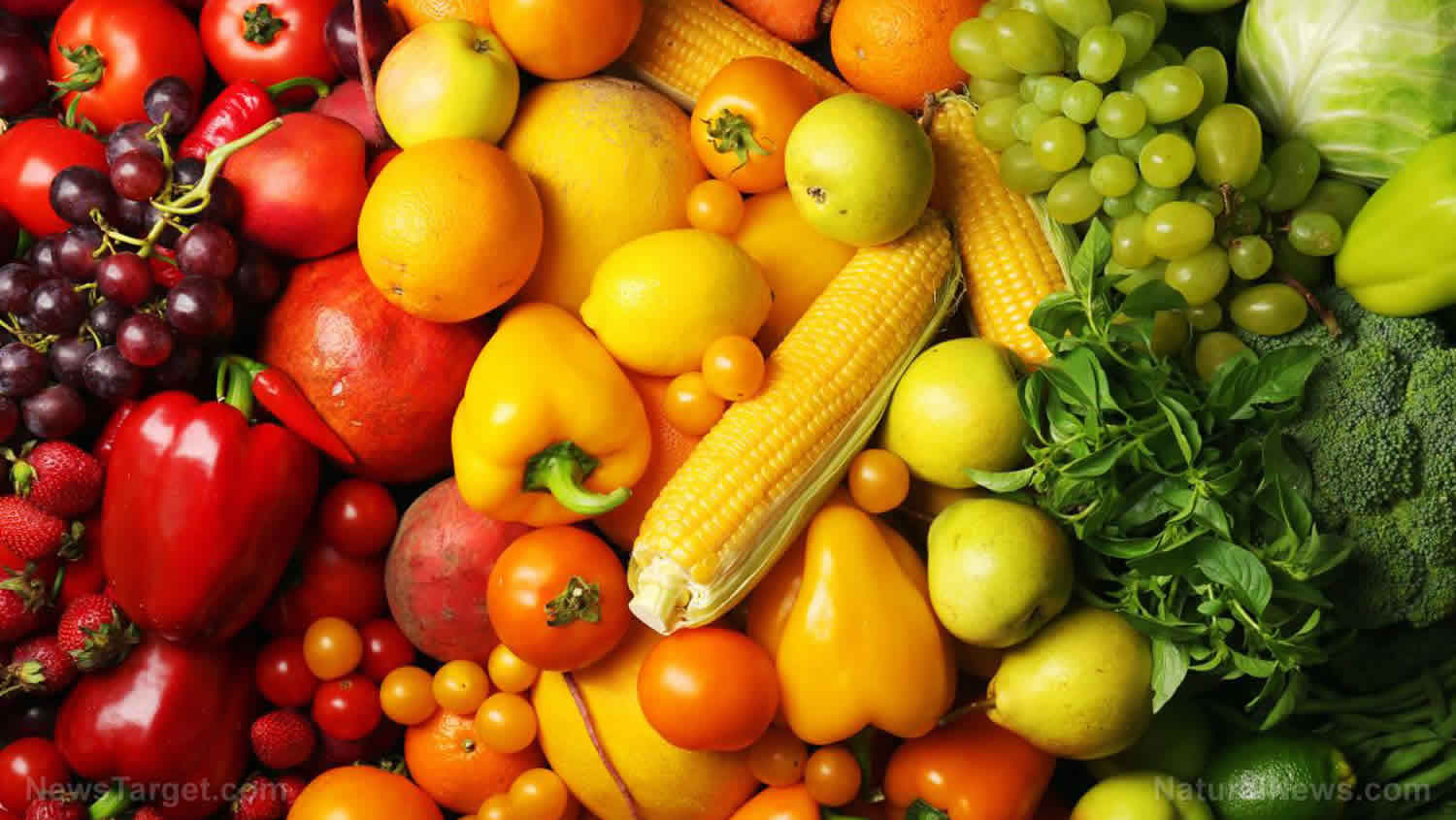
The major sources of carotenoids in the human diet are fruits and vegetables, which have various colors, such as green, red, orange, and yellow 7. Humans consume approximately 40 carotenoids from common fruits and vegetables 8. Dark green vegetables, such as broccoli, coriander, kale, and spinach, contain a large number of chloroplasts, in which most carotenoids exist; therefore, they possess high concentrations of carotenoids 9. As chloroplasts generally contain the most consistent carotenoid composition 10, the distribution of carotenoids is similar among different plant species in this group 8. On the other hand, in red-, orange-, or yellow-colored fruits and vegetables, carotenoids are mainly accumulated in chromoplasts, which are usually converted from chloroplasts during ripening 11. As chromoplasts in different plant species contain various carotenoids, the carotenoid distribution in this group is diverse 7. Some seafood and animal foods also contain carotenoids. Animals cannot synthesize carotenoids; instead, they ingest carotenoids through foods and accumulate these molecules in their bodies. As a result, some animal foods contain carotenoids. For example, high concentrations of lutein and zeaxanthin accumulate in egg yolks 12. Milk and dairy products, salmonid fish, and crustaceans also provide various carotenoids 13. The main carotenoid in bovine milk is β-carotene 14, whereas the major carotenoids in salmonid fish and crustaceans are astaxanthin and canthaxanthin 13. In addition, some edible brown seaweeds contain fucoxanthin as a major carotenoid 15.
Carotenoids are differentially distributed in various organs of the human body. Interestingly, xanthophylls account for 66–77% of the total carotenoids in the frontal and occipital lobes of the human brain 16, whereas less than 40% of the total carotenoids in most tissues and plasma are reported to be xanthophylls 17. It was reported that the human brain contains sixteen carotenoids, with the major carotenoids being anhydrolutein, α-carotene, α-cryptoxanthin, cis- and trans-β-carotene, β-cryptoxanthin, lutein, cis- and trans-lycopene, and zeaxanthin 16. Given carotenoids property of protecting tissues from oxidative stress and their localization in the brain, the role of carotenoids in preventing or treating oxidative stress-associated diseases, including neurodegenerative diseases, is of interest.
As carotenoids have various physiological activities, such as antioxidant activity, the amount of carotenoid in the human body is important for health. Therefore, the intake of carotenoids through the diet is associated with the prevention and treatment of various diseases, including age-related macular degeneration 18, cancer 19, cardiovascular diseases 20, and neurodegenerative diseases 5.
Types of carotenoids
The two main groups of carotenoids are carotenes and xanthophylls. Some familiar carotenes are β-carotene and lycopene—both these carotenoids are strict hydrocarbon carotenoids, and do not possess any substituent (or even oxygen) in their structures. Xanthophylls or oxycarotenoids, which belong to the second group, are oxygen-containing molecules. Lutein and zeaxanthin are two xanthophylls with –OH groups in their structures, whereas canthaxanthin and echinenone contain =O groups. Astaxanthin has both –OH and =O groups in its structure. Furthermore, some carotenoids such as violaxanthin and diadinoxanthin contain epoxy groups, and others such as dinoxanthin and fucoxanthin have acetyl groups in their structures. The two carotenoids with acetyl groups also contain the C=C=C (allene) group in their structures, which is unique to natural products 21. In addition, some carotenoids such as allo-, diato-, diadino-, hetero-, croco-, pyro-, and monadoxanthin contain C≡C (acetylene) groups in their structures.
Carotenoids foods
α-Carotene
- Banana, butternut, carrot, pumpkin
β-Carotene
- Apricots, banana, broccoli, cantaloupe, carrot, dairy products, honeydew, kale, mango, nectarine, peach, pumpkin, spinach, sweet potato, tomato
Crocetin
- Gardenia fruit, saffron stigma
Crocin
- Gardenia fruit, saffron stigma
β-Cryptoxanthin
- Apple, broccoli, celery, chili, crustaceans, grape, green beans, papaya, pea, peach, peppers, salmonid fish, squashes, tangerine
Lutein
- Apple, basil, broccoli, celery, crustaceans, cucumber, dairy products, grapes, green pepper, kale, kiwi, maize, parsley, pea, pumpkin, salmonid fish, spinach, squash
Lycopene
- Grapefruit, guava, tomato, watermelon
Zeaxanthin
- Basil, crustaceans, cucumber, dairy products, honeydew, kale, maize, mango, orange, parsley, salmonid fish, spinach
Astaxanthin
- Crustaceans, algae, salmonid fish
Fucoxanthin
- Brown seaweeds
Carotenoids function
Significant evidence has shown that carotenoids can reduce oxidative damage by scavenging reactive oxygen species (ROS) and exert anti-inflammatory effects in vivo 22.
One such carotenoid is beta-carotene.
- Beta-carotene is an antioxidant. Antioxidants protect cells from damage caused by substances called free radicals. Free radicals are believed to contribute to certain chronic diseases and play a role in the aging processes.
- Beta-carotene may have added benefits due its ability to be converted to vitamin A.
- Food sources of carotenoids such as beta-carotene may reduce the risk for cancer.
- But beta-carotene supplements do not seem to reduce cancer risk.
Additionally, lutein and zeaxanthin may be protective in eye disease because they absorb damaging blue light that enters the eye and they decreased the risk of functional vision loss among patients with category 3 or 4 age-related macular degeneration (AMD) 23.
Antioxidant activity
Carotenoids have been demonstrated to be one of the most potent natural singlet oxygen scavengers, with a fast quenching rate 24. They can effectively neutralize reactive oxygen species (ROS) and other free radicals to provide protection against oxidation in both photosynthetic and nonphotosynthetic organisms 7. However, each carotenoid shows different antioxidant activities, owing to the presence of functional groups with increasing polarities as well as the number of conjugated double bonds 25. The antioxidant property of carotenoids has inspired many epidemiological and clinical studies that have investigated if these pigment molecules are able to prevent various ROS-mediated disorders such as cancer, inflammation, retinal degeneration, and neurodegeneration. In the case of cancer, many studies have shown that carotenoid consumption is correlated with a reduced risk of several types of cancer; however, other studies have shown that the cancer-preventive effects of carotenoids are negligible or even that they are carcinogenic 26.
Lutein is a xanthophyll and the most abundant carotenoid in the human retina and brain 27. The Age-Related Eye Disease Study (AREDS) showed that a formulation consisting of vitamins C, E, β-carotene, and zinc is beneficial for the prevention of age-related macular degeneration (AMD) 28. In a second study, although primary analysis from the AREDS2 did not reveal a benefit of daily supplementation with lutein/zeaxanthin on AMD progression 29, secondary exploratory analyses suggested that lutein/zeaxanthin were helpful in reducing this risk 30. In addition, given that increased oxidative stress and inflammation are observed in age-related macular degeneration 31, lutein supplementation may improve visual function through antioxidant activity.
In addition to their antioxidant activities, carotenoids can protect cells from the oxidative stress induced by some stressors via activation of endogenous antioxidant enzymatic activities and a reduction in DNA damage. Crocetin, a pharmacologically active metabolite of Crocus sativus L., exerts cardioprotective effects by increasing superoxide dismutase (SOD) and glutathione peroxidase activities in cardiac hypertrophy induced by norepinephrine in rats 32. Crocin, another component of Crocus sativus L., has also been shown to increase superoxide dismutase (SOD) activity to prevent the death of PC-12 cells during serum/glucose deprivation 33. Recent studies have demonstrated that marine carotenoids such as astaxanthin and fucoxanthin also display antioxidant properties by activating the antioxidant network, including superoxide dismutase (SOD) and catalase 34. In addition, β-cryptoxanthin protects human cells from H2O2-induced damage by stimulating the repair of damage caused by DNA oxidation as well as by its antioxidant activity 35. Lycopene and β-carotene also provide protection against DNA damage at low concentrations 36. However, opposite effects have been shown at higher concentrations in cells with oxidative damage 36.
Antineuroinflammation
Neuroinflammation is a local response of the nervous system during neurodegeneration, trauma, and autoimmune disorders. A variety of cell types, including astrocytes, microglia, vascular cells, neutrophils, and macrophages, are involved in neuroinflammation 37. Growing evidence suggests that neuroinflammation is one of the pathological features of many neurodegenerative disorders and autoimmune diseases, such as multiple sclerosis 37. In the last decade, some carotenoids have been shown to have antineuroinflammatory effects in vivo. Among the polar xanthophylls, the ability of lutein to suppress inflammation has been demonstrated in murine retinal cells 38 and in a clinical trial studying retinal health in preterm infants 39. In addition, it has been shown that lutein reduces lipid peroxidation and proinflammatory cytokine release by suppressing the activation of the nuclear factor-κB (NF-κB) pathway in the presence of a variety of oxidative stressors 40. It has also been demonstrated that crocin and crocetin are able to suppress the production of proinflammatory cytokines and nitric oxide by lipopolysaccharide, interferon γ, and β-amyloid (Aβ) stimulation in microglial cells 41. Astaxanthin, a member of the xanthophyll family that confers the pink color in flamingos, has an anti-inflammatory effect and antioxidant activity similar to other carotenoids 42. Furthermore, astaxanthin has also been found to reduce hippocampal and retinal inflammation in streptozotocin-induced diabetic rats, alleviating cognitive deficits, retinal oxidative stress, and depression 43. Fucoxanthin, another member of the marine xanthophylls, exerts anti-inflammatory effects against various stimuli through Akt, NF-κB, and mitogen-activated protein kinase pathways 44.
Lycopene, one of the carotenes present in large amounts in tomatoes, has been demonstrated to reduce neuroinflammatory phenotypes, depression-like behaviors, and inflammation-induced cognitive function defects in murine models 45. As a whole, cellular and animal models have revealed that carotenoids are potent anti-inflammatory agents in the nervous system and act through the suppression of inflammation pathways.
Modulation of autophagy
Autophagy, a catabolic process necessary for the cleanup of damaged organelles, protein complexes, and even single proteins, as well as for the recycling of nutritional building blocks, has been implicated in numerous disorders and conditions such as aging, cancer, and neurodegeneration. A growing amount of evidence strongly suggests that autophagy removes misfolded or aggregated proteins, the main features of most neurodegenerative diseases, for example, tau fibrils in Alzheimer’s disease and Lewy bodies in Parkinson’s disease 46. Recent studies have shown that some carotenoids are able to modulate autophagy in cellular and animal models. It has been recently demonstrated that lutein attenuates cobalt chloride-induced autophagy via the mTOR pathway in rat Müller cells 47, whereas it induces autophagy through the upregulation of Beclin-1 in retinal pigment epithelial cells 48. Crocin has also been shown to have a paradoxical effect on autophagy. The induction of autophagy by crocin occurs during hypoxia, and the inhibition of autophagy by crocin occurs during reperfusion 49. Lycopene has also been shown to be involved in autophagy 50. Astaxanthin has been found to attenuate autophagy in hepatic cells 51. In a model of mice traumatic brain injury, fucoxanthin has the ability to protect neuronal cells from death through the activation of autophagy and the nuclear factor erythroid 2-related factor pathway 52. The modulation of autophagy by carotenoids remains a controversial topic, and the precise molecular mechanism of this modulation remains unclear.
Anti-angiogenic activity
Angiogenesis is the process of formation of new blood vessels from pre-existing capillaries and involves a sequence of events that are fundamental to many physiological and pathological processes 53. It occurs throughout life, during both healthy and diseased conditions, and is tightly regulated under normal physiological conditions such as during embryogenesis, ovary cycling, and wound healing. Chronic, unregulated angiogenesis can lead to several anomalous angiogenic conditions such as inflammatory diseases, rheumatoid arthritis, and tumor metastasis 54. Tumor growth and metastasis are processes that are highly dependent on the formation of new blood vessels. Therefore, preventing angiogenesis under pathological conditions (such as cancer and other angiogenesis related diseases) is a promising approach for controlling or eradicating such diseases.
Studies involving in vivo and in vitro experiments on male C57BL/6 mice and B16F-10 cells have been used to evaluate the anti-angiogenic effects of β-carotene by Guruvayoorappan and Kuttan 55. Their study found that treatment with β-carotene significantly reduces the number of tumor-directed capillaries (associated with altered serum cytokine levels) formed, and suppresses the proliferation, migration, and tube formation of endothelial cells. In addition, β-carotene treatment also inhibited the activation and nuclear translocation of p65, p50, and c-Rel sub-units of nuclear factor-κB (NF-κB), as well as other transcription factors such as c-fos, activated transcription factor-2, and cyclic adenosine monophosphate response element-binding protein in B16F-10 melanoma cells 55. This study clearly showed that the anti-angiogenic effect of β-carotene occurs by affecting serum cytokine levels, and that β-carotene could inhibit the activation and nuclear translocation of transcription factors.
In another study, Sugawara et al. 56 reported that fucoxanthin (at concentrations of >10 µM) can significantly inhibit tube formation and proliferation in human umbilical vein endothelial cells (HUVECs). Fucoxanthin significantly suppressed the differentiation of endothelial progenitor cells into endothelial cells during the formation of new blood vessel. Fucoxanthin and its metabolite fucoxanthinol also suppressed the growth of microvessels during in vitro and ex vivo experiments using rat aortic rings 56. In addition, a study in 2013 used HUVECs to understand the molecular mechanisms responsible for the anti-angiogenic activity of fucoxanthin 57. The results of this study showed that fucoxanthin significantly reduced the genetic expression, and hence, mRNA levels of fibroblast growth factor 2 (FGF-2), its receptor (FGFR-1), as well as their trans-activation factor, EGR-1. However, the study found no significant changes in the mRNA levels of the vascular endothelial growth factor receptor-2 (VEGFR-2). Furthermore, fucoxanthin was found to down-regulate the phosphorylation of FGF-2-mediated intracellular signaling proteins such as extracellular signal-reduced kinase and protein kinase B (ERK1/2 and Akt). Matrigel invasion assays showed that fucoxanthin not only inhibited the migration of endothelial cells, but also inhibited their differentiation into tube-like structures by suppressing the phosphorylation of the FGF-2-mediated intracellular signaling proteins. However, there was no evidence to indicate that carotenoids activate the angiopoietins 1 and 2 (Ang1 and Ang2) pathways.
Cardioprotective activity
A study in 2005 by Hussein et al. 58 reported the anti-hypertensive effects of astaxanthin in spontaneously hypertensive rats (SHRs). In their study, Hussein et al. 58 found that oral administration of astaxanthin (at a concentration of 50 mg/kg) for 14 days led to a significant decrease in blood pressure in the SHRs. In addition, long-term administration of astaxanthin (for 5 weeks) also considerably reduced blood pressure and postponed the occurrence of heart strokes in these rats. On the 4th day of treatment, 60% of the rats in the placebo group showed signs of heart stroke, whereas none of the rats in the astaxanthin-treated group showed any signs of heart stroke. In later studies, the same authors also reported the mechanism of how astaxanthin works to prevent heart strokes 59. The authors found that SHRs treated with astaxanthin showed significantly higher levels of vasorelaxation in response to nitric oxide (NO), which enhanced thoracic aorta contractions, as compared to rats not treated with astaxanthin. These results suggest that the anti-hypertensive effect of astaxanthin is mediated by NO-related mechanisms. In addition, another study carried out on SHRs to explore the beneficial effects of astaxanthin on blood rheology found that the transit times of whole blood in astaxanthin-treated SHRs were significantly lower than those of placebo-treated SHRs. Histopathological measures, such as levels of vascular elastin in the aorta and arterial wall thickness were also improved in SHRs treated with astaxanthin 59. A study by Preuss et al. 60 in Zucker fatty rats found that administration of 25 mg/kg of astaxanthin for one month significantly lowered systolic blood pressure. In addition, the astaxanthin treatment also decreased the activity of the renin-angiotensin system, which indicates that the lowering in blood pressure was dependent on changes in the renin-angiotensin, as well as the NO systems. Furthermore, in heat stress experiments, all rats fed with astaxanthin survived, whereas, >60% of the rats in the placebo group died.
In another study, female BALB/c mice treated with 800 mg/kg astaxanthin for eight weeks exhibited higher heart mitochondrial membrane potentials and contractility indices than mice in a placebo group 61. An ex vivo study of 24 adult humans showed that astaxanthin has the potential to prevent atherosclerosis by delaying the prolonged oxidation of low-density lipoprotein (LDL)-cholesterol. In this study, volunteers consumed astaxanthin at doses of 1.8, 3.6, 14.4, or 21.6 mg/day for 14 days and the LDL lag times were longer (5.0%, 26.2%, 42.3%, and 30.7%, respectively) compared with the initial day 62. Experiments by Miyawaki et al. 63 to determine the health benefits of astaxanthin extracted from Haematococcus pluvialis on human blood rheology were carried out on 20 adult men. After 10 days of astaxanthin (6 mg/day) administration, the whole blood transit time of the experimental group decreased from 52.8 ± 4.9 s to 47.6 ± 4.2 s, which is considerably lower than that of the control group (54.2 ± 6.7 s) 63. In addition, another study carried out on humans in an age group of 25–60 years [79] showed that 12 weeks of astaxanthin administration significantly decreased serum triglyceride levels, while significantly increasing high density lipoprotein (HDL)-cholesterol levels. However, LDL-cholesterol levels remained unchanged. Furthermore, astaxanthin intake increased serum adiponectin levels, which are positively correlated with changes in HDL-cholesterol levels independent of age and body mass index (BMI) 64. Fucoxanthin and its derivative fucoxanthinol show cardioprotective activity; administration of these carotenoids in an in vivo study reduced triglyceride levels in blood (high triglyceride levels in blood are related to the development of atherosclerotic vascular disease) 65. When rats were fed with 2 mg/kg of fucoxanthin or fucoxanthinol, they showed a significant reduction in triglyceride absorption in their jugular veins on being fed with non-pre-digested 10% soybean oil.
Anti-cancer activity
Numerous in vitro and in vivo studies have demonstrated the anti-cancer activities of carotenoids. The results of these studies indicate that carotenoids may prevent different types of cancers in humans, including bladder, breast, hepatic, intestinal, leukemic, lung, oral, and prostate cancer. The anti-cancer activity of carotenoids involves a variety of mechanisms, including induction of cell apoptosis and suppression of cell proliferation. In particular, one in vivo study showed that β-carotene, astaxanthin, canthaxanthin, and zeaxanthin help in reducing the sizes and numbers of liver neoplasias 66. Another study also reports that dietary intake of carotenoids can reduce the risk of developing colon cancer 67.
Many studies indicate that β-carotene shows great potential as an anti-tumor agent. In a study in China, administration of a combination of β-carotene, vitamin E, and selenium to humans was observed to decrease the incidence of mortality due to cancer 68. Many other studies have also reported an inverse relationship between ingesting carotenoids and cancer prevalence 69. Lycopene is one of the best studied carotenoids with respect to its potential health benefits 70; this is because it exhibits much higher anti-cancer potential than most other carotenoids 66. Several in vivo and in vitro studies using tumor cell lines indicate that lycopene can significantly reduce tumor cell growth 70. Nishino et al. 66 have reported that the carotenoids α-carotene, lutein, zeaxanthin, lycopene, β-cryptoxanthin, fucoxanthin, astaxanthin, capsanthin, crocetin, and phytoene exhibit greater anti-carcinogenic activity than β-carotene.
The anti-proliferative and cancer-preventive activities of fucoxanthin and fucoxanthinol are dependent on different molecules and pathways involved in the processes of cell cycle arrest, apoptosis, and metastasis 71. Furthermore, studies using human umbilical vein endothelial cells (HUVECs) have shown that fucoxanthin also has anti-angiogenic activity, which is helpful in preventing cancer. Fucoxanthin can potentially inhibit the proliferation of cancer cells by increasing intercellular communication through gap junctions in human cancer cells, which increases intracellular signaling mechanisms that promote cell cycle arrest and apoptosis. Therefore, fucoxanthin and its metabolites show great potential as chemotherapy agents if administered in the initial stages of cancer 71. In addition, fucoxanthin also lowers the viabilities of human leukemia (HL-60) cells. Fucoxanthin also shows anti-cancer activity against Caco-2, DLD-1, and HT-29, which are human colorectal adenocarcinoma cell lines. Although fucoxanthin treatment has been shown to reduce cell viability, the strength of the effect varies across cell types. After 72 h of fucoxanthin treatment (at a concentration of 15.2 mM), the viabilities of Caco-2, DLD-1, and HT-29 cells decreased to 14.8%, 29.4%, and 50.8%, respectively [89]. These remarkable reductions in cell viability levels were caused by a significant increase in cell apoptosis and DNA fragmentation 72. Kim et al. 73 reported that astaxanthin, β-carotene, and fucoxanthin show potent anti-cancer activities when tested on HL-60 cancer cells at a concentration of 7.6 mM. At this concentration, fucoxanthin caused high levels of DNA fragmentation, whereas the other two carotenoids (astaxanthin and β-carotene) did not show any significant effects on DNA fragmentation. Kim et al. 73 stated that the mechanism of fucoxanthin-induced apoptosis in HL-60 cells involves the generation of ROS, which leads to cytotoxicity and apoptosis involving the cleavage of caspases-3 and -9 and poly-ADP-ribose polymerase (PARP), coupled with reductions in levels of Bcl-x. Kotake-Nara et al. 74 investigated the effects of fucoxanthin (at concentrations of 5 and 10 mM) on the viabilities of six types of cancer cells. Incubation with fucoxanthin for 72 h showed that five of the cancer cell lines suffered significant reductions in cell viability. In addition, comparisons of the effects of fucoxanthin and lycopene on cancer cells indicate that at the same concentrations, fucoxanthin shows higher anti-cancer effects than lycopene. Fucoxanthin is a potential chemopreventive agent for urinary bladder cancers, as it inhibits the growth and causes apoptosis in EJ-1 cells (a urinary bladder cancer cell line). Treatment with fucoxanthin significantly reduced EJ-1 cell proliferation in a dose- and time-dependent manner. Treatment with 20 mM fucoxanthin for 72 h caused a high percentage of cells to undergo apoptosis (93%), which was evident by morphological changes, DNA fragmentation, increased percentages of hypodiploid cells, and caspase-3 activity 75.
Anti-diabetic activity
Recent work on carotenoids suggests that these molecules may be more effective in treating and controlling diabetes than antioxidants. Studies have shown that levels of dietary carotenoids and concentrations of β-carotene in blood are inversely associated with fasting blood glucose levels and insulin resistance, respectively 76. Numerous studies have reported that carotenoids reduce the risk of type 2 diabetes mellitus development in men and women 77. It has also been observed that carotenoid intake is inversely related to HbA1c levels 78. In addition, recent findings have confirmed that carotenoids such as lycopene, lutein, and zeaxanthin can protect against diabetic retinopathy 79.
Most studies on carotenoids and diabetes report the importance of carotenoids in dietary intake for the prevention and treatment of type 2 diabetes mellitus 80. A recent study by Sugiura et al. 81 shows that in middle-aged and older Japanese patients, serum levels of α-carotene and β-cryptoxanthin are associated with lower incidences of type 2 diabetes mellitus. In addition, another study that investigated the interactions between serum concentrations of carotenoids and smoking with the incidence of diabetes mellitus over a time span of 15 years 82 showed that the incidence of type 2 diabetes mellitus is inversely associated with serum concentrations of carotenoids in nonsmokers. A similar result was also obtained by Ylonen et al. 76, who showed that serum concentrations of lutein, zeaxanthin, lycopene, α-carotene, and β-carotene were significantly lower in diabetic subjects. Most of these studies also report that there is an association between carotenoid intake and reductions in the risk of developing type 2 diabetes mellitus 77.
Astaxanthin, which is one of the best studied carotenoids, shows great potential in preventing and treating diabetes. Astaxanthin has higher antioxidant activity than other carotenoids such as lutein, β-carotene, and zeaxanthin 83, and can be consumed safely by humans 84. In db/db mice (a well-known obesity model for type 2 diabetes mellitus), treatment with astaxanthin decreases glucose tolerance, enhances serum insulin levels, and attenuates blood glucose levels. These results indicate that astaxanthin has protective antioxidant effects that can help in the preservation of pancreatic β-cell function 85. Bhuvaneswari et al. 86 have also reported similar anti-diabetic effects in high-fat, high-fructose diet HFFD mice. The effect of astaxanthin on metabolic syndrome has also been investigated in a rat experimental model. Astaxanthin was found to decrease blood glucose and triglyceride levels, as well as enhance serum levels of HDL-cholesterol and adiponectin 87. Interestingly, recent studies have reported that astaxanthin primarily targets the peroxisome proliferator-activated receptor (PPARγ), which plays a pivotal role in carbohydrate metabolism. These studies also report that astaxanthin not only binds to PPARγ, but the carotenoid also affects the mRNA levels of this protein 88. These results are consistent with another study that reports the anti-hyperglycemic effects of astaxanthin 89.
Another important carotenoid, β-carotene, has been investigated in detail regarding its usefulness in the treatment of diabetes. Hozumi et al. 90 reported a significantly inverse correlation between serum concentrations of β-carotene and serum levels of HbA1c in diabetic patients. Arnlov et al. 91 also reported that impaired insulin sensitivity is linked to low serum concentrations of β-carotene. In a study conducted by the European Prospective Investigation into Cancer and Nutrition–Netherlands (EPIC), investigations on 37,846 men and women revealed an inverse association between dietary intake of β-carotene and the risk of type 2 diabetes mellitus development 77, a result similar to that obtained by Coyne et al. 92 in a population-based study in Queensland, Australia. Furthermore, serum levels of β-carotene are reported to be important determinants of metabolic syndrome outcome 93. Although β-carotene is well-studied with respect to its usefulness in preventing or treating diabetes, other carotenoids such as lutein have not been well investigated. Katyal et al. 94 found that lutein can lower streptozotocin (STZ)-induced hyperglycemia and shows significant antioxidant effects in the kidneys of diabetic rats.
Anti-obesity activity
Obesity is a condition where excessive accumulation and storage of fat in the body occurs, leading to inordinate increases in body weight 95. Obesity leads to, and exacerbates several conditions, particularly those related to cardiovascular diseases, type 2 diabetes mellitus, obstructive sleep apnea, certain types of cancer, osteoarthritis, and depression 96. Since the first half of this century, obesity has been one of the foremost issues of concern regarding public health. In developing countries, increased industrialization has increased the incidence of obesity in teenagers and senior citizens, causing a worrying health trend 97. Therefore, the search for safe anti-obesity agents is now of great importance.
Wang et al. 98 reported that in obese individuals, there is an excessive accumulation of adipose tissue in organs that have large numbers of fat cells. Obesity is thought to result from adipocyte hypertrophy and the recruitment of new adipocytes from precursor cells. For this reason, the regulation of adipogenesis may be a potential strategy for the treatment of obesity. Okada et al. 99 reported that the chemical structures of carotenoids are important for suppression of adipocyte differentiation; investigations on 13 naturally occurring carotenoids have revealed that molecules with keto or epoxy groups, as well as epoxy-hydroxy carotenoids, hydroxyl-carotenoid, and keto-hydroxy carotenoid have no suppressive effects on adipocyte differentiation. The study found that only fucoxanthin and neoxanthin could significantly suppress adipocyte differentiation, suggesting that the presence of the allenic bond is an important factor for carotenoids to exhibit anti-obesity functions. From these results, it could be hypothesized that carotenoids containing an allenic group and an additional hydroxyl group may be effective in controlling adipocyte differentiation.
Maeda et al. 100 used a mouse model to show that oral intake of fucoxanthin could significantly decrease the amount of abdominal white adipose tissue in obese mice. In addition, the study also found that this treatment had no such effects on normal mice kept on normal diets. This indicates that fucoxanthin specifically suppresses adiposity in obese mice. This study suggests that the anti-obesity effect of fucoxanthin is mediated by alterations in the functioning of lipid-regulating enzymes that could raise plasma adipokine levels and promote higher expression levels of uncoupling protein 1 (UCP1) and β3-adrenergic receptor (Adrb3) in abdominal fat tissues. UCP1, which is abundant in the inner membrane of the mitochondria, is specifically expressed at high levels in brown adipocytes. UCP1 can dissipate energy by uncoupling the process of oxidative phosphorylation, which then produces heat instead of ATP. It is well-known that brown adipose tissue (BAT) plays a vital role in the prevention and treatment of obesity 101. The role of UCP1 in BAT is known to be a significant component of the regulatory system governing whole-body energy expenditure, and the protein is thought to be important in preventing the development of obesity 102. Increasing UCP1 expression in BAT could be considered as a useful anti-obesity treatment option 103. However, in humans, most of the body fat is stored in white adipose tissue 104. Furthermore, white adipose tissue has now been recognized to function as an endocrine and active secretory organ as its produces biologically active mediators known as adipokines 105. Fucoxanthin is likely to emerge as an important and attractive anti-obesity agent 106. However, further studies are needed to clarify the various molecular mechanisms and intracellular signaling pathways that are involved in the anti-obesity activities of fucoxanthin. These studies indicate that natural pigments may play a vital role in the treatment and prevention of obesity, as these molecules may act as regulators of lipid metabolism in fat tissues. Natural pigments obtained from microalgae can be used in functional foods and pharmaceuticals, as these substances can be obtained at relatively low production costs, exhibit low cytotoxicity, and have gained wide acceptance as food supplements. Among the different types of carotenoids, fucoxanthin derived from marine algae may be considered a promising food supplement and weight-loss drug for the prevention and management of obesity.
Beauty-enhancing effect
Skin has naturally occurring antioxidant agents which can block the effects of ROS and suppress cell disruption and damage 107. However, when high levels of ROS are produced by ultraviolet (UV)-exposure, these defenses may not provide adequate protection. Apoptosis and necrosis are the two major modes of cell death that occur due to the accumulation of ROS in cells; excessive cell death can lead to wrinkling and dryness of skin. ROS accumulation also plays an important role in photo-aging conditions such as cutaneous inflammation, melanoma, and skin cancer 108. Natural pigments can be used as therapeutic agents to overcome these problems. As many consumers prefer naturally derived compounds in their cosmetics, there is an increasing global demand for naturally derived carotenoids rather than those synthetized chemically. Due to this demand, the price of natural pigments isolated from algae is roughly double (~700 Euros/kg) of that of synthetic products 109.
Astaxanthin is an excellent antioxidant, exhibiting higher antioxidant activity than vitamins C and E; furthermore, this molecule aids in the preservation of proteins and essential lipids in human lymphocytes as it boosts superoxide dismutase and catalase enzyme activities 110. Tominaga et al. 111 reported that both topical and oral use of astaxanthin can suppress skin hyper-pigmentation, inhibit synthesis of melanin, and improve the condition of all skin layers. Fucoxanthin has been reported to suppress tyrosinase activity in UVB-irradiated guinea pigs, and melanogenesis in UVB-irradiated mice. Studies have also found that oral administration of fucoxanthin decreases the mRNA levels of proteins linked to melanogenesis in skin cells. This indicates that fucoxanthin can negatively regulate melanogenesis factors at the transcriptional level 112. In addition, fucoxanthin has the ability to counteract oxidative stresses caused by UV radiation, due to which it is currently used in cosmeceuticals 113. Another important carotenoid exhibiting strong antioxidant activity is β-carotene, which helps in preventing the formation of free radicals that can cause premature aging in skin cells. In the epidermal and dermal layers of skin, the carotenoid lutein has been shown to protect against UV-induced oxidative damage, especially in combination with other antioxidant systems and immunoprotective substances 114.
A study conducted by Darvin et al. 115, which compared skin roughness with age in a cohort of women aged 40–50 years, indicated that there was no significant correlation between the two parameters. However, skin roughness was clearly correlated with the concentration of lycopene present in skin. Individuals with higher levels of antioxidants in their skin showed fewer furrows and wrinkles than those with lower levels of antioxidants 115. UV radiation from the sun is one of the major causes of premature skin aging. UV radiation from sun rays destroys elastin and collagen fibers in the skin 116. High concentrations of antioxidants such as carotenoids can efficiently neutralize free radicals before they can cause damage. These studies confirm the results of a study conducted by Heinrich et al. 117, which showed that a significant reduction in skin roughness could be achieved with supplements of antioxidant micronutrients such as lycopene.
- Carotenoid actions and their relation to health and disease. Krinsky NI, Johnson EJ. Mol Aspects Med. 2005 Dec; 26(6):459-516. https://www.ncbi.nlm.nih.gov/pubmed/16309738[↩]
- Vitamin A. https://ods.od.nih.gov/factsheets/VitaminA-HealthProfessional/[↩]
- Ross CA. Vitamin A. In: Coates PM, Betz JM, Blackman MR, et al., eds. Encyclopedia of Dietary Supplements. 2nd ed. London and New York: Informa Healthcare; 2010:778-91.[↩]
- Dietary Reference Intakes for Vitamin A, Vitamin K, Arsenic, Boron, Chromium, Copper, Iodine, Iron, Manganese, Molybdenum, Nickel, Silicon, Vanadium, and Zinc (2001) Chapter 4, Vitamin A. https://www.nap.edu/read/10026/chapter/6[↩]
- Widomska J., Zareba M., Subczynski W. K. Can xanthophyll-membrane interactions explain their selective presence in the retina and brain? Foods. 2016;5(4):p. 7. doi: 10.3390/foods5010007[↩][↩]
- Britton G. Structure and properties of carotenoids in relation to function. The FASEB Journal. 1995;9(15):1551–1558. doi: 10.1096/fasebj.9.15.8529834[↩]
- Guest J., Grant R. Carotenoids and neurobiological health. The Benefits of Natural Products for Neurodegenerative Diseases. 2016;12:199–228. doi: 10.1007/978-3-319-28383-8_11[↩][↩][↩]
- Khachik F., Beecher G. R., Goli M. B., Lusby W. R. Separation, identification, and quantification of carotenoids in fruits, vegetables and human plasma by high performance liquid chromatography. Pure and Applied Chemistry. 1991;63(1):71–80. doi: 10.1351/pac199163010071[↩][↩]
- Khoo H. E., Prasad K. N., Kong K. W., Jiang Y., Ismail A. Carotenoids and their isomers: color pigments in fruits and vegetables. Molecules. 2011;16(2):1710–1738. doi: 10.3390/molecules16021710[↩]
- Khachik F., Beecher G. R., Whittaker N. F. Separation, identification, and quantification of the major carotenoid and chlorophyll constituents in extracts of several green vegetables by liquid chromatography. Journal of Agricultural and Food Chemistry. 1986;34(4):603–616. doi: 10.1021/jf00070a006[↩]
- Li L., Yuan H. Chromoplast biogenesis and carotenoid accumulation. Archives of Biochemistry and Biophysics. 2013;539(2):102–109. doi: 10.1016/j.abb.2013.07.002[↩]
- Sommerburg O., Keunen J. E. E., Bird A. C., van Kuijk F. J. G. M. Fruits and vegetables that are sources for lutein and zeaxanthin: the macular pigment in human eyes. British Journal of Ophthalmology. 1998;82(8):907–910. doi: 10.1136/bjo.82.8.907[↩]
- Schweiggert R., Carle R. Carotenoid deposition in plant and animal foods and its impact on bioavailability. Critical Reviews in Food Science and Nutrition. 2017;57(9):1807–1830. doi: 10.1080/10408398.2015.1012756[↩][↩]
- Hulshof P. J. M., van Roekel-Jansen T., van de Bovenkamp P., West C. E. Variation in retinol and carotenoid content of milk and milk products in the Netherlands. Journal of Food Composition and Analysis. 2006;19(1):67–75. doi: 10.1016/j.jfca.2005.04.005[↩]
- Kanda H., Kamo Y., Machmudah S., Goto M. Extraction of fucoxanthin from raw macroalgae excluding drying and cell wall disruption by liquefied dimethyl ether. Marine Drugs. 2014;12(5):2383–2396. doi: 10.3390/md12052383[↩]
- Craft N., Haitema T., Garnett K., Fitch K., Dorey C. Carotenoid, tocopherol, and retinol concentrations in elderly human brain. The Journal of Nutrition, Health & Aging. 2004;8(3):156–162[↩][↩]
- Yeum K. J., Booth S. L., Sadowski J. A., et al. Human plasma carotenoid response to the ingestion of controlled diets high in fruits and vegetables. The American Journal of Clinical Nutrition. 1996;64(4):594–602. doi: 10.1093/ajcn/64.4.594[↩]
- Seddon J. M., Ajani U. A., Sperduto R. D., et al. Dietary carotenoids, vitamins A, C, and E, and advanced age-related macular degeneration. JAMA. 1994;272(18):1413–1420. doi: 10.1001/jama.1994.03520180037032[↩]
- Giovannucci E., Ascherio A., Rimm E. B., Stampfer M. J., Colditz G. A., Willett W. C. Intake of carotenoids and retino in relation to risk of prostate cancer. Journal of the National Cancer Institute. 1995;87(23):1767–1776. doi: 10.1093/jnci/87.23.1767[↩]
- Voutilainen S., Nurmi T., Mursu J., Rissanen T. H. Carotenoids and cardiovascular health. The American Journal of Clinical Nutrition. 2006;83(6):1265–1271. doi: 10.1093/ajcn/83.6.1265[↩]
- Dissing B. S., Nielsen M. E., Ersbøll B. K., Frosch S. Multispectral imaging for determination of astaxanthin concentration in salmonids. PLoS One. 2011;6(5, article e19032) doi: 10.1371/journal.pone.0019032[↩]
- Fiedor J., Burda K. Potential role of carotenoids as antioxidants in human health and disease. Nutrients. 2014;6(2):466–488. doi: 10.3390/nu6020466[↩]
- Kansagara D, Gleitsmann K, Gillingham M, et al. Nutritional Supplements for Age-Related Macular Degeneration: A Systematic Review [Internet]. Washington (DC): Department of Veterans Affairs (US); 2012 Jan. DISCUSSION. Available from: https://www.ncbi.nlm.nih.gov/books/NBK84271[↩]
- Edge R., McGarvey D., Truscott T. The carotenoids as anti-oxidants—a review. Journal of Photochemistry and Photobiology B: Biology. 1997;41(3):189–200. doi: 10.1016/S1011-1344(97)00092-4[↩]
- Miller N. J., Sampson J., Candeias L. P., Bramley P. M., Rice-Evans C. A. Antioxidant activities of carotenes and xanthophylls. FEBS Letters. 1996;384(3):240–242. doi: 10.1016/0014-5793(96)00323-7[↩]
- Athreya K., Xavier M. F. Antioxidants in the treatment of cancer. Nutrition and Cancer. 2017;69(8):1099–1104. doi: 10.1080/01635581.2017.1362445[↩]
- Johnson E. J. Role of lutein and zeaxanthin in visual and cognitive function throughout the lifespan. Nutrition Reviews. 2014;72(9):605–612. doi: 10.1111/nure.12133[↩]
- Age-Related Eye Disease Study Research Group. The relationship of dietary carotenoid and vitamin A, E, and C intake with age-related macular degeneration in a case-control study: AREDS report no. 22. Archives of Ophthalmology. 2007;125(9):1225–1232. doi: 10.1001/archopht.125.9.1225[↩]
- Age-Related Eye Disease Study 2 (AREDS2) Research Group, Chew E. Y., San Giovanni J. P., et al. Lutein/zeaxanthin for the treatment of age-related cataract: AREDS2 randomized trial report no. 4. JAMA Ophthalmology. 2013;131(7):843–850. doi: 10.1001/jamaophthalmol.2013.4412[↩]
- The Age-Related Eye Disease Study 2 (AREDS2) Research Group, Chew E. Y., Clemons T. E., et al. Secondary analyses of the effects of lutein/zeaxanthin on age-related macular degeneration progression: AREDS2 report no. 3. JAMA Ophthalmology. 2014;132(2):142–149. doi: 10.1001/jamaophthalmol.2013.7376[↩]
- Parmeggiani F., Romano M. R., Costagliola C., et al. Mechanism of inflammation in age-related macular degeneration. Mediators of Inflammation. 2012;2012:16. doi: 10.1155/2012/546786.435607[↩]
- Shen X. C., Qian Z. Y. Effects of crocetin on antioxidant enzymatic activities in cardiac hypertrophy induced by norepinephrine in rats. Die Pharmazie-An International Journal of Pharmaceutical Sciences. 2006;61(4):348–352[↩]
- Ochiai T., Ohno S., Soeda S., Tanaka H., Shoyama Y., Shimeno H. Crocin prevents the death of rat pheochromyctoma (PC-12) cells by its antioxidant effects stronger than those of α-tocopherol. Neuroscience Letters. 2004;362(1):61–64. doi: 10.1016/j.neulet.2004.02.067[↩]
- Galasso C., Corinaldesi C., Sansone C. Carotenoids from marine organisms: biological functions and industrial applications. Antioxidants. 2017;6(4):p. 96. doi: 10.3390/antiox6040096[↩]
- Lorenzo Y., Azqueta A., Luna L., Bonilla F., Dominguez G., Collins A. R. The carotenoid β-cryptoxanthin stimulates the repair of DNA oxidation damage in addition to acting as an antioxidant in human cells. Carcinogenesis. 2008;30(2):308–314. doi: 10.1093/carcin/bgn270[↩]
- Lowe G. M., Booth L. A., Young A. J., Bilton R. F. Lycopene and β-carotene protect against oxidative damage in HT29 cells at low concentrations but rapidly lose this capacity at higher doses. Free Radical Research. 1999;30(2):141–151. doi: 10.1080/10715769900300151[↩][↩]
- Masgrau R., Guaza C., Ransohoff R. M., Galea E. Should we stop saying “glia” and “neuroinflammation”? Trends in Molecular Medicine. 2017;23(6):486–500. doi: 10.1016/j.molmed.2017.04.005[↩][↩]
- Bian Q., Gao S., Zhou J., et al. Lutein and zeaxanthin supplementation reduces photooxidative damage and modulates the expression of inflammation-related genes in retinal pigment epithelial cells. Free Radical Biology & Medicine. 2012;53(6):1298–1307. doi: 10.1016/j.freeradbiomed.2012.06.024[↩]
- Rubin L. P., Chan G. M., Barrett-Reis B. M., et al. Effect of carotenoid supplementation on plasma carotenoids, inflammation and visual development in preterm infants. Journal of Perinatology. 2012;32(6):418–424. doi: 10.1038/jp.2011.87[↩]
- Liu T., Liu W. H., Zhao J. S., Meng F. Z., Wang H. Lutein protects against β-amyloid peptide-induced oxidative stress in cerebrovascular endothelial cells through modulation of Nrf-2 and NF-κB. Cell Biology and Toxicology. 2017;33(1):57–67. doi: 10.1007/s10565-016-9360-y[↩]
- Nam K. N., Park Y. M., Jung H. J., et al. Anti-inflammatory effects of crocin and crocetin in rat brain microglial cells. European Journal of Pharmacology. 2010;648(1–3):110–116. doi: 10.1016/j.ejphar.2010.09.003[↩]
- Franceschelli S., Pesce M., Ferrone A., et al. Astaxanthin treatment confers protection against oxidative stress in U937 cells stimulated with lipopolysaccharide reducing O2− production. PLoS One. 2014;9(2, article e88359) doi: 10.1371/journal.pone.0088359[↩]
- Zhou X., Zhang F., Hu X., et al. Inhibition of inflammation by astaxanthin alleviates cognition deficits in diabetic mice. Physiology & Behavior. 2015;151:412–420. doi: 10.1016/j.physbeh.2015.08.015[↩]
- Zhou X. Y., Zhang F., Hu X. T., et al. Depression can be prevented by astaxanthin through inhibition of hippocampal inflammation in diabetic mice. Brain Research. 2017;1657:262–268. doi: 10.1016/j.brainres.2016.12.018[↩]
- Zhao B., Ren B., Guo R., et al. Supplementation of lycopene attenuates oxidative stress induced neuroinflammation and cognitive impairment via Nrf2/Nf-κB transcriptional pathway. Food and Chemical Toxicology. 2017;109(Part 1):505–516. doi: 10.1016/j.fct.2017.09.050[↩]
- Rahman M. A., Rhim H. Therapeutic implication of autophagy in neurodegenerative diseases. BMB Reports. 2017;50(7):345–354. doi: 10.5483/BMBRep.2017.50.7.069[↩]
- Fung F. K., Law B. Y., Lo A. C. Lutein attenuates both apoptosis and autophagy upon cobalt (II) chloride-induced hypoxia in rat Műller cells. PLoS One. 2016;11(12, article e0167828) doi: 10.1371/journal.pone.0167828[↩]
- Chang C. J., Lin J. F., Hsiao C. Y., et al. Lutein induces autophagy via beclin-1 upregulation in IEC-6 rat intestinal epithelial cells. The American Journal of Chinese Medicine. 2017;45(6):1273–1291. doi: 10.1142/S0192415X17500707[↩]
- Zeng C., Li H., Fan Z., et al. Crocin-elicited autophagy rescues myocardial ischemia/reperfusion injury via paradoxical mechanisms. The American Journal of Chinese Medicine. 2016;44(3):515–530. doi: 10.1142/S0192415X16500282[↩]
- Zhang F., Xing S., Li Z. Antagonistic effects of lycopene on cadmium-induced hippocampal dysfunctions in autophagy, calcium homeostatis and redox. Oncotarget. 2017;8(27):44720–44731. doi: 10.18632/oncotarget.18249[↩]
- Li J., Wang F., Xia Y., et al. Astaxanthin pretreatment attenuates hepatic ischemia reperfusion-induced apoptosis and autophagy via the ROS/MAPK pathway in mice. Marine Drugs. 2015;13(6):3368–3387. doi: 10.3390/md13063368[↩]
- Zhang L., Wang H., Fan Y., et al. Fucoxanthin provides neuroprotection in models of traumatic brain injury via the Nrf2-ARE and Nrf2-autophagy pathways. Scientific Reports. 2017;7, article 46763 doi: 10.1038/srep46763[↩]
- Carmeliet P. Angiogenesis in health and disease. Nat. Med. 2003;9:653–660. doi: 10.1038/nm0603-653[↩]
- Kirk S., Frank J.A., Karlik S. Angiogenesis in multiple sclerosis: Is it good, bad or an epiphenomenon? J. Neurol. Sci. 2004;217:125–130. doi: 10.1016/j.jns.2003.10.016[↩]
- Guruvayoorappan C., Kuttan G. β-Carotene inhibits tumor-specific angiogenesis by altering the cytokine profile and inhibits the nuclear translocation of transcription factors in B16F-10 melanoma cells. Integr. Cancer Ther. 2007;6:258–270. doi: 10.1177/1534735407305978[↩][↩]
- Sugawara T., Matsubara K., Akagi R., Mori M., Hirata T. Antiangiogenic activity of brown algae fucoxanthin and its deacetylated product, fucoxanthinol. J. Agric. Food Chem. 2006;54:9805–9810. doi: 10.1021/jf062204q[↩][↩]
- Ganesan P., Matsubara K., Sugawara T., Hirata T. Marine algal carotenoids inhibit angiogenesis by down-regulating FGF-2-mediated intracellular signals in vascular endothelial cells. Mol. Cell. Biochem. 2013;380:1–9. doi: 10.1007/s11010-013-1651-5[↩]
- Hussein G., Nakamura M., Zhao Q., Iguchi T., Goto H., Sankawa U., Watanabe H. Antihypertensive and neuroprotective effects of astaxanthin in experimental animals. Biol. Pharm. Bull. 2005;28:47–52. doi: 10.1248/bpb.28.47[↩][↩]
- Hussein G., Goto H., Oda S., Sankawa U., Matsumoto K., Watanabe H. Antihypertensive potential and mechanism of action of astaxanthin: III. Antioxidant and histopathological effects in spontaneously hypertensive rats. Biol. Pharm. Bull. 2006;29:684–688. doi: 10.1248/bpb.29.684[↩][↩]
- Preuss H.G., Echard B., Yamashita E., Perricone N.V. High dose astaxanthin lowers blood pressure and increases insulin sensitivity in rats: Are these effects interdependent? Int. J. Med. Sci. 2011;8:126–138. doi: 10.7150/ijms.8.126[↩]
- Nakao R., Nelson O.L., Park J.S., Mathison B.D., Thompson P.A., Chew B.P. Effect of astaxanthin supplementation on inflammation and cardiac function in BALB/c mice. Anticancer Res. 2010;30:2721–2725[↩]
- Iwamoto T., Hosoda K., Hirano R., Kurata H., Matsumoto A., Miki W., Kondo K. Inhibition of low-density lipoprotein oxidation by astaxanthin. J. Atheroscler. Thromb. 2000;7:216–222. doi: 10.5551/jat1994.7.216[↩]
- Miyawaki H., Takahashi J., Tsukahara H., Takehara I. Effects of astaxanthin on human blood rheology. J. Clin. Biochem. Nutr. 2008;43:69–74. doi: 10.3164/jcbn.2008048[↩][↩]
- Yoshida H., Yanai H., Ito K., Tomono Y., Koikeda T., Tsukahara H., Tada N. Administration of natural astaxanthin increases serum HDL-cholesterol and adiponectin in subjects with mild hyperlipidemia. Atherosclerosis. 2010;209:520–523. doi: 10.1016/j.atherosclerosis.2009.10.012[↩]
- Matsumoto M., Hosokawa M., Matsukawa N., Hagio M., Shinoki A., Nishimukai M., Hara H. Suppressive effects of the marine carotenoids, fucoxanthin and fucoxanthinol on triglyceride absorption in lymph duct-cannulated rats. Eur. J. Nutr. 2010;49:243–249. doi: 10.1007/s00394-009-0078-y[↩]
- Nishino H., Murakoshi M., Li T., Takemura M., Kuchide M., Kanazawa M., Mou X., Wada S., Masuda M., Ohsaka Y., et al. Carotenoids in cancer chemoprevention. Cancer Metastasis Rev. 2002;21:257–264. doi: 10.1023/A:1021206826750[↩][↩][↩]
- Ramadas A., Kandiah M., Jabbar F., Zarida H. Dietary risk factors for colorectal adenomatous polyps: A mini review. J. Sci. Technol. 2010;18:321–349[↩]
- Blot W.J., Li J.Y., Taylor P.R., Guo W., Dawsey S., Wang G.Q., Yang C.S., Zheng S.F., Gail M., Yu G.Y.L.Y., et al. Nutrition intervention trials in Linxian, China: Supplementation with specific vitamin/mineral combinations, cancer incidence, and disease-specific mortality in the general population. J. Natl. Cancer Inst. 1993;85:1483–1492. doi: 10.1093/jnci/85.18.1483[↩]
- Rock C.L. Carotenoids: Biology and treatment. Pharmacol. Ther. 1997;75:185–197. doi: 10.1016/S0163-7258(97)00054-5[↩]
- Schwarz S., Obermüller-Jevic U., Hellmis E., Koch W., Jacobi G., Biesalski H.K. Lycopene inhibits disease progression in patients with benign prostate hyperplasia. J. Nutr. 2008;138:49–53[↩][↩]
- Kumar S.R., Hosokawa M., Miyashita K. Fucoxanthin: A marine carotenoid exerting anti-cancer effects by affecting multiple mechanisms. Mar. Drugs. 2013;11:5130–5147. doi: 10.3390/md11125130[↩][↩]
- Hosokawa M., Kudo M., Maeda H., Kohno H., Tanaka T., Miyashita K. Fucoxanthin induces apoptosis and enhances the antiproliferative effect of the PPAR c-ligand, troglitazone, on colon cancer cells. BBA-Gen. Subj. 2004;1675:113–119. doi: 10.1016/j.bbagen.2004.08.012[↩]
- Kim K.N., Heo S.J., Yoon W.J., Kang S.M., Ahn G., Yi T.H., Jeon Y.J. Fucoxanthin inhibits the inflammatory response by suppressing the activation of NF-κB and MAPKs in lipopolysaccharide-induced RAW 264.7 macrophages. Eur. J. Pharmacol. 2010;649:369–375. doi: 10.1016/j.ejphar.2010.09.032[↩][↩]
- Kotake-Nara E., Terasaki M., Nagao A. Characterization of apoptosis induced by fucoxanthin in human promyelocytic leukemia cells. Biosci. Biotechnol. Biochem. 2005;69:224–227. doi: 10.1271/bbb.69.224[↩]
- Zhang Z., Zhang P., Hamada M., Takahashi S., Xing G., Liu J., Sugiura N. Potential chemoprevention effect of dietary fucoxanthin on urinary bladder cancer EJ-1 cell line. Oncol. Rep. 2008;20:1099–1103. doi: 10.3892/or_00000115[↩]
- Ylonen K., Alfthan G., Groop L., Saloranta C., Aro A., Virtanen S.M. Dietary intakes and plasma concentrations of carotenoids and tocopherols in relation to glucose metabolism in subjects at high risk of type 2 diabetes: The Botnia dietary study. Am. J. Clin. Nutr. 2003;77:1434–1441[↩][↩]
- Sluijs I., Cadier E., Beulens J.W., van der A.D., Spijkerman A.M., van der Schouw Y.T. Dietary intake of carotenoids and risk of type 2 diabetes. Nutr. Metab. Cardiovasc. Dis. 2015;25:376–381. doi: 10.1016/j.numecd.2014.12.008[↩][↩][↩]
- Suzuki K., Ito Y., Nakamura S., Ochiai J., Aoki K. Relationship between serum carotenoids and hyperglycemia: A population-based cross-sectional study. J. Epidemiol. 2002;12:357–366. doi: 10.2188/jea.12.357[↩]
- Brazionis L., Rowley K., Itsiopoulos C., O’Dea K. Plasma carotenoids and diabetic retinopathy. Br. J. Nutr. 2009;101:270–277. doi: 10.1017/S0007114508006545[↩]
- Stahl W., Sies H. Bioactivity and protective effects of natural carotenoids. Biochim. Biophys. Acta. 2005;1740:101–107. doi: 10.1016/j.bbadis.2004.12.006[↩]
- Sugiura M., Nakamura M., Ogawa K., Ikoma Y., Yano M. High-serum carotenoids associated with lower risk for developing type 2 diabetes among Japanese subjects: Mikkabi cohort study. BMJ Open Diabetes Res. Care. 2015;3:e000147. doi: 10.1136/bmjdrc-2015-000147[↩]
- Hozawa A., Steffes D.R., Jr., Jacobs M.W., Gross M.D., Steffen L.M., Lee D.H. Associations of serum carotenoid concentrations with the development of diabetes and with insulin concentration: Interaction with smoking: The coronary artery risk development in young adults (CARDIA) study. Am. J. Epidemiol. 2006;163:929–937. doi: 10.1093/aje/kwj136[↩]
- Yeh P.T., Huang H.W., Yang C.M., Yang W.S., Yang C.H. Astaxanthin inhibits expression of retinal oxidative stress and inflammatory mediators in streptozotocin-induced diabetic rats. PLoS ONE. 2016;11:e0146438. doi: 10.1371/journal.pone.0146438[↩]
- Spiller G.A., Dewell A. Safety of an astaxanthin-rich Haematococcus pluvialis algal extract: A randomized clinical trial. J. Med. Food. 2003;6:51–56. doi: 10.1089/109662003765184741[↩]
- Uchiyama K., Naito Y., Hasegawa G., Nakamura N., Takahashi J., Yoshikawa T. Astaxanthin protects beta-cells against glucose toxicity in diabetic db/db mice. Redox Rep. 2002;7:290–293. doi: 10.1179/135100002125000811[↩]
- Bhuvaneswari S., Yogalakshmi B., Sreeja S., Anuradha C.V. Astaxanthin reduces hepatic endoplasmic reticulum stress and nuclear factor-κB-mediated inflammation in high fructose and high fat diet-fed mice. Cell Stress Chaperones. 2014;19:183–191. doi: 10.1007/s12192-013-0443-x[↩]
- Hussein G., Nakagawa T., Goto H., Shimada Y., Matsumoto K., Sankawa U., Watanabe H. Astaxanthin ameliorates features of metabolic syndrome in SHR/NDmcr-cp. Life Sci. 2007;80:522–529. doi: 10.1016/j.lfs.2006.09.041[↩]
- Inoue M., Tanabe H., Matsumoto A., Takagi M., Umegaki K., Amagaya S., Takahashi J. Astaxanthin functions differently as a selective peroxisome proliferator-activated receptor gamma modulator in adipocytes and macrophages. Biochem. Pharmacol. 2012;84:692–700. doi: 10.1016/j.bcp.2012.05.021[↩]
- Xu L., Zhu J., Yin W., Ding X. Astaxanthin improves cognitive deficits from oxidative stress, nitric oxide synthase and inflammation through upregulation of PI3K/Akt in diabetes rat. Int. J. Clin. Exp. Pathol. 2015;8:6083–6094[↩]
- Hozumi M., Murata T., Morinobu T., Manago M., Kuno T., Tokuda M., Konishi K., Mingci Z., Tamai H. Plasma beta-carotene retinol, and alpha-tocopherol levels in relation to glycemic control of children with insulin-dependent diabetes mellitus. J. Nutr. Sci. Vitaminol. 1998;44:1–9. doi: 10.3177/jnsv.44.1[↩]
- Arnlov J., Zethelius B., Riserus U., Basu S., Berne C., Vessby B., Alfthan G., Helmersson J. Serum and dietary β-carotene and α-tocopherol and incidence of type 2 diabetes mellitus in a community-based study of Swedish men: Report from the Uppsala Longitudinal Study of Adult Men (ULSAM) study. Diabetologia. 2009;52:97–105. doi: 10.1007/s00125-008-1189-3[↩]
- Coyne T., Ibiebele T.I., Baade P.D., Dobson A., McClintock C., Dunn S., Leonard D., Shaw J. Diabetes mellitus and serum carotenoids: Findings of a population-based study in Queensland, Australia. Am. J. Clin. Nutr. 2005;82:685–693[↩]
- Suzuki K., Ito Y., Inoue T., Hamajima N. Inverse association of serum carotenoids with prevalence of metabolic syndrome among Japanese. Clin. Nutr. 2011;30:369–375. doi: 10.1016/j.clnu.2010.12.006[↩]
- Katyal T., Singh G., Budhiraja R., Sachdeva M. Effect of lutein in development of experimental diabetic nephropathy in rats. Afr. J. Pharm. Pharmacol. 2013;7:2953–2959. doi: 10.5897/AJPP2012.1538[↩]
- Jung U.J., Choi M.S. Obesity and its metabolic complications: The role of adipokines and the relationship between obesity, inflammation, insulin resistance, dyslipidemia and nonalcoholic fatty liver disease. Int. J. Mol. Sci. 2014;15:6184–6223. doi: 10.3390/ijms15046184[↩]
- Luppino F.S., De Wit L.M., Bouvy P.F., Stijnen T., Cuijpers P., Penninx B.W., Zitman F.G. Overweight, obesity, and depression: A systematic review and meta-analysis of longitudinal studies. Arch. Gen. Psychiatry. 2010;67:220–229. doi: 10.1001/archgenpsychiatry.2010.2[↩]
- Kelishadi R. Childhood overweight, obesity, and the metabolic syndrome in developing countries. Epidemiol. Rev. 2007;29:62–76. doi: 10.1093/epirev/mxm003[↩]
- Wang T., Wang Y., Kontani Y., Kobayashi Y., Sato Y., Mori N., Yamashita N. Evodiamine improves diet-induced obesity in a uncoupling protein-1-independent manner: Involvement of antiadipogenic mechanism and extracellularly regulated kinase/mitogen-activated protein kinase signaling. Endocrinology. 2008;149:358–366. doi: 10.1210/en.2007-0467[↩]
- Okada T., Nakai M., Maeda H., Hosokawa M., Sashima T., Miyashita K. Suppressive effect of neoxanthin on the differentiation of 3T3-L1 adipose cells. J. Oleo Sci. 2008;57:345–351. doi: 10.5650/jos.57.345[↩]
- Maeda H., Hosokawa M., Sashima T., Miyashita K. Functional Food and Health. ACS Publications; Washington, DC, USA: 2008. Antiobesity effect of fucoxanthin from edible seaweeds and its multibiological functions; pp. 376–388[↩]
- Nicholls D.G., Locke R.M. Thermogenic mechanisms in brown fat. Physiol. Rev. 1984;64:1–64. doi: 10.1152/physrev.1984.64.1.1[↩]
- Lowell B.B., Susullc V.S., Hamann A., Lawitts J.A., Himms-Hagen J., Boyer B.B., Kozak L.P., Flier J.S. Development of obesity in transgenic mice after genetic ablation of brown adipose tissue. Nature. 1993;366:740–742. doi: 10.1038/366740a0[↩]
- Cederberg A., Gronning L.M., Ahren B., Tasken K., Carlsson P., Enerback S. FOXC2 is a winged helix gene that counteracts obesity, hypertriglyceridemia, and diet-induced insulin resistance. Cell. 2001;106:563–573. doi: 10.1016/S0092-8674(01)00474-3[↩]
- Trayhurn P., Wood I.S. Signalling role of adipose tissue: Adipokines and inflammation in obesity. Biochem. Soc. Trans. 2005;33:1078–1081. doi: 10.1042/BST0331078[↩]
- Curat C., Wegner V., Sengenes C., Miranville A., Tonus C., Busse R., Bouloumie A. Macrophages in human visceral adipose tissue: Increased accumulation in obesity and a source of resistin and visfatin. Diabetologia. 2006;49:744–747. doi: 10.1007/s00125-006-0173-z[↩]
- Miyashita K., Nishikawa S., Beppu F., Tsukui T., Abe M., Hosokawa M. The allenic carotenoid fucoxanthin, a novel marine nutraceutical from brown seaweeds. J. Sci. Food Agric. 2011;91:1166–1174. doi: 10.1002/jsfa.4353[↩]
- Masaki H. Role of antioxidants in the skin: Anti-aging effects. J. Dermatol. Sci. 2010;58:85–90. doi: 10.1016/j.jdermsci.2010.03.003[↩]
- Pallela R., Na-Young Y., Kim S.K. Anti-photoaging and photoprotective compounds derived from marine organisms. Mar. Drugs. 2010;8:1189–1202. doi: 10.3390/md8041189[↩]
- Guedes A.C., Amaro H.M., Malcata F.X. Microalgae as sources of carotenoids. Mar. Drugs. 2011;9:625–644. doi: 10.3390/md9040625[↩]
- Pan J.L., Wang H.M., Chen C.Y., Chang J.S. Extraction of astaxanthin from Haematococcus pluvialis by supercritical carbon dioxide fluid with ethanol modifier. Eng. Life Sci. 2012;12:638–647. doi: 10.1002/elsc.201100157[↩]
- Tominaga K., Hongo N., Karato M., Yamashita E. Cosmetic benefits of astaxanthin on humans subjects. Acta Biochim. Pol. 2012;59:43–47[↩]
- Thomas N.V., Kim S.K. Beneficial effects of marine algal compounds in cosmeceuticals. Mar. Drugs. 2013;11:146–164. doi: 10.3390/md11010146[↩]
- Wijesinghe W.A.J.P., Jeon Y.J. Biological activities and potential cosmeceutical applications of bioactive components from brown seaweeds: A review. Phytochem. Rev. 2011;10:431–443. doi: 10.1007/s11101-011-9214-4[↩]
- Lee E.H., Faulhaber D., Hanson K.M., Ding W., Peters S., Kodali S., Granstein R.D. Dietary lutein reduces ultraviolet radiation-induced inflammation and immunosuppression. J. Investig. Dermatol. 2004;122:510–517. doi: 10.1046/j.0022-202X.2004.22227.x[↩]
- Darvin M., Patzelt A., Gehse S., Schanzer S., Benderoth C., Sterry W., Lademann J. Cutaneous concentration of lycopene correlates significantly with the roughness of the skin. Eur. J. Pharm. Biopharm. 2008;6:943–947. doi: 10.1016/j.ejpb.2008.01.034[↩][↩]
- Kawaguchi Y., Tanaka H., Okada T., Konishi H., Takahashi M., Ito M., Asai J. Effect of reactive oxygen species on elastin mRNA expression in cultured human dermal fibroblast. Free Radic. Biol. Med. 1997;23:162–165. doi: 10.1016/S0891-5849(96)00570-9[↩]
- Heinrich U., Tronnier H., Stahl W., Béjot M., Maurette J.M. Antioxidant supplements improve parameters related to skin structure in humans. Skin Pharmacol. Physiol. 2006;19:224–231. doi: 10.1159/000093118[↩]


