Fascial hernia
Fascial hernia also known as myofascial herniations is where part of a muscle pokes through the surrounding fascia in your leg most commonly occurring between your knee and ankle 1. Both unilateral and bilateral fascial hernias have been observed in the muscles of persons whose legs had been exposed to severe chronic strain, such as athletes, skiers, mountain climbers, foresters and foot soldiers who have exercised in hilly or mountainous country 2. Hugo Idhe 3 is credited with providing the original investigation and groundwork on lower extremity muscle hernias in 1929. Symptomatic muscle fascial hernias in the leg are a rare cause of chronic leg pain and neuropathy, and are not routinely encountered in surgical practice 1.
Muscle fascial herniation in the legs is a rare clinical entity 4. Most commonly, fascial hernia occurs as a result of an acquired fascial defect, i.e. after trauma 3. In symptomatic patients, fascial hernia can appear as pain or discomfort on physical exertion of the affected limb, but also paresthesia by compression of nerves. It is, however, important to note, that the true incidence of the condition of muscle herniation of the lower extremities remains unclear 4. Many of these herniations are asymptomatic or may be misdiagnosed, e.g. a soft tissue tumor or successfully treated as another condition 5. Often, even MRI findings are non-specific detecting subtle fascial and muscle signal changes 6.
The true incidence of leg fascial hernias is not known. Interestingly, while rarely encountered, they are considered to be quite common 7. Most are likely asymptomatic and remain undiagnosed because they are never brought to the attention of a physician 8. Leg fascial hernias are associated with the development of exertional compartment syndromes. Of patients undergoing surgery for chronic exertional compartment syndrome, fascial defects have been found in 15% to 50%, even with normal preoperative examinations 9. Athletic men, such as military soldiers, athletes, mountain climbers, skiers, and those partaking in similar occupational and sporting activities, are the demographic population believed to be at the highest risk 9.
In the leg, the tibialis anterior is the most commonly involved muscle and the most reported in literature 1. The fascia of tibialis anterior is the most vulnerable to trauma because it is the weakest fascial point in the lower extremity 10. Additionally, reports have described involvement of peroneus longus 11, peroneus brevis 12, extensor digitorum longus 13, gastrocnemius 14 and flexor digitorum longus 15. Bilateral (usually symmetrical) involvement 16 and multiple hernias within the same muscle 17 have been described in the leg. Although located in the thigh, there have also been reports of iatrogenically induced hernias involving the vastus lateralis and rectus femoris as a complication following an anterolateral thigh perforator flap 18 and after fascia lata harvest for cruciate ligament repair 19.
A fascial hernia can be asymptomatic and does not always have to be painful and other reasons causing the complaints have to be excluded before surgery 4. A variety of differential diagnoses for which muscular hernias have been mistaken require exclusion, including hematomas, varicosities, angiomas, arteriovenous aneurysms, epidermoid cysts, lipomas, schwannomas, tumours, ankle sprains or fractures, ruptured muscle (pseudohernia) and central neuropathy 20. Although a clinical diagnosis, variable symptoms and a lengthy differential list may present a diagnostic challenge.
Different treatment options for symptomatic fascial herniation in the lower limb have been described 21. Asymptomatic fascial hernias usually require no treatment or can be treated conservatively 22. For mild cases, a support stocking, can be of benefit along with rest and activity modification 3. Most symptomatic muscle hernias are successfully treated with conservative therapy, including rest, activity restrictions and compression stockings 23. For patients with stronger symptoms or those in whom conservative treatment has failed to improve symptoms, surgery can be considered 24. There are different operative procedures, including direct repair 25, fascial grafting 26, fasciotomy 22 and more recently, mesh grafting 21. These techniques were mostly used for tibialis anterior muscle herniation and include conservative management (activity limitation, compressive stockings) as well as fasciotomy, direct approximation of the fascial defect, tibial periosteal flap, partial muscular excision, and patch repair with autologous fascia lata 27 or synthetic mesh 28.
Figure 1. Fascia and muscle compartments of the leg


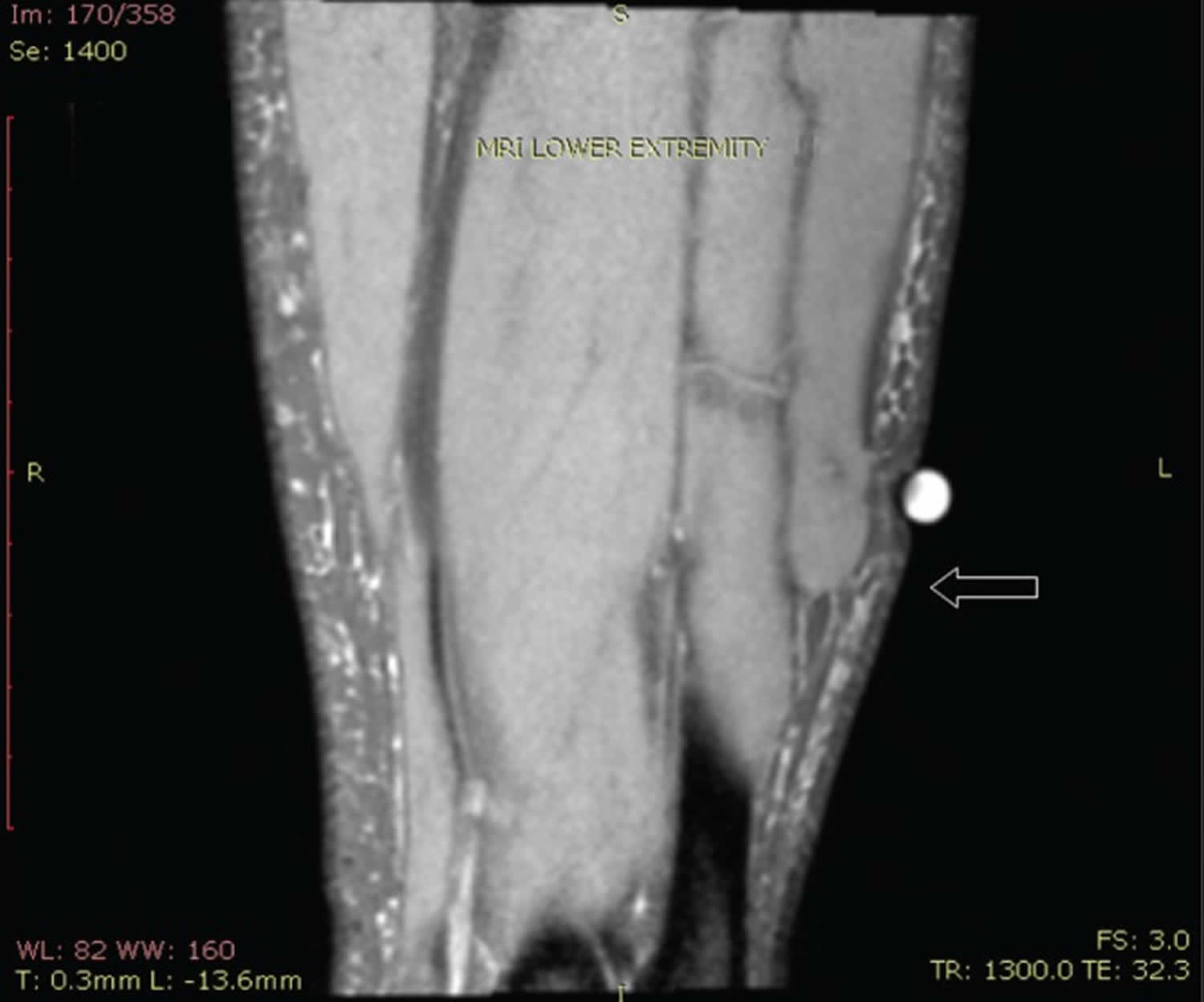
Figure 2. Fascial hernia

Footnote: Magnetic resonance imaging (MRI) of the left lower extremity T2 coronal plane. A skin marker had been placed directly over the peroneal musculature and demonstrated a focal contour irregularity on the anterior aspect, consistent with a fascial defect and herniation of peroneus brevis (arrow).
[Source 1 ]Fascial hernia causes
Fascial hernia is caused by a focal fascial sheath defect. Ihde 3 classified fascial hernias into two groups: constitutional (congenital) and traumatic (acquired). Congenital causes may be an overall general weakness in the muscular fascia (mesodermal insufficiency), or may occur at sites of perforating nerves and vessels. Acquired causes are usually secondary to trauma, occuring as either after direct or indirect trauma. Traumatic examples include penetrating trauma, closed fractures causing a fascial tear (direct trauma), or force applied to contracted muscle causing acute fascial rupture (indirect trauma) 29. In direct trauma, the fascia itself is injured resulting e.g. from fractures, wounds or contusion. Indirect trauma means injury to the contracted muscle that can cause rupture of the fascia 30. Herniation is potentiated by increases in intracompartmental pressures such as muscle hypertrophy or chronic exertional compartment syndrome (CECS). For perspective, regular cardiovascular exercise and physical activity can lead to muscle hypertrophy with a 20% increase in muscle volume 31. Chronic exertional compartment syndrome (CECS) is defined as a reversible form of abnormally increased intramuscular pressure during exercise or physical exertion secondary to noncompliance of osteofascial tissues to exercise-induced increases in muscle volume 31.
Acute compartment syndrome is attributed to various causes (i.e., fracture, ischemia, improper casting, etc.) and often presents with extreme pain in a passive state as an emergency. Fasciotomy is the gold standard for relieving symptoms associated with acute compartment syndrome 32. In contrast, chronic exertional compartment syndrome is less emergent but highly disruptive for people with an active lifestyle. Chronic exertional compartment syndrome usually occurs in the lower leg and most likely results from increased pressure in one or more muscle compartments. Chronic exertional compartment syndrome may result in neurovascular abnormalities causing extreme pain during and after exercise and often causes athletes to stop competing and exercising. The prevalence of chronic exertional compartment syndrome is unknown and it is likely that many people who suffer from chronic exertional compartment syndrome modify their activities in a way to reduce pain without ever seeking medical intervention. It is also possible that some people with chronic exertional compartment syndrome are not taken seriously by athletic trainers, coaches, and primary care physicians to whom symptoms are first presented 33. Chronic exertional compartment syndrome most often affects the lower leg, which is divided into four compartments: anterior, lateral, deep posterior, and superficial posterior (Figure 1). Of these four compartments, the anterior and lateral compartments are most commonly affected 34. Diagnostic criteria for chronic exertional compartment syndrome include one or more of the following intramuscular pressure readings: ≥15 mmHg at rest; ≥30 mmHg 1 minute after exercise; ≥20 mmHg 5 minutes after exercise 35.
Fascial hernia symptoms
A fascial hernia may clinically present as a visibly palpable bulge, soft tissue mass or subcutaneous nodule. Fascial hernias may be solitary, bilateral or multiple. They may or not be reducible and may present with strangulated muscle 36. Patients with fascial hernia usually suffer from pain or present due to cosmetic reasons or concerns of having a tumor 24. Patients may complain of tenderness or pain, cramping, discomfort, weakness or neuropathy such as hyposensitivity on the thigh and lower leg 37, 38. This may worsen with standing or physical activity. Tenderness may be elicited on examination and decreased sensation may occur with associated nerve involvement. If not readily apparent, herniation may be elicited with limb dependency, movements causing involved muscular contraction or certain stances. For example, pronounced herniation of the tibialis anterior may occur with resisted dorsiflexion of the foot or with the ‘lunge’ or ‘fencing’ position 3. If palpable and easily reducible, the outlined fascial defect may be appreciated. Patients may only initially present with cosmetic concerns or concerns of a tumour 39. Dermatologists have reported the incidental discovery of asymptomatic hernias during unrelated examination 40. Alhadeff et al. 41 reported an unusual case of pseudoradicular symptoms caused by compression of the common peroneal nerve in the popliteal area by gastrocnemius muscle herniation. The patient suffered from pseudoradicular symptoms that resembled sciatica 41. Therefore, neurologic symptoms such as hyposensitivity on the thigh and lower leg are possible in patients with fascial hernia occurring at the site of nerve perforation of the fascia 22.
Fascial hernia diagnosis
Radiologic imaging techniques, including magnetic resonance imaging (MRI) and CT and ultrasound have been used to have the definitive diagnosis of muscle herniation and to identify the defect 42.
Dynamic ultrasound must be the first imaging examination due to its low cost and ready availability 43. However, a fascial thinning is sometimes difficult to detect. It has been suggested the use of dynamic MRI in the evaluation of suspected muscle herniations to better delineate the fascial defect and the size of the muscle herniation, if dynamic ultrasound does not adequately define these features 44. It was hypothesized that MRI can be useful in planning operative treatment 45. MRI better visualizes musculofascial demarcation, allowing quantification of fascial splitting and muscle herniation 46. MRI is superior to computed tomography because muscle and fascia have similar attenuation, which is not as easily differentiable on computed tomography 46. MRI and ultrasonography are improved with dynamic imaging techniques, which incorporate fast imaging with forced muscular movements such as dorsiflexion and plantar flexion of the ankle, and enable better visualization and pinpointing of the hernia and fascial defect 14.
Fascial hernia treatment
Treatment of fascial hernias is mainly dependent on clinical symptoms and may range from conservative measures to operative intervention. Asymptomatic fascial hernias usually require no treatment or can be treated conservatively 22. For mild cases, a support stocking, can be of benefit along with rest and activity modification 3. Most symptomatic muscle hernias are successfully treated with conservative therapy, including rest, activity restrictions and compression stockings 23. Because asymptomatic fascial hernias do not necessitate treatment, a general guideline may suggest conservative therapy only for mildly symptomatic muscle hernias. Injections of sclerosing agents (sodium morrhuate) 47, local anesthetic (triamcinolone) 48 and botulinum toxin 19 have been described. Surgical referral is warranted for patients with moderate to severe symptoms that are not amenable to a conservative therapy trial. Operative repair for cosmetic concerns has also been described and is debated 49. For patients with stronger symptoms or those in whom conservative treatment has failed to improve symptoms, surgery can be considered 24.
Fascial hernia repair
Optimal surgical treatment is controversial. A variety of surgical techniques have been described, ranging from fasciotomy to anatomical repair of the fascial defect, with no consensus. Described treatments include decompressive fasciotomy 50, direct primary fascial repair 16, tibial periosteal flap 51, fascial patch grafting or stripping (woven strips of fascia) using autologous fascia lata 52 and the use of synthetic mesh 18. Partial muscle excision has been described as a solitary treatment and as an adjunct for excessive muscle volume interfering with repair 15.
Every operation has disadvantages and potential complications that must be weighed. Anatomical repair of the fascial defect (eg, primary repair, fascial grafting, synthetic mesh) requires close observation secondary to risks of acute or chronic compartment syndrome and hernia recurrence 29. Fascial grafting may require additional or longer incisions, and creates new potential sites for hernia formation 18. Synthetic mesh carries the risk of infection due to foreign body incorporation and may undesirably adhere to underlying structures.
Earlier rather than later elective surgical repair may be beneficial in patients with symptomatic muscle hernias and evidence of nerve involvement. Overstretched nerves cause severe pain, and even after the traction forces have been removed, pathological changes in the nerve may continue to progress secondary to continued inflammation and vascular degeneration 53. In their treatment of a symptomatic peroneus brevis hernia, Garfin et al 54 speculated that continued irritation of the superficial peroneal-nerve may account for failure of the fasciotomy to relieve symptoms.
The safest surgical option for the treatment of symptomatic muscular hernias of the leg is a longitudinal fasciotomy. This belief is the opinion of the authors and one shared by others 55. Almost all techniques involve surgical exploration, which allows for dissection and release of the involved nerve. Fasciotomy treats the muscle hernia by enlarging the defect and eliminating risks of muscle ischemia or strangulation, which are potential causes of pain. Most importantly, it eliminates any future risks of acute or chronic compartment syndrome, which can still occur with anatomical repair of the fascia. Potential complications with fasciotomies are universal to any surgery in the lower extremity that crosses a musculofascial plane and includes exposed tendon or bone, neuromuscular damage with dysesthesias or weakness, muscle herniation and venous disease from disruption of the calf muscle pump. Incomplete pain resolution may occur with fasciotomies 56. For the patient with high cosmetic concerns or expectations, enlarging the defect may cause more pronounced muscular bulging. Additionally, selective fasciotomy of only the involved compartment may benefit overall patient satisfaction compared with releasing multiple compartments 57. As with any surgery, the technique should be tailored to the individual and multiple viable options may exist. No absolute consensus exists regarding optimal surgical treatment.
Direct repair is possible when the fascial defect is small and the laxity of the borders permits approximation; this has been practiced in the past 30. However, because of reports of compartment syndrome after direct repair 24, this method should only be used when the defect is small and essential close postoperative follow up is assured 22. Some authors consider the longitudinal fasciotomy the safest method of treatment 43.
There are several reports about successful results after fascial defect coverage using artificial meshes 28, 21. They suggest that the operative procedure is simple, more rapid, and less complicated than other techniques and can be used for large defects. The mesh is fixed above and not under the fascia allowing the underlying muscle to slide without any friction by the mesh or sutures 28, 21.
Lee et al. 38 reported a patient who had multiple herniation of the tibialis anterior muscle. Large defects of the fascia were repaired using a mesh. A compartment syndrome as the typical complication after repair of fascial defects was not seen 58.
Using a permanent mesh in the treatment of hernia is advantageous because it is robust and very durable. Vicryl-Propylene composite meshes are made from 50% resorbable vicryl and 50% non-absorbable Polypropylene. It is therefore partially resorbable and makes a tension free fixation possible. Potential disadvantages may be an increased risk of infection as there is a synthetic nonabsorbable foreign body and there is the risk of adhesion between the mesh and the underlying structures.
The outcome in the follow up examination after 6 weeks showed a decrease of the preoperative pain and a good functionality with full physical load, complete mobilization and 100% employability postoperative. It can be assumed that approximately 2 months are needed to induce a stable scarring. This is similar to the results of Siliprandi et al. 28, who could show that this procedure can provide good functional results and a good cosmetic appearance without complications and complications. Results from various studies point towards a good long time outcome without recurrence of the hernia also after years 21.
- Nguyen JT, Nguyen JL, Wheatley MJ, Nguyen TA. Muscle hernias of the leg: A case report and comprehensive review of the literature. Can J Plast Surg. 2013;21(4):243-247. https://www.ncbi.nlm.nih.gov/pmc/articles/PMC3910527[↩][↩][↩][↩]
- Obermayer ME, Wilson JW. FASCIAL HERNIAS OF THE LEGS. JAMA. 1951;145(8):548–549. doi:10.1001/jama.1951.02920260016004[↩]
- Ihde H. On muscular hernia of the leg. Acta Chir Scand. 1929;65:97–120.[↩][↩][↩][↩][↩][↩]
- Bergmann G, Ciritsis BD, Wanner GA, Simmen HP, Werner CM, Osterhoff G. Gastrocnemius muscle herniation as a rare differential diagnosis of ankle sprain: case report and review of the literature. Patient Saf Surg. 2012;6(1):5. Published 2012 Mar 14. doi:10.1186/1754-9493-6-5 https://www.ncbi.nlm.nih.gov/pmc/articles/PMC3320538[↩][↩][↩]
- Alfageme F, Morales V, Garcia C, Miguelez AP, Dominguez E, Segurado A. Transfascial muscular hernia: an unusual cause for a “hide and seek” subcutaneous nodule. Dermatol Online J. 2011;17:4.[↩]
- Revelon G, Rahmouni A, Jazaerli N, Godeau B, Chosidow O, Authier J, Mathieu D, Roujeau JC, Vasile N. Acute swelling of the limbs: magnetic resonance pictorial review of fascial and muscle signal changes. Eur J Radiol. 1999;30:11–21. doi: 10.1016/S0720-048X(98)00119-3[↩]
- Oldfield MC. Athletes’ hernia of the tibialis anticus muscles. Br J Surg. 1949;36:405–8.[↩]
- Obermayer ME, Wilson JW. Fascial hernias of the legs. JAMA. 1951;145:548–9.[↩]
- Azar FM. Sports Medicine. In: Canale ST, Beaty JH, editors. Campbell’s Operative Orthopaedics. 12th edn. Vol. 3. Philadelphia: Mosby/Elsevier; 2012.[↩][↩]
- Verbov J. Muscle herniation of the lower legs. Br J Dermatol. 1976;95:329–30.[↩]
- Braunstein JT, Crues JV., III Magnetic resonance imaging of hereditary hernias of the peroneus longus muscle. Skelet Radiol. 1995;24:601–4.[↩]
- Mellado JM, Perez del Palomar L. Muscle hernias of the lower leg: MRI findings. Skeletal Radiol. 1999;28:465–9.[↩]
- Henning PT, Dahm DL, Smith J. Use of postexercise ultrasonography to identify a symptomatic extensor digitorum longus muscle hernia associated with running. PM R. 2009;1:1109–11.[↩]
- Tyson S, Subhas N. Radiologic case study. Gastrocnemius fascial defect and muscle herniation. Orthopedics. 2010;33:785.[↩][↩]
- McMaster PE. Muscle hernia of the leg: A study of 21 cases and 38 hernias. U S Naval Med Bull. 1943;41:404–9.[↩][↩]
- Lee HS, James M. Painful bilateral herniation of the anterior tibial muscle: A case report. Foot Ankle Int. 2006;27:552–5.[↩][↩]
- Kim M, Hong SP, Hwang SM, Park H, Ahn SK. Tibialis anterior muscle herniation developed after trauma. Int J Dermatol. 2008;47:845–7.[↩]
- Odili J, Wilson E, Chana JS. Muscle herniation: A complication at the anterolateral thigh perforator flap donor site. J Plast Reconstr Aesth Surg. 2009;62:1530–3.[↩][↩][↩]
- Burg D, Schnyder H, Buchmann R, Meyer VE. [Effective treatment of a large muscle hernia by local botulinum toxin administration] Handchir, Mikrochir, Plast Chir. 1999;31:75–8.[↩][↩]
- Ceyhan AM, Chen W, Yener M, Yildirim M, Yesildag A, Akkaya VB. Bilateral tibialis anterior muscle herniation simulating a soft tissue tumour in a young amateur football player. Australas J Dermatol. 2010;51:142–4.[↩]
- Marques A, Brenda E, Amarante MT. Bilateral multiple muscle hernias of the leg repaired with Marlex mesh. Br J Plast Surg. 1994;47:444–446. doi: 10.1016/0007-1226(94)90076-0[↩][↩][↩][↩][↩]
- Berglund HT, Stocks GW. Muscle hernia in a recreational athlete. Orthop Rev. 1993;22:1246–1248.[↩][↩][↩][↩][↩]
- Lane JE, Woody CM, Lesher JL. Tibialis anterior muscle herniation. Dermatol Surg. 2002;28:641–2.[↩][↩]
- Miniaci A, Rorabeck CH. Tibialis anterior muscle hernia: a rationale for treatment. Can J Surg. 1987;30:79–80.[↩][↩][↩][↩]
- Goldberg HC, Comstock GW. Herniation of muscles of the legs. War Medicine. 1944;5:365–367.[↩]
- Golshani SD, Lee C, Sydorak R. Symptomatic forearm muscle hernia: repair by autologous fascia lata inlay. Ann Plast Surg. 1999;43:204–206.[↩]
- Hartzell J. The use of living fascia transplant to repair a hernia of the tibialis anterior muscle. J Am Med Assoc. 1936;107:492–493. doi: 10.1001/jama.1936.92770330002007a[↩]
- Siliprandi L, Martini G, Chiarelli A, Mazzoleni F. Surgical repair of an anterior tibialis muscle hernia with Mersilene mesh. Plast Reconstr Surg. 1993;91:154–157. doi: 10.1097/00006534-199301000-00026[↩][↩][↩][↩]
- Berglund HT, Stocks GW. Muscle hernia in a recreational athlete. Ortho Rev. 1993;22:1246–8.[↩][↩]
- Simon HE, Sachet H. Muscle hernias of the leg: review of literature and report of twelve cases. Am J Surg. 1945;67:87–97. doi: 10.1016/0002-9610(45)90330-8[↩][↩]
- Schubert AG. Exertional compartment syndrome: Review of the literature and proposed rehabilitation guidelines following surgical release. Int J Sports Phys Ther. 2011;6:126–41.[↩][↩]
- Sheridan G. W., Matsen F. A., III Fasciotomy in the treatment of the acute compartment syndrome. The Journal of Bone & Joint Surgery—American Volume. 1976;58(1):112–115.[↩]
- Turnipseed W. D. Diagnosis and management of chronic compartment syndrome. Surgery. 2002;132(4):613–619. doi: 10.1067/msy.2002.128608[↩]
- Fraipont M. J., Adamson G. J. Chronic exertional compartment syndrome. The Journal of the American Academy of Orthopaedic Surgeons. 2003;11(4):268–276.[↩]
- Pedowitz R. A., Hargens A. R., Mubarak S. J., Gershuni D. H. Modified criteria for the objective diagnosis of chronic compartment syndrome of the leg. The American Journal of Sports Medicine. 1990;18(1):35–40. doi: 10.1177/036354659001800106[↩]
- Conwell HE, Alldredge RH. Ruptures and tears of muscles and tendons. Am J Surg. 1937;35:22–33.[↩]
- Lane JE, Woody CM, Lesher JL. Tibialis anterior muscle herniation. Dermatol Surg. 2002;28:641–642. doi: 10.1046/j.1524-4725.2002.01304.x[↩]
- Lee HS, James M. Painful bilateral herniation of the anterior tibial muscle: a case report. Foot Ankle Int. 2006;27:552–555.[↩][↩]
- Miniaci A, Rorabeck CH. Tibialis anterior muscle hernia: A rationale for treatment. Can J Surg. 1987;30:79–80.[↩]
- Alfageme F, Morales V, Garcia C, Miguelez AP, Dominguez E, Segurado A. Transfascial muscular hernia: An unusual cause for a “hide and seek” subcutaneous nodule. Dermatol Online J. 2011;17:4.[↩]
- Alhadeff J, Lee CK. Gastrocnemius muscle herniation at the knee causing peroneal nerve compression resembling sciatica. Spine (Phila Pa 1976) 1995;20:612–614. doi: 10.1097/00007632-199503010-00020[↩][↩]
- Hong JH. Herniation of the lateral head of the gastrocnemius muscle: is it the source of the posterolateral knee pain? Anesth Analg. 2007;104:1310–1311.[↩]
- Bates DG. Dynamic ultrasound findings of bilateral anterior tibialis muscle herniation in a pediatric patient. Pediatr Radiol. 2001;31:753–755. doi: 10.1007/s002470100534[↩][↩]
- Armfield DR, Kim DH, Towers JD, Bradley JP, Robertson DD. Sports-related muscle injury in the lower extremity. Clin Sports Med. 2006;25:803–842. doi: 10.1016/j.csm.2006.06.011[↩]
- Mellado JM, Perez del Palomar L. Muscle hernias of the lower leg: MRI findings. Skeletal Radiol. 1999;28:465–469. doi: 10.1007/s002560050548[↩]
- Zeiss J, Ebraheim NA, Woldenberg LS. Magnetic resonance imaging in the diagnosis of anterior tibialis muscle herniation. Clin Orthop Relat Res. 1989:249–53.[↩][↩]
- Schmier AA. Fascial hernia of both lower extremities: Injection with sodium morrhuate. JAMA. 1937;109:28–9.[↩]
- Hong JH. Herniation of the lateral head of the gastrocnemius muscle: Is it the source of the posterolateral knee pain? Anesth Analg. 2007;10:1310–1.[↩]
- Sherry RH. Herniation of peroneus brevis muscle: Report of a case. Bull Hosp Dis. 1942;3:69–72.[↩]
- Gupta RK, Singh D, Kansay R, Singh H. Cricket ball injury: A cause of symptomatic muscle hernia of the leg. Br J Sports Med. 2008;42:1002–3.[↩]
- Lee JK. Anterior tibial muscle hernia treated with local periosteal rotational flap – a case report. J Korean Fract Soc. 2012;25:331–4.[↩]
- Bloem JJ. The treatment of muscle hernias by fascial splitting. Br J Plast Surg. 1976;29:291–4.[↩]
- Nobel W. Peroneal palsy due to hematoma in the common peroneal nerve sheath after distal torsional fractures and inversion ankle sprains. J Bone Joint Surg. 1966;48:1484–95.[↩]
- Garfin S, Mubarak SJ, Owen CA. Exertional anterolateral-compartment syndrome. Case report with fascial defect, muscle herniation, and superficial peroneal-nerve entrapment. J Bone Joint Surg. 1977;59:404–5.[↩]
- Miniaci A, Rorabeck CH. Compartment syndrome as a complication of repair of a hernia of the tibialis anterior. A case report. J Bone Joint Surg. 1986;68:1444–5.[↩]
- Golshani SD, Lee C, Sydorak R. Symptomatic forearm muscle hernia: Repair by autologous fascia lata inlay. Ann Plast Surg. 1999;43:204–6.[↩]
- Packer JD, Day MS, Nguyen JT, Hobart SJ, Hannafin JA, Metzl JD. Functional outcomes and patient satisfaction after fasciotomy for chronic exertional compartment syndrome. Am J Sports Med. 2013;41:430–6.[↩]
- Miniaci A, Rorabeck CH. Compartment syndrome as a complication of repair of a hernia of the tibialis anterior. A case report. J Bone Joint Surg Am. 1986;68:1444–1445.[↩]


