
Fat embolism
Fat embolism is the presence of fat globules in microcirculation, whereas fat embolism syndrome is a systemic manifestation of dissemination of fat molecules or globules in microcirculation causing the classical triad of respiratory distress, cerebral signs and petechiae 1. Fat embolism syndrome is a continuum of fat embolism. Fat embolism syndrome can present in a wide variety of severity and symptoms. Usually presenting with a delay of 12-72 hours, with the classical triad of respiratory distress, cerebral signs and petechiae. Fat embolism syndrome can go unnoticed clinically or may present as an acute fatal event within hours of the inciting injury.
Fat embolism syndrome is commonly seen in trauma wards and is usually associated with fractures of long bones or multiple fractures 2. Fat embolism syndrome is estimated to occur in 3-4% of patients with long bone fracture 3. Apart from trauma, surgical procedures such as intramedullary reaming, pelvic or knee arthroplasty are important causes of fat embolism syndrome. Rarely, intra-osseous fluid administration, lipid soluble radio contrast, intravenous hyper alimentation, long term steroid administrations, liposuction, bone marrow harvesting and transplant are also implicated as iatrogenic causes for fat embolism syndrome 4.
Non-iatrogenic causes of fat embolism are very rare, but have been related to sickling crisis, pancreatitis, fat necrosis of omentum, diabetes, hepatic steatosis, osteomyelitis, panniculitis and bone tumor lysis 5.
Fat embolism and fat embolism syndrome are a clinical phenomenon that are characterized by systemic dissemination of fat emboli within the system circulation. The dissipation of fat emboli will disrupt the capillary bed and affect microcirculation, causing a systemic inflammatory response syndrome 6. End-organ manifestation typically will involve the following:
- Skin and integumentary organ
- Central nervous system
- Respiratory system lungs
- Eyes retina
Fat embolism syndrome is most common in patients with orthopedic trauma. It also can occur in nontraumatic patients. The following nontraumatic conditions can cause fat embolism syndrome:
- Acute or chronic pancreatitis
- Bone marrow transplant
- Liposuction
In most instances, diagnosis is usually established during the autopsy 1.
Fat embolism has a higher incidence than fat embolism syndrome. In the landmark study carried out by Gurd 7, using the established clinical criteria, an incidence of 19% of fat embolism syndrome was reported in a group of trauma patients. Since early open reduction and internal fixation has become the standard of care for repairing fractures of long bones, the incidence of fat embolism and fat embolism syndrome has gradually decreased. Most recent studies show an incidence of about 1% to 11% 1.
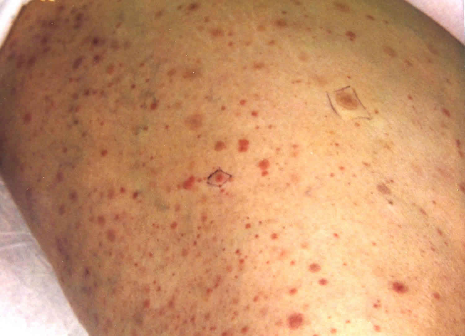
Figure 1. Fat embolism petechial rash
Fat embolism causes
Causes of fat embolism and fat embolism syndrome can be categorized as traumatic or nontraumatic.
Traumatic causes
Traumatic causes of fat embolism syndrome are more common that than nontraumatic causes. Clinical diagnosis of small fat embolism or mild cases of fat embolism syndrome may be missed and go away unnoticed. In one study, about 67% of orthopedic trauma patients have fat globules in their blood. If the blood sample was taken from a site close to the area of the fracture, the incidence is closer to 95%.
Trauma as a cause of fat embolism syndrome can occur from the following.
Fracture of the long bones, specifically:
- Femur
- Tibia
- Pelvis
The postoperative condition also can favor the development of fat embolism syndrome including the following:
- Pelvic arthroplasty
- Knee arthroplasty
- Intramedullary nailing and reaming
Fat embolism and fat embolism syndrome also can occur intraoperatively while repairing a long bone fracture. With a transesophageal echocardiogram, fat embolism has been detected in close to 41% of patients.
Other rare traumatic conditions that can cause fat embolism syndrome include the following:
- Massive soft tissue damage
- Crush injury
- Prolonged cardiopulmonary resuscitation
- Severe burn involving more than 50% of body surface area
- Bone marrow transplantation
- Liposuction
- Median sternotomy
Nontraumatic causes
Cases of nontraumatic fat embolism syndrome are very rare and include the following:
- Fatty Liver
- Acute or chronic pancreatitis
- Therapy with corticosteroid
- Infusion of fat emulsion
- Lymphography
- Hemoglobinopathies
- Sickle cell disease
- Thalassemia
Risk factors for developing fat embolism syndrome
Several risk factors are associated with the development of fat embolism syndrome. The following conditions increase the risk of developing fat embolism syndrome:
- Young age
- Closed fractures
- Multiple fractures
- Prolonged conservative management of long bone fracture
The technique of inserting the intramedullary nails also can contribute to the risk of developing fat embolism syndrome.
- Increased velocity in reaming
- Overzealousness in the nailing of the medullary cavity
- The widened gap between the nail and the cortex of the bone
Fat embolism and fat embolism syndrome pathophysiology
Fat particles or globules are released from the organs of primary origin and enter the microcirculation, causing damage to the capillary beds. The disruption affects the microcirculatory hemostasis in the following organs:
- Brain
- Skin
- Eyes
- Heart
Two main theories try to explain the development of fat embolism syndrome.
Mechanical theory
This theory was postulated by Gossling et al 8. According to Gassling et al 8, large fat droplets are released into the venous system. Elevation of the intramedullary from trauma or surgery leads to the release of fat into the venous sinusoids. From the venous system, these fat globules are deposited in the pulmonary capillary bed where they travel to the brain via the arteriovenous shunt into the brain. Initially, there was no valid explanation for the development of fat embolism syndrome in patients with no patent foramen ovale, but the presence of an arteriovenous shunt clarifies this. Also, intravascular fat droplets are deformable, hence their ability to transverse the pulmonary vasculature. The pathophysiological changes produced by fat droplets include the following:
- Elevated pulmonary artery pressure
- Impairment of oxygen exchange from ventilation-perfusion mismatch
- Systemic effects on end-organs such as the brain, kidney, and skin.
Deposition of the fat droplets in the brain produces a cascade of reactions and leads to a systemic inflammatory response syndrome, local inflammation, and ischemia as a result of disruption of microcirculation. The release of inflammatory mediators and vasoactive amines like histamine, serotonin leads to an increase in vascular permeability and vasodilation with ensuing hypotension and hypoperfusion.
Biochemical theory
Baker et al. proposed this theory for the development of fat embolism syndrome. According to this theory, the precipitating event, whether traumatic or nontraumatic, triggers a hormonal change in the body system. This leads to release of free fatty acid and chylomicrons. The presence of acute phase reactants, such as C-reactive protein, causes the chylomicron to coalesce and migrate. Baker et al. attribute the development of fat embolism syndrome to free fatty acid. Pneumocyte hydrolysis of fat particles generates free fatty acid which migrate to other organs, causing multiple organ dysfunction syndromes. The biochemical theory helps to explain the development of fat embolism syndrome in nontraumatic patients.
Fat embolism signs and symptoms
A fat embolism can travel to most of the organs in the body. Fat embolism and fat embolism syndrome are multiorgan diseases that can damage the kidneys, heart, skin, brain, and lungs. Fat embolism typically manifests at around 24 to 72 hours after the initial insult.
The symptoms in fat embolism and fat embolism syndrome are nonspecific. Patients might complain of the following 1:
- Pain related to bone fracture
- Nausea
- General weakness
- Malaise
- Difficulty breathing
- Headache
Fat embolism and fat embolism syndrome signs and symptoms
These include but is not limited to the following:
- Respiratory symptoms. These are usually the first presenting features. Hypoxemia, tachypnoea, and dyspnoea are the initial findings. In some cases, the patients may progress to respiratory failure, requiring mechanical ventilation 9. In other cases, if no ongoing embolism or infection occurs, the lung usually recovers by the third day.
- Tachypnea
- Tachycardia
- Diaphoresis
- Central nervous system symptoms. The symptoms may appear within 10-120 hours and are highly varying 10. Ranging from confusion, to seizures, these symptoms may even be the presenting symptoms of fat embolism syndrome 9. They are thought to be the direct result of cerebral hypoxia rather than embolization. The symptoms may include irritability, anxiety, agitation, confusion, delirium, and coma, which are described as progressive changes and as single manifestation in individual cases. These symptoms are usually non-lateralizing, transient and fully reversible 11. However, localizing signs due to fat embolism have also been reported. Localizing signs, such as aphasia, anisocoria, apraxia in a patient with poly-trauma warrants a computerized tomography of brain to rule out hematoma, although in fat embolism syndrome, a CT brain will be usually non-rewarding.
- Agitation from hypoxia
- Restlessness
- Change in mental status
- Seizure
- Coma
- Skin
- Petechial rash may be the last component to develop in fat embolism syndrome. Appearing within 36 hours, this is believed to be pathognomic feature of fat embolism syndrome 5. It is usually self limiting, and disappears within a week. It can be easily missed in dark skinned persons, and is to be actively sought on the upper portions of the chest and armpits. Other common sites of involvement are conjunctiva, oral cavity and shoulders. Petechiae are thought to develop due to extravasations of red blood cells from over distended capillaries 4. The fat particles floating in aortic arch vessels are embolized to non dependant parts via aortic arch vessels and hence the site predilection 12.
- Eye
- The obstruction of retinal capillaries may lead to cotton wool spots, macular edema and retinal hemorrhage but these changes are usually reversible 13.
- Other manifestations
A non-fulminant fat embolism, which is clinically apparent and milder than the fulminant form has also been described by Sevitt 15.
Subclinical fat embolism syndrome
Seen in almost all long bone fractures 8, it manifests as a decreased PaO2, minor hematological changes with no clinical signs or symptoms of respiratory insufficiency. It is often confused with postoperative symptoms such as pain, discomfort, or postoperative inflammatory process. Associated tachypnoea, tachycardia, and fever may be seen 16.
Non-fulminant (subacute) fat embolism syndrome
Clinically, apparent form in which respiratory and cerebral changes, petechiae are seen 9. Petechiae are seen on anterior portion of upper chest, and areas adjacent to the axillae, shoulder, mucosa and mouth 17. Radiologic abnormalities on chest skiagram and other lab abnormalities may be seen. Neurologic signs such as coma, confusion or stupor are present in up to 86% cases 9. They are usually transient and non-lateralizing. Respiratory symptoms are usually benign and lung parenchyma tends to improve from third day. They may vary from minimal tachypnoea to development of acute respiratory distress syndrome (ARDS). Changes similar to Purtscher’s Retinopathy may be present 5.
Fulminant fat embolism syndrome
This is a much rarer entity than the other two. Seen within hours of injury, it results in severe physiologic impairment including respiratory failure, and altered mental status. Cause of death, if occurs, is usually due to acute right heart failure.
Fat embolism diagnosis
Diagnosis of fat embolism syndrome can be very challenging because the signs and symptoms can be vague. There are no universally accepted diagnostic criteria. Several authors based on experience and research have proposed diagnostic criteria for fat embolism syndrome 18.
Gurd et al. in 1970 7 and later Wilson in 1974 19 put forward the following diagnostic criterion requiring two major criteria or at least one major criteria and four minor criteria.
Major Criteria
- Petechial rash
- Respiratory insufficiency
- Cerebral involvement in non-head injury patients
Minor Criteria
- Fever greater than 38.5 °C (101.3 °F)
- Tachycardia heart rate greater than 110 beats per minutes
- Retinal involvement
- Jaundice
- Renal signs
- Anemia
- Thrombocytopenia
- High erythrocyte sedimentation rate
- Fat macroglobulinemia
Schoenfeld Criteria
Another report by Schoenfeld et al. 20 proposed a quantitative means for the diagnosis of fat embolism syndrome.
A cumulative score greater than five is required for the diagnosis.
- 5 points – petechiae rash
- 4 points – diffuse infiltrate on x-ray
- 3 points – hypoxemia
- 1 point (for each) – fever, tachycardia, confusion
Lindeque Criteria
Lastly, Lindegue et al. 21 suggested the use of respiratory symptoms alone as the diagnostic criteria for fat embolism syndrome. This criterion has not gained worldwide acceptance when compared to the Gurd, Wilson, and Schoenfeld criteria.
- Sustained Pa02 less than 8 kilopascal
- Sustained PC02 greater than 7.3 kilopascal
- Sustained respiratory rate greater than 35 breaths per minute despite sedation
- Dyspnea, increased work of breathing, anxiety, tachycardia
Ancillary studies
Apart from the aforementioned diagnostic criteria, other ancillary studies are needed to aid in the diagnostic workup including the following:
- Complete blood count (CBC): Anemia and thrombocytopenia are very common in fat embolism syndrome.
- Comprehensive metabolic panel (CMP): Metabolic acidosis, increased level of BUN (blood urea nitrogen), and creatinine can be seen in patients with fat embolism syndrome.
- Arterial blood gas (ABG): Ventilation-perfusion mismatch is a hallmark of fat embolism syndrome. The arterial blood gas analysis usually has a low partial pressure of oxygen, causing hypoxemia. An increased alveolar-arterial (A-a) gradient is common in fat embolism syndrome. The A-a gradient is the difference in the partial pressure of oxygen in the alveolus and the partial pressure of oxygen in the pulmonary artery. In fat embolism syndrome, there is occlusion of the pulmonary blood vessels, causing impairment of perfusion with normal ventilation. The end result of this in fat embolism syndrome is a ventilation-perfusion mismatch.
To calculate the A-a gradient, use the formula:
- A-a gradient = PA02 – Pa02
Pa02 is the partial pressure of oxygen in the pulmonary artery
PA02 is the partial pressure of oxygen in the alveolar sac
To calculate the PAO2 and Pa02
- PAO2 = FiO2 (P atmospheric – P water vapor) – (PCO2/R).
- PaO2 = partial pressure of oxygen in the pulmonary artery.
PaO2 in arterial blood gas can be used as follows.
FiO2 is the concentration of inspired oxygen expressed as a fraction.
This is around 0.21 at room air.
- P atmospheric is the barometric pressure (760 mmHg at sea level).
- P water vapor is the water vapor pressure (48 mmHg at 37 C).
- PaCO2 is the partial pressure of alveolar carbon dioxide.
If approximated, this is presumed to be equal to arterial PCO2. PACO2 is presumed to be equal to 40 mmHg
- R is the respiratory quotient and is equated to about 0.8 on a regular diet.
The normal alveolar partial pressure of oxygen is as follows:
- PA02 = alveolar partial pressure of O2 = FiO2 × (patmospheric – P water vapor) – (PCO2/R).
- 0.21 × (760 – 48) – (40/0.8) = 150-50 = 100 mmHg
The normal partial pressure of oxygen in arterial blood is beween 75 to 100mmHg.
- PA02 – Pa02 = 100 mmHg – 75 mm = 25 mmHg
- PA02-Pa02 =100 mmHg -100 mmHg = 0 mmHg
This implies that the A-a gradient can have values that range from 0 to 25 mmHg
The normal A-a gradient is usually less than 10 mmHg. This can increased significantly in fat embolism syndrome because of ventilation-perfusion mismatch.
Bronchoalveolar Lavage
Bronchoalveolar Lavage (BAL) has been researching extensively as a diagnostic tool for fat embolism syndrome. Lipid inclusion in the macrophages might point to a diagnosis of fat embolism syndrome but is not specific, as this can be seen in other clinical conditions. Moreover, the procedure is time-consuming, invasive, and might not give the best diagnostic yield.
Attempts at developing biological markers for fat embolism syndrome have been disappointing because of low specificity. Lipase, free fatty acid, and phospholipase A2 have been demonstrated to be elevated in fat embolism syndrome, but this also can be seen in other disease conditions of the lung
Blood, urine, and sputum analysis might show the presence of fat globules. Again, this is nonspecific in fat embolism and fat embolism syndrome.
Imaging Studies
Chest x-ray
The chest X-ray will reveal the presence of the following:
- Diffuse interstitial marking
- Pulmonary edema
- Lung infiltrate
- Flake-like pulmonary marking (snowstorm appearance)31
CAT scan of the chest
- Area of increased vascular congestion
- Pulmonary edema
Imaging of the brain
CAT scan
This is not a very sensitive imaging study of the brain in fat embolism syndrome, but it can be used to exclude other causes of altered mental status such as epidural, subdural or subarachnoid bleed.
MRI
This is the most sensitive test that can be used to demonstrated changes in the brain related to fat embolism syndrome. Takahashi et al. 22 categorized these changes into the following four grades based on the size and distribution of the lesions in T2 weighted imaging:
- Grade 0 – normal
- Grade 1 – mild
- Grade 2 – moderate
- Grade 3 – severe
Lesions seen in fat embolism syndrome are distributed in the following areas of the brain:
- Centrum semi vale,
- Subcortical white matter
- Ganglionic regions
- Thalamus
The authors demonstrated that resolution of these lesions correlates well with clinical recovery from fat embolism syndrome 22. Some of these lesions develop as a result of vasogenic edema from free fatty acids which is potentially neurotoxic.
Transesophageal echocardiography may be utilized intraoperatively to monitor the release of fat globules or bone marrow materials into the bloodstream during the process of intramedullary nailing and reaming. Fat emboli in the pulmonary artery can increase the pulmonary artery wedge pressure as well as the right ventricular afterload.
Fat embolism treatment
There is no specific treatment for fat embolism or fat embolism syndrome 1. Based on experimental studies, an attempt was made to use dextrose infusion to decrease free fatty acid mobilization. Ethanol also was used as an agent to inhibit lipolysis. In clinical practice, there were no proven benefits 23.
Experimental use of heparin in an animal model was found to be beneficial but is no longer used in clinical practice because of the potential risk of bleeding. There has not been a proven clinical benefit with the use of heparin in fat embolism syndrome.
Therapy with corticosteroids has been proposed for the treatment of fat embolism syndrome based on the following effects of corticosteroids:
- Inhibition of complement activated leucocyte aggregation
- Limiting free fatty acid level
- Membrane stabilization
A meta-analysis of seven randomized control trials using corticosteroid prophylaxis showed close to 77% reduction in the risk for fat embolism syndrome in a patient with a long bone fracture. There is, however, no difference in mortality, infection, or avascular necrosis between the treatment group and control. For this reason, the use of corticosteroids is still very controversial.
Inferior Vena Cava Filter
This has been proposed as a measure to prevent the showering of fat emboli. As a prophylactic treatment for fat embolism syndrome, placement of an inferior vena cava filter has not been studied sufficiently.
Operative Measures
It is highly recommended to start early open reduction and internal fixation (ORIF) of long bone fractures. The incidence of fat embolism syndrome is higher in a patient with long bone fractures who are managed conservatively.
The use of internal fixation devices in the management of fractures of long bones significantly reduces the incidence of fat embolism syndrome.
During operative fixation of the long bone fracture, care must be taken to limit the intramedullary pressure, as a high pressure is associated with an increased amount of fat emboli entering the systemic circulation.
Some techniques utilized in orthopedic surgery to reduce embolization include:
- Lavage of bone marrow prior to fixation
- Venting of the femoral bone
- Drilling of small holes in the cortex of the bone to lower intramedullary pressure
None of these maneuvers has been shown to reduce fat embolism syndrome.
Supportive Care
This is the mainstay treatment once a patient develops fat embolism syndrome. Supportive care is geared towards adequately oxygenating the end organs.
Goals of Supportive Care
- Provision of adequate oxygenation and ventilation
- Maintenance of adequate hemodynamic stability
- Transfusion of packed red blood cells to improve oxygen delivery if indicated
- Prophylaxis of deep venous thrombosis with a sequential compression device
- Adequate nutrition and hydration
Supplemental oxygen might be required, and if the patient develops fulminant acute respiratory distress syndrome, intubation and mechanical ventilation might be required.
Albumin
Albumin is recommended as part of the resuscitation tools for hypovolemia. It restores intravascular volume and helps to bind free fatty acid. This prevents the systemic dissemination of fat globules.
Indications for Intubation
- Altered mental status with Glasgow coma score of less than 8
- Moderate to several respiratory distresses with no improvement on noninvasive support
Fat embolism syndrome might also cause pulmonary hypertension with right ventricular failure. Inotropic support with dobutamine or a phosphodiesterase inhibitor like milrinone might be required.
Cerebral edema if present might require management with the following:
- Mannitol
- Hypertonic saline
- Intracranial pressure monitors
Fat embolism prognosis
In patients with traumatic fat embolism syndrome, the prognosis depends on early open reduction and internal fixation of the long bone fracture. Most patients with adequate support therapy can recover from the neurological, respiratory, and retinal changes associated with fat embolism syndrome. The most recent studies approximate mortality between 5% to 15% 2.
The most common cause of morbidity or mortality include:
- Acute respiratory distress syndrome (ARDS)
- Cerebral edema.
- Adeyinka A, Pierre L. Fat Embolism. [Updated 2019 Feb 15]. In: StatPearls [Internet]. Treasure Island (FL): StatPearls Publishing; 2019 Jan-. Available from: https://www.ncbi.nlm.nih.gov/books/NBK499885[↩][↩][↩][↩][↩]
- George J, George R, Dixit R, Gupta RC, Gupta N. Fat embolism syndrome [published correction appears in Lung India. 2017 Jan-Feb;34(1):114]. Lung India. 2013;30(1):47–53. doi:10.4103/0970-2113.106133 https://www.ncbi.nlm.nih.gov/pmc/articles/PMC3644833[↩][↩]
- White T, Petrisor BA, Bhandari M. Prevention of fat embolism syndrome. Injury. 2006;37S:S59–67.[↩]
- Capan LM, Miller SM, Patel KP. Fat embolism. Anesthesiol Clin North Am. 1993;11:25–54.[↩][↩]
- Jain S, Mittal M, Kansal A, Singh Y, Kolar PR, Saigal R. Fat embolism syndrome. J Assoc Physicians India. 2008;56:245–9.[↩][↩][↩]
- Guerado E, Bertrand ML, Cano JR, Cerván AM, Galán A. Damage control orthopaedics: State of the art. World J Orthop. 2019 Jan 18;10(1):1-13.[↩]
- Gurd AR. Fat embolism: An aid to diagnosis. J Bone Joint Surg Br. 1970;52:732–7.[↩][↩]
- Gossling HR, Pellegrini VD., Jr Fat embolism syndrome: A review of the pathophysiology and physiological basis of treatment. Clin Orthop. 1982;165:68–82.[↩][↩][↩]
- Chhabra B, Kiran S, Senthilnathan TA, Gupta R. Fat embolism syndrome. Ind J Orthop. 2001;35:10–5.[↩][↩][↩][↩]
- Filomeno LT, Carelli CR, Silva NC, Filho TE, Amatuzzi MM. Embolia gordurosa: Uma revisāo para a prática ortopédica atual. Acta Ortop Bras. 2005;13:196–208.[↩]
- Gupta A, Reilly CS. Fat embolism. Cont Edu Anaesth Crit Care Pain. 2007;7:148–51.[↩]
- Alho A. Fat embolism syndrome, aetiology, pathogenesis, and treatment. Acta Chir Scand Suppl. 1980;499:75–85.[↩]
- Adams CB. The retinal manifestations of fat embolism. Injury. 1971;2:221–4.[↩][↩]
- Stephens JH, Fred HL. Petachiae associated with systemic fat embolism. Arch Dermatol. 1962;86:515–7.[↩]
- Sevitt S. The significance and pathology of fat embolism. Ann Clin Res. 1977;9:173–80.[↩]
- Fulde GW, Harrison P. Fat embolism: A review. Arch Emerg Med. 1991;8:233–9.[↩]
- Fischer JF, Turner RH, Herndon JH, Risenborough EJ. Massive steroid therapy in fat embolism. Surg Gynecol Obst. 1971;132:667–72.[↩]
- Berlot G, Bussani R, Shafiei V, Zarrillo N. Fulminant Cerebral Fat Embolism: Case Description and Review of the Literature. Case Rep Crit Care. 2018;2018:7813175 [↩]
- Gurd AR, Wilson RI. The fat embolism syndrome. J Bone Joint Surg Br. 1974;56:408–16.[↩]
- Schonfeld SA, Ploysongsang Y, DiLisio R, Crissman JD, Miller E, Hammerschmidt DE, et al. Fat embolism prophylaxis with corticosteroids. Ann Intern Med. 1983;99:438–43.[↩]
- Lindegue BY, Schoeman HS, Dommisse YF, Boeyens MC, Vlok AL. Fat embolism and the fat embolism syndrome: A double blind therapeutic study. J Bone Joint Surg Br. 1987;69:128–31.[↩]
- Takahashi M, Suzuki R, Osakabe Y, Asai JI, Miyo T, Nagashima G, et al. Magnetic resonance imaging findings in cerebral fat embolism: Correlation with clinical manifestations. J Trauma. 1999;46:324–7.[↩][↩]
- Blokhuis TJ, Pape HC, Frölke JP. Timing of definitive fixation of major long bone fractures: Can fat embolism syndrome be prevented? Injury. 2017 Jun;48 Suppl 1:S3-S6.[↩]


