Hutchinson sign
The Hutchinson sign can refer to two clinical signs.
Hutchinson sign nose (ophthalmology)
Hutchinson sign relates to involvement of the tip of the nose from facial herpes zoster 1. It implies involvement of the external nasal branch of the nasociliary nerve (branch of the ophthalmic division of the trigeminal nerve) and thus raises the spectre of involvement of the eye. Herpes zoster is caused by reactivation of a primary infection with varicella zoster virus (commonly known as chickenpox virus). After a primary infection or chickenpox, the varicella zoster virus lies dormant in dorsal root or cranial nerve ganglia. Reactivation causes the typical dermatomal pain and vesicular rash.
Figure 1. Hutchinson’s sign nose
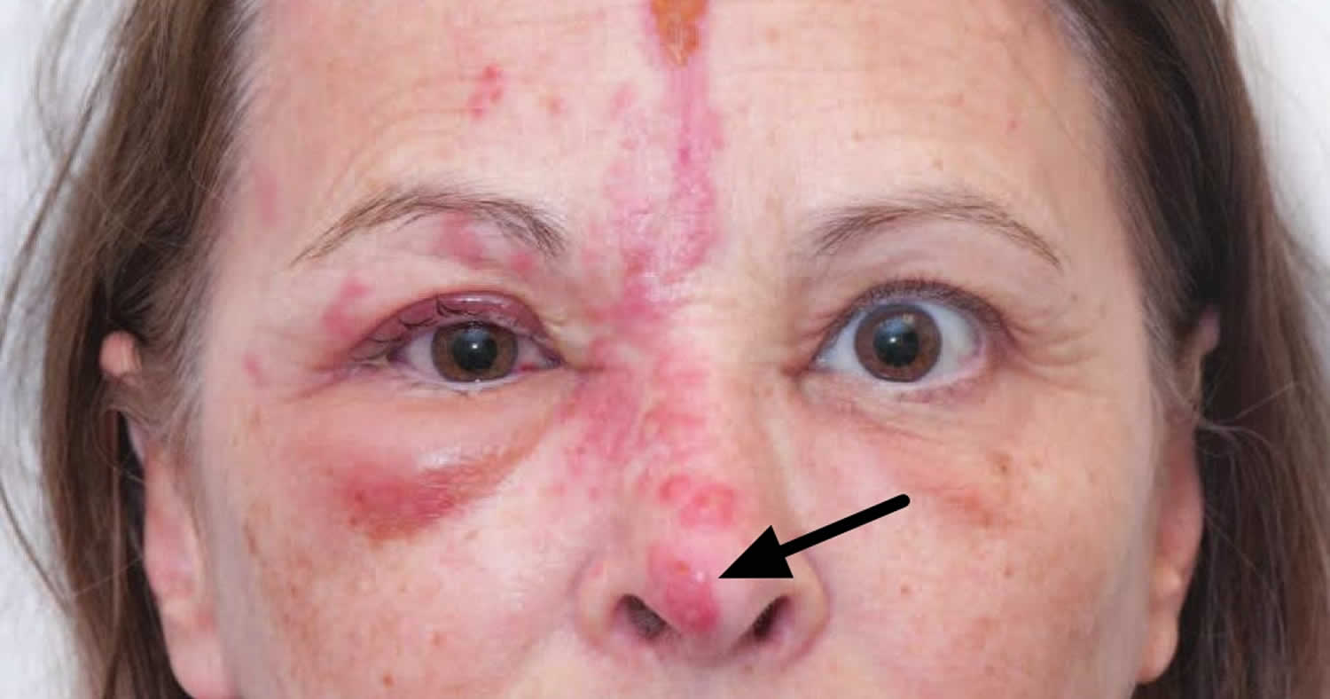
Footnote: This picture showed patient with herpes zoster along the right ophthalmic and maxillary division of the trigeminal nerve, and a crusted blister was found on the tip of her nose.
Hutchinson sign nail (dermatology)
Refers to pigmentation in the paronychial area suggesting aggressive subungual melanoma. Hutchinson’s nail sign is an important clinical clue to subungual melanoma and is characterized by extension of brown or black pigment from the nail bed, matrix, and nail plate to the adjacent cuticle and proximal or lateral nail folds.
Micro Hutchinson’s sign is cuticular pigmentation not easily seen by naked eye examination. Pseudo-Hutchinson’s sign is when the dark pigmentation is visible through the transparent nail fold.
Figure 2. Hutchinson’s sign toenail
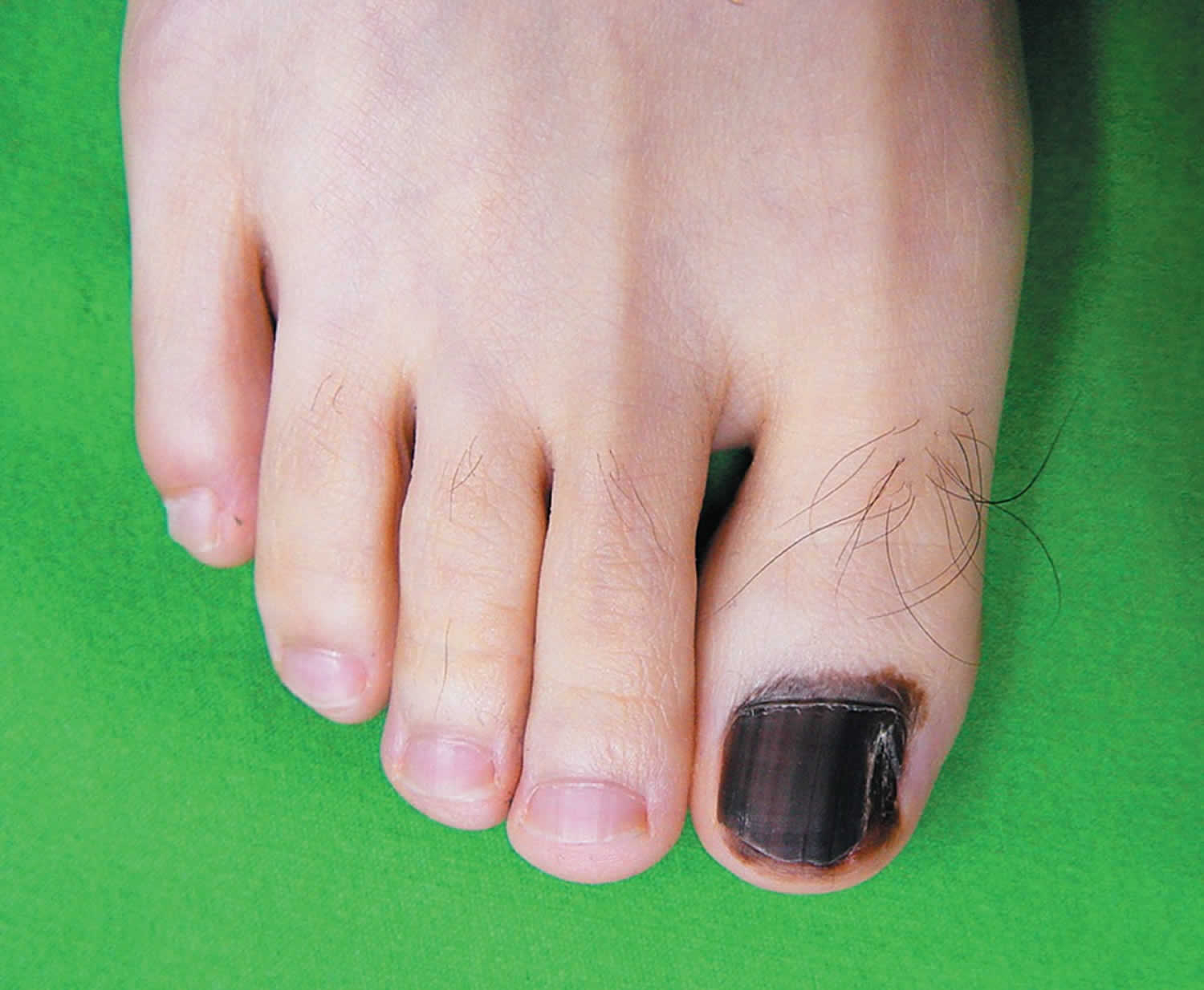
Footnote: An incisional biopsy of the nail matrix showed atypical melanocytes and inflammatory cells along the basal layer of the epidermis, findings consistent with acral lentiginous melanoma in situ. The patient underwent amputation of the great toe, and he remains healthy 8 years later.
- Herpes zoster infection. BMJ 2019;364:k5095 doi: https://doi.org/10.1136/bmj.k5095[↩]


