Hyperviscosity syndrome
Hyperviscosity syndrome is a combination of clinical signs and symptoms related to increased blood viscosity. Hyperviscosity syndrome is an oncologic emergency that classically presents with the triad of neurologic abnormalities, visual changes, and mucosal bleeding 1. Elevated blood viscosity is the result of either red blood cell shape deformity or a pathological increase in serum proteins, red blood cells (RBC), white blood cells (WBC), or platelets 1. The common causes of hyperviscosity syndrome is Waldenstrom macroglobulinemia, multiple myeloma or immune complexes (systemic lupus erythematosis) and therefore, the term hyperviscosity syndrome is typically used to describe an increase in serum proteins. Hyperviscosity syndrome can also be secondary to increased cellular components due to leukemia or myeloproliferative disorders. Management consists of supportive care with intravenous fluids, plasmapheresis, and treatment of the underlying hematological condition 2.
The diagnosis and management of hyperviscosity syndrome is done by an interprofessional team consisting of a hematologist, nephrologist, oncologist, internist, and an intensivist. Plasmapheresis is the mainstay of treatment for hyperviscosity secondary to increased plasma proteins. Leukapheresis, plateletpheresis and phlebotomy are used to treat leukostasis, thrombocytosis and polycythemia, respectively. These various modalities will only treat the symptoms of hyperviscosity syndrome. Definitive therapy is needed to address the underlying condition.
Hyperviscosity syndrome key points
- Hyperviscosity syndrome exists as a clinical triad of neurological symptoms, mucosal bleeding, and visual disturbances.
- Hyperviscosity syndrome can be caused by overproduction of nearly any hematologic component but is most common with hypergammaglobulinemia.
- Symptoms are varied and driven by decreased blood flow through capillary beds.
- Diagnosis of hyperviscosity syndrome is made by determining an elevated serum viscosity which can be suggested by Rouleax formation on peripheral smear or repeated malfunction of laboratory testing equipment.
- The key is to treat the primary cause of hyperviscosity syndrome. A hematology/oncology consultant should administer this, and it is strongly recommended to consult with this expert as soon as hyperviscosity syndrome is identified.
- Short-term management is directed at symptom control, whereas long-term management is directed at controlling the underlying hematologic condition. The mainstays of treatment include supportive therapy, plasma exchange or plasmapheresis, and chemotherapy. The patient must be kept well hydrated at all times to prevent complications. The more definitive short-term treatment is plasmapheresis. It can promptly reverse most clinical manifestations of hyperviscosity syndrome and is usually well tolerated and safe. Phlebotomy may be an option when one is unable to perform plasmapheresis.
Hyperviscosity syndrome causes
Any pathologic elevation of the cellular components (erythrocytes [red blood cells], leukocytes, or platelets) or acellular components (protein) of blood can cause hyperviscosity. Conditions responsible for hyperviscosity syndrome that involve cellular components of blood include polycythemia vera, leukemia, and thrombocytosis. Sickle cell disease and spherocytosis can also contribute to hyperviscosity syndrome due to deformed red blood cells. The pathologic rise of acellular components can either be monoclonal or polyclonal. Monoclonal diseases include myeloma, Waldenstrom macroglobulinemia and cryoglobulinemia. Rheumatic conditions such as seropositive rheumatoid arthritis, systemic lupus erythematosus (SLE) and Sjogren syndrome compromise polyclonal causes of hyperviscosity syndrome as well as Castleman disease and HIV infection 3.
Hypergammaglobulinemia is the most common cause of hyperviscosity syndrome, specifically the monoclonal condition Waldenstrom macroglobulinemia 1. More than 30% of all Waldenstrom macroglobulinemia patients develop hyperviscosity syndrome at some point in their life because of the large star-shaped IgM pentamers that are highly viscous. Myelomas are the second leading cause of hyperviscosity syndrome. About 25% of hyperviscosity syndrome cases secondary to multiple myeloma are caused by IgA myeloma, followed by IgG myelomas at less than 5% of cases.
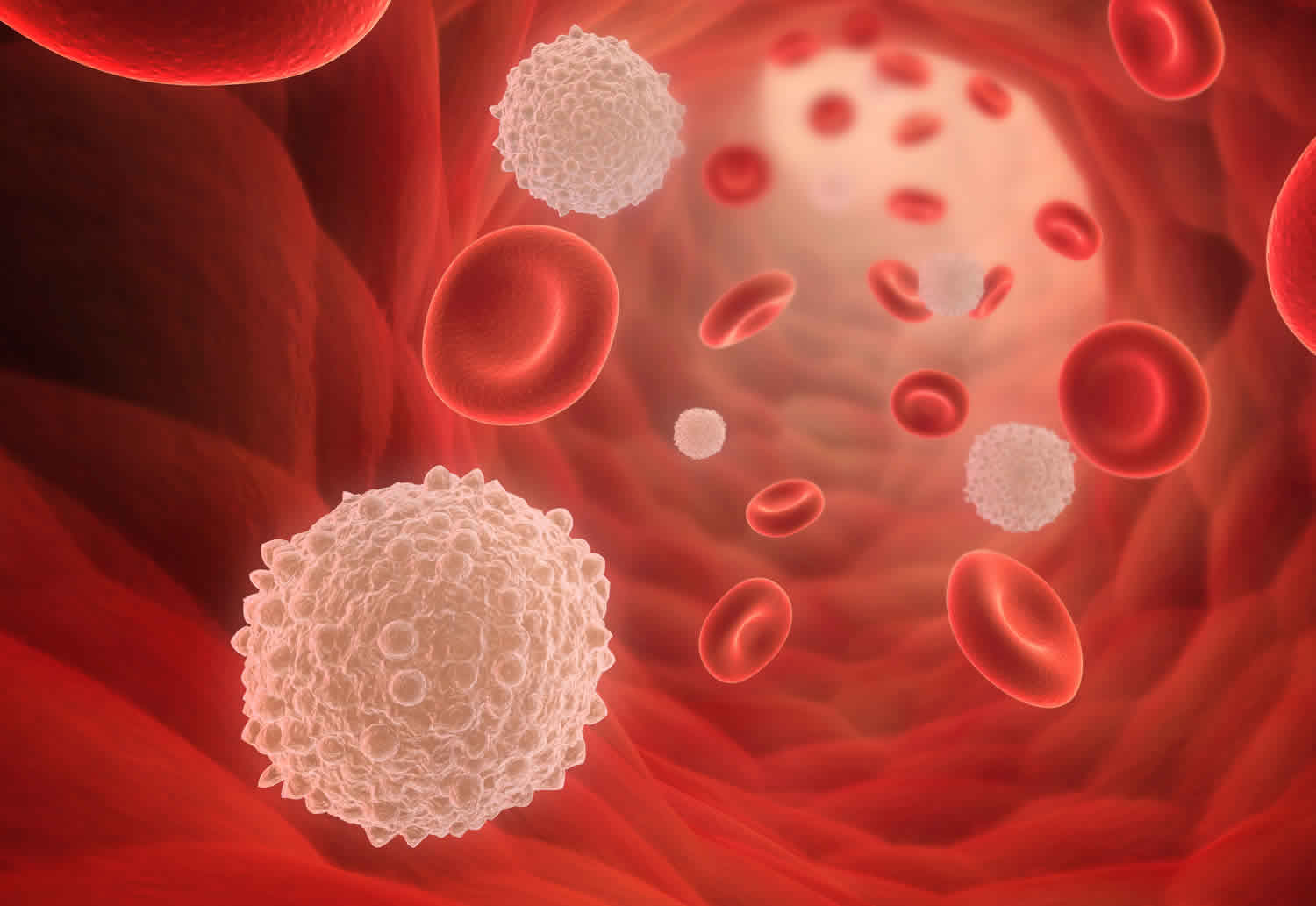
Viscosity is formally defined as the measurement of the internal resistance of a fluid to flow but can simply be thought of as the “thickness” or “stickiness” of a fluid. When fluid has low viscosity, it travels quickly and without much difficulty. Viscous fluids are thicker and travel more slowly. Hyperviscosity syndrome is the pathological condition in which blood is “thicker” than normal and therefore flow is reduced. An increase in blood viscosity can be caused either by a deformity of the shape of red blood cells (RBCs) which causes red blood cell aggregation and decreased blood flow or by any pathological elevation of the components of blood. This includes red blood cells, white blood cell, platelets, or serum proteins.
This increase in viscosity causes sluggish blood flow, relative decreased microvascular circulation, and hypoperfusion of tissues. An increase in circulating proteins can also affect platelet aggregation and cause prolonged bleeding time. The severity of clinical symptoms is directly related to the increased levels of serum viscosity, with progressively more severe symptoms occurring as the individual patient’s serum viscosity increases. The level of viscosity at which symptoms can initially present is variable from person to person depending on the underlying physiology, but for a given patient, symptoms will typically manifest about the same level of viscosity over time.
Hyperviscosity syndrome symptoms
Hyperviscosity syndrome classic triad of symptoms includes neurologic abnormalities, vision changes and mucocal bleeding.
- Neurologic abnormalities: headache, syncope, seizure, vertigo, hearing loss, stupor, stroke.
- Visual disturbances: diplopia, vision loss. Classic findings on fundoscopic examination are dilated, tortuous, “sausage-link” or “box-car” retinal veins.
- Mucosal bleeding: gingival, mucosal, nasal, vaginal.
Other clinical features may include:
- Dyspnea
- Chest pain
- High-output cardiac failure
- Myocardial infarction
Clinical manifestations of hyperviscosity syndrome 4:
Central nervous system
- Headache
- Dizziness and vertigo
- Impaired consciousness
- Somnolence
- Tinnitus and impaired hearing
- Ataxia
- Seizure
Ophthalmologic
- Blurred or loss of vision
- Diplopia
- Retinal vein occlusion
- Papilledema
- Retinal hemorrhage
Mucocutaneous
- Epistaxis
- Gingival bleeding
- Mucocutaneous bleeding
- Gastrointestinal bleeding
Cardiovascular
- High-output cardiac failure
- Renal impairment
- Priapism
Other
- Fatigue
- Malaise
- Shortness of breath
Hyperviscosity syndrome diagnosis
Hyperviscosity is a clinical manifestation of an underlying oncologic process, usually a plasma cell dyscrasia or acute leukemia, though it can sometimes be caused by immune complexes in patients with systemic lupus erythematosis.
There is no single diagnostic test for hyperviscosity syndrome. A high degree of clinical suspicion is required based on history and physical exam findings to diagnose hyperviscosity syndrome. During history taking, it is important to note any current or past hematological disorders as well as any family history of such conditions. Hyperviscosity syndrome should be suspected in any patient who presents with the classic triad of neurologic abnormalities, vision changes and mucosal bleeding; however, a variety of end-organ damages can also be observed as the initial presenting symptom.
Bleeding is the most common manifestation and typically arises from impaired platelet function resulting in oozing mucosal surfaces like epistaxis, bleeding gums, or gastrointestinal bleeding. Neurological findings can include a headache, neuropathic syndromes, generalized stupor, coma, dizziness, ataxia, hearing impairment, seizures, and stroke syndromes. These neurological manifestations are due to decreased blood flow to the central nervous system and deposition of paraproteins within the myelin sheath of peripheral nerves. Retinopathy and visual derangements such as blurred vision or double vision arise because of microvascular changes such as thrombosis or hemorrhage. The classic finding of “sausage link” or “boxcar” engorgement of retinal veins can be seen on the fundoscopic exam as well as papilledema, flame-shaped hemorrhages, or exudates. The eye examination is an important part of the physical exam because it can enable prompt diagnosis and treatment in the appropriate clinical setting. Less commonly seen are cardiopulmonary symptoms such as high output cardiac failure, shortness of breath, valvular dysfunction, or myocardial infarction. hyperviscosity syndrome can also cause acute kidney injury, likely resulting from a relative hypoperfusion state.
Laboratory tests
Laboratory evaluation includes a complete blood count (CBC), peripheral blood smear, serum electrolytes and serum viscosity. Laboratory evidence of high serum viscosity establishes the diagnosis. There is controversy over whether whole blood viscosity versus serum viscosity is superior, but most clinical laboratories measure the viscosity of the serum component of blood. Viscosity is measured in the unit of centipoise (cp). The viscosity of water is 1 cp. Normal serum viscosity relative to water is 1.4 to 1.8 centipoise (cp). Symptoms of hyperviscosity can appear with a serum viscosity as low as 3 cp, but usually, arise when it exceeds 4 to 5 cp 5.
Further testing should include a complete blood count (CBC), full serum chemistries, coagulation profile, and urinalysis. An elevated albumin-protein gap along with significant proteinuria on routine urinalysis suggest an underlying gammopathy. Rouleaux formation on a peripheral blood smear is highly suggestive of serum stasis. Serum stasis can also lead to the malfunction of laboratory testing equipment in which lab samples cannot be analyzed. This should raise suspicion for an underlying increase in serum viscosity. Measuring quantitative immunoglobulins is not necessary to establish a diagnosis of hyperviscosity syndrome, but can help guide long-term treatment if measured before and after the intervention.
- Complete blood count (CBC): patients with hyperviscosity syndrome are typically anemic due to their underlying disease. However, patients with polycythemia vera often present with elevated hemoglobin ( >16 g/dL) and hematocrit (can be as high as 70-80%). Signs and symptoms of leukostasis causing hyperviscosity syndrome are seen with white blood cell counts greater than 100,000, but can also be seen with a lower white blood cell.
- Peripheral blood smear: rouleaux formation can be seen in patient’s paraproteinemia.
- Serum electrolytes: hypercalcemia and hyponatremia are often present in paraproteinemia. The hyponatremia is usually an artifact of the paraproteinemia.
- Serum viscosity: clinical signs and symptoms of hyperviscosity usually occur when the viscosity reaches 4-5 cp (normal range is 1.4-1.8 cp).
Other laboratory studies that may be helpful
- Coagulation panel: prothrombin time (PT), partial thromboplastin time (PTT) and international normalized ratio (INR) are important, especially if the patient presents with bleeding.
- Serum/urine electrophoresis: the presence of a monoclonal spike confirms an underlying gammopathy, usually multiple myeloma or Waldenstrom’s macroglobulinemia
- Urinalysis: significant proteinuria suggests a gammopathy.
- Bone marrow biopsy: may reveal underlying disorder (plasma cell dyscrasia, leukemia, myeloproliferative disorder)
Hyperviscosity syndrome treatment
Hyperviscosity syndrome is medical emergency, and timely treatment can prevent life-threatening complications such as thromboembolic events, myocardial infarction, and catastrophic ischemia that results in multiple organ failure. Therapy should be based on the severity of signs and symptoms rather than the calculated degree of viscosity. Most signs and symptoms are reversible with prompt treatment. Short-term management is directed at symptom control, whereas long-term management is directed at controlling the underlying hematologic condition. The mainstays of treatment include supportive therapy, plasma exchange or plasmapheresis, and chemotherapy. Dehydration can worsen hyperviscosity syndrome, and these patients are usually dehydrated. Therefore, judicious fluid administration is advised. It is considered common practice to empirically administer 1 to 2 L of normal saline when hyperviscosity syndrome is suspected. The more definitive short-term treatment is plasmapheresis. It can promptly reverse most clinical manifestations of hyperviscosity syndrome and is usually well tolerated and safe. Plasmapheresis can decrease serum viscosity by 20% to 30% and can be done on a daily basis until clinical resolution of symptoms. Patients can present with concurrent anemia or acquire dilutional anemia secondary to fluid resuscitation, and it is important to note that transfusion of packed red blood cells can increase blood viscosity. Therefore, one should wait until after plasmapheresis has reduced serum viscosity before transfusing 6.
If prompt plasmapheresis cannot be obtained, a temporizing measure that can be performed emergently is intravenous phlebotomy. This involves phlebotomizing about 1 to 2 units of the patient’s blood and concurrently replacing it with normal saline. However, this has to be performed with caution because aggressive plasma exchange can cause the elimination of clotting factors, albumin, and platelets. Phlebotomy should only be completed in the presence of severe neurological deficits like seizure or coma.
The definitive treatment of hyperviscosity syndrome involves chemotherapy for the underlying hematologic condition. Plasmapheresis does not affect the underlying disease, so chemotherapy is often started concomitantly. A hematology/oncology consultant should administer this, and it is strongly recommended to consult with this expert as soon as hyperviscosity syndrome is identified. Since exchange therapies such as plasmapheresis and leukapheresis are the mainstay of management, these patients may require transfer to a higher level of care facility.
Hyperviscosity syndrome secondary to monoclonal gammopathies
The efficiency of plasmapheresis at removing paraproteins is dependent on the vascular distribution of the proteins. IgM protein is predominantly (70-80%) intravascular, thus a single plasmapheresis procedure can result in a dramatic response. Conversely, IgG and IgA proteins have a large extravascular volume, and several plasmapheresis procedures may be necessary to see the same effect.
In general, daily or every other day single volume plasmapheresis with 5% albumin replacement fluid is performed until symptoms improve. At the same time, disease-specific drug therapy can be started. Treatment options for Waldenstrom’s macroglobulinemia and multiple myeloma include alkylating agents, thalidomide-based therapy, rituximab, bortezomib and nucleoside analogs.
Maintenance plasmapheresis can be performed every 1-4 weeks for treatment of hyperviscosity that is not responsive to chemotherapy.
Hyperviscosity syndrome secondary tol eukemias and/or myeloproliferative disorders
These conditions include acute and chronic leukemias, polycythemia vera (polycythemia vera), and essential thrombocytosis (essential thrombocythemia).
Hyperleukocytosis: Prompt leukocytoreduction is the mainstay of treatment for hyperleukocytosis and is achieved with a combination of leukapheresis, hydroxyurea, and induction chemotherapy. Leukapheresis is usually performed when the white blood cell is greater than 100,000, or less if the patient is symptomatic. A single leaukapheresis can lower the white blood cell count by 20-60% and can be performed daily until the white blood cell count is less than 50,000/mm³. Hydroxyurea should also be given concomitantly at a dose of 50-100 mg/kg/day. This dose has been shown to reduce the white blood cell by 50-60% in 24-48 hours.
Induction chemotherapy should be initiated as soon as possible and is the definitive treatment. Supportive measures include hydration with careful monitoring of fluid balance, especially in patients with cardiac symptoms or comorbidities. It is important that hyperleukocytosis causing leukastasis be recognized and treated promptly, as the mortality rate, if untreated, is as high as 40%.
Erythrocytosis/thrombocytosis: Low-dose aspirin is recommended for all patients with polycythemia vera and essential thrombocythemia. It is recommended that all polycythemia vera patients be phlebotomized to a target hemacrit of 45%. Hydroxurea may be added for high-risk patients. Patients with polycythemia vera or essential thrombocythemia who are resistant to hydroxyurea may be treated with interferon-alpha or busulfan. Plateletpheresis is indicated in patients with essential thrombocythemia for acute thromboembolism or hemorrhage. The goal is normalization of the platelet count.
Hyperviscosity syndrome prognosis
The prognosis for patients with hyperviscosity syndrome depends on the primary tumor and extent of spread. For example, hyperviscosity secondary to an acute leukemia or multiple myeloma has a worse prognosis than that due to polycythemia vera or essential thrombocythemia. If the primary malignancy is beyond control, the outlook is grim 7.
Disease monitoring and follow-up
Hyperviscosity syndrome is a medical emergency, and most patients will remain hospitalized until their symptoms have resolved and their condition is stable. Additional follow-up will depend on the underlying disease.
Hyperviscosity syndrome secondary to monoclonal gammopathies
Plasmapheresis is very effective at removing paraproteins from the circulation, particularly IgM proteins. Because viscosity is logarithmically related to paraprotein levels, even small reductions can reduce viscosity and improve symptoms. Patients with Waldenstrom’s macroglobulinemia tend to respond better than patients with multiple myeloma.
Hyperviscosity syndrome secondary to leukemias and/or myeloproliferative disorders
Hyperleukocytosis: Though leukapheresis is very effective at lowering the blast count in acute leukemias, there appears to be no improvement in survival. Early death due to leukostasis may still occur. Predictors of poor prognosis are renal failure, respiratory distress, neurologic symptoms or coagulopathy.
Erythrocytosis/Thrombocytosis: Rapid improvement in clinical symptoms can be seen after one procedure.
If patients do not respond after 2-3 plasmapheresis/leukapheresis procedures, other causes for their symptoms should be investigated.
- Perez Rogers A, Estes M. Hyperviscosity Syndrome. [Updated 2020 Apr 27]. In: StatPearls [Internet]. Treasure Island (FL): StatPearls Publishing; 2020 Jan-. Available from: https://www.ncbi.nlm.nih.gov/books/NBK518963[↩][↩][↩]
- Georgakopoulos CD, Plotas P, Angelakis A, Kagkelaris K, Tzouvara E, Makri OE. Dexamethasone implant for immunogammopathy maculopathy associated with IgA multiple myeloma. Ther Adv Ophthalmol. 2019 Jan-Dec;11:2515841418820441[↩]
- Tedeschi A, Conticello C, Rizzi R, Benevolo G, Laurenti L, Petrucci MT, Zaja F, Varettoni M. Diagnostic framing of IgM monoclonal gammopathy: Focus on Waldenström macroglobulinemia. Hematol Oncol. 2019 Apr;37(2):117-128.[↩]
- Hyperviscosity Syndrome in Paraprotein Secreting Conditions Including Waldenstrom Macroglobulinemia. Front Oncol. 2020; 10: 815. doi: 10.3389/fonc.2020.00815 https://www.ncbi.nlm.nih.gov/pmc/articles/PMC7248405[↩]
- Castillo JJ, Garcia-Sanz R, Hatjiharissi E, Kyle RA, Leleu X, McMaster M, Merlini G, Minnema MC, Morra E, Owen RG, Poulain S, Stone MJ, Tam C, Varettoni M, Dimopoulos MA, Treon SP, Kastritis E. Recommendations for the diagnosis and initial evaluation of patients with Waldenström Macroglobulinaemia: A Task Force from the 8th International Workshop on Waldenström Macroglobulinaemia. Br. J. Haematol. 2016 Oct;175(1):77-86.[↩]
- Ballestri M, Ferrari F, Magistroni R, Mariano M, Ceccherelli GB, Milanti G, De Palma M, Albertazzi A. Plasma exchange in acute and chronic hyperviscosity syndrome: a rheological approach and guidelines study. Ann. Ist. Super. Sanita. 2007;43(2):171-5.[↩]
- Chen YY, Yen YF, Lin JX, Feng SC, Wei LC, Lai YJ, Shen YC. Risk of Ischemic Stroke, Hemorrhagic Stroke, and All-Cause Mortality in Retinal Vein Occlusion: A Nationwide Population-Based Cohort Study. J Ophthalmol. 2018;2018:8629429[↩]


