The immune system
The immune system is made up of a complex network of cells, chemicals, tissues and organs that cooperate to protect you from invasion and harm from a variety of infectious agents such as bacteria, viruses, fungi, parasites, toxins (chemicals produced by microbes) and other invaders (such as cancer cells) and also to provide you with a surveillance system to monitor the integrity of your tissues. The immune system recognizes invaders such as bacteria, viruses and fungi as well as abnormal cells. It mounts an immune response to help the body fight the invasion. The ability to recognize and respond to foreign entities is central to the operation of the immune system. Many levels of protection are involved in this process. When harmful microbes (tiny particles) enter and invade your body, your body produces white blood cells to fight the infection. The white blood cells identify the microbe, produce antibodies to fight it, and help other immune responses to occur. They also ‘remember’ the attack. This is how vaccinations work. Vaccines expose the immune system to a dead or weakened microbe or to proteins from a microbe, so that the body is able to recognize and respond very quickly to any future exposure to the same microbe. Although the immune system is quite elaborate, its function can be boiled down to four basic roles:
- Creating a barrier to prevent pathogens from entering your body. The barrier function of the immune system acts to prevent pathogens from entering your body from the external environment. Important initial barriers to infection are physical (e.g. the skin), enhanced by substances secreted by your body, such as saliva and tears, that contain molecules that can neutralize bacteria. Chemical barriers like the acid pH of the stomach; and biological barriers like the presence of commensal organisms on the skin and in the intestinal tract, secretions like immunoglobulin A (IgA) and antimicrobial proteins in saliva and tears, and the complement system. The internal mucosal tissues (e.g. lungs & airways, and the gut) are coated with mucus that is able to trap potential infectious agents. In the airways, mobile ciliate hairs work together to transport contaminants away from vulnerable areas. Tissues such as the skin, mucosal surfaces and airways also contain populations of immune cells that can respond to infectious agents that breach these physical defences.
- Identifying pathogens if they breech a barrier. Pathogens are recognized by cells of the innate immune system, such as macrophages, monocytes and dendritic cells. This is achieved through the presence of pattern recognition receptors (PRRs) that recognize general molecular structures that are broadly shared by groups of pathogens. These structures are termed pathogen-associated molecular patterns or PAMPs. When pattern recognition receptors (PRRs) recognize pathogen-associated molecular patterns (PAMPs), the first line of host defensive responses is activated (see Figure 3 below) 1. PRRs include Toll-like receptors (TLRs). More than 10 functional TLRs have been identified in humans, each one detecting distinct MAMPs from bacteria, viruses, fungi and parasites. The best described of these are TLR4 which recognizes the lipopolysaccharides from the cell wall of Gram-negative bacteria and TLR2 which recognizes the lipoteichoic acid from the cell wall of Gram-positive bacteria 1. Several Toll-like receptors (TLRs) are expressed on the cell surface of innate immune cells because the pathogens they recognize, mainly bacteria, are extracellular. Because viruses enter host cells, it is important that there are also intracellular TLRs. Indeed, intracellular TLRs that recognize viral DNA, viral double-stranded RNA and viral single-stranded RNA exist. Among these, TLR7 and TLR8 are found in macrophages, monocytes, dendritic cells and some other cell types and are likely to be important in innate recognition of the single-stranded RNA of coronaviruses 2.
- Eliminating pathogens by a diverse repertoire of cells and molecules that act in concert to neutralize the potential threat. Extracellular bacteria can be engulfed by phagocytic cells that include macrophages and dendritic cells. After digestion of internalized bacteria, peptide fragments, termed antigens, are presented on the surface of the phagocytic cells (via Major Histocompatibility Complex 2 [MHC-II]) to antigen-specific CD4+ helper T lymphocytes. The activated helper T lymphocytes (specifically the T helper 1 phenotype [TH1]) proliferate and produce cytokines including interleukin (IL)-2 and interferon (IFN)-γ. IFN-γ promotes antigen-specific antibody production by B lymphocytes. These antibodies coat the bacteria, neutralising them and making the process of phagocytosis more efficient. In parallel with phagocytosis, innate immune cell recognition of pathogens via PRRs triggers inflammatory signalling, activation of transcription factors like nuclear factor kappa-light-chain-enhancer of activated B cells (NFκB), inflammasome assembly, and production of classic inflammatory cytokines like tumor necrosis factor (TNF), IL-1β and IL-12. Viral infection of some cell types promotes release of type 1 IFNs (IFN-α and IFN-β) and these induce antiviral resistance, in part through activation of natural killer cells (NK cells) 3. Furthermore, virally infected cells directly activate natural killer (NK) cells which act to kill the infected cell. In addition, PRR signalling induces maturation of dendritic cells which are responsible for viral antigen processing and presentation, so initiating acquired immunity. Upregulation of MHC I on virally infected cells including both respiratory epithelial cells and dendritic cells results in presentation of viral antigens to CD8+ cytotoxic T lymphocytes. This activates them to kill virally infected cells through the release of pore forming proteins like perforin. Presentation of viral antigens via MHC II and the cytokine milieu lead to the activation of CD4+ helper T lymphocytes with switching to the T helper 1 phenotype. These cells produce IL-2, which promotes cytotoxic T lymphocyte activity, and IFN-γ, which promotes differentiation of B lymphocytes to plasma cells which produce antiviral antibodies. These antibodies can bind to free viruses neutralising them. The processes involved in antiviral immunity are summarised in figure 1.
- Generating an immunological memory. Immunological memory refers to the ability of the immune system to quickly and specifically recognize an antigen that your body has previously encountered and initiate a corresponding immune response. There are two aspects of immunological memory. First, antibodies can persist in the circulation for many months to many years, providing protection against reinfection. Second, after the cessation of an active immune response, a small number of memory T (both CD4+ and CD8+) and B lymphocytes remain; they are in a resting state but if they encounter the same antigen that triggered their formation they are able to respond immediately and lead to rapid elimination of the source of the antigen. Memory cells have a long life (up to several decades). Immunological memory is the basis of vaccination.
- Aging can be associated with a loss of immune competence, a process called immunosenescence 4. One factor linked to immunosenescence is decreased output of immune cells from bone marrow, the site of origin of all immune cells. In addition, involution of the thymus with age decreases output of naive T lymphocytes, resulting in reduced capacity to respond to new antigens. Immunosenescence means that, compared with younger adults, older people have increased susceptibility to infections including respiratory tract infections and pneumonia and poorer responses to vaccination 5. The gut mucosa is the largest site of immune tissue in humans and senescence of the gut mucosal immune system has been demonstrated in mice models, with reductions in secretory immunoglobulin A (IgA) responses, impaired oral tolerance to new antigens and impaired mucosal dendritic cell function 6. Paradoxically, ageing is also linked to an increase in blood concentrations of many inflammatory mediators, a situation termed inflammation in ageing or inflammageing 7. This state is considered to contribute to an increased risk of chronic conditions of aging like cardiovascular disease, metabolic disease (diabetes, non-alcoholic fatty liver disease), neurodegeneration and some cancers 7 and may predispose to mounting an excessive inflammatory response when infected. Although inflammation is part of the innate immune response and innate and acquired immunity should work in a coordinated and integrated way, an excessive inflammatory response can lead to impairments in acquired immunity 7.
- Obesity can be associated with a loss of immune competence 8, with impairments of the activity of helper T lymphocytes (TH), cytotoxic T lymphocytes, B lymphocytes and natural killer cells 9 and reduced antibody and IFN-γ production 10. This means that, compared with healthy weight individuals, the obese have increased susceptibility to a range of bacterial, viral and fungal infections 11 and poorer responses to vaccination 12. The impact of obesity has been well explored in relation to influenza infection and vaccination against influenza. During the 2009 H1N1 influenza A virus pandemic, obese individuals showed delayed and weakened antiviral responses to infection and showed poorer recovery from disease compared with healthy weight individuals 12. Animal studies and case studies in humans show that obesity is associated with prolonged shedding of influenza virus, indicating an impairment in viral control and killing, and the emergence of virulent minor variants 12. Green and Beck 13 note that compared with healthy weight individuals, vaccinated obese individuals have twice the risk of influenza or influenza-like illness, indicating poorer protection from vaccination in the obese. Sheridan et al 14 investigated the responses of immune cells from the blood of healthy weight, overweight and obese individuals to the influenza vaccine in vitro (test tubes). Exposure of the blood immune cells to the vaccine increased the number of activated cytotoxic T lymphocytes, the number of granzyme expressing cytotoxic T lymphocytes and the number of IFN-γ producing cytotoxic T lymphocytes. However, the responses of cells from obese individuals were blunted by 40%, almost 60% and 65%, respectively. Cells from overweight individuals showed responses intermediate between those from healthy weight and obese individuals. Similar findings for the response of blood cells to the pandemic H1N1 influenza A virus were reported by Paich et al 15. Paradoxically, obesity is also linked to an increase in blood concentrations of many inflammatory mediators, a state of chronic low-grade inflammation 16. This state is considered to contribute to an increased risk of chronic conditions of ageing 16 and may predispose to mounting an excessive inflammatory response when infected.
Therefore, a critical role of the immune system is to determine what is foreign (what immunologists often call “nonself”) from what is normally present in your body (i.e., self). As a consequence, the cells and molecules that comprise the innate immune system are preoccupied with detecting the presence of particular molecular patterns that are typically associated with infectious agents. Innate immunity involves the release of cytokines, complement, and chemokines, as well as neutrophils and macrophages to destroy the invading pathogens. When this is not enough, an antigen-specific or adaptive immune response becomes initiated, and antibodies, B cells, and T cells enter the battle 17. The generation of a specific response to an antigen is referred to as active immunity. Active immunity plays a vital role in immune responses in the event of re-exposure and your utilization of vaccines.
In addition to the immune system fundamental roles of recognition and elimination of infectious agents, it is also very useful to be able to learn from encounters with pathogens and to maintain a reserve of cells that are able to respond swiftly to a new infection with a previously encountered microbe. Forewarned is forearmed, and in this situation it may be possible to deliver a decisive blow that ends a nascent infection before it has begun. Fortunately, your immune systems have also acquired this ability, which is what your adaptive acquired immune system excels in, and this property is termed immunological memory.
Relevant terms and definitions for the immune system 17:
- Immunogen: Protein or carbohydrate that is recognized and sufficiently activates an immune response
- Antigen: A molecule that is recognized by a specific antibody or T-cell receptor (TCR)
- Antibodies also known as immunoglobulins (Ig) play an important role in the immune system mechanisms of defense against extracellular microorganisms such as bacteria. Antibodies (immunoglobulins) are produced by plasma cells, which as permanently differentiated B-cells that secrete antibodies. Antibodies (immunoglobulins) fight off extracellular pathogens, for instance, bacteria and can neutralize viruses when they are in the bloodstream and other body fluids. Normal individuals have 5 classes of immunoglobulins, which are IgM, IgG, IgA, IgD and IgE and immunoglobulin subclasses including IgG1, IgG2, IgG3, IgG4, IgA1, and IgA2 18. While they have overlapping roles, IgM generally is important for complement activation; IgD is involved in activating basophils; IgG is important for neutralization, opsonization, and complement activation; IgA is essential for neutralization in the gastrointestinal tract; and IgE is necessary for activating mast cells in parasitic and allergic responses. Antibodies are expressed in two ways. The B-cell receptor (BCR), which sits on the surface of a B cell, is actually an antibody. B cells also secrete antibodies to diffuse and bind to pathogens. This dual expression is important because the initial problem, for instance a bacterium, is recognized by a unique B-cell receptor (BCR) and activates the B cell. The activated B cell responds by secreting antibodies, essentially the BCR but in soluble form. This ensures that the response is specific against the bacterium that started the whole process. Antibody deficiencies may occur due to lack of B-cells maturation, missing enzymes or failure of T-cell stimulatory signals for appropriate antibody production. In transient hypogammaglobulinemia of infancy, recurrent bacterial infections occur in children until their immune system matures 19.
- Adjuvant: Prolongs the presence of antigen in tissue and enhances the immune response to an antigen; used in acquired or artificial immunization (vaccinations)
- Macrophages: Macrophages are white blood cells that swallow up and digest germs and dead or dying cells. The macrophages leave behind parts of the invading germs, called “antigens”. The body identifies antigens as dangerous and stimulates antibodies (also known as immunoglobulins [Ig]) to attack them.
- B-lymphocytes (B-cells): B-lymphocytes are defensive white blood cells. B-lymphocytes produce antibodies (also known as immunoglobulins [Ig]) that attack the pieces of the virus left behind by the macrophages.
- T-lymphocytes (T-cells): T-lymphocytes are another type of defensive white blood cell. T-lymphocytes attack cells in the body that have already been infected.
- Dendritic cells (Antigen-Presenting cells or APCs): Facilitate activation of an antigen-specific response by the innate immune system; present antigens via major histone complexes to activate CD8+ cytotoxic T cells and CD4+ helper T cells
- CD4+ helper T cells: Facilitate cell-to-cell interactions and cytokine release to activate and control immune and inflammatory responses. Initially, CD4+ helper T cells are naive and must be activated to begin immune functions. This activation occurs by interaction with pro-Antigen-Presenting cells (“professional” antigen-presenting cells), mainly dendritic cells in lymph nodules/follicles, which leads to an intracellular pathway that up-regulates more antigen-specific TCRs on the T cell and leads to effector functions. T cells can only recognize protein-based antigens. TCRs (T cell receptors) and their co-receptors, such as CD3 and CD4 found on these cells form a complex with the major histocompatibility complex 2 (MHC-II) receptor and the antigen in question. The CD4+ cells are then activated and produce cytokines to initiate immune responses from other white blood cells/other immune cells of cell-mediated immunity. They also activate the T cell-dependent branch of humoral immunity, in which CD4+ T cells recognize protein antigens (which normally would elicit a weak or absent B cell response) and activate B cells to produce immunoglobulin in response to the antigen 20. There are three different subtypes of CD4+ helper T cells, each with a unique function: TH1 CD4+ T cells, TH2 CD4+ T cells, and TH17 CD4+ T cells.
- CD8+ cytotoxic T cells also known as killer T cells, CD8+ T cells or cytotoxic lymphocytes (CTLs), are immune cells for cell-mediated immunity. CD8+ T cells are crucial for recognizing and removing virus-infected cells and cancer cells. CD8+ cytotoxic T cells have specialized compartments, or granules, containing cytotoxins that cause apoptosis, i.e., programmed cell death. Because of its potency, the release of granules is tightly regulated by the immune system. Initially, CD8+ T cells are naive and must be activated to begin immune functions. This activation occurs by interaction with pro-Antigen-Presenting cells (“professional” antigen-presenting cells), mainly dendritic cells in lymph nodules/follicles, and leads to an intracellular pathway that up-regulates more antigen-specific T-cell receptors (TCRs) on the T cell and leads to immune functions. T cells can only recognize protein-based antigens. T-cell receptors (TCRs) and their co-receptors, such as CD3 and CD8 found on these cells form a complex with the major histocompatibility complex 1 (MHC-I) receptor and the antigen in question. Once an activated CD8+ migrates into circulation looking for antigens presented by the major histocompatibility complex 1 (MHC-I) molecules present on all nucleated cells and finds its antigenic target (expressed on a virally infected cell or cell with intracellular bacteria, for example), it utilizes its killing function 21. The killing function of CD8+ T cells is mediated by 1 of 2 mechanisms. The first mechanism involves the use of the Fas/Fas Ligand (FasL). Activated CD8+ T cells express FasL which binds to Fas (CD95), a receptor found on many cell types, leading to the activation of caspases and subsequent apoptosis of the target cell (often a cell infected with intracellular bacteria, such as Listeria species or a virally infected cell). The second method that activated CD8+ T cells can use for their killing function involves the release of granzymes and perforin (two compounds that bypass cell walls and active caspases). Activated CD8+ T cells also secrete interferon gamma (IFN-γ) (a cytokine used in the activation of macrophages/other immune processes) 22.
- Major histocompatibility complex 1 (MHC-I) also known as human leukocyte antigen (HLA): Found on all nucleated cells, play a significant role in determining “self.” Responsible for presenting intracellular antigens to CD8 T cells
- Major histocompatibility complex 2 (MHC-II): Found on antigen-presenting cells that interact with CD4 T cells; responsible for presenting exogenous antigens
- TH0 or activated T helper cell: The initial role of activated CD4+ T cells, promotes cell immunity by activating dendritic cells and stimulating lymphocyte growth; releases cytokines IL-2, IL-4, and IFN-gamma; can develop into TH1, TH2, TH17, or other TH cells
- T helper lymphocyte 1 (TH1 CD4+ T cells): TH1 CD4+ cells are important in the activation of macrophages and fighting off intracellular infections especially bacteria and fungi. TH1 CD4+ cells secrete the cytokine IFN-gamma which activates macrophages, B cells also known as B lymphocytes (to produce immunoglobulin G or IgG) and increased surface expression of the MHC 2 markers on macrophages/antigen-presenting cells. TNF-a is also secreted by these cells to activate dendritic cell migration. The activated macrophages then produced IL-12, which increases differentiation/production of TH1 T cells, further amplifying the immune response. TH1 T cells also play roles in several autoimmune reactions and disease, notably in delayed-type hypersensitivity.
- T helper lymphocyte 2 (TH2 CD4+ T cells): The response that occurs in the absence of IL-12 and IFN-gamma; promotes systemic antibody driven response. TH2 CD4+ T cells are important in combating helminthic infections. TH2 CD4+ T cells produce IL-4, IL-5, IL-10 and IL-13 cytokines, which activate and expand mast cells and eosinophils to clear the parasitic infection 23. Macrophages are also activated by TH2 CD4+ T cells to begin clear of cellular debris and inflammation caused by large parasites. TH2 cells also appear to play a role in allergic diseases/allergies.
- T helper lymphocyte 17 (TH17 CD4+ T cells): TH17 CD4+ T cells are vital in mucosal immunity and are involved in combating extracellular bacteria and and fungal infections when IL-23 is released instead of IL-12. TH17 cells produce IL17A, IL17F, IL-22, TNF-alpha, and chemokines. These cytokines activate neutrophils and monocytes as well as driving increased inflammation. It is this pro-inflammatory function that appears to play a role in the development of autoimmune inflammatory disorders (such as inflammatory bowel disease and rheumatoid arthritis) when the response is pathogenic 24.
- T regulatory cells (T regs) also known as suppressor T cells. T regulatory cells as the name suggests, monitor and inhibit the activity of other T cells. T regulatory cells or suppressor T cells modulate immune responses and inhibit autoimmune processes such as autoreactive immune cells. T regulatory cells prevent adverse immune activation and maintain tolerance or the prevention of immune responses against the body’s own cells and antigens. T regulatory cells produce inhibitory IL-10 and IL-35 cytokines to control immune responses, as well as using CTLA-4 to inhibit B7 on antigen-presenting cells to decrease immune responses. Production of T regulatory cells is spurred by cytokine IL-2 (so much so that in autoimmune disease IL-2 is often absent) and TGF-beta. T regulatory cells express the T-cell receptors (TCRs) CD3, CD4 (they likely share a common lineage with CD4+ Helper T cells), CTLA-4 and CD25. T regulatory cells also express the biomarker FOXP3 (a transcription marker important in their development and immune functions). There has been a recent interest in T regulatory cells as a method to promote wound healing after surgery and to battle cancers 25.
- B cells also known as B lymphocytes. B cells have two major functions: they present antigens to T cells, and more importantly, B cells produce antibodies to neutralize infectious microbes. Antibodies coat the surface of a pathogen and serve three major roles: neutralization, opsonization, and complement activation. Neutralization occurs when the pathogen, because it is covered in antibodies, is unable to bind and infect host cells. In opsonization, an antibody-bound pathogen serves as a red flag to alert immune cells like neutrophils and macrophages, to engulf and digest the pathogen. Complement is a process for directly destroying, or lysing, bacteria.
- Plasma cells: Permanently differentiated B-cells that secrete antibodies
- Memory B cells: Long-lasting B-cells that are responsive to one particular antigen and become activated with re-exposure to the same antigen.
- Vaccination or immunization is a way to train your immune system against a specific pathogen. Vaccination achieves immune memory without an actual infection, so the body is prepared when the virus or bacterium enters. Saving time is important to prevent a pathogen from establishing itself and infecting more cells in the body. An effective vaccine will optimally activate both the innate and adaptive immune response. An immunogen is used to activate the adaptive immune response so that specific memory cells are generated. Because B-cell receptors (BCRs) and T-cell receptors (TCRs) are unique, some memory cells are simply better at eliminating the pathogen. The goal of vaccine design is to select immunogens that will generate the most effective and efficient memory response against a particular pathogen. Adjuvants, which are important for activating innate immunity, can be added to vaccines to optimize the immune response. Innate immunity recognizes broad patterns, and without innate responses, adaptive immunity cannot be optimally achieved.
Figure 1. Immune system
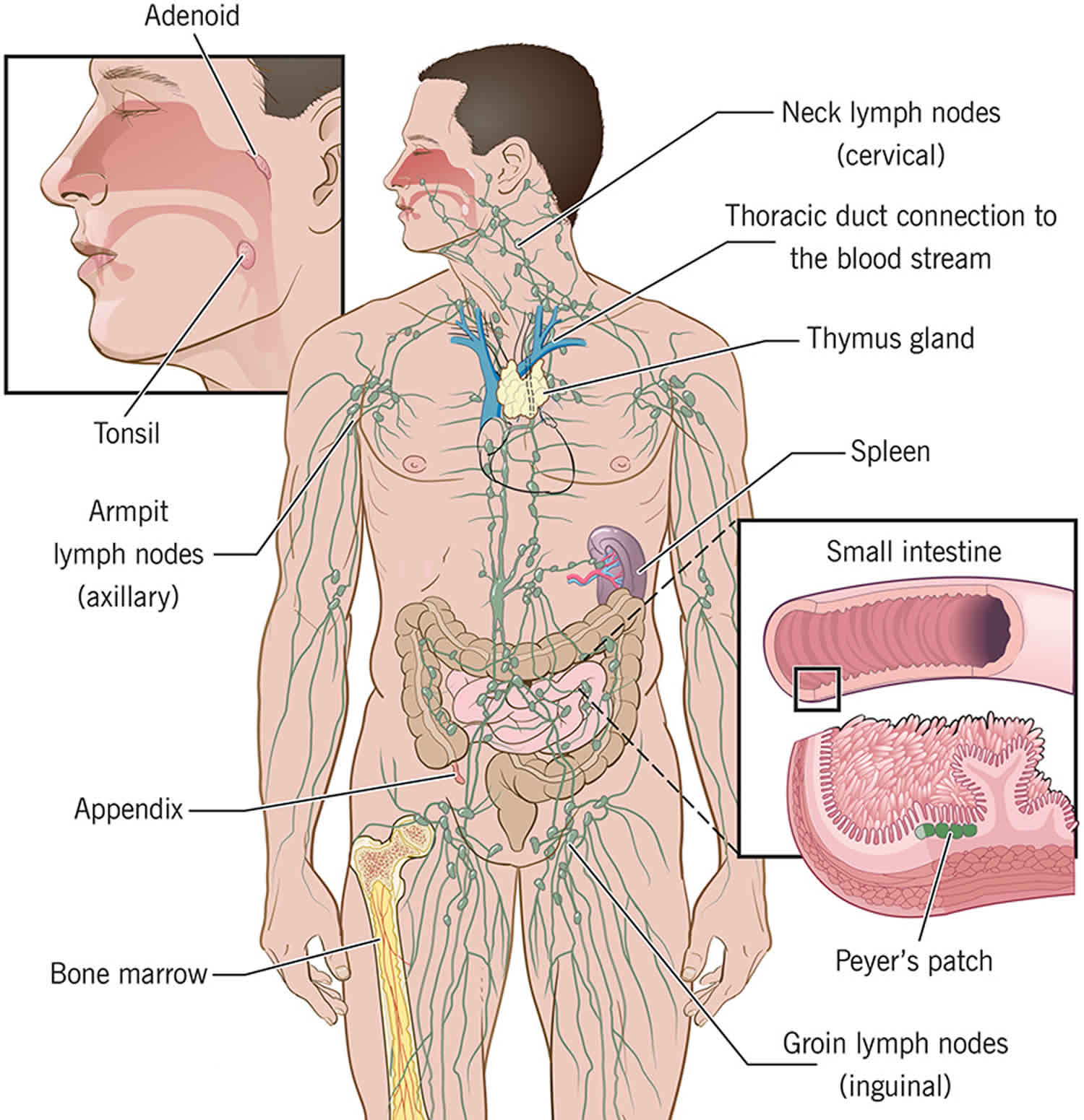
Figure 2. Cells of the immune system
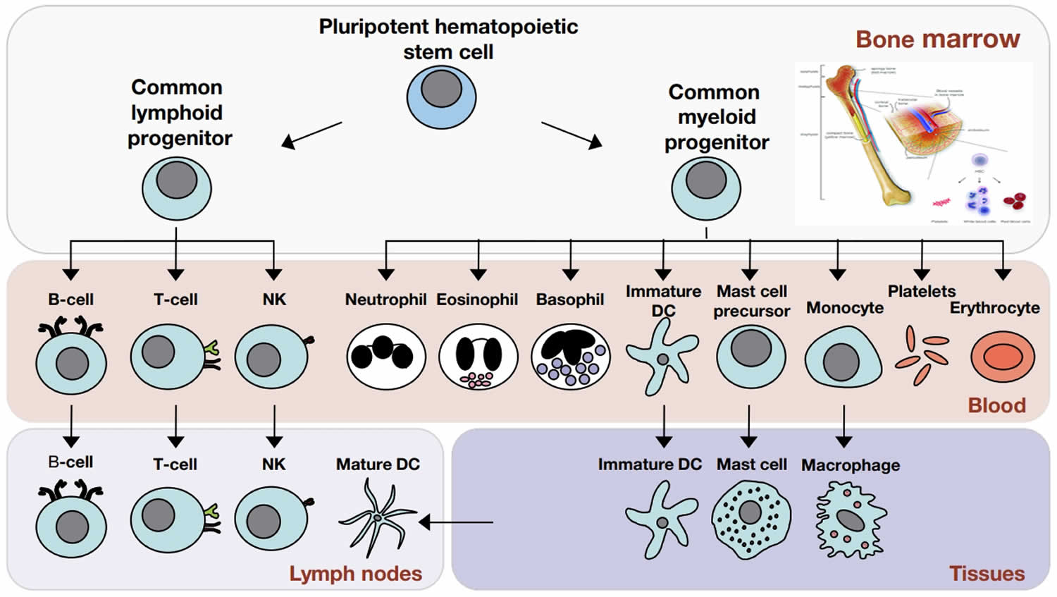
Footnote: The cells of the immune system originate in the bone marrow from pluripotent hematopoietic stem cells. Pluripotent hematopoietic stem cells give rise to a common lymphoid progenitor, which gives rise to all of the major lymphoid cell types (T‐cells, B‐cells, and Natural killer [NK] cells) or a common myeloid progenitor, which gives rise to all of the major myeloid cell types (neutrophils, eosinophils, basophils, dendritic cells (DCs), mast cells, and monocytes/macrophages) as well as the erythrocytes and megakaryocytes (which generate platelets).
- Granulocytes include basophils, eosinophils, and neutrophils. Basophils and eosinophils are important for host defense against parasites. They also are involved in allergic reactions.
- Neutrophils, the most numerous innate immune cell, patrol for problems by circulating in the bloodstream. Neutrophils can phagocytose, or ingest, bacteria, degrading them inside special compartments called vesicles.
- Mast cells also are important for defense against parasites. Mast cells are found in tissues and can mediate allergic reactions by releasing inflammatory chemicals like histamine.
- Monocytes, which develop into macrophages, also patrol and respond to problems. They are found in the bloodstream and in tissues. Macrophages, “big eater” in Greek, are named for their ability to ingest and degrade bacteria. Upon activation, monocytes and macrophages coordinate an immune response by notifying other immune cells of the problem. Macrophages also have important non-immune functions, such as recycling dead cells, like red blood cells, and clearing away cellular debris. These “housekeeping” functions occur without activation of an immune response.
- Dendritic cells (DC) are an important antigen-presenting cell (APC), and they also can develop from monocytes. Antigens are molecules from pathogens, host cells, and allergens that may be recognized by adaptive immune cells. APCs like DCs are responsible for processing large molecules into “readable” fragments (antigens) recognized by adaptive B or T cells. However, antigens alone cannot activate T cells. They must be presented with the appropriate major histocompatiblity complex (MHC) expressed on the APC. MHC provides a checkpoint and helps immune cells distinguish between host and foreign cells.
- Natural killer (NK) cells have features of both innate and adaptive immunity. They are important for recognizing and killing virus-infected cells or tumor cells. They contain intracellular compartments called granules, which are filled with proteins that can form holes in the target cell and also cause apoptosis, the process for programmed cell death. It is important to distinguish between apoptosis and other forms of cell death like necrosis. Apoptosis, unlike necrosis, does not release danger signals that can lead to greater immune activation and inflammation. Through apoptosis, immune cells can discreetly remove infected cells and limit bystander damage. Recently, researchers have shown in mouse models that NK cells, like adaptive cells, can be retained as memory cells and respond to subsequent infections by the same pathogen.
Figure 3. Innate immunity
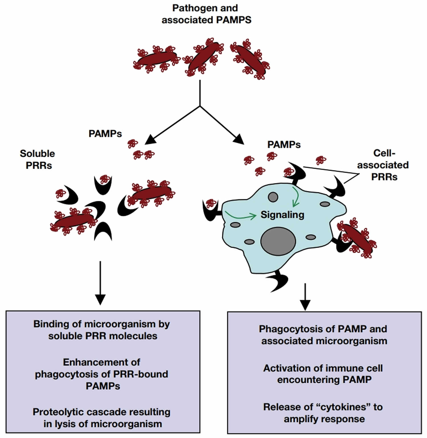
Footnote: Pattern recognition receptors (PRRs) detect pathogen‐associated molecular patterns (PAMPs) and initiate immune responses. PRRs can be either soluble or cell‐associated and can instigate a range of responses upon encountering their appropriate ligands.
How does the immune system work?
The immune system involves many parts of your body. Each part plays a role in recognizing foreign microbes, communicating with other parts of your body, and working to fight the infection. Parts of the immune system are:
- Skin – The skin is usually the first line of defense against microbes. Skin cells produce and secrete important antimicrobial proteins, and immune cells can be found in specific layers of skin.
- Bone marrow – helps produce immune cells. The bone marrow contains stems cells that can develop into a variety of cell types. The common myeloid progenitor stem cell in the bone marrow is the precursor to innate immune cells—neutrophils, eosinophils, basophils, mast cells, monocytes, dendritic cells, and macrophages—that are important first-line responders to infection. The common lymphoid progenitor stem cell leads to adaptive immune cells—B cells and T cells—that are responsible for mounting responses to specific microbes based on previous encounters (immunological memory). Natural killer (NK) cells also are derived from the common lymphoid progenitor and share features of both innate and adaptive immune cells, as they provide immediate defenses like innate cells but also may be retained as memory cells like adaptive cells. B, T, and NK cells also are called lymphocytes.
- Bloodstream: Immune cells constantly circulate throughout the bloodstream, patrolling for problems. When blood tests are used to monitor white blood cells, another term for immune cells, a snapshot of the immune system is taken. If a cell type is either scarce or overabundant in the bloodstream, this may reflect a problem.
- The thymus, a small gland in your upper chest where some immune T cells mature.
- Lymphatic system is a network of of tiny vessels which allows immune cells to travel between tissues and the bloodstream. The lymphatic system contains lymphocytes (white blood cells; mostly T cells and B cells), which try to recognize any bacteria, viruses or other foreign substances in the body and fight them. They are carried in a milky fluid called lymph. Immune cells are carried through the lymphatic system and converge in lymph nodes, which are found throughout the body.
- Lymph nodes, small lumps in the groin, armpit, around the neck and elsewhere that help the lymphatic system to communicate. Lymph nodes are a communication hub where immune cells sample information brought in from the body. They can become swollen when the body mounts an immune response. For instance, if adaptive immune cells in the lymph node recognize pieces of a microbe brought in from a distant area, they will activate, replicate, and leave the lymph node to circulate and address the pathogen. Thus, doctors may check patients for swollen lymph nodes, which may indicate an active immune response.
- The spleen is an organ under the ribs behind the stomach on the left that processes information from the blood. While it is not directly connected to the lymphatic system, it is important for processing information from the bloodstream. Immune cells are enriched in specific areas of the spleen, and upon recognizing blood-borne pathogens, they will activate and respond accordingly.
- Mucous membranes, like the lining of the inside of your mouth are prime entry points for pathogens, and specialized immune hubs are strategically located in mucosal tissues like the respiratory tract and gut. For instance, Peyer’s patches are important areas in the small intestine where immune cells can access samples from the gastrointestinal tract.
Immune organs
The organ systems involved in the immune response are primarily lymphoid organs which include, spleen, thymus, bone marrow, lymph nodes, tonsils, and liver. The lymphoid organ system classifies according to the following 26:
- Primary lymphoid organs (thymus and bone marrow), where T and B cells first express antigen receptors and become mature functionally.
- Secondary lymphoid organs like the spleen, tonsils, lymph nodes, the cutaneous and mucosal immune system; this is where B and T lymphocytes recognize foreign antigens and develop appropriate immune responses.
All immune cells originate in the bone marrow, deriving from hematopoietic stem cells, but an important set of immune cells (T lymphocytes) undergo maturation in an organ known as the thymus. The thymus and bone marrow are known as primary lymphoid tissues. T lymphocytes mature in the thymus, where these cells reach a stage of functional competence while B lymphocytes mature in the bone marrow the site of generation of all circulating blood cells. Excessive release of cytokines stimulated by these organisms can cause tissue damage, such as endotoxin shock syndrome.
Secondary lymphoid tissues, namely the lymph nodes, spleen and mucosa-associated lymphoid tissues (MALT) are important sites for generating adaptive immune responses and contain the lymphocytes (key adaptive cells). The lymphatic system is a system of vessels draining fluid (derived from blood plasma) from body tissues. Lymph nodes, that house lymphocytes, are positioned along draining lymph vessels, and monitor the lymph for signs of infection. MALT tissues are important in mucosal immune responses, and reflect the particular importance of the gut and airways in immune defence. The spleen essentially serves as a ‘lymph node’ for the blood.
Immune cells communication
Immune cells communicate in a number of ways, either by cell-to-cell contact or through secreted signaling molecules. Receptors and ligands are fundamental for cellular communication.
- Receptors are protein structures that may be expressed on the surface of a cell or in intracellular compartments. The molecules that activate receptors are called ligands, which may be free-floating or membrane-bound.
- Ligand-receptor interaction leads to a series of events inside the cell involving networks of intracellular molecules that relay the message. By altering the expression and density of various receptors and ligands, immune cells can dispatch specific instructions tailored to the situation at hand.
Cytokines are small proteins with diverse functions. In immunity, there are several categories of cytokines important for immune cell growth, activation, and function.
- Colony-stimulating factors are essential for cell development and differentiation.
- Interferons (IFNs) are necessary for immune-cell activation. Type I interferons mediate antiviral immune responses, and type II interferon is important for antibacterial responses.
- Interleukins (ILs), which come in over 30 varieties, provide context-specific instructions, with activating or inhibitory responses.
- Chemokines are made in specific locations of the body or at a site of infection to attract immune cells. Different chemokines will recruit different immune cells to the site needed.
- Tumor necrosis factor (TNF) family of cytokines stimulates immune-cell proliferation and activation. They are critical for activating inflammatory responses, and as such, TNF blockers are used to treat a variety of disorders, including some autoimmune diseases.
Toll-like receptors (TLRs) are expressed on innate immune cells, like macrophages and dendritic cells. They are located on the cell surface or in intracellular compartments because microbes may be found in the body or inside infected cells. TLRs recognize general microbial patterns, and they are essential for innate immune-cell activation and inflammatory responses.
B-cell receptors (BCRs) and T-cell receptors (TCRs) are expressed on adaptive immune cells. They are both found on the cell surface, but BCRs also are secreted as antibodies to neutralize pathogens. The genes for BCRs and TCRs are randomly rearranged at specific cell-maturation stages, resulting in unique receptors that may potentially recognize anything. Random generation of receptors allows the immune system to respond to unforeseen problems. They also explain why memory B or T cells are highly specific and, upon re-encountering their specific pathogen, can immediately induce a neutralizing immune response.
Major histocompatibility complex (MHC) or human leukocyte antigen (HLA), proteins serve two general roles.
Major histocompatibility complex (MHC) proteins function as carriers to present antigens on cell surfaces. MHC class I proteins are essential for presenting viral antigens and are expressed by nearly all cell types, except red blood cells. Any cell infected by a virus has the ability to signal the problem through MHC class I proteins. In response, CD8+ T cells (also called CTLs) will recognize and kill infected cells. MHC class II proteins are generally only expressed by antigen-presenting cells like dendritic cells and macrophages. MHC class II proteins are important for presenting antigens to CD4+ T cells. MHC class II antigens are varied and include both pathogen- and host-derived molecules.
MHC proteins also signal whether a cell is a host cell or a foreign cell. They are very diverse, and every person has a unique set of MHC proteins inherited from his or her parents. As such, there are similarities in MHC proteins between family members. Immune cells use MHC to determine whether or not a cell is friendly. In organ transplantation, the MHC or HLA proteins of donors and recipients are matched to lower the risk of transplant rejection, which occurs when the recipient’s immune system attacks the donor tissue or organ. In stem cell or bone marrow transplantation, improper MHC or HLA matching can result in graft-versus-host disease, which occurs when the donor cells attack the recipient’s body.
Complement refers to a unique process that clears away pathogens or dying cells and also activates immune cells. Complement consists of a series of proteins found in the blood that form a membrane-attack complex. Complement proteins are only activated by enzymes when a problem, like an infection, occurs. Activated complement proteins stick to a pathogen, recruiting and activating additional complement proteins, which assemble in a specific order to form a round pore or hole. Complement literally punches small holes into the pathogen, creating leaks that lead to cell death. Complement proteins also serve as signaling molecules that alert immune cells and recruit them to the problem area.
What happens when a microbe enters your body?
When an infectious agent enters your body through a break in your skin, an open wound or intravenously, the immune system will immediately recognize it as a foreign body that must be eliminated. The first cells to detect the foreign agent are phagocytes and lymphocytes, which are constantly navigating the body’s tissues. The phagocytes and lymphocytes detect the intruder, capture it inside the cell and start destroying it in small pieces. They also release molecules to alert the other system’s actors to the fact that there is something strange going on in the body.
Sometimes this first barrier of cells alone can eliminate the intruder. However, when the infectious agent is more powerful, reinforcement is needed.
The next line of defense is the production of antibodies in the white blood cells, which are proteins that stick to the foreign agent and are used to attack, weaken and destroy infectious agents. Antibodies keep in memory everything they have attacked and are trained to fight it again.
Therefore, if the same antigen enters the body a second time, the immune system is able to give a faster and more adequate response to it. In short, the body creates immunity.
Another protective barrier is that of the lymph nodes (small organs in the neck, armpits, abdomen and groin), which work as filters for germs. When the lymph nodes cells recognize a foreign agent, they become activated, replicate and seek for the infection. As an immune response, these nodes become inflamed so doctors usually check them to see if there is an infection.
Nevertheless, there are germs and viruses that manage to adapt to survive in the body, prevent the immune system from recognizing them and create an autoimmune disease.
Types of immunity
In its most complex forms, the immune system consists of two branches:
- Innate immune system also known as non-specific immunity utilizes certain ‘hard-wired’ strategies to provide a rapid, general, response when alerted by certain typical signals of infection (essentially forming a first-line of defence). The innate immunity, often your first line of defense against anything foreign, defends your body against a pathogen in a similar fashion at all times. These natural mechanisms include the skin barrier, saliva, tears, various cytokines, complement proteins, lysozyme, bacterial flora, and numerous cells including neutrophils, basophils, eosinophils, monocytes, macrophages, reticuloendothelial system, natural killer cells (NK cells), epithelial cells, endothelial cells, red blood cells, and platelets. The inflammatory immune response is an example of innate immunity as it blocks the entry of invading pathogens through the skin, respiratory or gastrointestinal tract. If pathogens can breach the epithelial surfaces, they encounter macrophages in the subepithelial tissues that will not only attempt to engulf them but also produce cytokines to amplify the inflammatory response.
- Acquired immunity also known as adaptive immune system is able to develop highly specific responses and a persistent ‘immune memory’ to target infection with extraordinary accuracy. The adaptive acquired immunity will utilize the ability of specific lymphocytes and their products (immunoglobulins, and cytokines) to generate a response against the invading microbes and its typical features are 27:
- Specificity: as the triggering mechanism is a particular pathogen, immunogen or antigen.
- Heterogeneity: signifies the production of millions of different effectors of the immune response (antibodies) against millions of intruders.
- Memory: The immune system has the ability not only to recognize the pathogen on its second contact but to generate a faster and stronger response.
Both systems work in close cooperation and to an important extent, the adaptive immune system relies upon the innate immune system to alert it to potential targets, and shape its response to them.
Table 1. Types of immunity
| Innate immunity (Non-Specific immunity) | Acquired immunity (Specific immunity) |
| Response is antigen-independent | Response is antigen-dependent |
| There is immediate maximal response | There is a lag time between exposure and maximal response |
| Not antigen-specific | Antigen-specific |
| Exposure results in no immunologic memory | Exposure results in immunologic memory |
Innate immunity
Innate immunity also known as nonspecific immunity, is the defense system with which you were born. Innate immunity protects you against all antigens. Innate immunity involves barriers, secretory molecules and cellular components that keep harmful materials from entering your body. Barriers form the first line of defense in the immune response. Among the mechanical anatomical barriers are the skin and internal epithelial layers, the movement of the intestines and the oscillation of broncho-pulmonary cilia. Associated with these protective surfaces are chemical and biological agents. Examples of innate immunity include:
- Cough reflex
- Enzymes in tears and skin oils
- Mucus, which traps bacteria and small particles
- Skin
- Stomach acid
Innate immunity also comes in a protein chemical form, called innate humoral immunity. Examples include the body’s complement system and substances called interferon and interleukin-1 (IL-1) which causes fever.
If an antigen gets past these barriers, it is attacked and destroyed by other parts of the immune system.
Mast cells and basophils are innate cell types that, when activated, secrete histamine, which can be an important inflammatory mediator produced in response to initial tissue damage as a result of infection. Mast cells are tissue resident (e.g. in mucosal tissues) whilst basophils are found in the blood. In particular, they play a key role in the so-called allergic response.
Innate immunity comprises both cellular and humoral (‘in solution’) elements. The cellular elements are represented notably by phagocytes (specifically neutrophils and macrophages) that can respond to signs of infection (i.e. inflammation) in the tissues and home-in on infective bacteria before neutralising and engulfing them (‘phagocytosis’). Recognition of microorganisms by the innate system occurs via characteristic pathogen-associated molecular patterns (PAMPs) on microbial surfaces, and an important family of innate receptors called pattern-recognition receptors (PRRs) are responsible for this (notably including Toll-like receptors [TLRs]). The natural killer (NK) cell is another important innate cell that is able to detect and target intracellular infection of body cells by viruses. A further specialised innate cell is the eosinophil that plays a particular role in targeting larger infective organisms, such as parasitic worms.
The complement system represents the humoral arm of innate immunity, and consists of a number of proteins (found in solution in the blood) that can interact directly, or indirectly, with infective bacteria (through different activation pathways). Inflammation, as a result of infection, allows plasma, containing complement proteins, to enter infected tissues. Once activated, the member proteins assemble to form complexes on the surface of microbes that punch holes in the membrane. The complement activation pathways are termed: the classical pathway, the alternative pathway, and the mannose-binding lectin pathway.
Anatomical barriers to infections
- Mechanical factors: The epithelial surfaces form a physical barrier that is very impermeable to most infectious agents. Thus, the skin acts as our first line of defense against invading organisms. The desquamation of skin epithelium also helps remove bacteria and other infectious agents that have adhered to the epithelial surfaces. Movement due to cilia or peristalsis helps to keep air passages and the gastrointestinal tract free from microorganisms. The flushing action of tears and saliva helps prevent infection of the eyes and mouth. The trapping effect of mucus that lines the respiratory and gastrointestinal tract helps protect the lungs and digestive systems from infection.
- Chemical factors: Fatty acids in sweat inhibit the growth of bacteria. Lysozyme and phospholipase found in tears, saliva and nasal secretions can breakdown the cell wall of bacteria and destabilize bacterial membranes. The low pH of sweat and gastric secretions prevents growth of bacteria. Defensins (low molecular weight proteins) found in the lung and gastrointestinal tract have antimicrobial activity. Sweat also contains low molecular weight anti-microbial peptides that interact with bacterial cell membranes (including MRSA) in which they form a channel that allows the passage of water and ions, disrupting the transmembrane potential, leading to the death of the cell. Surfactants in the lung act as opsonins (substances that promote phagocytosis of particles by phagocytic cells).
- Biological factors: The normal flora of the skin and in the gastrointestinal tract can prevent the colonization of pathogenic bacteria by secreting toxic substances or by competing with pathogenic bacteria for nutrients or attachment to cell surfaces.
Humoral barriers to infection
The anatomical barriers are very effective in preventing colonization of tissues by microorganisms. However, when there is damage to tissues the anatomical barriers are breached and infection may occur. Once infectious agents have penetrated tissues, another innate defense mechanism comes into play, namely acute inflammation. Humoral factors play an important role in inflammation, which is characterized by edema and the recruitment of phagocytic cells. These humoral factors are found in serum or they are formed at the site of infection.
- Complement system: The complement system is the major humoral non-specific defense mechanism (see complement chapter). Once activated complement can lead to increased vascular permeability, recruitment of phagocytic cells, and lysis and opsonization of bacteria.
- Coagulation system: Depending on the severity of the tissue injury, the coagulation system may or may not be activated. Some products of the coagulation system can contribute to the non-specific defenses because of their ability to increase vascular permeability and act as chemotactic agents for phagocytic cells. In addition, some of the products of the coagulation system are directly antimicrobial. For example, beta-lysin, a protein produced by platelets during coagulation can lyse many Gram positive bacteria by acting as a cationic detergent.
- Lactoferrin and transferrin: By binding iron, an essential nutrient for bacteria, these proteins limit bacterial growth.
- Interferons: Interferons are proteins that can limit virus replication in cells.
- Lysozyme: Lysozyme breaks down the cell wall of bacteria.
- Interleukin-1: Interleukin-1 (IL-1) induces fever and the production of acute phase proteins, some of which are antimicrobial because they can opsonize bacteria.
Cellular barriers to infection
Part of the inflammatory response is the recruitment of polymorphonuclear eosinophiles and macrophages to sites of infection. These cells are the main line of defense in the non-specific immune system.
- Neutrophils: Polymorphonuclear cells (PMNs) are recruited to the site of infection where they phagocytose invading organisms and kill them intracellularly. In addition, neutrophils contribute to collateral tissue damage that occurs during inflammation.
- Macrophages: Tissue macrophages and newly recruited monocytes, which differentiate into macrophages, also function in phagocytosis and intracellular killing of microorganisms. In addition, macrophages are capable of extracellular killing of infected or altered self target cells. Furthermore, macrophages contribute to tissue repair and act as antigen-presenting cells, which are required for the induction of specific immune responses.
- Natural killer (NK) and lymphokine activated killer (LAK) cells: Natural killer (NK) and lymphokine activated killer (LAK) cells can nonspecifically kill virus infected and tumor cells. These cells are not part of the inflammatory response but they are important in nonspecific immunity to viral infections and tumor surveillance.
- Eosinophils: Eosinophils have proteins in granules that are effective in killing certain parasites.
Acquired immunity (adaptive immunity)
Acquired immunity is also called adaptive immunity, is immunity that develops with exposure to various antigens. Your immune system builds a defense against that specific antigen. Key to the adaptive immune response is the lymphocyte. There are several lymphocyte subtypes, however these fall under two broad designations: T lymphocytes and B lymphocytes, commonly known as T cells and B cells respectively. Although both originate in the bone marrow, T cells mature in the thymus, whilst B cells mature in the bone marrow. During an organism’s early development a large number of B- and T cells are produced, each of which has the ability to recognize a specific, and essentially unique, molecular target. An important aspect of this maturation process is that, for both of these cell types, cells that recognize targets within the body (‘self’ tissue) are identified and weeded-out. An additional aspect of the maturation process for T cells is that further distinct subsets are produced – helper T cells (also called CD4+ T cells) and cytotoxic T cells (also called CD8+ T cells). The individual specificity of lymphocytes is key to the generation of adaptive responses.
Adaptive immunity utilizes many kinds of receptor to coordinate its activities. T cells carry T-cell receptors (TCR), whilst B cells carry B-cell receptors (BCR), and variations in the fine structure of these receptors account for the individual specificity described above. In addition, another set of receptors, encoded by the major histocompatibility complex (MHC), play an important role in adaptive immunity. Major histocompatibility complex class 1 (MHC-I) receptors are displayed on a majority of body cells, whilst major histocompatibility complex class 2 (MHC-II) receptors are restricted to antigen-presenting cells (APCs). Both of these receptor types interact with T-cell receptors (TCRs).
The adaptive immune response consists of two branches, a cellular adaptive response (effected by cytotoxic T cells) and a humoral adaptive response (effected by B cells). The cellular adaptive immune response is directed especially towards pathogens that have colonised body cells or body cells that have become malignant (as in cancer). The humoral adaptive immune response generally targets pathogens or molecules (antigens) that are free in the bloodstream or present at mucosal surfaces. As suggested by its name, the helper T cell plays a central role in both of these responses since, once activated, it can shape the subsequent immune response through the particular molecules that it secretes – in particular, controlling the activation of other cell types – as such it is an important ‘gatekeeper’. Two subtypes of helper T cells (Th1 and Th2) have been identified as being responsible for guiding adaptive responses towards either a cellular profile (Th1) or a humoral profile (Th2). Th17 cells have recently been identified and are thought to play a further specialised role. Effective regulation of immune responses is also vital to ensure that they don’t themselves cause unnecessary tissue damage, and regulatory T cells (Tregs) are a subset of T cell that play an important role in this process.
Immune memory is a feature of the adaptive immune response. After B or T cells are activated, they expand rapidly. As the problem resolves, cells stop dividing and are retained in the body as memory cells. The next time this same pathogen enters the body, a memory cell is already poised to react and can clear away the pathogen before it establishes itself.
Initiation of adaptive immunity
Antigen-presenting cells (APCs) are functionally-defined cells that are able to initiate adaptive immune responses by presenting antigen to T cells. Major APCs are dendritic cells (DCs), which are found throughout your body – however macrophages and B cells may also serve as APCs, with the former providing an important link from innate immunity. Dendritic cells continuously monitor the bodily environment by absorbing protein fragments that they acquire from their surroundings, and presenting them on the their cell surface in association with MHC receptors. Dendritic cells (DCs) may be activated by local innate immune signals (induced by infection) causing them to migrate through the lymph (or blood) to lymph nodes where they present antigen to T cells. If a protein fragment is recognized by a particular cytotoxic T cell this will suggest that it is of foreign origin (due to elimination of cells recognising ‘’self’’) leading to a cellular adaptive response. Similarly, B cells in the lymph node may encounter free antigen carried in the lymph, leading to a humoral adaptive response. In both cases, concurrent activation of helper T cells is usually necessary to ensure an effective overall response.
The cellular adaptive response
Body cells are continuously processing protein derived from the internal cellular environment and presenting it in association with MHC class I receptors. This will typically be ‘self’ antigen (that is ignored by the immune system), but can also be peptides derived from infecting viruses or bacteria, or aberrant cancer peptides. Activated cytotoxic T cells of a given specificity proliferate in the lymph and then migrate to sites of infection where they monitor body cells for signs of intracellular infection or aberrant self proteins associated with cancer – presented on MHC class I molecules – using their T-cell receptors (TCRs). If they encounter antigen that they recognize, this indicates infection or malignancy, and they are then able to induce apoptosis (autodestruction) of targeted body cells. This constitutes the cellular adaptive response.
The humoral adaptive response
As already stated, B cells can recognize antigen through direct recognition of antigen via their BCRs, without the need for prior processing or presentation via a receptor – so they are key to identifying extracellular pathogens (e.g. bacteria in the lymph). Once activated, B cells differentiate into plasma cells that are capable of secreting antibody molecules into the circulation (small molecules that match the individual specificity of the parent cell) that are then able to find their targets elsewhere in the body. Once bound to a target, antibody molecules can activate the classical pathway of the complement system, thereby directing it to neutralize its targets with great specificity. Binding of antibody also enhances phagocytosis.
Immune memory
It is important to note that an effective primary adaptive response (e.g. relating to a pathogen that hasn’t previously been encountered) takes some time to develop, since only small numbers of target-specific B- and T cells are present initially and, once activated, they must first proliferate through a process known as clonal selection, to form effector cells. A proportion of these effector cells go on to form a stock of long-lived memory cells ensuring that if a particular pathogen is encountered again, any subsequent secondary adaptive response (or ‘memory response’) develops more quickly and is thus more effective.
Active immunity
Active immunity also known as adaptive immune response, results from the immune system’s response to an antigen and therefore is acquired immunity. Active immunity can be achieved naturally or acquired through vaccines. An example of this is a child who becomes ill with chickenpox (varicella-zoster) infection. During this illness, the child’s immune system will mount a specific response to the varicella-zoster virus, and the child will have immunity moving forward. This process is a natural, active immune response. An example of acquired immunity against varicella is through vaccination with the live attenuated varicella vaccine. With this method, the individual has never actually had an infection with the organism 28.
The adaptive (active) immune response takes 1 to 2 weeks to reach its full functioning capacity, much longer compared to the twelve hours required to activate the innate immunity completely. With the development of the adaptive immune response, comes a phenomenon called immunologic memory, an immune defense that can last a lifetime to provide future protection if re-exposed to the same antigen.
Passive immunity
Passive immunity is immunity resulting from the transfer of immune cells or antibodies from an immunized individual. Passive immunity is due to antibodies that are produced in a body other than your own. Infants have passive immunity because they are born with antibodies that are transferred through the placenta from their mother. These antibodies disappear between ages 6 and 12 months.
Passive immunization may also be due to injection of antiserum, which contains antibodies that are formed by another person or animal. It provides immediate protection against an antigen, but does not provide long-lasting protection. Immune serum globulin (given for hepatitis exposure) and tetanus antitoxin are examples of passive immunization.
Immune system booster
The immune system is always active, carrying out surveillance, but its activity is enhanced if an individual becomes infected. This heightened activity is accompanied by an increased rate of metabolism, requiring energy sources, substrates for biosynthesis and regulatory molecules, which are all ultimately derived from the diet. Although no drug or supplement can maximize your immune system and make it run perfectly, you can take steps to optimize how well yours works. Through experimental research and studies of people with immune deficiencies, a number of vitamins (A, B6, B12, folate, C, D and E) and trace elements (zinc, copper, selenium, iron) have been demonstrated to have key roles in supporting the human immune system and reducing risk of infections 29. Other essential nutrients including other vitamins and trace elements, amino acids and fatty acids are also important in this regard. All of nutrients named above have roles in supporting antibacterial and antiviral defences but zinc and selenium seem to be particularly important for the latter. In essence, good nutrition creates an environment in which the immune system is able to respond appropriately to challenge, irrespective of the nature of the challenge. Conversely poor nutrition creates an environment in which the immune system cannot respond well.
Just like the rest of your body, your immune system needs nourishment, rest, and a healthy environment to stay strong. Certain lifestyle changes have been proven to boost immune systems and help you avoid illness. To keep your immune system running smoothly, you should:
- Quit smoking. Smoking is the single, biggest, most avoidable threat to your immunological health.
- Lose weight or maintain a healthy body mass. If you’re overweight, drop those pounds because they are known to boost inflammation.
- Eat a healthy diet that includes lots of fruits and vegetables.
- Avoid alcohol or use it only in moderation.
- Avoid carcinogens as much as you can, including too much sun exposure.
- Get enough sleep and exercise regularly.
- Wash your hands often.
- Try to stress less and focus on mind/body wellness.
- Make sure you’re up to date on your vaccines.
Table 2. Micronutrients and respiratory infections (summary of selected recent meta-analyses)
| Micronutrient | Sample size | Main findings | Stated conclusion in abstract | Reference |
| Vitamin A | 47 randomized controlled trials (1 223 856 children) | Vitamin A did not affect incidence of, or mortality from, respiratory disease; Note: vitamin A decreased all cause mortality and mortality from diarrhoea and decreased incidence of diarrhoea and measles | Vitamin A supplementation is associated with a clinically meaningful reduction in morbidity and mortality in children. | Imdad et al 30 |
| Vitamin A | 15 randomized controlled trials (3021 children) | Vitamin A did not affect mortality of children with pneumonia. Vitamin A decreased pneumonia morbidity, increased the clinical response rate, shortened clearance time of signs and shortened length of hospital stay. | Vitamin A supplementation helps to relieve clinical symptoms and signs (of pneumonia) and shorten the length of hospital stay. | Hu et al 31 |
| Vitamin C | 3 prophylactic trials (2335 participants) two therapeutic trials (197 patients) | All three trials found vitamin C decreased the incidence of pneumonia. One trial found vitamin C decreased severity and mortality from pneumonia; the other trial found vitamin C shortened duration of pneumonia. | Hemila and Louhiala 32 | |
| Vitamin C | 29 prophylactic randomized controlled trials investigating incidence (11 306 participants) 31 prophylactic randomized controlled trials investigating duration (9745 episodes) | Vitamin C did not affect incidence of the common cold in the general population (24 randomized controlled trials) but decreased incidence in people under heavy short-term physical stress (5 randomized controlled trials). Vitamin C shortened duration of common cold in all studies (31 randomized controlled trials), in adults (13 randomized controlled trials) and in children (10 randomized controlled trials) and decreased severity of colds. | Hemila and Chalker 33 | |
| Vitamin D | 11 randomized controlled trials (5660 participants) | Vitamin D decreased the risk of respiratory tract infections. | Vitamin D has a positive effect against respiratory tract infections and dosing once daily seems most effective. | Bergman et al 34 |
| Vitamin D | 25 randomized controlled trials (11 321 participants) | Vitamin D decreased the risk of acute respiratory tract infection, effects greater in those with low starting status | Vitamin D supplementation was safe and it protected against respiratory tract infection. | Martineau et al 35 |
| Vitamin D | 24 studies; 14 included in meta-analysis of risk of acute respiratory tract infections and 5 in the meta-analysis of severity | Serum vitamin D was inversely associated with risk and severity of acute respiratory tract infections. | There is an inverse non-linear association between 25-hydroxyvitamin D concentration and acute respiratory tract infection. | Pham et al 36 |
| Vitamin D | 8 observational studies (20 966 participants) | Participants with vitamin D deficiency had increased risk of community-acquired pneumonia. | There is an association between vitamin D deficiency and increased risk of community-acquired pneumonia. | Zhou et al 37 |
| Zinc, copper and iron | 13 studies in Chinese children | Children with recurrent respiratory tract infection had lower hair levels of zinc, copper and iron. | The deficiency of zinc, copper and iron may be a contributing factor for the susceptibility of recurrent respiratory tract infection in Chinese children. | Mao et al 38 |
| Zinc | 7 randomized controlled trials (575 participants) | Zinc shortened duration of common cold. | Hemila 39 | |
| Zinc | 17 randomized controlled trials (2121 adults and children) | Zinc decreased duration of common cold symptoms overall and in adults but not in children. | Oral zinc formulations may shorten the duration of symptoms of the common cold. | Science et al 40 |
| Zinc | 6 randomized controlled trials (5193 children) | Zinc decreased incidence of pneumonia. Zinc decreased prevalence of pneumonia. | Zinc supplementation in children is associated with a reduction in the incidence and prevalence of pneumonia. | Lassi et al 41 |
| Zinc | 6 randomized controlled trials (2216 adults with severe pneumonia) | Zinc given as an adjunct therapy decreased mortality. No effect of zinc on treatment failure or antibiotic treatment. | Zinc given as an adjunct to the treatment of severe pneumonia is effective in reducing mortality. | Wang and Song 42 |
What are the best ways to boost your immune system?
Unfortunately and despite what you might hear or read on the internet there are no quick fixes to boost your immune health. The good news, however, is that you can gain immune strength by focusing on exercise, diet and nutrition, and managing your mental health and stress levels. An exciting amount of work is going on in each of these areas proving that, like other physiologic systems, you can train and maintain your immune system for optimal health. It’s essential that each of these critical areas be optimized to achieve improved health.
Vitamins for immune system
A number of vitamins (A, B6, B12, folate, C, D and E) and trace elements (zinc, copper, selenium, iron) are vital for supporting immune function 1. Other essential nutrients including other vitamins and trace elements, amino acids and fatty acids are also important in this regard.
Table 3. Important dietary sources of nutrients that support the immune system
| Nutrient | Good dietary sources |
| Vitamin A (or equivalents) | Milk and cheese, eggs, liver, oily fish, fortified cereals, dark orange or green vegetables (eg, carrots, sweet potatoes, pumpkin, squash, kale, spinach, broccoli), orange fruits (eg, apricots, peaches, papaya, mango, cantaloupe melon), tomato juice |
| Vitamin B6 | Fish, poultry, meat, eggs, whole grain cereals, fortified cereals, many vegetables (especially green leafy) and fruits, soya beans, tofu, yeast extract |
| Vitamin B12 | Fish, meat, some shellfish, milk and cheese, eggs, fortified breakfast cereals, yeast extract |
| Folate | Broccoli, brussels sprouts, green leafy vegetables (spinach, kale, cabbage), peas, chick peas, fortified cereals |
| Vitamin C | Oranges and orange juice, red and green peppers, strawberries, blackcurrants, kiwi, broccoli, brussels sprouts, potatoes |
| Vitamin D | Oily fish, liver, eggs, fortified foods (spreads and some breakfast cereals) |
| Vitamin E | Many vegetable oils, nuts and seeds, wheat germ (in cereals) |
| Zinc | Shellfish, meat, cheese, some grains and seeds, cereals, seeded or wholegrain breads |
| Selenium | Fish, shellfish, meat, eggs, some nuts especially brazil nuts |
| Iron | Meat, liver, beans, nuts, dried fruit (eg, apricots), wholegrains (eg, brown rice), fortified cereals, most dark green leafy vegetables (spinach, kale) |
| Copper | Shellfish, nuts, liver, some vegetables |
| Essential amino acids | Meat, poultry, fish, eggs, milk and cheese, soya, nuts and seeds, pulses |
| Essential fatty acids | Many seeds, nuts and vegetable oils |
| Long chain omega-3 fatty acids (eicosapentaenoic acid [EPA] and docosahexaenoic acid [DHA]) | Oily fish |
Vitamin A
Vitamin A is name of a group of fat-soluble vitamin (retinoids, including retinol, retinal, and retinyl esters) 43, 44, 45, that is naturally present in many foods.
Vitamin A is important for normal vision, gene expression, the immune system, embryonic development, growth, and reproduction. Vitamin A also helps the heart, lungs, kidneys, and other organs work properly 46.
There are two different types of vitamin A 47.
- The first type, preformed vitamin A (retinol and its esterified form, retinyl ester), is found in meat (especially liver), poultry, fish, and dairy products.
- The second type, provitamin A carotenoids (beta-carotene, alpha-carotene and beta-cryptoxanthin), is found in fruits, vegetables, and other plant-based products (oily fruits and red palm oil). The most common type of provitamin A carotenoids in foods and dietary supplements is beta-carotene (β-carotene). The body converts these plant pigments into vitamin A.
There are a number of reviews of the role of vitamin A and its metabolites (eg, 9-cis-retinoic acid) in immunity and in host susceptibility to infection 48. Vitamin A is important for normal differentiation of epithelial tissue and for immune cell maturation and function. Thus, vitamin A deficiency is associated with impaired barrier function, altered immune responses and increased susceptibility to a range of infections. Vitamin A-deficient mice show breakdown of the gut barrier and impaired mucus secretion (due to loss of mucus-producing goblet cells), both of which would facilitate entry of pathogens. Many aspects of innate immunity, in addition to barrier function, are modulated by vitamin A and its metabolites. Vitamin A controls neutrophil maturation and in vitamin A deficiency blood neutrophil numbers are increased, but they have impaired phagocytic function. Therefore, the ability of neutrophils to ingest and kill bacteria is impaired. Vitamin A also supports phagocytic activity and oxidative burst of macrophages, so promoting bacterial killing. Natural killer cell activity is diminished by vitamin A deficiency, which would impair antiviral defences. The impact of vitamin A on acquired immunity is less clear and may depend on the exact setting and the vitamin A metabolite involved. Vitamin A controls dendritic cell and CD4+ T lymphocyte maturation and its deficiency alters the balance between T helper 1 and T helper 2 lymphocytes. Studies in experimental model systems indicate that the vitamin A metabolite 9-cis retinoic acid enhances T helper 1 responses. Retinoic acid promotes movement (homing) of T lymphocytes to the gut-associated lymphoid tissue. Interestingly, some gut-associated immune cells are able to synthesise retinoic acid. Retinoic acid is required for CD8+ T lymphocyte survival and proliferation and for normal functioning of B lymphocytes including antibody generation. Thus, vitamin A deficiency can impair the response to vaccination, as discussed elsewhere 49. In support of this, vitamin A-deficient Indonesian children provided with vitamin A showed a higher antibody response to tetanus vaccination than seen in vitamin A-deficient children 50. Vitamin A deficiency predisposes to respiratory infections, diarrhoea and severe measles. Systematic reviews and meta-analyses of trials in children with vitamin A report reduced all-cause mortality 30, reduced incidence, morbidity and mortality from measles 30 and from infant diarrhoea 30 and improved symptoms in acute pneumonia 31.
You can get recommended amounts of vitamin A by eating a variety of foods, including the following:
- Beef liver and other organ meats (but these foods are also high in cholesterol, so limit the amount you eat).
- Some types of fish, such as salmon.
- Green leafy vegetables and other green, orange, and yellow vegetables, such as broccoli, carrots, and squash.
- Fruits, including cantaloupe, apricots, and mangos.
- Dairy products, which are among the major sources of vitamin A for Americans.
- Fortified breakfast cereals.
Table 4 suggests many dietary sources of vitamin A. The foods from animal sources contain primarily preformed vitamin A, the plant-based foods have provitamin A, and the foods with a mixture of ingredients from animals and plants contain both preformed vitamin A and provitamin A.
Table 4: Selected Food Sources of Vitamin A
| Food | mcg RAE per serving | IU per serving | Percent DV* |
|---|---|---|---|
| Sweet potato, baked in skin, 1 whole | 1,403 | 28,058 | 561 |
| Beef liver, pan fried, 3 ounces | 6,582 | 22,175 | 444 |
| Spinach, frozen, boiled, ½ cup | 573 | 11,458 | 229 |
| Carrots, raw, ½ cup | 459 | 9,189 | 184 |
| Pumpkin pie, commercially prepared, 1 piece | 488 | 3,743 | 249 |
| Cantaloupe, raw, ½ cup | 135 | 2,706 | 54 |
| Peppers, sweet, red, raw, ½ cup | 117 | 2,332 | 47 |
| Mangos, raw, 1 whole | 112 | 2,240 | 45 |
| Black-eyed peas (cowpeas), boiled, 1 cup | 66 | 1,305 | 26 |
| Apricots, dried, sulfured, 10 halves | 63 | 1,261 | 25 |
| Broccoli, boiled, ½ cup | 60 | 1,208 | 24 |
| Ice cream, French vanilla, soft serve, 1 cup | 278 | 1,014 | 20 |
| Cheese, ricotta, part skim, 1 cup | 263 | 945 | 19 |
| Tomato juice, canned, ¾ cup | 42 | 821 | 16 |
| Herring, Atlantic, pickled, 3 ounces | 219 | 731 | 15 |
| Ready-to-eat cereal, fortified with 10% of the DV for vitamin A, ¾–1 cup (more heavily fortified cereals might provide more of the DV) | 127–149 | 500 | 10 |
| Milk, fat-free or skim, with added vitamin A and vitamin D, 1 cup | 149 | 500 | 10 |
| Baked beans, canned, plain or vegetarian, 1 cup | 13 | 274 | 5 |
| Egg, hard boiled, 1 large | 75 | 260 | 5 |
| Summer squash, all varieties, boiled, ½ cup | 10 | 191 | 4 |
| Salmon, sockeye, cooked, 3 ounces | 59 | 176 | 4 |
| Yogurt, plain, low fat, 1 cup | 32 | 116 | 2 |
| Pistachio nuts, dry roasted, 1 ounce | 4 | 73 | 1 |
| Tuna, light, canned in oil, drained solids, 3 ounces | 20 | 65 | 1 |
| Chicken, breast meat and skin, roasted, ½ breast | 5 | 18 | 0 |
Footnote: *DV = Daily Value. DVs were developed by the FDA to help consumers compare the nutrient contents of products within the context of a total diet. The DV for vitamin A is 5,000 IU for adults and children age 4 and older. Foods providing 20% or more of the DV are considered to be high sources of a nutrient.
[Source 51]B-group vitamins
There is a recent comprehensive review of B vitamins and immunity 52. B vitamins are involved in intestinal immune regulation, thus contributing to gut barrier function. Folic acid (vitamin B9) deficiency in animals causes thymus and spleen atrophy, and decreases circulating T lymphocyte numbers. Spleen lymphocyte proliferation is also reduced but the phagocytic and bactericidal capacity of neutrophils appears unchanged. In contrast, vitamin B12 deficiency decreases phagocytic and bacterial killing capacity of neutrophils, while vitamin B6 deficiency causes thymus and spleen atrophy, low blood T lymphocyte numbers and impaired lymphocyte proliferation and T lymphocyte-mediated immune responses. Vitamins B6 and B12 and folate all support the activity of natural killer cells and CD8+ cytotoxic T lymphocytes, effects which would be important in antiviral defence. Patients with vitamin B12 deficiency had low blood numbers of CD8+ T lymphocytes and low natural killer cell activity 53. In a study in healthy older humans 54, a vitamin B6-deficient diet for 21 days resulted in a decreased percentage and total number of circulating lymphocytes, and a decrease in T and B lymphocyte proliferation and IL-2 production. Repletion over 21 days using vitamin B6 at levels below those recommended did not return immune function to starting values, while repletion at the recommended intake (22.5 µg/kg body weight per day, which would be 1.575 mg/day in a 70 kg individual) did 54. Providing excess vitamin B6 (33.75 µg/kg body weight per day, which would be 2.362 mg/day in a 70 kg individual) for 4 days caused a further increase in lymphocyte proliferation and IL-2 production.
Thiamin (Vitamin B1)
Thiamin (or thiamine) is one of the water-soluble B vitamins. It is also known as vitamin B1. Thiamin is naturally present in some foods, added to some food products, and available as a dietary supplement. This vitamin plays a critical role in energy metabolism and, therefore, in the growth, development, and function of cells 55.
Thiamin (also called vitamin B1) helps turn the food you eat into the energy you need. Thiamin is important for the growth, development, and function of the cells in your body.
Table 5: Selected Food Sources of Thiamine
| Food | Milligrams (mg) per serving | Percent DV* |
| Breakfast cereals, fortified with 100% of the DV for thiamin, 1 serving | 1.5 | 100 |
| Rice, white, long grain, enriched, parboiled, ½ cup | 1.4 | 73 |
| Egg noodles, enriched, cooked, 1 cup | 0.5 | 33 |
| Pork chop, bone-in, broiled, 3 ounces | 0.4 | 27 |
| Trout, cooked, dry heat, 3 ounces | 0.4 | 27 |
| Black beans, boiled, ½ cup | 0.4 | 27 |
| English muffin, plain, enriched, 1 muffin | 0.3 | 20 |
| Mussels, blue, cooked, moist heat, 3 ounces | 0.3 | 20 |
| Tuna, Bluefin, cooked, dry heat, 3 ounces | 0.2 | 13 |
| Macaroni, whole wheat, cooked, 1 cup | 0.2 | 13 |
| Acorn squash, cubed, baked, ½ cup | 0.2 | 13 |
| Rice, brown, long grain, not enriched, cooked, ½ cup | 0.1 | 7 |
| Bread, whole wheat, 1 slice | 0.1 | 7 |
| Orange juice, prepared from concentrate, 1 cup | 0.1 | 7 |
| Sunflower seeds, toasted, 1 ounce | 0.1 | 7 |
| Beef steak, bottom round, trimmed of fat, braised, 3 ounces | 0.1 | 7 |
| Yogurt, plain, low fat, 1 cup | 0.1 | 7 |
| Oatmeal, regular and quick, unenriched, cooked with water, ½ cup | 0.1 | 7 |
| Corn, yellow, boiled, 1 medium ear | 0.1 | 7 |
| Milk, 2%, 1 cup | 0.1 | 7 |
| Barley, pearled, cooked, 1 cup | 0.1 | 7 |
| Cheddar cheese, 1½ ounces | 0 | 0 |
| Chicken, meat and skin, roasted, 3 ounces | 0 | 0 |
| Apple, sliced, 1 cup | 0 | 0 |
Footnote: *DV = Daily Value. DVs were developed by the U.S. Food and Drug Administration (FDA) to help consumers compare the nutrient contents of products within the context of a total diet. The DV for thiamine is 1.5 mg for adults and children age 4 and older. Foods providing 20% or more of the DV are considered to be high sources of a nutrient.
[Source 56]Vitamin B2 (Riboflavin)
Riboflavin also called vitamin B2 is one of the B vitamins, which are all water soluble and it’s important for the growth, development, and function of the cells in your body. It also helps turn the food you eat into the energy you need.
More than 90% of dietary riboflavin is in the form of flavin adenine dinucleotide (FAD) or flavin mononucleotide (FMN); the remaining 10% is comprised of the free form and glycosides or esters 57, 58. Most riboflavin is absorbed in the proximal small intestine 59. The body absorbs little riboflavin from single doses beyond 27 mg and stores only small amounts of riboflavin in the liver, heart, and kidneys. When excess amounts are consumed, they are either not absorbed or the small amount that is absorbed is excreted in urine 58.
Bacteria in the large intestine produce free riboflavin that can be absorbed by the large intestine in amounts that depend on the diet. More riboflavin is produced after ingestion of vegetable-based than meat-based foods 57.
Riboflavin is yellow and naturally fluorescent when exposed to ultraviolet light 60. Moreover, ultraviolet and visible light can rapidly inactivate riboflavin and its derivatives. Because of this sensitivity, lengthy light therapy to treat jaundice in newborns or skin disorders can lead to riboflavin deficiency. The risk of riboflavin loss from exposure to light is the reason why milk is not typically stored in glass containers 58, 61.
Table 6: Selected Food Sources of Riboflavin
| Food | Milligrams (mg) per serving | Percent DV* |
| Beef liver, pan fried, 3 ounces | 2.9 | 171 |
| Breakfast cereals, fortified with 100% of the DV for riboflavin, 1 serving | 1.7 | 100 |
| Oats, instant, fortified, cooked with water, 1 cup | 1.1 | 65 |
| Yogurt, plain, fat free, 1 cup | 0.6 | 35 |
| Milk, 2% fat, 1 cup | 0.5 | 29 |
| Beef, tenderloin steak, boneless, trimmed of fat, grilled, 3 ounces | 0.4 | 24 |
| Clams, mixed species, cooked, moist heat, 3 ounces | 0.4 | 24 |
| Mushrooms, portabella, sliced, grilled, ½ cup | 0.3 | 18 |
| Almonds, dry roasted, 1 ounce | 0.3 | 18 |
| Cheese, Swiss, 3 ounces | 0.3 | 18 |
| Rotisserie chicken, breast meat only, 3 ounces | 0.2 | 12 |
| Egg, whole, scrambled, 1 large | 0.2 | 12 |
| Quinoa, cooked, 1 cup | 0.2 | 12 |
| Bagel, plain, enriched, 1 medium (3½”–4” diameter) | 0.2 | 12 |
| Salmon, pink, canned, 3 ounces | 0.2 | 12 |
| Spinach, raw, 1 cup | 0.1 | 6 |
| Apple, with skin, 1 large | 0.1 | 6 |
| Kidney beans, canned, 1 cup | 0.1 | 6 |
| Macaroni, elbow shaped, whole wheat, cooked, 1 cup | 0.1 | 6 |
| Bread, whole wheat, 1 slice | 0.1 | 6 |
| Cod, Atlantic, cooked, dry heat, 3 ounces | 0.1 | 6 |
| Sunflower seeds, toasted, 1 ounce | 0.1 | 6 |
| Tomatoes, crushed, canned, ½ cup | 0.1 | 6 |
| Rice, white, enriched, long grain, cooked, ½ cup | 0.1 | 6 |
| Rice, brown, long grain, cooked, ½ cup | 0 | 0 |
Footnote: *DV = Daily Value. DVs were developed by the U.S. Food and Drug Administration (FDA) to help consumers compare the nutrient contents of products within the context of a total diet. The DV for riboflavin is 1.7 mg for adults and children age 4 and older. Foods providing 20% or more of the DV are considered to be high sources of a nutrient.
[Source 56]Vitamin B6 (Pyridoxine)
Vitamin B6 includes a group of closely related compounds: pyridoxine, pyridoxal, and pyridoxamine. Substantial proportions of the naturally occurring pyridoxine in fruits, vegetables, and grains exist in glycosylated forms that exhibit reduced bioavailability 62. The body needs vitamin B6 for more than 100 enzyme reactions involved in metabolism. They are metabolized in the body to pyridoxal phosphate, which acts as a coenzyme in many important reactions in blood, CNS, and skin metabolism. Vitamin B6 is important in heme and nucleic acid biosynthesis and in lipid, carbohydrate, and amino acid metabolism. Vitamin B6 is also involved in brain development during pregnancy and infancy as well as immune function.
Vitamin B6 in coenzyme forms performs a wide variety of functions in the body and is extremely versatile, with involvement in more than 100 enzyme reactions, mostly concerned with protein metabolism. Both pyridoxal 5’ phosphate and pyridoxamine 5’ phosphate are involved in amino acid metabolism, and pyridoxal 5’ phosphate is also involved in the metabolism of one-carbon units, carbohydrates, and lipids 62. Vitamin B6 also plays a role in cognitive development through the biosynthesis of neurotransmitters and in maintaining normal levels of homocysteine, an amino acid in the blood 62. Vitamin B6 is involved in gluconeogenesis and glycogenolysis, immune function (for example, it promotes lymphocyte and interleukin-2 production), and hemoglobin formation 62.
The human body absorbs vitamin B6 in the jejunum. Phosphorylated forms of the vitamin are dephosphorylated, and the pool of free vitamin B6 is absorbed by passive diffusion 63.
Table 7: Selected Food Sources of Vitamin B6
| Food | Milligrams (mg) per serving | Percent DV* |
| Chickpeas, canned, 1 cup | 1.1 | 55 |
| Beef liver, pan fried, 3 ounces | 0.9 | 45 |
| Tuna, yellowfin, fresh, cooked, 3 ounces | 0.9 | 45 |
| Salmon, sockeye, cooked, 3 ounces | 0.6 | 30 |
| Chicken breast, roasted, 3 ounces | 0.5 | 25 |
| Breakfast cereals, fortified with 25% of the DV for vitamin B6 | 0.5 | 25 |
| Potatoes, boiled, 1 cup | 0.4 | 20 |
| Turkey, meat only, roasted, 3 ounces | 0.4 | 20 |
| Banana, 1 medium | 0.4 | 20 |
| Marinara (spaghetti) sauce, ready to serve, 1 cup | 0.4 | 20 |
| Ground beef, patty, 85% lean, broiled, 3 ounces | 0.3 | 15 |
| Waffles, plain, ready to heat, toasted, 1 waffle | 0.3 | 15 |
| Bulgur, cooked, 1 cup | 0.2 | 10 |
| Cottage cheese, 1% low-fat, 1 cup | 0.2 | 10 |
| Squash, winter, baked, ½ cup | 0.2 | 10 |
| Rice, white, long-grain, enriched, cooked, 1 cup | 0.1 | 5 |
| Nuts, mixed, dry-roasted, 1 ounce | 0.1 | 5 |
| Raisins, seedless, ½ cup | 0.1 | 5 |
| Onions, chopped, ½ cup | 0.1 | 5 |
| Spinach, frozen, chopped, boiled, ½ cup | 0.1 | 5 |
| Tofu, raw, firm, prepared with calcium sulfate, ½ cup | 0.1 | 5 |
| Watermelon, raw, 1 cup | 0.1 | 5 |
Footnote: *DV = Daily Value. DVs were developed by the U.S. Food and Drug Administration (FDA) to help consumers compare the nutrient contents of products within the context of a total diet. The DV for vitamin B6 is 2 mg for adults and children age 4 and older. However, the FDA does not require food labels to list vitamin B6 content unless a food has been fortified with this nutrient. Foods providing 20% or more of the DV are considered to be high sources of a nutrient.
[Source 56]Vitamin B12 (Cyanocobalamin)
Vitamin B12 is also known as Cyanocobalamin is a nutrient that helps keep the body’s nerve and blood cells healthy and helps make DNA, the genetic material in all cells. Vitamin B12 also helps prevent a type of anemia called megaloblastic anemia that makes people tired and weak.
Vitamin B12 is a water-soluble vitamin that is naturally present in some foods, added to others, and available as a dietary supplement and a prescription medication. Vitamin B12 exists in several forms and contains the mineral cobalt 64, 65, 66, 67, so compounds with vitamin B12 activity are collectively called “cobalamins”. Methylcobalamin and 5-deoxyadenosylcobalamin are the forms of vitamin B12 that are active in human metabolism 68.
Two steps are required for the body to absorb vitamin B12 from food.
- First, food-bound vitamin B12 is released in the stomach’s acid environment (hydrochloric acid and and gastric protease in the stomach separate vitamin B12 from the protein to which vitamin B12 is attached in food) and is bound to R protein (haptocorrin) 68. When synthetic vitamin B12 is added to fortified foods and dietary supplements, it is already in free form and thus, does not require this separation step.
- Second, pancreatic enzymes cleave this B12 complex (B12-R protein) in the small intestine. After cleavage, intrinsic factor (a protein made by the stomach), secreted by parietal cells in the gastric mucosa, binds with the free vitamin B12. Intrinsic factor is required for absorption of vitamin B12, which takes place in the terminal ileum 68, 69. Approximately 56% of a 1 mcg oral dose of vitamin B12 is absorbed, but absorption decreases drastically when the capacity of intrinsic factor is exceeded (at 1–2 mcg of vitamin B12) 70. Some people have pernicious anemia, a condition where they cannot make intrinsic factor. As a result, they have trouble absorbing vitamin B12 from all foods and dietary supplements.
Several food sources of vitamin B12 are listed in Table 8.
Table 8: Selected Food Sources of Vitamin B12
| Food | Micrograms (mcg) per serving | Percent DV* |
| Clams, cooked, 3 ounces | 84.1 | 1402 |
| Liver, beef, cooked, 3 ounces | 70.7 | 1178 |
| Breakfast cereals, fortified with 100% of the DV for vitamin B12, 1 serving | 6 | 100 |
| Trout, rainbow, wild, cooked, 3 ounces | 5.4 | 90 |
| Salmon, sockeye, cooked, 3 ounces | 4.8 | 80 |
| Trout, rainbow, farmed, cooked, 3 ounces | 3.5 | 58 |
| Tuna fish, light, canned in water, 3 ounces | 2.5 | 42 |
| Cheeseburger, double patty and bun, 1 sandwich | 2.1 | 35 |
| Haddock, cooked, 3 ounces | 1.8 | 30 |
| Breakfast cereals, fortified with 25% of the DV for vitamin B12, 1 serving | 1.5 | 25 |
| Beef, top sirloin, broiled, 3 ounces | 1.4 | 23 |
| Milk, low-fat, 1 cup | 1.2 | 18 |
| Yogurt, fruit, low-fat, 8 ounces | 1.1 | 18 |
| Cheese, Swiss, 1 ounce | 0.9 | 15 |
| Beef taco, 1 soft taco | 0.9 | 15 |
| Ham, cured, roasted, 3 ounces | 0.6 | 10 |
| Egg, whole, hard boiled, 1 large | 0.6 | 10 |
| Chicken, breast meat, roasted, 3 ounces | 0.3 | 5 |
Footnote: *DV = Daily Value. DVs were developed by the U.S. Food and Drug Administration (FDA) to help consumers determine the level of various nutrients in a standard serving of food in relation to their approximate requirement for it. The DV for vitamin B12 is 6.0 mcg. However, the FDA does not require food labels to list vitamin B12 content unless a food has been fortified with this nutrient. Foods providing 20% or more of the DV are considered to be high sources of a nutrient, but foods providing lower percentages of the DV also contribute to a healthful diet.
[Source 56]Folate (Vitamin B9)
Folate is also known vitamin B9 (Folacin, Folic Acid, Pteroylglutamic acid) that is naturally present in many foods. Folic Acid is a form of folate that is manufactured and used in dietary supplements and fortified foods 71. Everyone needs folic acid. Our bodies need folate to make DNA and other genetic material. Folate is also needed for the body’s cells to divide.
Folic acid and folate also help your body make healthy new red blood cells. Red blood cells carry oxygen to all the parts of your body. If your body does not make enough red blood cells, you can develop anemia. Anemia happens when your blood cannot carry enough oxygen to your body, which makes you pale, tired, or weak. Also, if you do not get enough folic acid, you could develop a type of anemia called folate-deficiency anemia 72.
Folate-deficiency anemia is most common during pregnancy. Other causes of folate-deficiency anemia include alcoholism and certain medicines to treat seizures, anxiety, or arthritis.
In women and pregnant mothers, folic acid is very important because it can help prevent some major birth defects of the baby’s brain and spine (anencephaly and spina bifida) 73.
Every woman needs folic acid every day, whether she’s planning to get pregnant or not, for the healthy new cells the body makes daily. Think about the skin, hair, and nails. These – and other parts of the body – make new cells each day.
Centers for Disease Control and Prevention (CDC) urges women to take 400 mcg of folic acid every day, starting at least one month before getting pregnant and while she is pregnant, to help prevent major birth defects of the baby’s brain and spine.
Folate is naturally present in many foods and food companies add folic acid to other foods, including bread, cereal, and pasta. You can get recommended amounts by eating a variety of foods, including the following:
- Leafy Green Vegetables (especially asparagus, Brussels sprouts, and dark green leafy vegetables such as spinach and mustard greens).
- Fruits and fruit juices (especially oranges and orange juice).
- Nuts, beans, and peas (such as peanuts, black-eyed peas, and kidney beans).
- Grains (including whole grains; fortified cold cereals; enriched flour products such as bread, bagels, cornmeal, and pasta; and rice).
- Folic acid is added to many grain-based products, enriched breads, cereals and corn masa flour (used to make corn tortillas and tamales, for example). To find out whether folic acid has been added to a food, check the product label.
Beef liver is high in folate but is also high in cholesterol, so limit the amount you eat. Only small amounts of folate are found in other animal foods like meats, poultry, seafood, eggs, and dairy products.
Table 9. Selected Food Sources of Folate and Folic Acid
| Food | mcg DFE per serving | Percent DV* |
| Beef liver, braised, 3 ounces | 215 | 54 |
| Spinach, boiled, ½ cup | 131 | 33 |
| Black-eyed peas (cowpeas), boiled, ½ cup | 105 | 26 |
| Breakfast cereals, fortified with 25% of the DV† | 100 | 25 |
| Rice, white, medium-grain, cooked, ½ cup† | 90 | 23 |
| Asparagus, boiled, 4 spears | 89 | 22 |
| Spaghetti, cooked, enriched, ½ cup† | 83 | 21 |
| Brussels sprouts, frozen, boiled, ½ cup | 78 | 20 |
| Lettuce, romaine, shredded, 1 cup | 64 | 16 |
| Avocado, raw, sliced, ½ cup | 59 | 15 |
| Spinach, raw, 1 cup | 58 | 15 |
| Broccoli, chopped, frozen, cooked, ½ cup | 52 | 13 |
| Mustard greens, chopped, frozen, boiled, ½ cup | 52 | 13 |
| Green peas, frozen, boiled, ½ cup | 47 | 12 |
| Kidney beans, canned, ½ cup | 46 | 12 |
| Bread, white, 1 slice† | 43 | 11 |
| Peanuts, dry roasted, 1 ounce | 41 | 10 |
| Wheat germ, 2 tablespoons | 40 | 10 |
| Tomato juice, canned, ¾ cup | 36 | 9 |
| Crab, Dungeness, 3 ounces | 36 | 9 |
| Orange juice, ¾ cup | 35 | 9 |
| Turnip greens, frozen, boiled, ½ cup | 32 | 8 |
| Orange, fresh, 1 small | 29 | 7 |
| Papaya, raw, cubed, ½ cup | 27 | 7 |
| Banana, 1 medium | 24 | 6 |
| Yeast, baker’s, ¼ teaspoon | 23 | 6 |
| Egg, whole, hard-boiled, 1 large | 22 | 6 |
| Vegetarian baked beans, canned, ½ cup | 15 | 4 |
| Cantaloupe, raw, 1 wedge | 14 | 4 |
| Fish, halibut, cooked, 3 ounces | 12 | 3 |
| Milk, 1% fat, 1 cup | 12 | 3 |
| Ground beef, 85% lean, cooked, 3 ounces | 7 | 2 |
| Chicken breast, roasted, ½ breast | 3 | 1 |
Footnote: * DV = Daily Value. The FDA developed DVs to help consumers compare the nutrient contents of products within the context of a total diet. The DV for folate is 400 mcg for adults and children aged 4 and older. However, the FDA does not require food labels to list folate content unless a food has been fortified with this nutrient. Foods providing 20% or more of the DV are considered to be high sources of a nutrient.
† Fortified with folic acid as part of the folate fortification program.
[Source 56]Vitamin C
Vitamin C also known as ascorbic acid or ascorbate, is a water-soluble vitamin that is naturally present in some foods, added to others, and available as a dietary supplement. Vitamin C is synthesized from D-glucose or D-galactose by many plants and animals. However, humans lack the enzyme L-gulonolactone oxidase required for ascorbic acid synthesis and must obtain vitamin C through food or supplements 74, 75. Vitamin C is found in many fruits and vegetables, including citrus fruits, tomatoes, potatoes, red and green peppers, kiwifruit, broccoli, strawberries, brussels sprouts, and cantaloupe. In the body, vitamin C acts as an antioxidant, helping to protect cells from the damage caused by free radicals. Free radicals are compounds formed when our bodies convert the food we eat into energy. People are also exposed to free radicals in the environment from cigarette smoke, air pollution, and ultraviolet light from the sun.
The Recommended Dietary Allowance (RDA; average daily level of intake sufficient to meet the nutrient requirement of 97–98% healthy individuals) for vitamin C ranges from 15 to 115 mg for infants and children (depending on age) and from 75 to 120 mg for nonsmoking adults; people who smoke need 35 mg more per day 76. The intestinal absorption of vitamin C is regulated by at least one specific dose-dependent, active transporter 77. Cells accumulate vitamin C via a second specific transport protein. In vitro studies have found that oxidized vitamin C, or dehydroascorbic acid, enters cells via some facilitated glucose transporters and is then reduced internally to ascorbic acid. The physiologic importance of dehydroascorbic acid uptake and its contribution to overall vitamin C economy is unknown.
Vitamin C plays a role in collagen, carnitine, hormone, and amino acid formation. It is essential for wound healing and facilitates recovery from burns. Vitamin C is also an antioxidant, supports immune function, and facilitates the absorption of iron 78. Vitamin C also plays an important role in both innate and adaptive immunity, probably because of its antioxidant effects, antimicrobial and antiviral actions, and effects on immune system modulators 79. Vitamin C helps maintain epithelial integrity, enhance the differentiation and proliferation of B cells and T cells, enhance phagocytosis, normalize cytokine production, and decrease histamine levels 80. Vitamin C might also inhibit viral replication 81.
Vitamin C deficiency impairs immune function and increases susceptibility to infections 80. Some research suggests that supplemental vitamin C enhances immune function 82, but its effects might vary depending on an individual’s vitamin C status 83.
Vitamin C deficiency is uncommon in the United States, affecting only about 7% of individuals aged 6 years and older 84. People who smoke and those whose diets include a limited variety of foods (such as some older adults and people with alcohol or drug use disorders) are more likely than others to obtain insufficient amounts of vitamin C 82.
High-Dose vitamin C, when taken by intravenous (IV) infusion, vitamin C can reach much higher levels in the blood than when it is taken by mouth. Studies suggest that these higher levels of vitamin C may cause the death of cancer cells in the laboratory. Surveys of healthcare practitioners at United States complementary and alternative medicine conferences in recent years have shown that high-dose IV vitamin C is frequently given to patients as a treatment for infections, fatigue, and cancers, including breast cancer 85.
There are reviews of the role of vitamin C in immunity and in host susceptibility to infection 86. Vitamin C is required for collagen biosynthesis and is vital for maintaining epithelial integrity. It also has roles in several aspects of immunity, including leucocyte migration to sites of infection, phagocytosis and bacterial killing, natural killer cell activity, T lymphocyte function (especially of CD8+ cytotoxic T lymphocytes) and antibody production. Jacob et al 87 showed that a vitamin C-deficient diet in healthy young adult humans decreased mononuclear cell vitamin C content by 50% and decreased the T lymphocyte-mediated immune responses to recall antigens. Vitamin C deficiency in animal models increases susceptibility to a variety of infections 86. People deficient in vitamin C are susceptible to severe respiratory infections such as pneumonia. A meta-analysis 32 reported a significant reduction in the risk of pneumonia with vitamin C supplementation, particularly in individuals with low dietary intakes (see Table 2). Vitamin C supplementation has also been shown to decrease the duration and severity of upper respiratory tract infections, such as the common cold, especially in people under enhanced physical stress 33.
Vitamin C is required for the biosynthesis of collagen, L-carnitine, and certain neurotransmitters; vitamin C is also involved in protein metabolism 75, 88. Collagen is an essential component of connective tissue, which plays a vital role in wound healing. Vitamin C is also an important physiological antioxidant 89 and has been shown to regenerate other antioxidants within the body, including alpha-tocopherol (vitamin E) 90. Ongoing research is examining whether vitamin C, by limiting the damaging effects of free radicals through its antioxidant activity, might help prevent or delay the development of certain cancers, cardiovascular disease, and other diseases in which oxidative stress plays a causal role. In addition to its biosynthetic and antioxidant functions, vitamin C plays an important role in immune function 90 and improves the absorption of nonheme iron 91, the form of iron present in plant-based foods. Insufficient vitamin C intake causes scurvy, which is characterized by fatigue or lassitude, widespread connective tissue weakness, and capillary fragility 75, 88, 90, 92, 93, 94, 95.
Table 10: Selected Food Sources of Vitamin C
| Food | Milligrams (mg) per serving | Percent (%) DV* |
| Red pepper, sweet, raw, ½ cup | 95 | 158 |
| Orange juice, ¾ cup | 93 | 155 |
| Orange, 1 medium | 70 | 117 |
| Grapefruit juice, ¾ cup | 70 | 117 |
| Kiwifruit, 1 medium | 64 | 107 |
| Green pepper, sweet, raw, ½ cup | 60 | 100 |
| Broccoli, cooked, ½ cup | 51 | 85 |
| Strawberries, fresh, sliced, ½ cup | 49 | 82 |
| Brussels sprouts, cooked, ½ cup | 48 | 80 |
| Grapefruit, ½ medium | 39 | 65 |
| Broccoli, raw, ½ cup | 39 | 65 |
| Tomato juice, ¾ cup | 33 | 55 |
| Cantaloupe, ½ cup | 29 | 48 |
| Cabbage, cooked, ½ cup | 28 | 47 |
| Cauliflower, raw, ½ cup | 26 | 43 |
| Potato, baked, 1 medium | 17 | 28 |
| Tomato, raw, 1 medium | 17 | 28 |
| Spinach, cooked, ½ cup | 9 | 15 |
| Green peas, frozen, cooked, ½ cup | 8 | 13 |
Footnote: *DV = Daily Value. DVs were developed by the U.S. Food and Drug Administration (FDA) to help consumers compare the nutrient contents of products within the context of a total diet. The DV for vitamin C is 60 mg for adults and children aged 4 and older. The FDA requires all food labels to list the percent DV for vitamin C. Foods providing 20% or more of the DV are considered to be high sources of a nutrient.
[Source 56]Vitamin D
Vitamin D is a fat-soluble vitamin that is naturally present in very few foods, added to others, and available as a dietary supplement. It is also produced endogenously when ultraviolet rays from sunlight strike the skin and trigger vitamin D synthesis. Vitamin D obtained from sun exposure, food, and supplements is biologically inert and must undergo two hydroxylations in the body for activation 96. The first occurs in the liver and converts vitamin D to 25-hydroxyvitamin D [25(OH)D], also known as calcidiol. The second occurs primarily in the kidney and forms the physiologically active 1,25-dihydroxyvitamin D [1,25(OH)2D], also known as calcitriol 97.
Vitamin D is a nutrient found in some foods that is needed for health and to maintain strong bones. It does so by helping the body absorb calcium (one of bone’s main building blocks) from food and supplements. People who get too little vitamin D may develop soft, thin, and brittle bones, a condition known as rickets in children and osteomalacia in adults.
Vitamin D is important to the body in many other ways as well. Muscles need it to move, for example, nerves need it to carry messages between the brain and every body part, and the immune system needs vitamin D to fight off invading bacteria and viruses. Together with calcium, vitamin D also helps protect older adults from osteoporosis. Vitamin D is found in cells throughout the body.
Vitamin D promotes calcium absorption in the gut and maintains adequate serum calcium and phosphate concentrations to enable normal mineralization of bone and to prevent hypocalcemic tetany. It is also needed for bone growth and bone remodeling by osteoblasts and osteoclasts 97, 98. Without sufficient vitamin D, bones can become thin, brittle, or misshapen. Vitamin D sufficiency prevents rickets in children and osteomalacia in adults 97. Together with calcium, vitamin D also helps protect older adults from osteoporosis.
Vitamin D has other roles in the body, including modulation of cell growth, neuromuscular and immune function, and reduction of inflammation 97, 99, 100. Many genes encoding proteins that regulate cell proliferation, differentiation, and apoptosis are modulated in part by vitamin D 97. Many cells have vitamin D receptors, and some convert 25(OH)D to 1,25(OH)2D.
There are a number of reviews of the role of vitamin D and its metabolites in immunity and in host susceptibility to infection 101. The active form of vitamin D (1,25-dihydroxyvitamin D3) is referred to here as vitamin D. Vitamin D receptors have been identified in most immune cells and some cells of the immune system can synthesise the active form of vitamin D from its precursor, suggesting that vitamin D is likely to have important immunoregulatory properties. Vitamin D enhances epithelial integrity and induces antimicrobial peptide (eg, cathelicidin) synthesis in epithelial cells and macrophages, directly enhancing host defence 102. However, the effects of vitamin D on the cellular components of immunity are rather complex. Vitamin D promotes differentiation of monocytes to macrophages and increases phagocytosis, superoxide production and bacterial killing by innate immune cells. It also promotes antigen processing by dendritic cells although antigen presentation may be impaired. Vitamin D is also reported to inhibit T-cell proliferation and production of cytokines by T helper 1 lymphocytes and of antibodies by B lymphocytes, highlighting the paradoxical nature of its effects. Effects on T helper 2 responses are not clear and vitamin D seems to increase number of regulatory T lymphocytes. Vitamin D seems to have little impact on CD8+ T lymphocytes. A systematic review and meta-analysis of the influence of vitamin D status on influenza vaccination (nine studies involving 2367 individuals) found lower seroprotection rates to influenza A virus subtype H3N2 and to influenza B virus in those who were vitamin D deficient 103. Berry et al 104 described an inverse linear relationship between vitamin D levels and respiratory tract infections in a cross-sectional study of 6789 British adults. In agreement with this, data from the US Third National Health and Nutrition Examination Survey which included 18 883 adults showed an independent inverse association between serum 25(OH)-vitamin D and recent upper respiratory tract infection 105. Other studies also report that individuals with low vitamin D status have a higher risk of viral respiratory tract infections 106. Supplementation of Japanese schoolchildren with vitamin D for 4 months during winter decreased the risk of influenza by about 40% 107. Meta-analyses have concluded that vitamin D supplementation can reduce the risk of respiratory tract infections 37, 36.
Table 11: Selected Food Sources of Vitamin D
| Food | IUs per serving* | Percent DV** |
| Cod liver oil, 1 tablespoon | 1360 | 340 |
| Swordfish, cooked, 3 ounces | 566 | 142 |
| Salmon (sockeye), cooked, 3 ounces | 447 | 112 |
| Tuna fish, canned in water, drained, 3 ounces | 154 | 39 |
| Orange juice fortified with vitamin D, 1 cup (check product labels, as amount of added vitamin D varies) | 137 | 34 |
| Milk, nonfat, reduced fat, and whole, vitamin D-fortified, 1 cup | 115-124 | 29-31 |
| Yogurt, fortified with 20% of the DV for vitamin D, 6 ounces (more heavily fortified yogurts provide more of the DV) | 80 | 20 |
| Margarine, fortified, 1 tablespoon | 60 | 15 |
| Sardines, canned in oil, drained, 2 sardines | 46 | 12 |
| Liver, beef, cooked, 3 ounces | 42 | 11 |
| Egg, 1 large (vitamin D is found in yolk) | 41 | 10 |
| Ready-to-eat cereal, fortified with 10% of the DV for vitamin D, 0.75-1 cup (more heavily fortified cereals might provide more of the DV) | 40 | 10 |
| Cheese, Swiss, 1 ounce | 6 | 2 |
Footnotes: * IUs = International Units. ** DV = Daily Value. DVs were developed by the U.S. Food and Drug Administration to help consumers compare the nutrient contents among products within the context of a total daily diet. The DV for vitamin D is currently set at 400 IU for adults and children age 4 and older. Food labels, however, are not required to list vitamin D content unless a food has been fortified with this nutrient. Foods providing 20% or more of the DV are considered to be high sources of a nutrient, but foods providing lower percentages of the DV also contribute to a healthful diet.
[Source 56]Vitamin E
Naturally occurring vitamin E exists in eight chemical forms (alpha-, beta-, gamma-, and delta-tocopherol and alpha-, beta-, gamma-, and delta-tocotrienol) that have varying levels of biological activity 108. Alpha- (or α-) tocopherol is the only form that is recognized to meet human requirements, but beta-, gamma-, and delta-tocopherols, 4 tocotrienols, and several stereoisomers may also have important biologic activity. These compounds act as antioxidants, which prevent lipid peroxidation of polyunsaturated fatty acids in cellular membranes 109.
Serum concentrations of vitamin E (alpha-tocopherol) depend on the liver, which takes up the nutrient after the various forms are absorbed from the small intestine.
Vitamin E is a fat-soluble antioxidant that stops the production of reactive oxygen species formed when fat undergoes oxidation. Scientists are investigating whether, by limiting free-radical production and possibly through other mechanisms, vitamin E might help prevent or delay the chronic diseases associated with free radicals.
Antioxidants protect cells from the damaging effects of free radicals, which are molecules that contain an unshared electron. Free radicals damage cells and might contribute to the development of cardiovascular disease and cancer 110. Unshared electrons are highly energetic and react rapidly with oxygen to form reactive oxygen species. The body forms reactive oxygen species endogenously when it converts food to energy, and antioxidants might protect cells from the damaging effects of reactive oxygen species. The body is also exposed to free radicals from environmental exposures, such as cigarette smoke, air pollution, and ultraviolet radiation from the sun. Reactive oxygen species are part of signaling mechanisms among cells.
The body also needs vitamin E to boost its immune system so that it can fight off invading bacteria and viruses. It helps to widen blood vessels and keep blood from clotting within them.
In addition to its activities as an antioxidant, vitamin E is involved in immune function and, as shown primarily by in vitro studies of cells, cell signaling, regulation of gene expression, and other metabolic processes 108. Alpha-tocopherol inhibits the activity of protein kinase C, an enzyme involved in cell proliferation and differentiation in smooth muscle cells, platelets, and monocytes 111. Vitamin-E–replete endothelial cells lining the interior surface of blood vessels are better able to resist blood-cell components adhering to this surface. Vitamin E also increases the expression of two enzymes that suppress arachidonic acid metabolism, thereby increasing the release of prostacyclin from the endothelium, which, in turn, dilates blood vessels and inhibits platelet aggregation 111.
There are a number of reviews of the role of vitamin E in immunity and host susceptibility to infection 112. In laboratory animals, vitamin E deficiency decreases lymphocyte proliferation, natural killer cell activity, specific antibody production following vaccination and phagocytosis by neutrophils. Vitamin E deficiency also increases susceptibility of animals to infectious pathogens. Vitamin E supplementation of the diet of laboratory animals enhances antibody production, lymphocyte proliferation, T helper 1-type cytokine production, natural killer cell activity and macrophage phagocytosis. Vitamin E promotes interaction between dendritic cells and CD4+ T lymphocytes. There is a positive association between plasma vitamin E and cell-mediated immune responses, and a negative association has been demonstrated between plasma vitamin E and the risk of infections in healthy adults over 60 years of age 113. There appears to be particular benefit of vitamin E supplementation for the elderly 114. Studies by Meydani et al 115 demonstrated that vitamin E supplementation at high doses, one study used 800 mg/day 116 and the other used doses of 60, 200 and 800 mg/day 115 enhanced T helper 1 cell-mediated immunity (lymphocyte proliferation, IL-2 production) and improved vaccination responses, including to hepatitis B virus. Supplementation of older adults with vitamin E (200 mg/day) improved neutrophil chemotaxis and phagocytosis, natural killer cell activity and mitogen-induced lymphocyte proliferation 114. Secondary analysis of data from the Alpha-Tocopherol, Beta Carotene Cancer Prevention Study identified that daily vitamin E supplements for 5 to 8 years reduced the incidence of hospital treated, community-acquired pneumonia in smokers 117. One study reported that vitamin E supplementation (200 IU/day~135 mg/day) for 1 year decreased risk of upper respiratory tract infections in the elderly 118, but another study did not see an effect of supplemental vitamin E (200 mg/day) on the incidence, duration or severity of respiratory infections in an elderly population 119.
Table 12: Selected Food Sources of Vitamin E (Alpha-Tocopherol)
| Food | Milligrams (mg) per serving | Percent DV* |
| Wheat germ oil, 1 tablespoon | 20.3 | 100 |
| Sunflower seeds, dry roasted, 1 ounce | 7.4 | 37 |
| Almonds, dry roasted, 1 ounce | 6.8 | 34 |
| Sunflower oil, 1 tablespoon | 5.6 | 28 |
| Safflower oil, 1 tablespoon | 4.6 | 25 |
| Hazelnuts, dry roasted, 1 ounce | 4.3 | 22 |
| Peanut butter, 2 tablespoons | 2.9 | 15 |
| Peanuts, dry roasted, 1 ounce | 2.2 | 11 |
| Corn oil, 1 tablespoon | 1.9 | 10 |
| Spinach, boiled, ½ cup | 1.9 | 10 |
| Broccoli, chopped, boiled, ½ cup | 1.2 | 6 |
| Soybean oil, 1 tablespoon | 1.1 | 6 |
| Kiwifruit, 1 medium | 1.1 | 6 |
| Mango, sliced, ½ cup | 0.7 | 4 |
| Tomato, raw, 1 medium | 0.7 | 4 |
| Spinach, raw, 1 cup | 0.6 | 3 |
Footnote: *DV = Daily Value. DVs were developed by the FDA to help consumers compare the nutrient content of different foods within the context of a total diet. The DV for vitamin E is 30 IU (approximately 20 mg of natural alpha-tocopherol) for adults and children age 4 and older. However, the FDA does not require food labels to list vitamin E content unless a food has been fortified with this nutrient. Foods providing 20% or more of the DV are considered to be high sources of a nutrient, but foods providing lower percentages of the DV also contribute to a healthful diet.
[Source 56]Zinc
Zinc is involved in numerous aspects of cellular metabolism. It is required for the catalytic activity of approximately 100 enzymes, including many nicotinamide adenine dinucleotide (NADH) dehydrogenases, RNA and DNA polymerases, and DNA transcription factors as well as alkaline phosphatase, superoxide dismutase, and carbonic anhydrase 120, 121 and it plays a role in immune function 122, 123, protein synthesis 123, wound healing 124, DNA synthesis 121, 123 and cell division 123. Zinc also supports normal growth and development during pregnancy, childhood, and adolescence 125, 126, 127 and is required for proper sense of taste and smell 128. A daily intake of zinc is required to maintain a steady state because the body has no specialized zinc storage system 129.
Most Americans get enough zinc from the foods they eat.
There are a number of reviews of the role of zinc in immunity and host susceptibility to infection 130. Of note, Read et al 131 have recently provided a very insightful evaluation of the role of zinc in antiviral immunity. Zinc inhibits the RNA polymerase required by RNA viruses, like coronaviruses, to replicate 132, suggesting that zinc may play a key role in host defence against RNA viruses. In vitro replication of influenza virus was inhibited by the zinc ionophore pyrrolidine dithiocarbamate 133 and there are indications that zinc might inhibit replication of SARS-associated coronavirus (SARS-CoV) in vitro 134. In addition, as discussed by Read et al 131, the zinc-binding metallothioneins seem to play an important role in antiviral defence 135. Zinc deficiency has a marked impact on bone marrow, decreasing the number immune precursor cells, with reduced output of naive B lymphocytes and causes thymic atrophy, reducing output of naive T lymphocytes. Therefore, zinc is important in maintaining T and B lymphocyte numbers. Zinc deficiency impairs many aspects of innate immunity, including phagocytosis, respiratory burst and natural killer cell activity. Zinc also supports the release of neutrophil extracellular traps that capture microbes 136. There are also marked effects of zinc deficiency on acquired immunity. Circulating CD4+ T lymphocyte numbers and function (eg, IL-2 and IFN-γ production) are decreased and there is a disturbance in favour of T helper 2 cells. Likewise, B lymphocyte numbers and antibody production are decreased in zinc deficiency. Zinc supports proliferation of CD8+ cytotoxic T lymphocytes, key cells in antiviral defence. Many of the in vitro immune effects of zinc are prevented by zinc chelation 137. Moderate or mild zinc deficiency or experimental zinc deficiency in humans result in decreased natural killer cell activity, T lymphocyte proliferation, IL-2 production and cell-mediated immune responses which can all be corrected by zinc repletion 138. In patients with zinc deficiency related to sickle cell disease, natural killer cell activity is decreased, but can be returned to normal by zinc supplementation 139. Patients with the zinc malabsorption syndrome acrodermatitis enteropathica display severe immune impairments 140 and increased susceptibility to bacterial, viral and fungal infections. Zinc supplementation (30 mg/day) increased T lymphocyte proliferation in elderly care home residents in the USA, an effect mainly due to an increase in numbers of T lymphocytes 141. The wide ranging impact of zinc deficiency on immune components is an important contributor to the increased susceptibility to infections, especially lower respiratory tract infection and diarrhoea, seen in zinc deficiency. Correcting zinc deficiency lowers the likelihood of diarrhea and of respiratory and skin infections, although some studies fail to show benefit of zinc supplementation in respiratory disease 142. Meta-analysis of studies in Chinese children showed that those with recurrent respiratory tract infection were more likely to have low hair zinc 38. Recent systematic reviews and meta-analyses of trials with zinc report shorter duration of common cold in adults 39, reduced incidence and prevalence of pneumonia in children 41 and reduced mortality when given to adults with severe pneumonia 42.
Table 13: Selected Food Sources of Zinc
| Food | Milligrams (mg) per serving | Percent DV* |
| Oysters, cooked, breaded and fried, 3 ounces | 74 | 493 |
| Beef chuck roast, braised, 3 ounces | 7 | 47 |
| Crab, Alaska king, cooked, 3 ounces | 6.5 | 43 |
| Beef patty, broiled, 3 ounces | 5.3 | 35 |
| Breakfast cereal, fortified with 25% of the DV for zinc, ¾ cup serving | 3.8 | 25 |
| Lobster, cooked, 3 ounces | 3.4 | 23 |
| Pork chop, loin, cooked, 3 ounces | 2.9 | 19 |
| Baked beans, canned, plain or vegetarian, ½ cup | 2.9 | 19 |
| Chicken, dark meat, cooked, 3 ounces | 2.4 | 16 |
| Yogurt, fruit, low fat, 8 ounces | 1.7 | 11 |
| Cashews, dry roasted, 1 ounce | 1.6 | 11 |
| Chickpeas, cooked, ½ cup | 1.3 | 9 |
| Cheese, Swiss, 1 ounce | 1.2 | 8 |
| Oatmeal, instant, plain, prepared with water, 1 packet | 1.1 | 7 |
| Milk, low-fat or non fat, 1 cup | 1 | 7 |
| Almonds, dry roasted, 1 ounce | 0.9 | 6 |
| Kidney beans, cooked, ½ cup | 0.9 | 6 |
| Chicken breast, roasted, skin removed, ½ breast | 0.9 | 6 |
| Cheese, cheddar or mozzarella, 1 ounce | 0.9 | 6 |
| Peas, green, frozen, cooked, ½ cup | 0.5 | 3 |
| Flounder or sole, cooked, 3 ounces | 0.3 | 2 |
Footnote: * DV = Daily Value. DVs were developed by the U.S. Food and Drug Administration to help consumers compare the nutrient contents of products within the context of a total diet. The DV for zinc is 15 mg for adults and children age 4 and older. Food labels, however, are not required to list zinc content unless a food has been fortified with this nutrient. Foods providing 20% or more of the DV are considered to be high sources of a nutrient.
[Source 56]Copper
Copper is an essential mineral that you need to stay healthy. Your body uses copper to carry out many important functions, including making energy, connective tissues, and blood vessels. Copper also helps maintain the nervous, pigmentation, and immune systems, and activates genes. Your body also needs copper for brain development 143. In addition, defense against oxidative damage depends mainly on the copper-containing superoxide dismutases 144. Copper is a cofactor for several enzymes known as “cuproenzymes” involved in energy production, iron metabolism, neuropeptide activation, connective tissue synthesis, and neurotransmitter synthesis 143. One abundant cuproenzyme is ceruloplasmin, which plays a role in iron metabolism and carries more than 95% of the total copper in healthy human plasma 145.
There are a number of reviews of the role of copper in immunity and host susceptibility to infection 146. Copper itself has antimicrobial properties. Copper supports neutrophil, monocyte and macrophage function and natural killer cell activity. It promotes T lymphocyte responses such as proliferation and IL-2 production. Copper deficiency in animals impairs a range of immune functions and increases susceptibility to bacterial and parasitic challenges. Human studies show that subjects on a low copper diet have decreased lymphocyte proliferation and IL-2 production, with copper administration reversing these effects 147. Children with Menke’s syndrome, a rare congenital disease with complete absence of the circulating copper-carrying protein caeruloplasmin, show immune impairments and have increased bacterial infections, diarrhea and pneumonia 148. Meta-analysis of studies in Chinese children showed that those with recurrent respiratory tract infection were more likely to have low hair copper 38.
A wide variety of plant and animal foods contain copper, and the average human diet provides approximately 1,400 mcg/day for men and 1,100 mcg/day for women that is primarily absorbed in the upper small intestine 149. Almost two-thirds of the body’s copper is located in the skeleton and muscle 143.
Only small amounts of copper are typically stored in the body, and the average adult has a total body content of 50–120 mg copper 143. Most copper is excreted in bile, and a small amount is excreted in urine. Total fecal losses of copper of biliary origin and nonabsorbed dietary copper are about 1 mg/day 143. Copper levels in the body are homeostatically maintained by copper absorption from the intestine and copper release by the liver into bile to provide protection from copper deficiency and toxicity 150.
Copper status is not routinely assessed in clinical practice, and no biomarkers that accurately and reliably assess copper status have been identified 151. Human studies typically measure copper and cuproenzyme activity in plasma and blood cells because individuals with known copper deficiency often have low blood levels of copper and ceruloplasmin 151. However, plasma ceruloplasmin and copper levels can be influenced by other factors, such as estrogen status, pregnancy, infection, inflammation, and some cancers 151. Normal serum concentrations are 10–25 mcmol/L (63.5–158.9 mcg/dL) for copper and 180–400 mg/L for ceruloplasmin 152.
The amount of copper you need each day depends on your age. Typical diets in the United States meet or exceed the copper recommended dietary allowance (RDA), which is the average daily level of intake sufficient to meet the nutrient requirements of nearly all (97%–98%) healthy individuals; often used to plan nutritionally adequate diets for individuals. Mean dietary intakes of copper from foods range from 800 to 1,000 mcg per day for children aged 2–19 153. In adults aged 20 and older, average daily intakes of copper from food are 1,400 mcg for men and 1,100 mcg for women. Total intakes from supplements and foods are 900 to 1,100 mcg/day for children and 1,400 to 1,700 mcg/day for adults aged 20 and over.
Copper deficiency is uncommon in humans 151. Based on studies in animals and humans, the effects of copper deficiency include anemia, hypopigmentation, hypercholesterolemia, connective tissue disorders, osteoporosis and other bone defects, abnormal lipid metabolism, ataxia, and increased risk of infection 154.
Copper is available in dietary supplements containing only copper, in supplements containing copper in combination with other ingredients, and in many multivitamin/multimineral products 155. These supplements contain many different forms of copper, including cupric oxide, cupric sulfate, copper amino acid chelates, and copper gluconate. To date, no studies have compared the bioavailability of copper from these and other forms 156. The amount of copper in dietary supplements typically ranges from a few micrograms to 15 mg (about 17 times the daily value [DV] for copper) 155.
Table 14: Selected Food Sources of Copper
| Food | Micrograms (mcg) per serving | Percent DV* |
| Beef, liver, pan fried (3 ounces) | 12400 | 1378 |
| Oysters, eastern, wild, cooked, 3 ounces | 4850 | 539 |
| Baking chocolate, unsweetened, 1 ounce | 938 | 104 |
| Potatoes, cooked, flesh and skin, 1 medium potato | 675 | 75 |
| Mushrooms, shiitake, cooked, cut pieces, ½ cup | 650 | 72 |
| Cashew nuts, dry roasted, 1 ounce | 629 | 70 |
| Crab, Dungeness, cooked, 3 ounces | 624 | 69 |
| Sunflower seed kernels, toasted, ¼ cup | 615 | 68 |
| Turkey, giblets, simmered, 3 ounces | 588 | 65 |
| Chocolate, dark, 70%-85% cacao solids, 1 ounce | 501 | 56 |
| Tofu, raw, firm, ½ cup | 476 | 53 |
| Chickpeas, mature sees, ½ cup | 289 | 32 |
| Millet, cooked, 1 cup | 280 | 31 |
| Salmon, Atlantic, wild, cooked, 3 ounces | 273 | 30 |
| Pasta, whole wheat, cooked, 1 cup (not packed) | 263 | 29 |
| Avocado, raw, ½ cup | 219 | 24 |
| Figs, dried, ½ cup | 214 | 24 |
| Spinach, boiled, drained, ½ cup | 157 | 17 |
| Asparagus, cooked, drained, ½ cup | 149 | 17 |
| Sesame seeds, ¼ cup | 147 | 16 |
| Turkey, ground, cooked, 3 ounces | 128 | 14 |
| Cereals, Cream of Wheat, cooked with water, stove-top, 1 cup | 104 | 12 |
| Tomatoes, raw, chopped, ½ cup | 53 | 6 |
| Yogurt, Greek, plain, lowfat, 7-ounce container | 42 | 5 |
| Milk, nonfat, 1 cup | 27 | 3 |
| Apples, raw, with skin, ½ cup slices | 17 | 2 |
Footnote: *DV = Daily Value. The U.S. Food and Drug Administration (FDA) developed DVs to help consumers compare the nutrient contents of foods and dietary supplements within the context of a total diet. The DV for copper is 0.9 mg (900 mcg) for adults and children age 4 years and older [13]. The FDA does not require food labels to list copper content unless copper has been added to the food. Foods providing 20% or more of the DV are considered to be high sources of a nutrient, but foods providing lower percentages of the DV also contribute to a healthful diet.
[Source 56]Selenium
Selenium is a trace element that is naturally present in many foods, added to others, and available as a dietary supplement. Selenium, which is nutritionally essential for humans, is a constituent of more than two dozen selenoproteins that play critical roles in reproduction, thyroid hormone metabolism, DNA synthesis, and protection from oxidative damage and infection 157.
Selenium exists in two forms:
- inorganic (selenate and selenite) and
- organic (selenomethionine and selenocysteine) 158.
Both forms can be good dietary sources of selenium 159. Soils contain inorganic selenites and selenates that plants accumulate and convert to organic forms, mostly selenocysteine and selenomethionine and their methylated derivatives.
Selenium is found naturally in many foods. The amount of selenium in plant foods depends on the amount of selenium in the soil where they were grown. The amount of selenium in animal products depends on the selenium content of the foods that the animals ate. You can get recommended amounts of selenium by eating a variety of foods, including the following:
- Seafood
- Meat, poultry, eggs, and dairy products
- Breads, cereals, and other grain products.
Selenium is important for reproduction, thyroid gland function, DNA production, and protecting the body from damage caused by free radicals and from infection 160, 161. Selenium is incorporated into selenoproteins that have a wide range of pleiotropic effects, ranging from antioxidant and anti-inflammatory effects to the production of active thyroid hormone 162. In the past 10 years, the discovery of disease-associated polymorphisms in selenoprotein genes has drawn attention to the relevance of selenoproteins to health. Low selenium status has been associated with increased risk of mortality, poor immune function, and cognitive decline. Higher selenium status or selenium supplementation has antiviral effects, is essential for successful male and female reproduction, and reduces the risk of autoimmune thyroid disease. Prospective studies have generally shown some benefit of higher selenium status on the risk of prostate, lung, colorectal, and bladder cancers, but findings from trials have been mixed, which probably emphasises the fact that supplementation will confer benefit only if intake of a nutrient is inadequate. Supplementation of people who already have adequate intake with additional selenium might increase their risk of type-2 diabetes. The crucial factor that needs to be emphasised with regard to the health effects of selenium is the inextricable U-shaped link with status; whereas additional selenium intake may benefit people with low status, those with adequate-to-high status might be affected adversely and should not take selenium supplements.
There are a number of reviews of the role of selenium in immunity and host susceptibility to infection 163. Selenium deficiency in laboratory animals adversely affects several components of both innate and acquired immunity, including T and B lymphocyte function including antibody production and increases susceptibility to infections. Lower selenium concentrations in humans have been linked with diminished natural killer cell activity and increased mycobacterial disease. Selenium deficiency was shown to permit mutations of coxsackievirus, polio virus and murine influenza virus increasing virulence 164. These latter observations suggest that poor selenium status could result in the emergence of more pathogenic strains of virus, thereby increasing the risks and burdens associated with viral infection. Selenium supplementation (100 to 300 µg/day depending on the study) has been shown to improve various aspects of immune function in humans 165, including in the elderly 166. Selenium supplementation (50 or 100 µg/day) in adults in the UK with low selenium status improved some aspects of their immune response to a poliovirus vaccine 167.
Table 15: Selected Food Sources of Selenium
| Food | Micrograms (mcg) per serving | Percent DV* |
| Brazil nuts, 1 ounce (6–8 nuts) | 544 | 777 |
| Tuna, yellowfin, cooked, dry heat, 3 ounces | 92 | 131 |
| Halibut, cooked, dry heat, 3 ounces | 47 | 67 |
| Sardines, canned in oil, drained solids with bone, 3 ounces | 45 | 64 |
| Ham, roasted, 3 ounces | 42 | 60 |
| Shrimp, canned, 3 ounces | 40 | 57 |
| Macaroni, enriched, cooked, 1 cup | 37 | 53 |
| Beef steak, bottom round, roasted, 3 ounces | 33 | 47 |
| Turkey, boneless, roasted, 3 ounces | 31 | 44 |
| Beef liver, pan fried, 3 ounces | 28 | 40 |
| Chicken, light meat, roasted, 3 ounces | 22 | 31 |
| Cottage cheese, 1% milkfat, 1 cup | 20 | 29 |
| Rice, brown, long-grain, cooked, 1 cup | 19 | 27 |
| Beef, ground, 25% fat, broiled, 3 ounces | 18 | 26 |
| Egg, hard-boiled, 1 large | 15 | 21 |
| Puffed wheat ready-to-eat cereal, fortified, 1 cup | 15 | 21 |
| Bread, whole-wheat, 1 slice | 13 | 19 |
| Baked beans, canned, plain or vegetarian, 1 cup | 13 | 19 |
| Oatmeal, regular and quick, unenriched, cooked with water, 1 cup | 13 | 19 |
| Spinach, frozen, boiled, 1 cup | 11 | 16 |
| Milk, 1% fat, 1 cup | 8 | 11 |
| Yogurt, plain, low fat, 1 cup | 8 | 11 |
| Lentils, boiled, 1 cup | 6 | 9 |
| Bread, white, 1 slice | 6 | 9 |
| Spaghetti sauce, marinara, 1 cup | 4 | 6 |
| Cashew nuts, dry roasted, 1 ounce | 3 | 4 |
| Corn flakes, 1 cup | 2 | 3 |
| Green peas, frozen, boiled, 1 cup | 2 | 3 |
| Bananas, sliced, 1 cup | 2 | 3 |
| Potato, baked, flesh and skin, 1 potato | 1 | 1 |
| Peaches, canned in water, solids and liquids, 1 cup | 1 | 1 |
| Carrots, raw, 1 cup | 0 | 0 |
| Lettuce, iceberg, raw, 1 cup | 0 | 0 |
Footnote: *DV = Daily Value. DVs were developed by the U.S. Food and Drug Administration (FDA) to help consumers compare the nutrient contents of products within the context of a total diet. The DV for selenium is 70 mcg for adults and children aged 4 and older. Foods providing 20% or more of the DV are considered to be high sources of a nutrient. The U.S. Department of Agriculture’s (USDA’s) Nutrient Database Web site 168 lists the nutrient content of many foods and provides a comprehensive list of foods containing selenium arranged by nutrient content and by food name.
[Source 56]Iron
Iron is a mineral that our bodies need for many functions. In the human body, iron is present in all cells and has several vital functions — as a carrier of oxygen to the tissues from the lungs in the form of hemoglobin (Hb), as a facilitator of oxygen use and storage in the muscles as myoglobin, as a transport medium for electrons within the cells in the form of cytochromes, and as an integral part of enzyme reactions in various tissues. Too little iron can interfere with these vital functions and lead to morbidity and mortality 169, 170.
In adults, the recommended dietary allowance of iron is 8 to 11 mg per day for men and 8 to 18 mg for women in whom higher levels are recommended during pregnancy (27 mg per day) 171. Iron is poorly absorbed and body and tissue iron stores are controlled largely by modifying rates of absorption. Adequate amounts of iron are found in most Western diets, with highest levels found in red meats and moderate levels in fish, poultry, green vegetables, cereals and grains (some of which are fortified with iron).
Your body needs the right amount of iron. If you have too little iron, you may develop iron deficiency anemia. Iron deficiency is usually due to loss of iron, predominantly as a result of blood loss in the gastrointestinal tract or from menstruation and is rarely due to deficiency in intake or an inability to absorb enough iron from foods. People at higher risk of having too little iron are young children and women who are pregnant or have periods.
There are a number of reviews of the role of iron in immunity and host susceptibility to infection 172. Iron deficiency induces thymus atrophy, reducing output of naive T lymphocytes, and has multiple effects on immune function in humans. The effects are wide ranging and include impairment of respiratory burst and bacterial killing, natural killer cell activity, T lymphocyte proliferation and production of T helper 1 cytokines. T lymphocyte proliferation was lower by 50% to 60% in iron-deficient than in iron-replete housebound older Canadian women 173. These observations would suggest a clear case for iron deficiency increasing susceptibility to infection. However, the relationship between iron deficiency and susceptibility to infection remains complex 174. Evidence suggests that infections caused by organisms that spend part of their life-cycle intracellularly, such as plasmodia and mycobacteria, may actually be enhanced by iron. In the tropics, in children of all ages, iron at doses above a particular threshold has been associated with increased risk of malaria and other infections, including pneumonia. Thus, iron intervention in malaria-endemic areas is not advised, particularly high doses in the young, those with compromised immunity and during the peak malaria transmission season. There are different explanations for the detrimental effects of iron administration on infections. First, iron overload causes impairment of immune function 175. Second, excess iron favors damaging inflammation. Third, micro-organisms require iron and providing it may favor the growth of the pathogen. Perhaps for the latter reasons several host immune mechanisms have developed for withholding iron from a pathogen 172. In a recent study giving iron (50 mg on each of 4 days a week) to iron-deficient school children in South Africa increased the risk of respiratory infections 176; coadministration of omega-3 fatty acids (500 mg on each of 4 days a week) mitigated the effect of iron. Meta-analysis of studies in Chinese children showed that those with recurrent respiratory tract infection were more likely to have low hair iron 38.
What foods provide iron?
Iron is found naturally in many foods and is added to some fortified food products. You can get recommended amounts of iron by eating a variety of foods, including the following:
- Lean meat, seafood, and poultry.
- Iron-fortified breakfast cereals and breads.
- White beans, lentils, spinach, kidney beans, and peas.
- Nuts and some dried fruits, such as raisins.
Iron in food comes in two forms: heme iron and nonheme iron. Nonheme iron is found in plant foods and iron-fortified food products. Meat, seafood, and poultry have both heme and nonheme iron.
Heme iron has higher bioavailability than nonheme iron, and other dietary components have less effect on the bioavailability of heme than nonheme iron 177. The bioavailability of iron is approximately 14% to 18% from mixed diets that include substantial amounts of meat, seafood, and vitamin C (ascorbic acid, which enhances the bioavailability of nonheme iron) and 5% to 12% from vegetarian diets 178. In addition to ascorbic acid, meat, poultry, and seafood can enhance nonheme iron absorption, whereas phytate (present in grains and beans) and certain polyphenols in some non-animal foods (such as cereals and legumes) have the opposite effect 179. Unlike other inhibitors of iron absorption, calcium might reduce the bioavailability of both nonheme and heme iron. However, the effects of enhancers and inhibitors of iron absorption are attenuated by a typical mixed western diet, so they have little effect on most people’s iron status.
Several food sources of iron are listed in Table 13. Some plant-based foods that are good sources of iron, such as spinach, have low iron bioavailability because they contain iron-absorption inhibitors, such as polyphenols 180.
Your body absorbs iron from plant sources better when you eat it with meat, poultry, seafood, and foods that contain vitamin C, like citrus fruits, strawberries, sweet peppers, tomatoes, and broccoli.
Table 16: Selected Food Sources of Iron
| Food | Milligrams per serving | Percent DV* |
| Breakfast cereals, fortified with 100% of the DV for iron, 1 serving | 18 | 100 |
| Oysters, eastern, cooked with moist heat, 3 ounces | 8 | 44 |
| White beans, canned, 1 cup | 8 | 44 |
| Chocolate, dark, 45%–69% cacao solids, 3 ounces | 7 | 39 |
| Beef liver, pan fried, 3 ounces | 5 | 28 |
| Lentils, boiled and drained, ½ cup | 3 | 17 |
| Spinach, boiled and drained, ½ cup | 3 | 17 |
| Tofu, firm, ½ cup | 3 | 17 |
| Kidney beans, canned, ½ cup | 2 | 11 |
| Sardines, Atlantic, canned in oil, drained solids with bone, 3 ounces | 2 | 11 |
| Chickpeas, boiled and drained, ½ cup | 2 | 11 |
| Tomatoes, canned, stewed, ½ cup | 2 | 11 |
| Beef, braised bottom round, trimmed to 1/8” fat, 3 ounces | 2 | 11 |
| Potato, baked, flesh and skin, 1 medium potato | 2 | 11 |
| Cashew nuts, oil roasted, 1 ounce (18 nuts) | 2 | 11 |
| Green peas, boiled, ½ cup | 1 | 6 |
| Chicken, roasted, meat and skin, 3 ounces | 1 | 6 |
| Rice, white, long grain, enriched, parboiled, drained, ½ cup | 1 | 6 |
| Bread, whole wheat, 1 slice | 1 | 6 |
| Bread, white, 1 slice | 1 | 6 |
| Raisins, seedless, ¼ cup | 1 | 6 |
| Spaghetti, whole wheat, cooked, 1 cup | 1 | 6 |
| Tuna, light, canned in water, 3 ounces | 1 | 6 |
| Turkey, roasted, breast meat and skin, 3 ounces | 1 | 6 |
| Nuts, pistachio, dry roasted, 1 ounce (49 nuts) | 1 | 6 |
| Broccoli, boiled and drained, ½ cup | 1 | 6 |
| Egg, hard boiled, 1 large | 1 | 6 |
| Rice, brown, long or medium grain, cooked, 1 cup | 1 | 6 |
| Cheese, cheddar, 1.5 ounces | 0 | 0 |
| Cantaloupe, diced, ½ cup | 0 | 0 |
| Mushrooms, white, sliced and stir-fried, ½ cup | 0 | 0 |
| Cheese, cottage, 2% milk fat, ½ cup | 0 | 0 |
| Milk, 1 cup | 0 | 0 |
Footnote: * DV = Daily Value. DVs were developed by the U.S. Food and Drug Administration (FDA) to help consumers compare the nutrient contents of products within the context of a total diet. The DV for iron is 18 mg for adults and children age 4 and older. Foods providing 20% or more of the DV are considered to be high sources of a nutrient.
[Source 56]Disorders of the Immune System
Disorders of the immune system fall into three broad categories: underactivity of the immune system (immunodeficiency), overactivity of the immune system and hypersensitivities 181.
Overactivity of the immune system
Overactivity of the immune system is related to disorders such as allergies and autoimmune diseases.
- Allergies involve an immune response to something considered harmless in most people, such as pollen or a certain food. Allergies are a form of hypersensitivity reaction. In allergic diseases the immune system makes an excessive response to proteins in substances (known as allergens). Allergic diseases are extremely common and include food, drug or insect allergy, hay fever (allergic rhinitis), sinus disease, asthma, hives (urticaria) and eczema (atopic dermatitis). Severe allergic reactions (anaphylaxis) are potentially life-threatening.
- Autoimmune diseases, such as multiple sclerosis and rheumatoid arthritis, occur when the immune system attacks normal components of the body. Autoimmune diseases occur when self-tolerance is broken. Self-tolerance breaks when adaptive immune cells that recognize host cells persist unchecked. B cells may produce antibodies targeting host cells, and active T cells may recognize self-antigen. This amplifies when they recruit and activate other immune cells. Autoimmune diseases range from common to rare, and include type 1 diabetes, systemic lupus erythematosus (lupus), rheumatoid arthritis and vasculitis. Autoimmunity is also classified as either organ-specific or systemic, meaning it affects the whole body. For instance, type I diabetes is organ-specific and caused by immune cells erroneously recognizing insulin-producing pancreatic β cells as foreign. However, systemic lupus erythematosus, commonly called lupus, can result from antibodies that recognize antigens expressed by nearly all healthy cells. Autoimmune diseases have a strong genetic component, and with advances in gene sequencing tools, researchers have a better understanding of what may contribute to specific diseases.
- If you feel like you’re always sick or you have symptoms that never seem to go away, you should visit your doctor. Some symptoms could be signs of an autoimmune disease. These symptoms include:
- Exhaustion or fatigue (always feeling tired).
- Sore, aching muscles, especially if you also have a fever.
- Difficulty concentrating or paying attention.
- Hair loss.
- Inflammation, rashes, or redness anywhere on your body.
- Fingers or toes that tingle or are numb.
- If you feel like you’re always sick or you have symptoms that never seem to go away, you should visit your doctor. Some symptoms could be signs of an autoimmune disease. These symptoms include:
- Cancer. Some forms of cancer are directly caused by the uncontrolled growth of immune cells. Leukemia is cancer caused by white blood cells, which is another term for immune cells. Lymphoma is cancer caused by lymphocytes, which is another term for adaptive B or T cells. Myeloma is cancer caused by plasma cells, which are mature B cells. Unrestricted growth of any of these cell types causes cancer. In addition, an emerging concept is that cancer progression may partially result from the ability of cancer cells to avoid immune detection. The immune system is capable of removing infectious pathogens and dangerous host cells like tumors. Cancer researchers are studying how the tumor microenvironment may allow cancer cells to evade immune cells. Immune evasion may result from the abundance of suppressive, regulatory immune cells, excessive inhibitory cytokines, and other features that are not well understood.
- Sepsis. Sepsis may refer to an infection of the bloodstream or it can refer to a systemic inflammatory state caused by the uncontrolled, broad release of cytokines that quickly activate immune cells throughout the body. Sepsis is an extremely serious condition and is typically triggered by an infection. However, the damage itself is caused by cytokines, the adverse response is sometimes referred to as a “cytokine storm”. The systemic release of cytokines may lead to loss of blood pressure, resulting in septic shock and possible multi-organ failure.
Underactivity of the immune system (immunodeficiency)
Underactivity of the immune system or immunodeficiency, can increase your risk of infection and/or swellings that can be life threatening in severe cases. You may be born with an immunodeficiency or acquire it due to medical treatment or another disease.
- Primary immunodeficiencies are caused by defects in the genes that control the immune system, and are usually inherited.
- Acquired immunodeficiencies include AIDS (acquired immunodeficiency syndrome), that is due to human immunodeficiency virus (HIV).
- Secondary immunodeficiencies may be caused by immunosuppression treatment, that is often required for recipients of cancer chemotherapy and transplants, to prevent rejection or graft versus host disease.
Hypersensitivity
A hypersensitivity reaction is an inappropriate or overreactive immune response to an antigen resulting in undesirable effects. The symptoms typically appear in an individual who had at least one previous exposure to the antigen. Hypersensitivity is divided into four types (Type I – IV) by Coombs and Gell, based on the mechanisms involved and the time course of the hypersensitive reaction 182. Type 1, type 2, and type 3 hypersensitivity reactions are known as immediate hypersensitivity reactions because they occur within 24 hours of exposure to the antigen or allergen. Immediate hypersensitivity reactions are predominantly mediated by IgE, IgM, and IgG antibodies 183.
- Type 1 hypersensitivity reaction is an immediate reaction or anaphylactic response, often associated with allergy. Type 1 hypersensitivity is mediated by immunoglobulin E (IgE) antibodies that are produced by the immune system in response to environmental proteins (allergens) such as pollens, animal danders, dust mites, cats, dust mite, German cockroaches, grass, rats, fungi, plants, and drugs. Bee and wasp venoms, tree nuts (e.g., almond, hazelnut, walnut, and cashew), eggs, milk, latex, antibiotics (e.g., cephalosporins), heterologous antisera, hormones (e.g., insulin) and others including shellfish and anesthetics can trigger anaphylaxis 184. These antibodies (IgE) bind to mast cells and basophils, which contain histamine granules that are released in the reaction and cause inflammation. Type I hypersensitivity reactions can be seen in bronchial asthma, allergic rhinitis, allergic dermatitis, food allergy, allergic conjunctivitis, and anaphylactic shock 185. Symptoms can range from mild discomfort to death.
- Type 2 hypersensitivity reaction also called antibody-dependent or cytotoxic hypersensitivity, occurs when IgG or IgM antibodies bind to antigens on the individual’s own cells, marking them for destruction. Type 2 hypersensitivity is mediated by IgG and IgM antibodies. In type 2 hypersensitivity reactions, the antigens can be found in the membrane of erythrocytes (e.g., A, B, O, C, c, D, d, E, e, K, k, Fy, M, and N). In transfusion reactions, all blood groups are not equally antigenic, e.g., A or B evoke stronger hypersensitivity reactions in an incompatible recipient than other antigens such as Fy 186. Immune complexes (aggregations of antigens, complement proteins, and IgG and IgM antibodies) deposited in various tissues trigger type 3 hypersensitivity reactions.
- Type 3 hypersensitivity reaction. In type 3 hypersensitivity reaction, an abnormal immune response is mediated by the formation of antigen-antibody aggregates called “immune complexes.” They can precipitate in various tissues such as skin, joints, vessels, or glomeruli, and trigger the classical complement pathway. Complement activation leads to the recruitment of inflammatory cells (monocytes and neutrophils) that release lysosomal enzymes and free radicals at the site of immune complexes, causing tissue damage. The persistence of antigen from chronic infection or autoimmune diseases can develop immune complex-mediated diseases, including vasculitis, glomerulonephritis, serositis, arthritis, and skin manifestations of autoimmunity such as malar rash, which is due to photosensitivity. Penicillin as an antigen can produce any hypersensitivity reactions, e.g., anaphylactic shock, hemolytic anemia, and serum sickness 187. The most common diseases involving a type 3 hypersensitivity reaction are serum sickness, post-streptococcal glomerulonephritis, systemic lupus erythematosus, farmers’ lung (hypersensitivity pneumonitis), and rheumatoid arthritis. The principle feature that separates type 3 reactions from other hypersensitivity reactions is that in type 3 hypersensitivity reaction, the antigen-antibody complexes are pre-formed in the circulation before their deposition in tissues 188.
- Type 4 hypersensitivity reaction also known as cell-mediated or delayed type hypersensitivity reaction usually takes between two and three days to develop. Type 4 hypersensitivity reactions are involved in many autoimmune and infectious diseases, but may also involve contact dermatitis. These reactions are mediated by T cells, monocytes, and macrophages as a result of cytokine release, leading to tissue damage.
Hypersensitivity reactions are very common. Fifteen percent of the world population will be affected by any type of allergic reaction during their lives. In the second half of this century, allergic diseases have increased. The cause of the increase is unknown, but it may reflect lifestyle changes, decreased breastfeeding, and air pollution. The hygiene hypothesis proposes that since IgE is no longer needed to protect against parasites in the Western world, the IgE-mast cell axis has evolved in type I hypersensitivity reaction 189.
European data estimate that 0.3% of the population will be troubled by anaphylaxis at some point in their lives. In addition, 1 out of 3000 inpatients in the United States experiences a severe allergic reaction every year. However, the prevalence of bronchial asthma was 1.5% in Korea. Fernández-Soto et al. 190, 2018 reported that fungal infections could be as high as 50% in inner cities and constitute a risk factor predisposed to the development of allergic bronchial asthma. Worldwide epidemiological data of anaphylaxis are scanty and remain unavailable in many countries.
Immunotherapy
Immunotherapy is the use of drugs (e.g., immunosuppressors), biologicals (e.g., cytokines, monoclonal antibodies, and antisera), vitamins and minerals (e.g., zinc, vitamin C, and vitamin B6), transplantation (e.g., bone marrow) and immunizations (e.g., prophylactic and therapeutic vaccines) to control immune responses in diverse direction 191. Immunotherapy is used to upregulate or downregulate the immune system to achieve a therapeutic effect in immunological mediated disorders including immunodeficiencies, hypersensitivity reactions, autoimmune diseases, tissue and organ transplantations, malignancies, inflammatory disorders, infectious diseases, and any other disease, where immunotherapy can improve the quality and life expectancy 192.
Doctors use immunotherapy in some essential disorders of the immune system such as 191:
- Intravenous Immunoglobulins (IVIG) therapy 193, 194
- X- linked agammaglobulinemia
- Transient hypogammaglobulinemia of infancy
- Variable common immunodeficiency
- Selective immunoglobulin deficiencies, except for IgA
- Hyper-IgM syndrome
- Lupus-like syndromes
- Transfer Factor (Dialysable Leukocyte Extract)
- Interstitial pneumonia in acquired immunodeficient states
- Recurrent viral infections in immunodeficiency syndromes
- Chronic mucocutaneous candidiasis
- Primary tuberculosis with immunodeficiency
- Wiskott-Aldrich syndrome
- Severe combined immunodeficiency disease
- Chronic active hepatitis
- Coccidioidomycosis
- Behcet disease
- Aphthous stomatitis
- Familial keratoacanthoma
- Malignancy
- Immunosuppressors 195
- Systemic lupus erythematosus (SLE)
- Wiskott-Aldrich syndrome
- Autoimmune polyendocrinopathy candidiasis ectodermal dystrophy
- Autoimmune lymphoproliferative syndrome
- Idiopathic CD4+ lymphocytopenia
- Complement system deficiencies
- Various malignancies
- Transplantation 196
- Bone marrow transplant
- RAG-1/RAG-2 severe combined immunodeficiency (SCID)
- ADA-SCID
- Artemis SCID
- Wiskott-Aldrich syndrome
- X-linked agammaglobulinemia
- Acute leukemia
- Thymus transplant
- DiGeorge syndrome
- Bone marrow transplant
- Immunizations
- Diphtheria, tetanus, and pertussis (DTP)
- Inactivated Polio vaccine
- Measles, Mumps, and Rubella (MMR)
- Pneumococcal conjugate
- Hemophilus B conjugate
- Hepatitis B
- Varicella
- Bacille Calmette-Guerin (BCG)
- Human Papillomavirus (HPV)
- Meningococcal vaccine
- Cholera vaccine
- Rotavirus vaccine
- Yellow fever vaccine
- Dengue vaccine
- Cytokines in the Immunotherapy of Advanced Malignancies
- Interleukin-2
- Interleukin-7
- Interleukin-12
- Interleukin-18
- Interleukin-21
- Nutritional Supplements (Vitamins A, C, E and B6, Iron, Zinc, Selenium, and Copper)
- Primary immunodeficiency with malnutrition
- Lymphoma
- Malignancies in general
- Graft-versus-host reaction
- Diseases with impaired cell-mediated immunity
- Recurrent and chronic bacterial infections
- Severe combined immunodeficiency (SCID)
- HIV/AIDS
- Burns
- Phase III Clinical Trials of the Bruton’s Tyrosine Kinase (BTK) Inhibitor Ibrutinib
- Relapsed or refractory chronic lymphocytic leukemia
- Small lymphocytic lymphoma
- Relapsed or refractory Mantle cell lymphoma
- Newly diagnosed non-germinal center B-cell subtype of diffuse large B-cell lymphoma
- Interferon Gamma
- Chronic granulomatous disease
- Bladder carcinoma
- Melanoma
- Chagas disease
- Lepromatous leprosy
- HIV/AIDS
- Cryptococcal meningitis
- Immune Checkpoint Inhibitors
- Ipilimumab
- Nivolumab
- Pembrolizumab
- Atezolizumab
- Avelumab
- Durvalumab
- Cytokine Antagonists (IL-1RA)
- Septic shock
- Inflammatory bowel disease
- Ischemia-reperfusion injury
- Adult respiratory distress syndrome
- Osteoporosis
- Polyarteritis nodosa
- Glomerulonephritis
- Granulocyte-Macrophage Colony-Stimulating Factor (GM-CSF)
- Accelerate bone marrow recovery after autologous bone marrow transplantation
- Primary neutropenia
- Myelodysplasia
- Myeloproliferative disorders
- AIDS
- Aplastic anemia
- Neutropenia associated with Felty syndrome
Patients with T-cell deficiencies, including severe combined immunodeficiency (SCID), should not vaccinate with the live-attenuated vaccine, because there is a danger that the antigen reverses its pathogenicity and causes physical illness. Patients with IgA deficiency should not receive IgG preparations that are not highly purified, because there is a danger of a hypersensitivity reaction, If the immune system does not recognize the IgA in the preparation, this can be life-threatening. Patients with DiGeorge syndrome should not be transplanted with a thymus older than 14 weeks, because a graft-versus-host reaction may occur. The donor can be one of the siblings or a parent if genetic compatibility exists. Blood group compatibility for major antigens such as the ABO system and Rh system must match 197.
- Calder PC. Nutrition, immunity and COVID-19. BMJ Nutr Prev Health. 2020 May 20;3(1):74-92. doi: 10.1136/bmjnph-2020-000085[↩][↩][↩]
- Al-Qahtani AA, Lyroni K, Aznaourova M, et al. . Middle east respiratory syndrome corona virus spike glycoprotein suppresses macrophage responses via DPP4-mediated induction of IRAK-M and PPARγ. Oncotarget 2017;8:9053–66. 10.18632/oncotarget.14754[↩]
- García-Sastre A, Biron CA. Type 1 interferons and the virus-host relationship: a lesson in Detente. Science 2006;312:879–82. 10.1126/science.1125676[↩]
- Ventura MT, Casciaro M, Gangemi S, et al. . Immunosenescence in aging: between immune cells depletion and cytokines up-regulation. Clin Mol Allergy 2017;15:21 10.1186/s12948-017-0077-0[↩]
- Pera A, Campos C, López N, et al. . Immunosenescence: implications for response to infection and vaccination in older people. Maturitas 2015;82:50–5. 10.1016/j.maturitas.2015.05.004[↩]
- Ogra PL. Ageing and its possible impact on mucosal immune responses. Ageing Res Rev 2010;9:101–6. 10.1016/j.arr.2009.07.007[↩]
- Calder PC, Bosco N, Bourdet-Sicard R, et al. . Health relevance of the modification of low grade inflammation in ageing (inflammageing) and the role of nutrition. Ageing Res Rev 2017;40:95–119. 10.1016/j.arr.2017.09.001[↩][↩][↩]
- Andersen CJ, Murphy KE, Fernandez ML. Impact of obesity and metabolic syndrome on immunity. Adv Nutr 2016;7:66–75. 10.3945/an.115.010207[↩]
- O’Shea D, Hogan AE. Dysregulation of natural killer cells in obesity. Cancers 2019;11:E573 10.3390/cancers11040573[↩]
- Frasca D, Diaz A, Romero M, et al. . Ageing and obesity similarly impair antibody responses. Clin Exp Immunol 2017;187:64–70. 10.1111/cei.12824[↩]
- Frasca D, Blomberg BB. The Impact of Obesity and Metabolic Syndrome on Vaccination Success. Interdiscip Top Gerontol Geriatr. 2020;43:86-97. doi: 10.1159/000504440[↩]
- Honce R, Schultz-Cherry S. Impact of obesity on influenza A virus pathogenesis, immune response, and evolution. Front Immunol 2019;10:1071 10.3389/fimmu.2019.01071[↩][↩][↩]
- Green WD, Beck MA. Obesity impairs the adaptive immune response to influenza virus. Ann Am Thorac Soc 2017;14:S406–9. 10.1513/AnnalsATS.201706-447AW[↩]
- Sheridan PA, Paich HA, Handy J, et al. . Obesity is associated with impaired immune response to influenza vaccination in humans. Int J Obes 2012;36:1072–7. 10.1038/ijo.2011.208[↩]
- Paich HA, Sheridan PA, Handy J, et al. . Overweight and obese adult humans have a defective cellular immune response to pandemic H1N1 influenza A virus. Obesity 2013;21:2377–86. 10.1002/oby.20383[↩]
- Calder PC, Ahluwalia N, Brouns F, et al. . Dietary factors and low-grade inflammation in relation to overweight and obesity. Br J Nutr 2011;106:S5–78. 10.1017/S0007114511005460[↩][↩]
- Grubbs H, Kahwaji CI. Physiology, Active Immunity. [Updated 2020 Sep 1]. In: StatPearls [Internet]. Treasure Island (FL): StatPearls Publishing; 2021 Jan-. Available from: https://www.ncbi.nlm.nih.gov/books/NBK513280[↩][↩]
- Justiz Vaillant AA, Jamal Z, Ramphul K. Immunoglobulin. [Updated 2021 Mar 7]. In: StatPearls [Internet]. Treasure Island (FL): StatPearls Publishing; 2021 Jan-. Available from: https://www.ncbi.nlm.nih.gov/books/NBK513460[↩]
- Justiz Vaillant AA, Qurie A. Immunodeficiency. [Updated 2020 Dec 30]. In: StatPearls [Internet]. Treasure Island (FL): StatPearls Publishing; 2021 Jan-. Available from: https://www.ncbi.nlm.nih.gov/books/NBK500027[↩]
- Bourne N, Perry CL, Banasik BN, Miller AL, White M, Pyles RB, Schäfer H, Milligan GN. Increased Frequency of Virus Shedding by Herpes Simplex Virus 2-Infected Guinea Pigs in the Absence of CD4+ T Lymphocytes. J Virol. 2019 Feb 5;93(4):e01721-18. doi: 10.1128/JVI.01721-18[↩]
- Lyu F, Ozawa T, Hamana H, Kobayashi E, Muraguchi A, Kishi H. A novel and simple method to produce large amounts of recombinant soluble peptide/major histocompatibility complex monomers for analysis of antigen-specific human T cell receptors. N Biotechnol. 2019 Mar 25;49:169-177. doi: 10.1016/j.nbt.2018.11.005[↩]
- Nakiboneka R, Mugaba S, Auma BO, Kintu C, Lindan C, Nanteza MB, Kaleebu P, Serwanga J. Interferon gamma (IFN-γ) negative CD4+ and CD8+ T-cells can produce immune mediators in response to viral antigens. Vaccine. 2019 Jan 3;37(1):113-122. doi: 10.1016/j.vaccine.2018.11.024[↩]
- Hirata H, Yukawa T, Tanaka A, Miyao T, Fukuda T, Fukushima Y, Kurasawa K, Arima M. Th2 cell differentiation from naive CD4+ T cells is enhanced by autocrine CC chemokines in atopic diseases. Clin Exp Allergy. 2019 Apr;49(4):474-483. doi: 10.1111/cea.13313[↩]
- Osborne LM, Brar A, Klein SL. The role of Th17 cells in the pathophysiology of pregnancy and perinatal mood and anxiety disorders. Brain Behav Immun. 2019 Feb;76:7-16. doi: 10.1016/j.bbi.2018.11.015[↩]
- Batista-Duharte A, Téllez-Martínez D, Roberto de Andrade C, Portuondo DL, Jellmayer JA, Polesi MC, Carlos IZ. Sporothrix brasiliensis induces a more severe disease associated with sustained Th17 and regulatory T cells responses than Sporothrix schenckii sensu stricto in mice. Fungal Biol. 2018 Dec;122(12):1163-1170. doi: 10.1016/j.funbio.2018.08.004[↩]
- Justiz Vaillant AA, Sabir S, Jan A. Physiology, Immune Response. [Updated 2020 Sep 27]. In: StatPearls [Internet]. Treasure Island (FL): StatPearls Publishing; 2021 Jan-. Available from: https://www.ncbi.nlm.nih.gov/books/NBK539801[↩]
- Arce-Sillas A, Álvarez-Luquín DD, Tamaya-Domínguez B, Gomez-Fuentes S, Trejo-García A, Melo-Salas M, Cárdenas G, Rodríguez-Ramírez J, Adalid-Peralta L. Regulatory T Cells: Molecular Actions on Effector Cells in Immune Regulation. J Immunol Res. 2016;2016:1720827. doi: 10.1155/2016/1720827[↩]
- Baxter D. Active and passive immunity, vaccine types, excipients and licensing. Occup Med (Lond). 2007 Dec;57(8):552-6. doi: 10.1093/occmed/kqm110[↩]
- EFSA Panel on Dietetic Products, Nutrition and Allergies (NDA) Guidance on the scientific requirements for health claims related to the immune system, the gastrointestinal tract and defence against pathogenic microorganisms. Efsa J 2016;14:4369.[↩]
- Imdad A, Mayo-Wilson E, Herzer K, et al. . Vitamin A supplementation for preventing morbidity and mortality in children from six months to five years of age. Cochrane Database Syst Rev 2017;3:CD008524. 10.1002/14651858.CD008524.pub3[↩][↩][↩][↩]
- Hu N, QB L, Zou SY. Effect of vitamin A as an adjuvant therapy for pneumonia in children: a meta analysis. Zhongguo Dang Dai Er. Ke. Za Zhi 2018;20:146–53.[↩][↩]
- Hemilä H, Louhiala P. Vitamin C for preventing and treating pneumonia. Cochrane Database Syst Rev 2013:CD005532. 10.1002/14651858.CD005532.pub3[↩][↩]
- Hemilä H, Chalker E. Vitamin C for preventing and treating the common cold. Cochrane Database Syst Rev 2013:CD000980. 10.1002/14651858.CD000980.pub4[↩][↩]
- Bergman P, Lindh Åsa U., Björkhem-Bergman L, et al. . Vitamin D and respiratory tract infections: a systematic review and meta-analysis of randomized controlled trials. PLoS One 2013;8:e65835 10.1371/journal.pone.0065835[↩]
- Martineau AR, Jolliffe DA, Hooper RL, et al. . Vitamin D supplementation to prevent acute respiratory tract infections: systematic review and meta-analysis of individual participant data. BMJ 2017;356:i6583 10.1136/bmj.i6583[↩]
- Pham H, Rahman A, Majidi A, et al. . Acute respiratory tract infection and 25-hydroxyvitamin D concentration: a systematic review and meta-analysis. Int J Environ Res Public Health 2019;16:3020 10.3390/ijerph16173020[↩][↩]
- Zhou YF, Luo BA, Qin LL. The association between vitamin D deficiency and community-acquired pneumonia: a meta-analysis of observational studies. Medicine 2019;98:17252.[↩][↩]
- Mao S, Zhang A, Huang S. Meta-Analysis of Zn, Cu and Fe in the hair of Chinese children with recurrent respiratory tract infection. Scand J Clin Lab Invest 2014;74:561–7. 10.3109/00365513.2014.921323[↩][↩][↩][↩]
- Hemilä H. Zinc lozenges and the common cold: a meta-analysis comparing zinc acetate and zinc gluconate, and the role of zinc dosage. JRSM Open 2017;8:205427041769429. 10.1177/2054270417694291[↩][↩]
- Science M, Johnstone J, Roth DE, et al. . Zinc for the treatment of the common cold: a systematic review and meta-analysis of randomized controlled trials. Can Med Assoc J 2012;184:E551–61. 10.1503/cmaj.111990[↩]
- Lassi ZS, Moin A, Bhutta ZA. Zinc supplementation for the prevention of pneumonia in children aged 2 months to 59 months. Cochrane Database Syst Rev 2016;12:CD005978. 10.1002/14651858.CD005978.pub3[↩][↩]
- Wang L, Song Y. Efficacy of zinc given as an adjunct to the treatment of severe pneumonia: a meta-analysis of randomized, double-blind and placebo-controlled trials. Clin Respir J 2018;12:857–64. 10.1111/crj.12646[↩][↩]
- Johnson EJ, Russell RM. Beta-Carotene. In: Coates PM, Betz JM, Blackman MR, et al., eds. Encyclopedia of Dietary Supplements. 2nd ed. London and New York: Informa Healthcare; 2010:115-20.[↩]
- Ross CA. Vitamin A. In: Coates PM, Betz JM, Blackman MR, et al., eds. Encyclopedia of Dietary Supplements. 2nd ed. London and New York: Informa Healthcare; 2010:778-91.[↩]
- Ross A. Vitamin A and Carotenoids. In: Shils M, Shike M, Ross A, Caballero B, Cousins R, eds. Modern Nutrition in Health and Disease. 10th ed. Baltimore, MD: Lippincott Williams & Wilkins; 2006:351-75.[↩]
- Institute of Medicine, US Panel on Micronutrients. Dietary reference intakes for vitamin A, boron, chromium, copper, iodine, iron, manganese, molybdenum, nickel, silicon, vanadium, and zinc. National Academies Press. Washington, DC, 2001. PMID: 25057538 www.ncbi.nlm.nih.gov/pubmed/25057538.[↩]
- National Institute of Health. Vitamin A. https://ods.od.nih.gov/factsheets/VitaminA-HealthProfessional[↩]
- Oliveira LdeM, Teixeira FME, Sato MN. Impact of retinoic acid on immune cells and inflammatory diseases. Mediators Inflamm 2018;2018:1–17. 10.1155/2018/3067126[↩]
- Ross AC. Vitamin A deficiency and retinoid repletion regulate the antibody response to bacterial antigens and the maintenance of natural killer cells. Clin Immunol Immunopathol 1996;80:S63–72. 10.1006/clin.1996.0143[↩]
- Semba RD, Scott AL, et al. , Muhilal . Depressed immune response to tetanus in children with vitamin A deficiency. J Nutr 1992;122:101–7. 10.1093/jn/122.1.101[↩]
- U.S. Department of Agriculture, Agricultural Research Service. USDA National Nutrient Database for Standard Reference, Release 24. Nutrient Data Laboratory Home Page, 2011. https://www.ars.usda.gov/northeast-area/beltsville-md/beltsville-human-nutrition-research-center/nutrient-data-laboratory/[↩]
- Yoshii K, Hosomi K, Sawane K, et al. . Metabolism of dietary and microbial vitamin B family in the regulation of host immunity. Front. Nutr. 2019;6:48 10.3389/fnut.2019.00048[↩]
- Tamura J, Kubota K, Murakami H, et al. . Immunomodulation by vitamin B12: augmentation of CD8+ T lymphocytes and natural killer (NK) cell activity in vitamin B12-deficient patients by methyl-B12 treatment. Clin Exp Immunol 1999;116:28–32. 10.1046/j.1365-2249.1999.00870.x[↩]
- Meydani SN, Ribaya-Mercado JD, Russell RM, et al. . Vitamin B − 6 deficiency impairs interleukin 2 production and lymphocyte proliferation in elderly adults. Am J Clin Nutr 1991;53:1275–80. 10.1093/ajcn/53.5.1275[↩][↩]
- Said HM. Thiamin. In: Coates PM, Betz JM, Blackman MR, et al., eds. Encyclopedia of Dietary Supplements. 2nd ed. London and New York: Informa Healthcare; 2010:748-53.[↩]
- U.S. Department of Agriculture, Agricultural Research Service. USDA National Nutrient Database for Standard Reference, Release 27. Nutrient Data Laboratory home page, 2014. https://ndb.nal.usda.gov/ndb/[↩][↩][↩][↩][↩][↩][↩][↩][↩][↩][↩][↩]
- Said HM, Ross AC. Riboflavin. In: Ross AC, Caballero B, Cousins RJ, Tucker KL, Ziegler TR, eds. Modern Nutrition in Health and Disease. 11th ed. Baltimore, MD: Lippincott Williams & Wilkins; 2014:325-30.[↩][↩]
- Institute of Medicine. Food and Nutrition Board. Dietary Reference Intakes for Thiamin, Riboflavin, Niacin, Vitamin B6, Folate, Vitamin B12, Pantothenic Acid, Biotin, and Choline 1998. https://www.nap.edu/catalog/6015/dietary-reference-intakes-for-thiamin-riboflavin-niacin-vitamin-b6-folate-vitamin-b12-pantothenic-acid-biotin-and-choline[↩][↩][↩]
- McCormick DB. Riboflavin. In: Erdman JW, Macdonald IA, Zeisel SH, eds. Present Knowledge in Nutrition. 10th ed. Washington, DC: Wiley-Blackwell; 2012:280-92.[↩]
- Rivlin RS. Riboflavin. In: Coates PM, Betz JM, Blackman MR, et al., eds. Encyclopedia of Dietary Supplements. 2nd ed. London and New York: Informa Healthcare; 2010:691-9.[↩]
- Gaylord AM, Warthesen JJ, Smith DE. Influence of milk fat, milk solids, and light intensity on the light stability of vitamin A and riboflavin in lowfat milk. J Dairy Sci 1986;69:2779-84. https://www.ncbi.nlm.nih.gov/pubmed/3805455?dopt=Abstract[↩]
- Mackey A, Davis S, Gregory J. Vitamin B6. In: Shils M, Shike M, Ross A, Caballero B, Cousins R, eds. Modern Nutrition in Health and Disease. 10th ed. Baltimore, MD: Lippincott Williams & Wilkins; 2005.[↩][↩][↩][↩]
- McCormick D. Vitamin B6. In: Bowman B, Russell R, eds. Present Knowledge in Nutrition. 9th ed. Washington, DC: International Life Sciences Institute; 2006.[↩]
- Herbert V. Vitamin B12 in Present Knowledge in Nutrition. 17th ed. Washington, DC: International Life Sciences Institute Press, 1996.[↩]
- Herbert V, Das K. Vitamin B12 in Modern Nutrition in Health and Disease. 8th ed. Baltimore, MD: Williams & Wilkins, 1994.[↩]
- Combs G. Vitamin B12 in The Vitamins. New York: Academic Press, Inc., 1992.[↩]
- Zittoun J, Zittoun R. Modern clinical testing strategies in cobalamin and folate deficiency. Sem Hematol 1999;36:35-46. https://www.ncbi.nlm.nih.gov/pubmed/9930567?dopt=Abstract[↩]
- Institute of Medicine. Food and Nutrition Board. Dietary Reference Intakes: Thiamin, Riboflavin, Niacin, Vitamin B6, Folate, Vitamin B12, Pantothenic Acid, Biotin, and Choline. Washington, DC: National Academy Press, 1998.[↩][↩][↩]
- Klee GG. Cobalamin and folate evaluation: measurement of methylmalonic acid and homocysteine vs vitamin B(12) and folate. Clin Chem 2000;46:1277-83. https://www.ncbi.nlm.nih.gov/pubmed/10926922?dopt=Abstract[↩]
- Carmel R. How I treat cobalamin (vitamin B12) deficiency. Blood.2008;112:2214-21. https://www.ncbi.nlm.nih.gov/pubmed/18606874?dopt=Abstract[↩]
- U.S. National Library of Medicine, MedlinePlus – Folic Acid – https://medlineplus.gov/folicacid.html[↩]
- Office on Women’s Health, U.S. Department of Health and Human Services – Folic acid – https://www.womenshealth.gov/a-z-topics/folic-acid[↩]
- Centers for Disease Control and Prevention – Facts About Folic Acid – https://www.cdc.gov/ncbddd/folicacid/about.html[↩]
- Naidu KA: Vitamin C in human health and disease is still a mystery? An overview. Nutr J 2: 7, 2003. https://www.ncbi.nlm.nih.gov/pubmed/14498993?dopt=Abstract[↩]
- Li Y, Schellhorn HE. New developments and novel therapeutic perspectives for vitamin C. J Nutr 2007;137:2171-84. https://www.ncbi.nlm.nih.gov/pubmed/17884994?dopt=Abstract[↩][↩][↩]
- Institute of Medicine. Food and Nutrition Board. Dietary Reference Intakes for Vitamin C, Vitamin E, Selenium, and Carotenoids Washington, DC: National Academy Press; 2000.[↩]
- Jacob RA, Sotoudeh G. Vitamin C function and status in chronic disease. Nutr Clin Care 2002;5:66-74. https://www.ncbi.nlm.nih.gov/pubmed/12134712[↩]
- Merck Sharp & Dohme Corp., Merck Manual. Vitamin C (Ascorbic Acid). https://www.merckmanuals.com/professional/nutritional-disorders/vitamin-deficiency,-dependency,-and-toxicity/vitamin-c[↩]
- Holford P, Carr AC, Jovic TH, Ali SR, Whitaker IS, Marik PE, Smith AD. Vitamin C-An Adjunctive Therapy for Respiratory Infection, Sepsis and COVID-19. Nutrients. 2020 Dec 7;12(12):3760. doi: 10.3390/nu12123760[↩]
- Carr AC, Maggini S. Vitamin C and Immune Function. Nutrients. 2017 Nov 3;9(11):1211. doi: 10.3390/nu9111211[↩][↩]
- Hemilä H, Chalker E. Vitamin C for preventing and treating the common cold. Cochrane Database Syst Rev. 2013 Jan 31;2013(1):CD000980. doi: 10.1002/14651858.CD000980.pub4[↩]
- Johnston CS. Vitamin C. In: Marriott BP, Birt DF, Stallings VA, Yates AA, eds. Present Knowledge in Nutrition 11th ed. Cambridge, MA: Elsevier; 2020:155-69.[↩][↩]
- Hemilä H. Vitamin C and Infections. Nutrients. 2017 Mar 29;9(4):339. doi: 10.3390/nu9040339[↩]
- Schleicher RL, Carroll MD, Ford ES, Lacher DA. Serum vitamin C and the prevalence of vitamin C deficiency in the United States: 2003-2004 National Health and Nutrition Examination Survey (NHANES). Am J Clin Nutr. 2009 Nov;90(5):1252-63. doi: 10.3945/ajcn.2008.27016[↩]
- National Cancer Institute. High-Dose Vitamin C–Patient Version. https://www.cancer.gov/about-cancer/treatment/cam/patient/vitamin-c-pdq#link/_5[↩]
- Hemilä H. Vitamin C and infections. Nutrients 2017;9:339 10.3390/nu9040339[↩][↩]
- Jacob RA, Kelley DS, Pianalto FS, et al. . Immunocompetence and oxidant defense during ascorbate depletion of healthy men. Am J Clin Nutr 1991;54:1302S–9. 10.1093/ajcn/54.6.1302s[↩]
- Carr AC, Frei B. Toward a new recommended dietary allowance for vitamin C based on antioxidant and health effects in humans. Am J Clin Nutr 1999;69:1086-107. https://www.ncbi.nlm.nih.gov/pubmed/10357726?dopt=Abstract[↩][↩]
- Proc Natl Acad Sci U S A. 1989 Aug;86(16):6377-81. Ascorbate is an outstanding antioxidant in human blood plasma. https://www.ncbi.nlm.nih.gov/pubmed/2762330%20?dopt=Abstract[↩]
- Jacob RA, Sotoudeh G. Vitamin C function and status in chronic disease. Nutr Clin Care 2002;5:66-74. https://www.ncbi.nlm.nih.gov/pubmed/12134712?dopt=Abstract[↩][↩][↩]
- Gershoff SN. Vitamin C (ascorbic acid): new roles, new requirements? Nutr Rev 1993;51:313-26. https://www.ncbi.nlm.nih.gov/pubmed/8108031?dopt=Abstract[↩]
- Weinstein M, Babyn P, Zlotkin S. An orange a day keeps the doctor away: scurvy in the year 2000. Pediatrics 2001;108:E55. https://www.ncbi.nlm.nih.gov/pubmed/11533373?dopt=Abstract[↩]
- Wang AH, Still C. Old world meets modern: a case report of scurvy. Nutr Clin Pract 2007;22:445-8. https://www.ncbi.nlm.nih.gov/pubmed/17644699?dopt=Abstract[↩]
- Institute of Medicine. Food and Nutrition Board. Dietary Reference Intakes for Vitamin C, Vitamin E, Selenium, and Carotenoids. Washington, DC: National Academy Press, 2000. https://www.nap.edu/catalog/9810/dietary-reference-intakes-for-vitamin-c-vitamin-e-selenium-and-carotenoids[↩]
- Stephen R, Utecht T. Scurvy identified in the emergency department: a case report. J Emerg Med 2001;21:235-7. https://www.ncbi.nlm.nih.gov/pubmed/11604276?dopt=Abstract[↩]
- National Institute of Health. Vitamin D. https://ods.od.nih.gov/factsheets/VitaminD-HealthProfessional/[↩]
- Institute of Medicine, Food and Nutrition Board. Dietary Reference Intakes for Calcium and Vitamin D. Washington, DC: National Academy Press, 2010[↩][↩][↩][↩][↩]
- Cranney C, Horsely T, O’Donnell S, Weiler H, Ooi D, Atkinson S, et al. Effectiveness and safety of vitamin D. Evidence Report/Technology Assessment No. 158 prepared by the University of Ottawa Evidence-based Practice Center under Contract No. 290-02.0021. AHRQ Publication No. 07-E013. Rockville, MD: Agency for Healthcare Research and Quality, 2007. https://www.ncbi.nlm.nih.gov/pubmed/18088161[↩]
- Holick MF. Vitamin D. In: Shils ME, Shike M, Ross AC, Caballero B, Cousins RJ, eds. Modern Nutrition in Health and Disease, 10th ed. Philadelphia: Lippincott Williams & Wilkins, 2006.[↩]
- Norman AW, Henry HH. Vitamin D. In: Bowman BA, Russell RM, eds. Present Knowledge in Nutrition, 9th ed. Washington DC: ILSI Press, 2006.[↩]
- Medrano M, Carrillo-Cruz E, Montero I, et al. . Vitamin D: effect on haematopoiesis and immune system and clinical applications. Int J Mol Sci 2018;19:2663 10.3390/ijms19092663[↩]
- Gombart AF. The vitamin D-antimicrobial peptide pathway and its role in protection against infection. Future Microbiol 2009;4:1151–65. 10.2217/fmb.09.87[↩]
- Lee M-D, Lin C-H, Lei W-T, et al. . Does vitamin D deficiency affect the immunogenic responses to influenza vaccination? A systematic review and meta-analysis. Nutrients 2018;10:409 10.3390/nu10040409[↩]
- Berry DJ, Hesketh K, Power C, et al. . Vitamin D status has a linear association with seasonal infections and lung function in British adults. Br J Nutr 2011;106:1433–40. 10.1017/S0007114511001991[↩]
- Ginde AA, Mansbach JM, Camargo CA. Association between serum 25-hydroxyvitamin D level and upper respiratory tract infection in the third National Health and Nutrition Examination Survey. Arch Intern Med 2009;169:384–90. 10.1001/archinternmed.2008.560[↩]
- Sabetta JR, DePetrillo P, Cipriani RJ, et al. . Serum 25-hydroxyvitamin D and the incidence of acute viral respiratory tract infections in healthy adults. PLoS One 2010;5:e11088 10.1371/journal.pone.0011088[↩]
- Urashima M, Segawa T, Okazaki M, et al. . Randomized trial of vitamin D supplementation to prevent seasonal influenza A in schoolchildren. Am J Clin Nutr 2010;91:1255–60. 10.3945/ajcn.2009.29094[↩]
- Traber MG. Vitamin E. In: Shils ME, Shike M, Ross AC, Caballero B, Cousins R, eds. Modern Nutrition in Health and Disease. 10th ed. Baltimore, MD: Lippincott Williams & Wilkins, 2006;396-411.[↩][↩]
- Merck Sharp & Dohme Corp., Merck Manual. Vitamin E (Tocopherol). https://www.merckmanuals.com/professional/nutritional-disorders/vitamin-deficiency,-dependency,-and-toxicity/vitamin-e[↩]
- Verhagen H, Buijsse B, Jansen E, Bueno-de-Mesquita B. The state of antioxidant affairs. Nutr Today 2006;41:244-50.[↩]
- Institute of Medicine. Food and Nutrition Board. Dietary Reference Intakes: Vitamin C, Vitamin E, Selenium, and Carotenoids. Washington, DC: National Academy Press, 2000. https://www.nap.edu/catalog/9810/dietary-reference-intakes-for-vitamin-c-vitamin-e-selenium-and-carotenoids[↩][↩]
- Lee G, Han S. The role of vitamin E in immunity. Nutrients 2018;10:614 10.3390/nu10111614[↩]
- Chavance M, Herbeth B, Fournier C, et al. . Vitamin status, immunity and infections in an elderly population. Eur J Clin Nutr 1989;43:827–35.[↩]
- De la Fuente M, Hernanz A, Guayerbas N, et al. . Vitamin E ingestion improves several immune functions in elderly men and women. Free Radic Res 2008;42:272–80. 10.1080/10715760801898838[↩][↩]
- Meydani SN, et al. Vitamin E supplementation and in vivo immune response in healthy elderly subjects. JAMA 1997;277:1380–6. 10.1001/jama.1997.03540410058031[↩][↩]
- Meydani SN, Barklund MP, Liu S, et al. . Vitamin E supplementation enhances cell-mediated immunity in healthy elderly subjects. Am J Clin Nutr 1990;52:557–63. 10.1093/ajcn/52.3.557[↩]
- Hemilä H. Vitamin E administration may decrease the incidence of pneumonia in elderly males. Clin Interv Aging 2016;11:1379–85. 10.2147/CIA.S114515[↩]
- Meydani SN, Leka LS, Fine BC, et al. . Vitamin E and respiratory tract infections in elderly nursing home residents: a randomized controlled trial. JAMA 2004;292:828–36. 10.1001/jama.292.7.828[↩]
- Graat JM, Schouten EG, Kok FJ. Effect of daily vitamin E and multivitamin-mineral supplementation on acute respiratory tract infections in elderly persons: a randomized controlled trial. JAMA 2002;288:715–21. 10.1001/jama.288.6.715[↩]
- Sandstead HH. Understanding zinc: recent observations and interpretations. J Lab Clin Med 1994;124:322-7. https://www.ncbi.nlm.nih.gov/pubmed/8083574?dopt=Abstract[↩]
- Institute of Medicine, Food and Nutrition Board. Dietary Reference Intakes for Vitamin A, Vitamin K, Arsenic, Boron, Chromium, Copper, Iodine, Iron, Manganese, Molybdenum, Nickel, Silicon, Vanadium, and Zinc (2001). Washington, DC: National Academy Press, 2001. https://www.nap.edu/catalog/10026/dietary-reference-intakes-for-vitamin-a-vitamin-k-arsenic-boron-chromium-copper-iodine-iron-manganese-molybdenum-nickel-silicon-vanadium-and-zinc[↩][↩]
- Solomons NW. Mild human zinc deficiency produces an imbalance between cell-mediated and humoral immunity. Nutr Rev 1998;56:27-8. https://www.ncbi.nlm.nih.gov/pubmed/9481116?dopt=Abstract[↩]
- Prasad AS. Zinc: an overview. Nutrition 1995;11:93-9. https://www.ncbi.nlm.nih.gov/pubmed/7749260?dopt=Abstract[↩][↩][↩][↩]
- Heyneman CA. Zinc deficiency and taste disorders. Ann Pharmacother 1996;30:186-7. https://www.ncbi.nlm.nih.gov/pubmed/8835055?dopt=Abstract[↩]
- Simmer K, Thompson RP. Zinc in the fetus and newborn. Acta Paediatr Scand Suppl 1985;319:158-63. https://www.ncbi.nlm.nih.gov/pubmed/3868917?dopt=Abstract[↩]
- Fabris N, Mocchegiani E. Zinc, human diseases and aging. Aging (Milano) 1995;7:77-93. https://www.ncbi.nlm.nih.gov/pubmed/7548268?dopt=Abstract[↩]
- Maret W, Sandstead HH. Zinc requirements and the risks and benefits of zinc supplementation. J Trace Elem Med Biol 2006;20:3-18. https://www.ncbi.nlm.nih.gov/pubmed/16632171?dopt=Abstract[↩]
- Prasad AS, Beck FW, Grabowski SM, Kaplan J, Mathog RH. Zinc deficiency: changes in cytokine production and T-cell subpopulations in patients with head and neck cancer and in noncancer subjects. Proc Assoc Am Physicians 1997;109:68-77. https://www.ncbi.nlm.nih.gov/pubmed/9010918?dopt=Abstract[↩]
- Rink L, Gabriel P. Zinc and the immune system. Proc Nutr Soc 2000;59:541-52. https://www.ncbi.nlm.nih.gov/pubmed/11115789?dopt=Abstract[↩]
- Maywald M, Wessels I, Rink L. Zinc signals and immunity. Int J Mol Sci 2017;18:2222 10.3390/ijms18102222[↩]
- Read SA, Obeid S, Ahlenstiel C, et al. . The role of zinc in antiviral immunity. Adv Nutr 2019;10:696–710. 10.1093/advances/nmz013[↩][↩]
- Kaushik N, Subramani C, Anang S, et al. . Zinc salts block hepatitis E virus replication by inhibiting the activity of viral RNA-dependent RNA polymerase. J. Virol 2017;91i:e00754–17[↩]
- Uchide N, Ohyama K, Bessho T, et al. . Effect of antioxidants on apoptosis induced by influenza virus infection: inhibition of viral gene replication and transcription with pyrrolidine dithiocarbamate. Antiviral Res 2002;56:207–17. 10.1016/S0166-3542(02)00109-2[↩]
- te Velthuis AJW, van den Worm SHE, Sims AC, et al. . Zn2+ inhibits coronavirus and arterivirus RNA polymerase activity in vitro and zinc ionophores block the replication of these viruses in cell culture. PLoS Pathog 2010;6:e1001176 10.1371/journal.ppat.1001176[↩]
- Subramanian Vignesh K, Deepe Jr. G. Metallothioneins: emerging modulators in immunity and infection. Int J Mol Sci 2017;18:2197 10.3390/ijms18102197[↩]
- Hasan R, Rink L, Haase H. Zinc signals in neutrophil granulocytes are required for the formation of neutrophil extracellular traps. Innate Immun 2013;19:253–64. 10.1177/1753425912458815[↩]
- Hasan R, Rink L, Haase H. Chelation of free Zn²⁺ impairs chemotaxis, phagocytosis, oxidative burst, degranulation, and cytokine production by neutrophil granulocytes. Biol Trace Elem Res 2016;171:79–88. 10.1007/s12011-015-0515-0[↩]
- Kahmann L, Uciechowski P, Warmuth S, et al. . Zinc supplementation in the elderly reduces spontaneous inflammatory cytokine release and restores T cell functions. Rejuvenation Res 2008;11:227–37. 10.1089/rej.2007.0613[↩]
- Tapazoglou E, Prasad AS, Hill G, et al. . Decreased natural killer cell activity in patients with zinc deficiency with sickle cell disease. J Lab Clin Med 1985;105:19–22.[↩]
- Sandström B, et al. Acrodermatitis enteropathica, zinc metabolism, copper status, and immune function. Arch Pediatr Adolesc Med 1994;148:980–5. 10.1001/archpedi.1994.02170090094017[↩]
- Barnett JB, Dao MC, Hamer DH, et al. . Effect of zinc supplementation on serum zinc concentration and T cell proliferation in nursing home elderly: a randomized, double-blind, placebo-controlled trial. Am J Clin Nutr 2016;103:942–51. 10.3945/ajcn.115.115188[↩]
- Gammoh NZ, Rink L. Zinc in infection and inflammation. Nutrients 2017;9:624. 10.3390/nu9060624[↩]
- Collins JF. Copper. In: Ross AC, Caballero B, Cousins RJ, Tucker KL, Ziegler TR, eds. Modern Nutrition in Health and Disease. 11th ed. Baltimore, MD: Lippincott Williams & Wilkins; 2014:206-16.[↩][↩][↩][↩][↩]
- Allen KG, Klevay LM. Copper: an antioxidant nutrient for cardiovascular health. Curr Opin Lipidol. 1994 Feb;5(1):22-8. doi: 10.1097/00041433-199402000-00005[↩]
- Hellman NE, Gitlin JD. Ceruloplasmin metabolism and function. Annu Rev Nutr. 2002;22:439-58. doi: 10.1146/annurev.nutr.22.012502.114457[↩]
- Li C, Li Y, Ding C. The role of copper homeostasis at the host-pathogen axis: from bacteria to fungi. Int J Mol Sci 2019;20:175 10.3390/ijms20010175[↩]
- Hopkins RG, Failla ML. Copper deficiency reduces interleukin-2 (IL-2) production and IL-2 mRNA in human T-lymphocytes. J Nutr 1997;127:257–62. 10.1093/jn/127.2.257[↩]
- Vyas D, Chandra RK. Thymic factor activity, lymphocyte stimulation response and antibody producing cells in copper deficiency. Nutr Res 1983;3:343–9. 10.1016/S0271-5317(83)80084-0[↩]
- Klevay LM. Is the Western diet adequate in copper? J Trace Elem Med Biol. 2011 Dec;25(4):204-12. doi: 10.1016/j.jtemb.2011.08.146[↩]
- Institute of Medicine, Food and Nutrition Board. Dietary Reference Intakes for Vitamin A, Vitamin K, Arsenic, Boron, Chromium, Copper, Iodine, Iron, Manganese, Molybdenum, Nickel, Silicon, Vanadium, and Zinc. Washington, DC: National Academies Press; 2001.[↩]
- Prohaska JR. Copper. In: Erdman JW, Macdonald IA, Zeisel SH, eds. Present Knowledge in Nutrition. 10th ed. Washington, DC: Wiley-Blackwell; 2012:540-53.[↩][↩][↩][↩]
- Institute of Medicine. Food and Nutrition Board. Dietary Reference Intakes: Thiamin, Riboflavin, Niacin, Vitamin B6, Folate, Vitamin B12, Pantothenic Acid, Biotin, and Choline. Washington, DC: National Academy Press; 1998.[↩]
- U.S. Department of Agriculture, Agricultural Research Service. What We Eat in America, 2013-2014. https://www.ars.usda.gov/northeast-area/beltsville-md-bhnrc/beltsville-human-nutrition-research-center/food-surveys-research-group/docs/wweia-data-tables[↩]
- Prohaska JR. Impact of copper deficiency in humans. Ann N Y Acad Sci. 2014 May;1314:1-5. doi: 10.1111/nyas.12354[↩]
- National Institutes of Health. Dietary Supplement Label Database. 2021. https://dsld.od.nih.gov/dsld[↩][↩]
- Rosado JL. Zinc and copper: proposed fortification levels and recommended zinc compounds. J Nutr. 2003 Sep;133(9):2985S-9S. doi: 10.1093/jn/133.9.2985S[↩]
- Sunde RA. Selenium. In: Ross AC, Caballero B, Cousins RJ, Tucker KL, Ziegler TR, eds. Modern Nutrition in Health and Disease. 11th ed. Philadelphia, PA: Lippincott Williams & Wilkins; 2012:225-37[↩]
- Sunde RA. Selenium. In: Bowman B, Russell R, eds. Present Knowledge in Nutrition. 9th ed. Washington, DC: International Life Sciences Institute; 2006:480-97[↩]
- Terry EN, Diamond AM. Selenium. In: Erdman JW, Macdonald IA, Zeisel SH, eds. Present Knowledge in Nutrition. 10th ed. Washington, DC: Wiley-Blackwell; 2012:568-87[↩]
- Environ Health Prev Med. 2008 Mar; 13(2): 102–108. Selenium: its role as antioxidant in human health. https://www.ncbi.nlm.nih.gov/pmc/articles/PMC2698273/[↩]
- Br J Nutr. 2008 Aug;100(2):254-68. doi: 10.1017/S0007114508939830. Epub 2008 Mar 18. Food-chain selenium and human health: emphasis on intake. https://www.ncbi.nlm.nih.gov/pubmed/18346308?dopt=Abstract[↩]
- Lancet. 2012 Mar 31;379(9822):1256-68. doi: 10.1016/S0140-6736(11)61452-9. Epub 2012 Feb 29. Selenium and human health. https://www.ncbi.nlm.nih.gov/pubmed/22381456?dopt=Abstract[↩]
- Guillin OM, Vindry C, Ohlmann T, et al. . Selenium, selenoproteins and viral infection. Nutrients 2019;11:2101 10.3390/nu11092101[↩]
- Beck M, Handy J, Levander O. Host nutritional status: the neglected virulence factor. Trends Microbiol 2004;12:417–23. 10.1016/j.tim.2004.07.007[↩]
- Hawkes WC, Kelley DS, Taylor PC. The effects of dietary selenium on the immune system in healthy men. Biol Trace Elem Res 2001;81:189–213. 10.1385/BTER:81:3:189[↩]
- Roy M, Kiremidjian-Schumacher L, Wishe HI, et al. . Supplementation with selenium restores age-related decline in immune cell function. Exp Biol Med 1995;209:369–75. 10.3181/00379727-209-43909[↩]
- Broome CS, McArdle F, Kyle JAM, et al. . An increase in selenium intake improves immune function and poliovirus handling in adults with marginal selenium status. Am J Clin Nutr 2004;80:154–62. 10.1093/ajcn/80.1.154[↩]
- https://ndb.nal.usda.gov/ndb/[↩]
- U.S. National Library of Medicine. Medline Plus. Iron. https://medlineplus.gov/iron.html#cat_51[↩]
- Centers for Disease Control and Prevention. Recommendations to Prevent and Control Iron Deficiency in the United States. https://www.cdc.gov/mmwr/preview/mmwrhtml/00051880.htm[↩]
- U.S. National Library of Medicine. Iron. https://livertox.nlm.nih.gov/Iron.htm[↩]
- Nairz M, Dichtl S, Schroll A, et al. . Iron and innate antimicrobial immunity-Depriving the pathogen, defending the host. J Trace Elem Med Biol 2018;48:118–33. 10.1016/j.jtemb.2018.03.007[↩][↩]
- Ahluwalia N, Sun J, Krause D, et al. . Immune function is impaired in iron-deficient, homebound, older women. Am J Clin Nutr 2004;79:516–21. 10.1093/ajcn/79.3.516[↩]
- Nairz M, Theurl I, Swirski FK, et al. . “Pumping iron”—how macrophages handle iron at the systemic, microenvironmental, and cellular levels. Pflugers Arch – Eur J Physiol 2017;469:397–418. 10.1007/s00424-017-1944-8[↩]
- Ganz T. Iron and infection. Int J Hematol 2018;107:7–15. 10.1007/s12185-017-2366-2[↩]
- Malan L, Baumgartner J, Calder PC, et al. . n–3 long-chain PUFAs reduce respiratory morbidity caused by iron supplementation in iron-deficient South African schoolchildren: a randomized, double-blind, placebo-controlled intervention. Am J Clin Nutr 2015;101:668–79. 10.3945/ajcn.113.081208[↩]
- Murray-Kolbe LE, Beard J. Iron. In: Coates PM, Betz JM, Blackman MR, et al., eds. Encyclopedia of Dietary Supplements. 2nd ed. London and New York: Informa Healthcare; 2010:432-8.[↩]
- Aggett PJ. Iron. In: Erdman JW, Macdonald IA, Zeisel SH, eds. Present Knowledge in Nutrition. 10th ed. Washington, DC: Wiley-Blackwell; 2012:506-20.[↩]
- Hurrell R, Egli I. Iron bioavailability and dietary reference values. Am J Clin Nutr 2010;91:1461S-7S. http://ajcn.nutrition.org/content/91/5/1461S.long[↩]
- Rutzke CJ, Glahn RP, Rutzke MA, Welch RM, Langhans RW, Albright LD, et al. Bioavailability of iron from spinach using an in vitro/human Caco-2 cell bioassay model. Habitation 2004;10:7-14. https://www.ncbi.nlm.nih.gov/pubmed/15880905[↩]
- Sompayrac L (2019). How the immune system works. Hoboken, NJ: Wiley-Blackwell. ISBN 978-1-119-54212-4.[↩]
- Usman N, Annamaraju P. Type III Hypersensitivity Reaction. [Updated 2020 Dec 14]. In: StatPearls [Internet]. Treasure Island (FL): StatPearls Publishing; 2021 Jan-. Available from: https://www.ncbi.nlm.nih.gov/books/NBK559122[↩]
- Tomasiak-Łozowska MM, Klimek M, Lis A, Moniuszko M, Bodzenta-Łukaszyk A. Markers of anaphylaxis – a systematic review. Adv Med Sci. 2018 Sep;63(2):265-277. doi: 10.1016/j.advms.2017.12.003[↩]
- Croote D, Darmanis S, Nadeau KC, Quake SR. High-affinity allergen-specific human antibodies cloned from single IgE B cell transcriptomes. Science. 2018 Dec 14;362(6420):1306-1309.[↩]
- Son JH, Park SY, Cho YS, Chung BY, Kim HO, Park CW. Immediate Hypersensitivity Reactions Induced by Triamcinolone in a Patient with Atopic Dermatitis. J Korean Med Sci. 2018 Mar 19;33(12):e87. doi: 10.3346/jkms.2018.33.e87[↩]
- Okamoto T, Hashimoto M, Samejima H, Mori A, Wakabayashi M, Takeda A, Nakamura H, Naruse H, Bouike Y, Araki N. Mechanisms responsible for delayed and immediate hemolytic transfusion reactions in a patient with anti-E + Jk(b)+ Di(b) and anti-HLA alloantibodies. Immunopharmacol Immunotoxicol. 2004;26(4):645-52. doi: 10.1081/iph-200042369[↩]
- Kim DH, Choi YH, Kim HS, Yu JE, Koh YI. A case of serum sickness-like reaction and anaphylaxis – induced simultaneously by rifampin. Allergy Asthma Immunol Res. 2014 Mar;6(2):183-5. doi: 10.4168/aair.2014.6.2.183[↩]
- Fan H, Zhang S, Li N, Fan P, Hu X, Liang K, Cheng X, Wu Y. Stable expression ratios of five pyroptosis-inducing cytokines in the spleen and thymus of mice showed potential immune regulation at the organ level. Lupus. 2020 Mar;29(3):290-302. doi: 10.1177/0961203320903802[↩]
- Tanno LK, Bierrenbach AL, Simons FER, Cardona V, Thong BY, Molinari N, Calderon MA, Worm M, Chang YS, Papadopoulos NG, Casale T, Demoly P; on behalf the Joint Allergy Academies. Critical view of anaphylaxis epidemiology: open questions and new perspectives. Allergy Asthma Clin Immunol. 2018 Apr 4;14:12. doi: 10.1186/s13223-018-0234-0[↩]
- Fernández-Soto R, Navarrete-Rodríguez EM, Del-Rio-Navarro BE, Sienra-Monge JJL, Meneses-Sánchez NA, Saucedo-Ramírez OJ. Fungal Allergy: Pattern of sensitization over the past 11 years. Allergol Immunopathol (Madr). 2018 Nov-Dec;46(6):557-564. doi: 10.1016/j.aller.2018.01.005[↩]
- Justiz Vaillant AA, Nessel TA, Zito PM. Immunotherapy. [Updated 2020 Dec 30]. In: StatPearls [Internet]. Treasure Island (FL): StatPearls Publishing; 2021 Jan-. Available from: https://www.ncbi.nlm.nih.gov/books/NBK519046[↩][↩]
- Karkhah A, Javanian M, Ebrahimpour S. The role of regulatory T cells in immunopathogenesis and immunotherapy of viral infections. Infect Genet Evol. 2018 Apr;59:32-37. doi: 10.1016/j.meegid.2018.01.015[↩]
- Apte S, Navarro-Puerto J, Damodar S, Ramanan V, John J, Kato G, Ross C, Shah C, Torres M, Fu C’, Rucker K, Pinciaro P, Barrera G, Aragonés ME, Ayguasanosa J. Safety and efficacy of intravenous immunoglobulin (Flebogamma® 10% DIF) in patients with immune thrombocytopenic purpura. Immunotherapy. 2019 Feb;11(2):81-89. doi: 10.2217/imt-2018-0165[↩]
- Martínez T, Garcia-Robledo JE, Plata I, Urbano MA, Posso-Osorio I, Rios-Serna LJ, Barrera MC, Tobón GJ. Mechanisms of action and historical facts on the use of intravenous immunoglobulins in systemic lupus erythematosus. Autoimmun Rev. 2019 Mar;18(3):279-286. doi: 10.1016/j.autrev.2018.10.002[↩]
- Cantarovich D, Vistoli F, Soulillou JP. Immunosuppression minimization in kidney transplantation. Front Biosci. 2008 Jan 1;13:1413-32. doi: 10.2741/2771[↩]
- Miller HK, Hanley PJ, Lang H, Lazarski CA, Chorvinsky EA, McCormack S, Roesch L, Albihani S, Dean M, Hoq F, Adams RH, Bollard CM, Keller MD. Antiviral T Cells for Adenovirus in the Pretransplant Period: A Bridge Therapy for Severe Combined Immunodeficiency. Biol Blood Marrow Transplant. 2018 Sep;24(9):1944-1946. doi: 10.1016/j.bbmt.2018.04.030[↩]
- Alexander KE, Tong PL, Macartney K, Beresford R, Sheppeard V, Gupta M. Live zoster vaccination in an immunocompromised patient leading to death secondary to disseminated varicella zoster virus infection. Vaccine. 2018 Jun 22;36(27):3890-3893. doi: 10.1016/j.vaccine.2018.05.078[↩]


