What is intraductal papilloma
Intraductal papillomas are benign (non-cancerous), wart-like tumors that grow within the milk ducts of the breast as a result of papillary proliferation of the ductal epithelium. They are made up of gland tissue along with fibrous tissue and blood vessels (called fibrovascular tissue). A papilloma is a growth a bit like a wart. These can grow inside the ducts of the breast, often near to the nipple. Intraductal papilloma is not a cancer and is very unlikely to develop into a cancer. But the cells of the papilloma should be examined under the microscope after it has been removed.
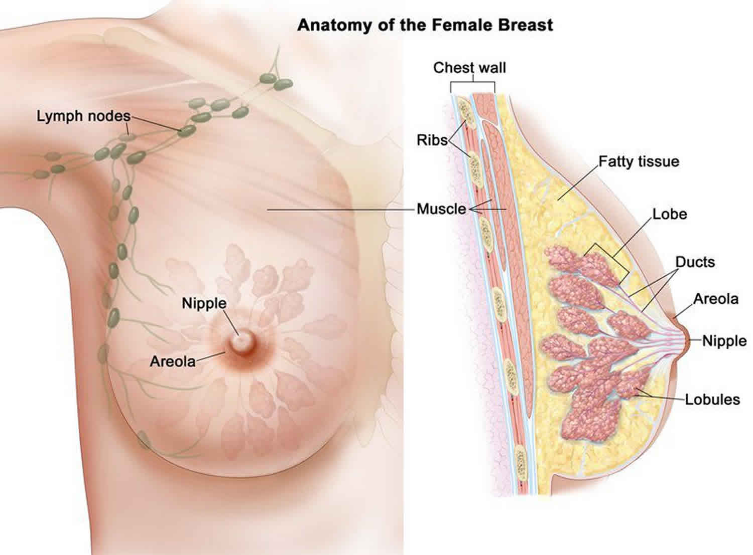
Solitary intraductal papillomas (solitary papillomas) are single tumors that often grow in the large milk ducts near the nipple. They are a common cause of clear or bloody nipple discharge, especially when it comes from only one breast. They may be felt as a small lump behind or next to the nipple. Sometimes they cause pain.
Intraductal papillomas may also be found in small ducts in areas of the breast farther from the nipple. In this case, there are often several growths (multiple papillomas). These are less likely to cause nipple discharge.
In papillomatosis, there are very small areas of cell growth within the ducts, but they aren’t as distinct as intraductal papillomas are.
Two types of intraductal papillomas are generally distinguished: central and peripheral. Central type is a solitary intraductal papilloma which settles in the subareaolar region, and it is observed in perimenauposal women. Peripheral type is situated on the proximal parts of the laciferous ducts of young female patients and tends to be multiple 1.
Usually intraductal papillomas are between 1 and 2cm in size. Sometimes they can be double that, about 4cm. Often there is only one papilloma which can be easily removed. Sometimes there are many of them. In these cases, the whole area containing the intraductal papillomas can be removed.
Intraductal papillomas incidence is 2-3%, and it is seen between 30, and 77 years of age 2. Intraductal papillomas are most common in women over the age of 40, and usually occur as the breast ages and changes 3.
Although intraductal papillomas tumors are benign, there is a great deal of controversy surrounding their diagnosis 4. This is due to the fact that intraductal papillomatous breast lesions are associated with a broad range of histological types, with different characteristics. At one end of the spectrum are intraductal papillomas; at the other end are lesions such as intraductal papillary carcinomas 5.
Can intraductal papilloma go away?
No. Having a single (solitary) intraductal papilloma does not raise your breast cancer risk unless it contains other breast changes, such as atypical hyperplasia. However, having multiple papillomas increases your breast cancer risk slightly.
Intraductal papilloma can be associated with another condition called atypical hyperplasia which means an abnormal growth of cells. There is a risk that the atypical hyperplasia could develop into a breast cancer over time if it is not treated. If there are any atypical cells in the papilloma when the biopsy is examined, they will usually be seen under the microscope.
Multiple papillomas are more likely to be associated with atypical hyperplasia, but this is not always the case. You will need to talk to your doctor about your biopsy result to make sure.
Intraductal papilloma causes
The causes and risk factors for intraductal papilloma are unknown. Intraductal papilloma occurs most often in women ages 35 to 55.
Recent molecular evidence has shown that papillomas frequently have loss of heterozygosity at loci on chromosome 16 6. Specifically, Di Cristofano et al. 7 found loss of heterozygosity at telomeric 16p (marker D16S423) in 3 of 11 informative papillomas, as well as at centromeric 16q (marker D16S310) in 1 of 3 informative papillomas. Similarly, Lininger et al. 8 found loss of heterozygosity at 16p13 in 6 of 10 informative intraductal papillomas with florid epithelial hyperplasia.
Extensive studies of usual ductal hyperplasia have shown it to have frequent, although inconsistent, chromosomal imbalances, and it has become accepted that usual ductal hyperplasia is a truly neoplastic process 9. Given that allelic imbalances are also characteristic of breast papillomas, as preliminary studies indicate, it is likely they are also truly neoplastic. It should be recognized, however, that these studies did not distinguish between small, peripheral intraductal papilloma and large, central intraductal papilloma, and that genetic difference may exist between these lesions.
In addition, peripheral intraductal papilloma is frequently present in breasts involved by other benign proliferative lesions, including columnar cell hyperplasia, sclerosing adenosis, and usual ductal hyperplasia. Therefore, it stands to reason these breasts are somehow the “fertile soil” from which proliferative lesions of all types arise. However, what makes this “fertile soil” is an enormous question that remains unanswered.
Intraductal papilloma symptoms
Intraductal papilloma might cause the following symptoms:
- a lump
- a clear or bloodstained discharge coming from the nipple
- pain or discomfort. For the most part, intraductal papillomas do not cause pain.
Clinically, 72% of all cases present with bloody discharge from the nipple 10 caused by the fragility of the proliferating, disorganized epithelium, which tends to breakdown and bleed 11.
Intraductal papilloma diagnosis
Intraductal papillomas are sometimes found at routine breast screening examinations when you have a mammogram, or following breast surgery. Intraductal papillomas can also be found if you visit your doctor with symptoms. Your doctor will refer you to a breast clinic where you can be seen by a specialist.
At the specialist clinic you are likely to have:
- a breast examination
- a mammogram and/or an ultrasound
- a fine needle aspiration (FNA), core biopsy or vacuum assisted biopsy.
An ultrasound and/or mammogram may be done to learn more about the size and location of papillomas. If you’re a woman under 40, you may have an ultrasound scan rather than a mammogram. This is because younger women’s breast tissue can be dense which can make the x-ray image in a mammogram less clear. But some women under 40 still have a mammogram as part of their assessment.
Ductograms (x-rays of the breast ducts) are sometimes helpful in finding papillomas. If the papilloma is large enough to be felt, a biopsy can be done. This is where tissue is removed from the papilloma and looked at under microscope.
Intraductal papilloma treatment
The usual treatment is surgery to remove the intraductal papilloma and part of the duct that it’s in. If intramammary intraductal papilloma lesion can be located using radiological techniques, then excision, without needle biopsy is recommended. Core biopsy is recommended for cases with radiologically suspect malignancy or in the presence of microcalcification, and distorted tissue ultrastructure 12. If small papillary lesions can be totally excised, and histopathologic examination does not reveal any evidence of atypia, then the patient can be followed up with ultrasound, and mammograms 13.
Intraductal papilloma possible complications
Complications of surgery can include bleeding, infection, and anesthesia risks. If the biopsy shows cancer, you may need further surgery.
Intraductal papilloma prognosis
For the most part, intraductal papillomas do not appear to increase the risk for developing breast cancer.
The outcome is excellent for people with one papilloma. The risk for cancer may be higher for:
- Women with many intraductal papillomas
- Women who get them at an early age
- Women with a family history of cancer
- Women who have abnormal cells in the biopsy.
- Masciadri N, Ferranti C. Benign breast lesions: Ultrasound. J Ultrasound 2011;14:55-65[↩]
- Ganesan S, Karthik G, Joshi M, Damodaran V. Ultrasound spec-trum in intraductal papillary neoplasms of breast. Br J Radiol 2006;79:843-9[↩]
- Intraductal papilloma. https://www.cancerresearchuk.org/about-cancer/other-conditions/ductal-papilloma[↩]
- Richter-Ehrenstein C., Tombokan F., Fallenberg E.M., Schneider A., Denkert C. Intraductal papillomas of the breast: diagnosis and management of 151 patients. Breast. 2011;20:501–504.[↩]
- Brookes M.J., Bourke A.G. Radiological appearances of papillary breast lesions. Clin Radiol. 2008;63:1265–1273[↩]
- Pathology of Small, Peripheral Intraductal Papillomas. https://emedicine.medscape.com/article/1873858-overview[↩]
- Di Cristofano C, Mrad K, Zavaglia K, et al. Papillary lesions of the breast: a molecular progression?. Breast Cancer Res Treat. 2005 Mar. 90(1):71-6[↩]
- Lininger RA, Park WS, Man YG, et al. LOH at 16p13 is a novel chromosomal alteration detected in benign and malignant microdissected papillary neoplasms of the breast. Hum Pathol. 1998 Oct. 29(10):1113-8[↩]
- O’Connell P, Pekkel V, Fuqua SA, Osborne CK, Clark GM, Allred DC. Analysis of loss of heterozygosity in 399 premalignant breast lesions at 15 genetic loci. J Natl Cancer Inst. 1998 May 6. 90(9):697-703[↩]
- Poma S., Longo A. The clinician’s role in the diagnosis of breast disease. J Ultrasound. 2011;14(2):47–54[↩]
- Tarallo V, Canepari E, Bortolotto C. Intraductal papilloma of the breast: A case report. J Ultrasound. 2012;15(2):99-101. https://www.ncbi.nlm.nih.gov/pmc/articles/PMC3558092/[↩]
- A prolapsed intraductal papilloma:a case report. North Clin Istanbul 2015;2(1):59-61 doi: 10.14744/nci.2014.18209 https://pdfs.semanticscholar.org/3854/b63769d5605445fe9078a660307b4d889805.pdf[↩]
- Richter-Ehrenstein C, Tombokan F, Fallenberg EM, Schneider A, Denkert C. Intraductal papillomas of the breast: diagnosis and management of 151 patients. Breast 2011;20:501-4[↩]


