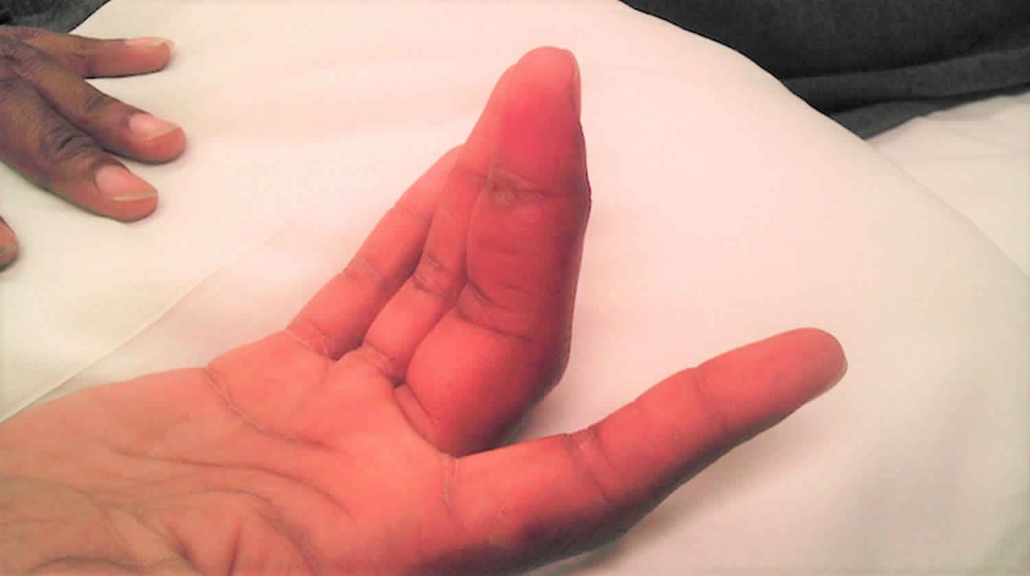
Kanavel’s sign is a clinical sign found in patients with infection of a flexor tendon sheath in the hand (pyogenic flexor tenosynovitis), a serious condition which can cause rapid loss of function of the affected finger. Kanavel sign of flexor sheath infection (pyogenic flexor tenosynovitis) is a finger held in slight flexed position, fusiform swelling of the affected digit, tenderness along the flexor tendon sheath, and pain with passive extension of the digit. Dr Allen B. Kanavel (1874–1938) initially described three cardinal signs of pyogenic flexor tenosynovitis in his seminal work in 1912 as “1. Exquisite tenderness over the course of the sheath, limited to the sheath. 2. Flexion of the finger. 3. Exquisite pain on extending the finger, most marked at the proximal end” 1. Although not noted in his initial description as a cardinal sign, he explained “the whole of the involved finger is uniformally swollen,” and fusiform swelling later became the fourth cardinal sign 2. The constellation of the four signs, commonly known as “Kanavel’s signs,” is frequently used as the primary clinical tool for diagnosing pyogenic flexor tenosynovitis, because advanced imaging and laboratory studies often are nonspecific 3. However, other hand ailments including septic arthritis, crystal-induced arthritides, and stenosing flexor tenosynovitis can have similar presentations to pyogenic flexor tenosynovitis 3.
Future studies are needed to evaluate the sensitivity and specificity of Kanavel’s signs, as this could aid in preventing the incorrect diagnosis of pyogenic flexor tenosynovitis leading to overtreatment in addition to avoiding delayed or misdiagnosed pyogenic flexor tenosynovitis 4.
Pyogenic flexor tenosynovitis is an infection of the flexor tendon sheath of the finger that can result in tendon necrosis and adhesions leading to marked loss of motion, deformity, and loss of limb, particularly if treatment is delayed 5. Pyogenic flexor tenosynovitis comprises 2.5% – 9.4% of all hand infections 6 and is diagnosed primarily using four criteria described by Kanavel in the 1930s (Table 1) 7. In one large series, pyogenic flexor tenosynovitis was reported to represent 9.4% (13/138) of hand infections 8. The advent of antibiotics and appropriate surgical treatment has decreased the risk of serious complications secondary to pyogenic flexor tenosynovitis. However, early recognition and clinical suspicion remain paramount to minimizing potentially devastating consequences from delayed treatment of these infections.
Table 1. Kanavel signs used to diagnose acute flexor tenosynovitis
| Kanavel sign | |
|---|---|
| 1. | Fusiform swelling of the affected finger |
| 2. | Tenderness along the course of the tendon sheath |
| 3. | Digit held in semi-flexed posture |
| 4. | Pain on passive extension of the affected digit |
Infectious flexor tenosynovitis remains an orthopedic emergency. Many advocate early surgical therapy for all cases. The literature clearly shows, however, that medical treatment can be used initially for early, uncomplicated infections, but timing is controversial 9.
Some authors have used single-incision irrigation and drainage. For stage 1 (increased fluid in tendon sheath, mainly a serous exudate) and stage 2 (purulent fluid, granulomatous synovium) infections, the authors advise proximal and distal incisions, with sterile saline intraoperative irrigation in conjunction with empiric intravenous (IV) antibiotics. The authors prefer repeat surgical irrigation and débridement rather than postoperative indwelling catheter irrigation.
Strong evidence and agreement exist for open treatment of stage 3 (necrosis of the tendon, pulleys, or tendon sheath) infections. Some physicians still advocate radical tenosynovectomy for Mycobacterium infections, while others adhere to partial tenosynovectomy with a multiple antibiotic regimen and close observation. The devastating potential complication of infectious flexor tenosynovitis warrants prompt aggressive treatment.
Dailiana et al 10, in a retrospective study of 41 patients with purulent flexor tenosynovitis, found that the best functional outcome associated with this condition resulted from early diagnosis, drainage through small incisions, and continuous postoperative irrigation. Worse outcomes resulted in cases of delayed treatment and infections with specific pathogens. Staphylococcus aureus was detected in most cases.
The indication for surgical drainage includes history and physical examination consistent with acute or chronic flexor tenosynovitis. In certain circumstances when acute flexor tenosynovitis presents within the first 24 hours of infection development, medical management may initially be used. Prompt improvement of symptoms and physical findings must follow within the ensuing 12 hours; otherwise, surgical intervention is necessary.
- Kanavel AB. The symptoms, signs, and diagnosis of tenosynovitis and fascial-space abscesses. In Infections of the Hand. 1st ed. Philadelphia, PA: Lea & Febiger; 1912:201–226.[↩]
- Kanavel AB. Infections of the Hand: A Guide to the Surgical Treatment of Acute and Chronic Suppurative Processes in the Fingers, Hand and Forearm. 7. Philadelphia, PA: Lea & Febiger; 1939.[↩]
- Draeger RW, Bynum DK., Jr Flexor tendon sheath infections of the hand. J Am Acad Orthop Surg. 2012;20:373–382. doi: 10.5435/JAAOS-20-06-373[↩][↩]
- Kennedy CD, Huang JI, Hanel DP. In Brief: Kanavel’s Signs and Pyogenic Flexor Tenosynovitis. Clin Orthop Relat Res. 2016;474(1):280–284. doi:10.1007/s11999-015-4367-x https://www.ncbi.nlm.nih.gov/pmc/articles/PMC4686527[↩]
- Stevanovic MV, Sharpe F. Acute infections. In: Wolfe SW, Pederson WC, Hotchkiss RN, Kozin SH, editors. Green’s Operative Hand Surgery. 6. Philadelphia, PA: Elsevier Churchill Livingstone; 2011. pp. 41–84.[↩]
- Factors affecting the prognosis of pyogenic flexor tenosynovitis. Pang HN, Teoh LC, Yam AK, Lee JY, Puhaindran ME, Tan AB. J Bone Joint Surg Am. 2007 Aug; 89(8):1742-8.[↩]
- Flexor tendon sheath infections of the hand. Draeger RW, Bynum DK Jr. J Am Acad Orthop Surg. 2012 Jun; 20(6):373-82.[↩]
- Glass KD. Factors related to the resolution of treated hand infections. J Hand Surg Am. 1982;7:388–394. doi: 10.1016/S0363-5023(82)80150-0[↩]
- Giladi AM, Malay S, Chung KC. A systematic review of the management of acute pyogenic flexor tenosynovitis. J Hand Surg Eur Vol. 2015;40(7):720–728. doi:10.1177/1753193415570248 https://www.ncbi.nlm.nih.gov/pmc/articles/PMC4804717[↩][↩]
- Dailiana ZH, Rigopoulos N, Varitimidis S, Hantes M, Bargiotas K, Malizos KN. Purulent flexor tenosynovitis: factors influencing the functional outcome. J Hand Surg Eur Vol. 2008 Jun. 33 (3):280-5.[↩]


