What is a lipoma
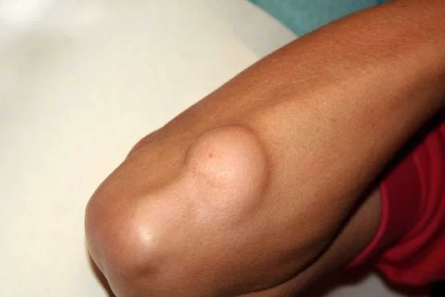
A lipoma is a common benign (not cancer) slow-growing, fatty lump that’s most often situated between your skin and the underlying muscle layer and are most commonly seen on the trunk 1. A lipoma, which feels doughy, soft and usually isn’t painful, moves readily with slight finger pressure. Lipomas are usually painless and are most often found on the upper back, shoulders, arms, buttocks, and upper thighs. Less commonly, lipomas can be found in deeper tissue of the thigh, shoulder, or calf.
Lipomas are usually detected in middle age. Some people have more than one lipoma. The presence of multiple lipomas may be the presenting feature of a variety of syndromes 2.
Lipomas are common. They:
- feel soft and squishy
- can be anything from the size of a pea to a few centimeters across
- may move slightly under your skin if you press them
- aren’t usually painful
- often appear on your shoulders, chest, arms, back, bottom or thighs
- grow slowly
A lipoma isn’t cancer and usually is harmless. Some lumps that look like lipomas can be cysts. Cysts tend to be closer to the skin surface and firm to the touch.
Lipomas have a slightly higher incidence in males compared to females. Although they can occur at any age, they are often noted between the fourth to sixth decades of life. Lipomas are the most common soft tissue tumor found in adults. It is possible to have more than one lipoma.
In most cases, people only develop 1 or 2 lipomas. Multiple lipomas are present in 5% to 10% of affected patients and are usually associated with familial lipomatosis or numerous other genetic disorders. The use of protease inhibitors in HIV patients may induce lipomas and lipodystrophy; therefore, a thorough past medical and medication history should be obtained 3.
Lipoma treatment generally isn’t necessary, but if the lipoma bothers you, is painful or is growing, you may want to have it removed.
A lipoma is rarely a serious medical condition.
See a doctor if:
- you get a lump anywhere on your body
- your lump is painful, red or hot
- your lump is hard and doesn’t move
Your doctor will usually be able to tell if the lump is a lipoma. If there is any doubt, they may refer you for a scan to check it out.
In rare cases lumps under your skin can be a sign of something more serious.
Types of Lipoma
While all lipomas are made up of fat, there are subtypes based on the way they appear under the microscope. Some varieties include:
- Conventional lipoma (common, mature white fat)
- Hibernoma (brown fat instead of the usual white fat)
- Fibrolipoma (fat plus fibrous tissue)
- Angiolipoma (fat plus a large amount of blood vessels)
- Myelolipoma (fat plus tissue that makes blood cells)
- Spindle cell lipoma (fat with cells that look like rods)
- Pleomorphic lipoma (fat with cells of all different shapes and sizes)
- Atypical lipoma (deeper fat with a larger number of cells)
There is ongoing research to learn more about the various subtypes of lipomas and why they form in the first place. In the future, there may be specific treatment recommendations for various lipoma subtypes.
Lipoma vs Cyst
A cyst is a benign (not cancer), round, dome-shaped encapsulated lesion that contains fluid or semi-fluid material. A cyst may be firm or fluctuant and often distends the overlying skin. There are several types of cyst.
Cysts are very common, affecting at least 20% of adults. They may be present at birth or appear later in life. They arise in all races. Most types of cyst are more common in males than in females.
The cause of many cysts is unknown.
- Epidermoid cysts are due to the proliferation of epidermal cells within the dermis. Their origin is the follicular infundibulum. Multiple epidermoid cysts may indicate Gardner syndrome.
- An epidermal inclusion cyst is a response to an injury. Skin is tucked in to form a sac that is lined by healthy epidermal cells that continue to multiply, mature and form keratin.
- The origin of a trichilemmal cyst is hair root sheath. Inheritance is autosomal dominant (the affected gene is within the short arm of chromosome 3) or sporadic.
- The origin of steatocystoma is the sebaceous duct within the hair follicle. Steatocystoma multiplex is sometimes an autosomal dominantly inherited disorder due to mutations localised to the keratin 17 (K17) gene, when it may be associated with pachyonychia congenita. More often, steatocysts are sporadic, when these mutations are not present.
- The origin of the eruptive vellus hair cyst is follicular infundibulum. It may be inherited as an autosomal dominant disorder due to mutations in the keratin gene.
- A dermoid cyst is a hamartoma, a developmental error.
- The origin of a ganglion cyst is degeneration of the mucoid connective tissue of a joint.
- Occlusion of pilosebaceous units (hair follicles) or eccrine sweat ducts leads to a build-up of secretions, which can present as milia.
- Occlusion of the orifice of a mucous gland can lead to a fluid-filled cyst in a mucous membrane (lip, vulva, vagina).
- A milium is a pseudocyst due to failure to release keratin from an adnexal structure. The origin of primary milium is infundibulum of the vellus hair follicle at the level of the sebaceous gland and is a miniature version of an epidermoid cyst. The source of secondary milium is a retention cyst within a vellus hair follicle, sebaceous duct, sweat duct or epidermis.
- Pseudocysts in acne are formed by occlusion of the follicle by keratin and sebum.
Lipoma causes
The precise cause of lipomas is unknown. Genetics appear to play a role since 2% to 3% of affected patients have multiple lesions inherited in a familial pattern 3. There also are several genetic syndromes that feature lipomas as a clinical manifestation. The incidence of lipomas is increased in patients with obesity, hyperlipidemia, and diabetes mellitus.
The exact pathophysiology of lipomas is unclear. However, several cytogenetic abnormalities have been identified including the following:
- Mutations in chromosome 12q13-15, 65% of cases
- Deletions of 13q (10% of cases), rearrangements of 6p21-33, 5% of cases
- Unidentified mutations or normal karyotype, 15% to 20% of cases
Rearrangements of the 12q13-15 result in fusion of the high-mobility group AT-hook 2 (HMGA2) gene to a variety of transcription regulatory domains that promote tumorigenesis 4.
Histologic examination of lipomas reveals mature, normal-appearing adipocytes with a small eccentric nucleus. Adipocytes are intermixed among thin fibrous septa containing blood vessels. These features are indistinguishable from adipocytes in the subcutaneous tissue. Histologic subtypes of lipomas include angiolipomas, myelolipomas, angiomyolipomas, myelolipomas, fibrolipomas, ossifying lipoma, hibernomas, spindle cell lipomas, pleomorphic lipomas, chondroid lipomas, and neural fibrolipomas. Common lipomas and its variants must be distinguished from liposarcomas which are a malignant lipomatous neoplasm containing lipoblasts, which are characterized by coarse vacuoles and one or more scalloped, hyperchromatic nuclei.
Risk factors for lipoma
Several factors may increase your risk of developing a lipoma, including:
- Being between 40 and 60 years old. Although lipomas can occur at any age, they’re most common in this age group.
- Genetics. Lipomas tend to run in families.
Lipoma symptoms
Lipomas typically present as soft, doughy, solitary, painless, subcutaneous nodules that are mobile and not associated with epidermal change. They are typically slow-growing and grow to a final stable size of 2 to 3 centimeters. However, they are occasionally greater than 10 centimeters and referred to as “giant lipomas.” Lipomas may appear anywhere on the body but tend to favor the fatty areas of the trunk, neck, forearms, and proximal extremities. They are rarely seen in acral areas. Lipomas may affect many cutaneous and noncutaneous sites, including dermal, subcutaneous, and subfascial tissues along with intermuscular, intramuscular, synovial, bone, nervous, or retroperitoneal sites.
Lipomas can occur anywhere in the body. They are typically:
- Situated just under the skin. They commonly occur in the neck, shoulders, back, abdomen, arms and thighs.
- Soft and doughy to the touch. They also move easily with slight finger pressure.
- Generally small. Lipomas are typically less than 2 inches (5 centimeters) in diameter, but they can grow larger (5-10cm).
- Sometimes painful. Lipomas can be painful if they grow and press on nearby nerves or if they contain many blood vessels.
See your doctor if you have a lump that you are concerned about.
Lipoma diagnosis
To diagnose a lipoma, your doctor may perform:
- A physical exam
- A tissue sample removal (biopsy) for lab examination. A biopsy is sometimes necessary to confirm the diagnosis of lipoma. In a biopsy, a tissue sample of the tumor is taken and examined under a microscope. Your doctor may give you a local anesthetic to numb the area and take a sample using a needle. Biopsies can also be performed as a small operation. In most lipoma cases, a biopsy is not necessary to confirm the diagnosis. After the lipoma is removed, a biopsy will be done on a sample of the tissue. Under a microscope, lipomas often have a classic appearance with abundant mature fat cells. Sometimes there can be a small amount of other cell types, too, such as cartilage or bone.
- An X-ray or other imaging test, such as an MRI or CT scan, if the lipoma is large, has unusual features or appears to be deeper than the fatty.
- Computerized tomography (CT) scans. These scans are more detailed than x-rays and will often show a fatty mass to confirm the diagnosis of lipoma.
- Magnetic resonance imaging (MRI) scans. The best information for diagnosing lipomas comes from an MRI scan, which can create better images of soft tissues like a lipoma. MRI scanning will show a fatty mass from all perspectives. Oftentimes, doctors can make the diagnosis of lipoma based on MRI imaging alone, and a biopsy is not required.
There’s a very small chance that a lump resembling a lipoma may actually be a form of cancer called liposarcoma. Liposarcomas — cancerous tumors in fatty tissues — grow rapidly, don’t move under the skin and are usually painful. A biopsy or an MRI or CT scan is typically done if your doctor suspects liposarcoma.
Radiologic imaging before surgery may be prudent in cases featuring the following:
- Giant size (greater than 10 centimeters),
- Rapid growth
- Pain
- Fixation to underlying tissues
- Location in deep tissues, the thigh, or retroperitoneal space
Lipoma treatment
Lipomas are harmless. No treatment is usually necessary for a lipoma. However, if the lipoma bothers you, is painful or is growing, your doctor might recommend that it be removed. Lipoma treatments include:
- Surgical removal. Most lipomas are removed surgically by cutting them out. Recurrences after removal are uncommon. Possible side effects are scarring and bruising. A technique known as minimal excision extraction may result in less scarring.
- Liposuction. This treatment uses a needle and a large syringe to remove the fatty lump 5.
Lipomas are almost always cured by simple excision. It is unusual for a lipoma to grow back but, if it does recur, excision is again the best treatment option.
Lipoma surgery
The only treatment that will completely remove a lipoma is a surgical procedure called excision.
In this procedure, a local anesthetic is typically injected around the tumor to numb the area. Large lipomas or those that are deep may require regional anesthesia or general anesthesia. Regional anesthesia numbs a large area by injecting numbing medicine into specific nerves. General anesthesia puts you to sleep.
After the anesthesia is given, your doctor will make an incision in your skin and cut the tumor out.
Figure 1. Lipoma removal
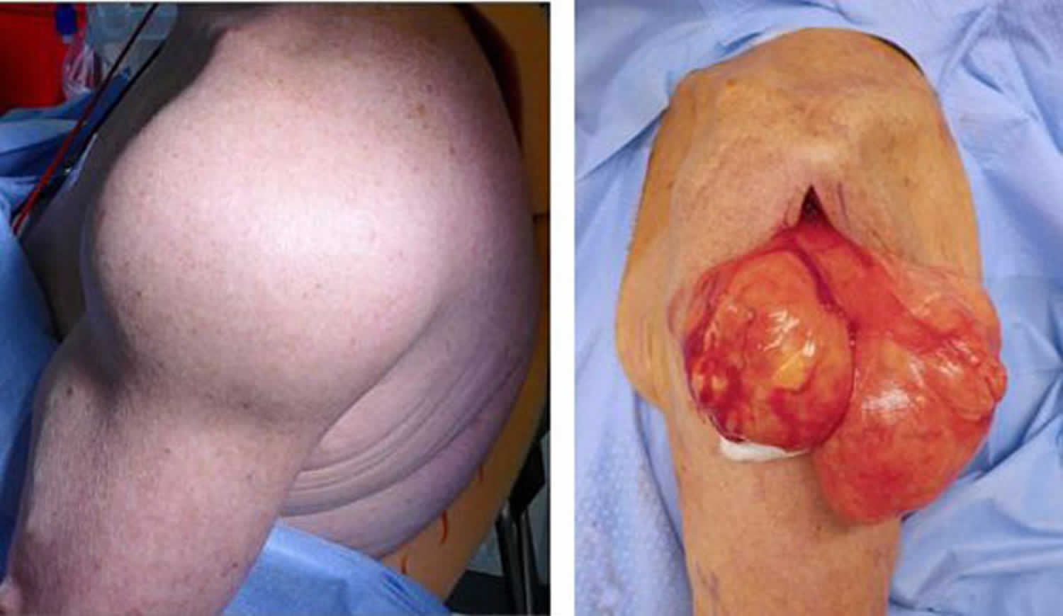
Footnote: (Left) Clinical photo shows a patient with a large lipoma in his shoulder. (Right) The tumor is a large mass of yellowish fat tissue. Here it has been dissected out of the arm just before it is removed by the surgeon.
Recovery
You should be able to go home soon after the procedure if it is a small or superficial mass. You will have a few stitches, which your doctor will remove within a couple of weeks.
How long it takes you to return to most daily activities will depend on the size and location of your lipoma. If you have any pain or discomfort, you may want to limit some activity. Your doctor will provide you with specific instructions to guide your recovery.
- Tong KN, Seltzer S, Castle JT. Lipoma of the Parotid Gland. Head Neck Pathol. 2019 Mar 19[↩]
- Putra J, Al-Ibraheemi A. Adipocytic tumors in Children: A contemporary review. Semin Diagn Pathol. 2019 Mar;36(2):95-104[↩]
- Kolb L, Rosario-Collazo JA. Lipoma. [Updated 2019 Mar 25]. In: StatPearls [Internet]. Treasure Island (FL): StatPearls Publishing; 2019 Jan-. Available from: https://www.ncbi.nlm.nih.gov/books/NBK507906[↩][↩]
- Burkes JN, Campos L, Williams FC, Kim RY. Laryngeal Spindle Cell/Pleomorphic Lipoma: A Case Report. An In-Depth Review of the Adipocytic Tumors. J. Oral Maxillofac. Surg. 2019 Feb 07[↩]
- Johnson CN, Ha AS, Chen E, Davidson D. Lipomatous Soft-tissue Tumors. J Am Acad Orthop Surg. 2018 Nov 15;26(22):779-788[↩]


