What is microphthalmia
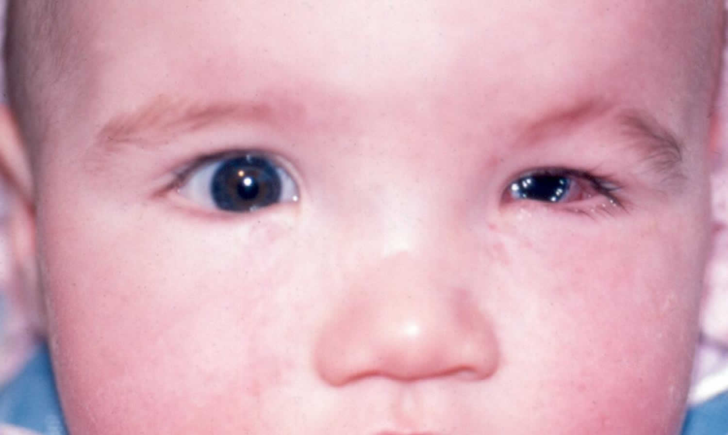
Microphthalmia also called microphthalmos, is a congenital (present at birth) eye abnormality where one or both eyeballs are abnormally small and have anatomic malformations 1. Microphthalmia is also defined as eyeball with a total axial length that is at least two standard deviations below the mean for age 2. In some affected individuals, the eyeball may appear to be completely missing; however, even in these cases some remaining eye tissue is generally present. Such severe microphthalmia should be distinguished from another condition called anophthalmia, in which no eyeball forms at all. However, the terms anophthalmia and severe microphthalmia are often used interchangeably 3. Microphthalmia may or may not result in significant vision loss. Anophthalmia and microphthalmia are rare disorders that develop during pregnancy and can be associated with other birth defects.
Microphthalmia occurs in approximately 14 in 100,000 individuals and affects 3-11% of blind children 4. Microphthalmia is commonly bilateral and it does not present differences according to sex and race 5.
People with microphthalmia may also have a condition called coloboma. Colobomas are missing pieces of tissue in structures that form the eye. They may appear as notches or gaps in the colored part of the eye called the iris; the retina, which is the specialized light-sensitive tissue that lines the back of the eye; the blood vessel layer under the retina called the choroid; or in the optic nerves, which carry information from the eyes to the brain. Colobomas may be present in one or both eyes and, depending on their size and location, can affect a person’s vision.
People with microphthalmia may also have other eye abnormalities, including clouding of the lens of the eye (cataract) and a narrowed opening of the eye (narrowed palpebral fissure). Additionally, affected individuals may have an abnormality called microcornea, in which the clear front covering of the eye (cornea) is small and abnormally curved.
Between one-third and one-half of affected individuals have microphthalmia as part of a syndrome that affects other organs and tissues in the body. These forms of the condition are described as syndromic. When microphthalmia occurs by itself, it is described as nonsyndromic or isolated.
What causes microphthalmia?
The precise cause of microphthalmia remains unknown. Microphthalmia may be caused by changes in many genes involved in the early development of the eye, most of which have not been identified 6. Microphthalmia may also result from a chromosomal abnormality affecting one or more genes. Most genetic changes associated with isolated microphthalmia have been identified only in very small numbers of affected individuals.
Most of cases of microphthalmos are sporadic, but autosomal dominant, autosomal recessive and X linked modes of inheritance have been described. Mutations in genes SOX2, OTX2, BMP4, CHD7, GDF6, RARB and SHH present an autosomal dominant inheritance pattern, while mutations in genes PAX6, STRA6, FOXE3, RAX, SMOC1, VSX2 are associated to autosomal recessive mode of inheritance and BCOR, HCCS and NAA10 mutations are associated to X-linked mode of inheritance 2.
It was suggested that, during post-natal ocular growth, the decreased size of the optic cup, altered proteoglycans in the vitreous, low intraocular pressure, abnormal growth factor production and inadequate production of secondary vitreous may contribute to microphthalmia. Some cases of microphthalmia associated with a cyst may result from failure of the optic fissure to close 7.
Environmental and heritable factors can contribute to microphthalmia. Environmental risk factors are maternal age over 40, multiple births, infants of low birth weight, low gestational age, gestational-acquired infections (rubella, toxoplasmosis, varicella, cytomegalovirus, parvovirus B19, influenza virus, and coxsackie A9), maternal vitamin A deficiency, fever, hyperthermia, exposure to X-rays, exposure to substances that cause birth defects (teratogens), solvent misuse and exposure to drugs like thalidomide, warfarin and alcohol 5.
Microphthalmia inheritance pattern
Isolated microphthalmia is sometimes inherited in an autosomal recessive pattern, which means both copies of the gene in each cell have mutations. The parents of an individual with an autosomal recessive condition each carry one copy of the mutated gene, but they typically do not show signs and symptoms of the condition. In some cases, parents of affected individuals have less severe eye abnormalities.
When microphthalmia occurs as a feature of a genetic syndrome or chromosomal abnormality, it may cluster in families according to the inheritance pattern for that condition, which may be autosomal recessive or other patterns.
Often microphthalmia is not inherited, and there is only one affected individual in a family.
Microphthalmia diagnosis
Family history
As already mentioned, microphthalmia often exhibits a hereditary pattern and it is fundamental to complete eye examination of both parents and to obtain a three-generation family history of eye anomalies, including microphthalmia and coloboma.
Ocular evaluation
Microphthalmia is defined by an eye with anatomical malformation and whose axial length is two standard deviations below the mean for age, what corresponds to an axial length below 21 mm in adult eyes 8.
Ocular disorders may affect the anterior segment and/or the posterior segment. It may be associated with uveal coloboma, hence the general classification into colobomatous and non-colobomatous categories. Ocular abnormalities are microcornea, corneal opacification, corectopia, ectopia lentis, aniridia, cataract, persistent fetal vasculature and/or retinal dysplasia. Microphthalmia may be associated to an orbital cyst (tipically located in the inferior orbit) originated from the optic nerve where it usually communicates with the subarachnoid space 8.
Visual acuity will depend on the type of ocular malformations and especially on the retinal involvement. A good visual acuity can be present in eyes with small iris or choroidal colobomas. However, eyes with macular and optic nerve head involvement have poor vision.
Microphthalmic eyes are usually highly hypermetropic, but sometimes can be highly myopic because of staphyloma formation in the area of the coloboma.
Systemic evaluation
Microphthalmia can be associated to mental retardation, craniofacial malformations (as cleft lip/palate or microcephaly) and malformations of hands and feet (polydactyly). This ocular disorder can occur in isolation or be syndromic (33-50%). Syndromes associated to Microphthalmia are CHARGE syndrome, Duker syndrome, Lenz microphthalmia syndrome, Oculo-Dento-Osseous Dysplasia, Cryptophthalmos syndrome, Cerebro-Oculo-Facial Syndrome, Goltz syndrome, Lowe syndrome, Meckel-Gruber syndrome, Basal cell nevus syndrome of Gorlin-Goltz, Cross syndrome and Microphthalmia with linear skin defects 9.
Since Microphthalmia can be associated to these non-ocular anomalies, physical examination (including dysmorphology examination) is mandatory to determine the presence of distinguishing clinical features.
Imaging
Ultrasound is most commonly used to determine the length of the globe in microphthalmic eyes and to make examination of the orbits 8.
Magnetic resonance imaging (MRI) is extremely useful because there is higher resolution of the structures of interest and no radiation exposure. It shows a small and abnormal globe and it’s useful to orbital evaluation. If there is an orbital cyst, it produces a homogeneous signal that varies from isointense to hypointense on MRI T1-weighted image, while on T2-weighted image the cyst appears hyperintense and there is no enhancement with gadolinium 8.
Electrophysiological tests are critical for assessing the severity of visual impairment and help to determine at which level the abnormality is. In cases of severe microphthalmia, a flash visual evoked potential will establish if any visual function is present. A pattern visual evoked potential will determine the severity of the disease and detect any optic nerve dysfunction while an electroretinogram will identify if there is retinal dysfunction 8.
Since microphthalmia can be associated with systemic anomalies, it is important to consider an endocrine evaluation, echocardiogram and renal ultrasound examination.
Microphthalmia treatment
Because of the variable phenotype spectrum associated to microphthalmia, patients should be evaluated by multidisciplinary teams composed by ophthalmologists, pediatricians, and clinical geneticists. If no syndrome is identified in childhood, further examination after three or four years should be performed as many syndromes become more apparent at this age 10.
There is no treatment for severe anophthalmia or microphthalmia that will create a new eye or restore vision. However, some less severe forms of microphthalmia may benefit from medical or surgical treatments. In almost all cases improvements to a child’s appearance are possible. Children can be fitted for a prosthetic (artificial) eye for cosmetic purposes and to promote socket growth. A newborn with anophthalmia or microphthalmia will need to visit several eye care professionals, including those who specialize in pediatrics, vitreoretinal disease, orbital and oculoplastic surgery, ophthalmic genetics, and prosthetic devices for the eye. Each specialist can provide information and possible treatments resulting in the best care for the child and family. The specialist in prosthetic diseases for the eye will make conformers, plastic structures that help support the face and encourage the eye socket to grow. As the face develops, new conformers will need to be made. A child with anophthalmia may also need to use expanders in addition to conformers to further enlarge the eye socket. Once the face is fully developed, prosthetic eyes can be made and placed. Prosthetic eyes will not restore vision.
Medical therapy
If retinal function is detectable, refraction and treatment of any underlying amblyopia is critical.
Children with microphthalmia may have some residual vision (limited sight). In these cases, the good eye can be patched to strengthen vision in the microphthalmic eye. A prosthesis can be made to cap the microphthalmic eye to help with cosmetic appearance, while preserving the remaining sight.
Surgery
Microphthalmia leads to the appearance of hemifacial asymmetry due to a small orbital volume compared to age-matched controls. Reconstructive strategies aim simultaneous management of both soft tissue hypoplasia and asymmetric bone growth 11.
If the axial length of the eye is over 16 mm, orbital growth is more likely to be normal. However, if the axial length is less than 16 mm, it is unlikely to promote normal orbital growth alone and it is necessary to increase the socket volume early on to prevent pronouncing asymmetry as the child grows 8. Mild/moderate microphthalmia is generally managed conservatively with insertion of a conformer (like a prosthetic eye but not painted) while in severe microphthalmia is necessary to provide endo-orbital volume replacement using implants of progressively increasing size 8. Orbital osteotomies are indicated in more severe cases.
When an orbital cyst is present its expansion properties are harnessed, and surgery is postponed until it reaches 90% of the orbital volume, allowing removal for cosmetic reasons at about the time the child starts school 8.
Microphthalmia prognosis
The potential for visual development depends on the ocular structures affected and the severity of the malformations. The treatment aims to maximize the existing vision and provide improvement at the aesthetic level.
- Microphthalmos. https://eyewiki.org/Microphthalmos[↩]
- Bardakjian T, Weiss A, Schneider A. Microphthalmia/Anophthalmia/Coloboma Spectrum. 2004 Jan 29 [Updated 2015 Jul 9]. In: Adam MP, Ardinger HH, Pagon RA, et al., editors. GeneReviews® [Internet]. Seattle (WA): University of Washington, Seattle; 1993-2019. Available from: https://www.ncbi.nlm.nih.gov/books/NBK1378[↩][↩]
- Facts About Anophthalmia and Microphthalmia. https://nei.nih.gov/health/anoph/anophthalmia[↩]
- Dharmasena, Aruna; Keenan, Tiarnan; Goldacre, Raph; Hall, Nick; Goldacre, Michael J (2017). “Trends over time in the incidence of congenital anophthalmia, microphthalmia and orbital malformation in England: Database study”. British Journal of Ophthalmology. 101 (6): 735–739.[↩]
- Shaw GM, Carmichael SL, Yang W, Harris JA, Finnell RH, Lammer EJ. Epidemiologic characteristics of anophthalmia and bilateral microphthalmia among 2.5 million births in California, 1989–1997. Am J Med Genet A. 2005;137:36–40.[↩][↩]
- Microphthalmia. https://ghr.nlm.nih.gov/condition/microphthalmia[↩]
- Forrester MB, Merz RD. Descriptive epidemiology of anophthalmia and microphthalmia, Hawaii, 1986–2001. Birth Defects Res A Clin Mol Teratol. 2006;76:187–92.[↩]
- N K Ragge, I D Subak-Sharpe, J R O Collin, A practical guide to the management of anophthalmia and microphthalmia. Eye. 2007 21:1290–1300[↩][↩][↩][↩][↩][↩][↩][↩]
- Traboulsi EI. Compendium of Inherited Disorders and the Eye. New York: Oxford University Press; 2005 [↩]
- Verma AS, Fitzpatrick DR. Anophthalmia and microphthalmia. Orphanet J Rare Dis. 2007;2:47. Published 2007 Nov 26. doi:10.1186/1750-1172-2-47[↩]
- Hintschich C, Zonneveld F, Baldeschi L, Bunce C, Koornneef L. Bony orbital development after early enucleation in humans. Br J Ophthalmol. 2001;85:205–208.[↩]


