
Nuclear sclerosis
Nuclear sclerosis is an age-related change in the opacification of the crystalline lens nucleus which typically occurs as a part of normal aging. Nuclear sclerosis is caused by compression of older lens fibers in the center of the lens (nucleus) as a consequence of the production of new fibers 1. The lens is a transparent structure made up of fibers (modified epithelial cells) enclosed in a membranous structure called lens capsule. Lens matter consists of two main parts 2:
- Cortex (superficial part) – containing younger fibers
- Nucleus (deeper part) – containing older fibers
Some degree of nuclear sclerosis and yellowing is normal in adult patients past middle age. In general, nuclear sclerosis interferes only minimally with visual function. An excessive amount of light scattering and yellowing is called a nuclear cataract, which causes a central opacity (Figure 1). The nuclear cataract per se can never – technically – become a mature cataract 3. To be mature all of the lens including the cortex must be cataractous.
The stages of a nuclear cataract are:
- Nuclear sclerosis – a slight loss of normal clarity which does not elicit a circular reflex with the direct ophthalmoscope
- Incipient nuclear cataract (slight nuclear cataract) with the first appearance of the circular reflex with the direct ophthalmoscope with the pupil fully dilated. That reflex determined that nuclear sclerosis had advanced to nuclear cataract.
- Definite nuclear cataract with a definite increase in density,
- Cataracta brunescens, and finally
- Cataracta nigra – cataract appears black in the pupil, but is seen to be mahogany in color when extracted from the eye. Prior to the slit-lamp, it frequently was not recognized that the patient’s loss of vision was due to a cataract. It was concluded that a black pupil indicated the absence of a cataract. The slit-lamp revolutionized the evaluation of cataracts.
A nuclear sclerotic cataract may at first cause more nearsightedness or even a temporary improvement in your reading vision. But with time, the lens gradually turns more densely yellow and further clouds your vision. As the nuclear sclerotic cataract slowly progresses, the lens may even turn brown. Advanced yellowing or browning of the lens can lead to difficulty distinguishing between shades of color.
To determine whether nuclear sclerosis has entered the realm of nuclear cataract, the ophthalmologist can evaluate the degree of increased color and of opacification by using a slit-lamp biomicroscope. However, there is another method: by examining the orange-red reflex with the pupil dilated with the direct ophthalmoscope at arm’s length from the eye. If the nuclear sclerosis has reached at least the incipient stage of a nuclear cataract, a circular reflex encircling the cataract will be elicited.
Nuclear sclerotic cataracts tend to progress slowly. Although they are usually bilateral, they may be asymmetric. Nuclear sclerotic cataracts typically cause greater impairment of distance vision than of near vision. In the early stages, the progressive hardening of the lens nucleus frequently causes an increase in the refractive index of the lens and thus a myopic shift in refraction (lenticular myopia). In hyperopic eyes, the myopic shift enables otherwise presbyopic individuals to read without spectacles, a condition referred to as second sight. Occasionally, the abrupt change in refractive index between the sclerotic nucleus (or other lens opacities) and the lens cortex can cause monocular diplopia. Progressive yellowing or browning of the lens causes poor hue discrimination, especially at the blue end of the visible light spectrum. Photopic retinal function may decrease with advanced nuclear cataract. In very advanced cases, the lens nucleus becomes opaque and brown and is called a brunescent nuclear cataract.
Histopathologically, the nucleus in nuclear sclerotic cataract is difficult to distinguish from the nucleus of normal, aged lenses. Investigations by electron microscopy have identified an increased number of lamellar membrane whorls in some nuclear cataracts. The degree to which protein aggregates or these membrane modifications contribute to the increased light scattering of nuclear cataracts is unclear.
Figure 1. Eye lens anatomy
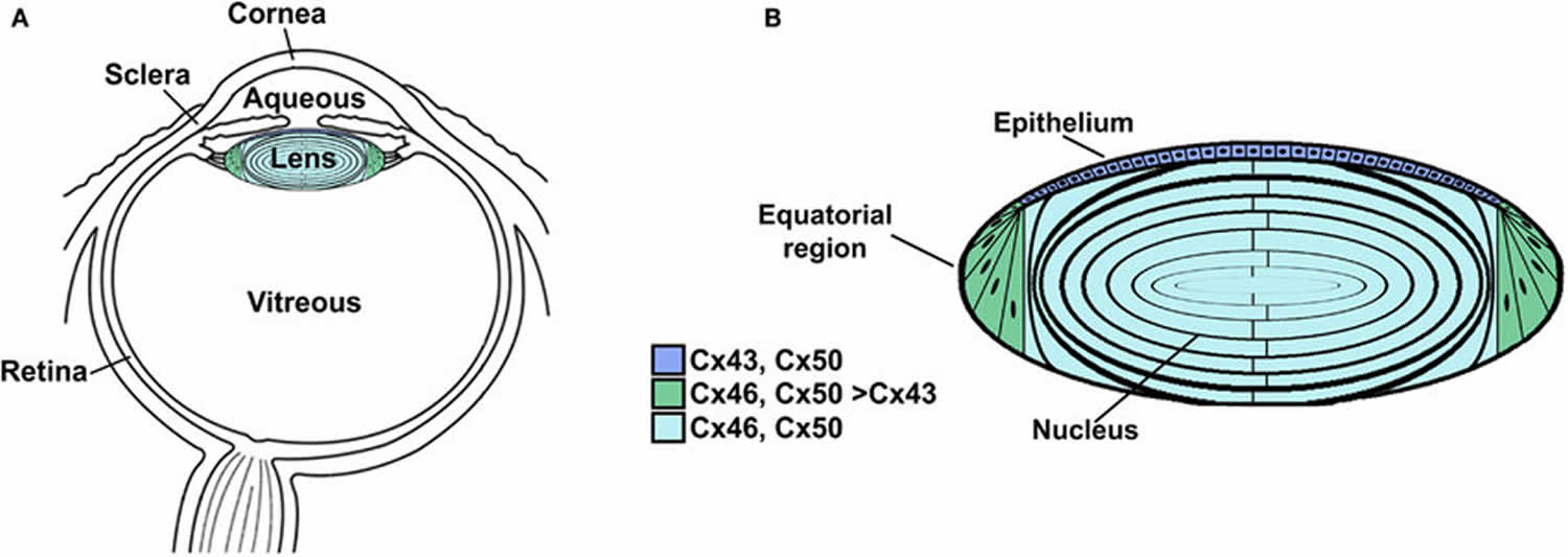
Footnote: Anatomy of the eye (A) and structure of the lens (B). (A) Diagram of the eye illustrating the location of the lens in relation to other structures. The lens is suspended between two transparent fluids, the aqueous and vitreous humors. The aqueous humor fills the space between the cornea and the lens (as well as the posterior chamber). It contains water, oxygen, carbon dioxide, inorganic and organic ions, carbohydrates, glutathione, urea, amino acids, and proteins (including immunoglobulins and growth factors) and is very dynamic (with turnover rates of 1–1.5% per minute). The aqueous humor stabilizes the ocular structure and contributes to the homeostasis of the avascular structures of the anterior part of the eye by providing nutrition and removing metabolic products. The vitreous is a viscous, gelatinous liquid that fills the space between the lens and the retina. It is essentially a specialized extracellular fluid containing collagen fibers, hyaluronic acid and other soluble proteins, and glycoproteins. The vitreous is rather stagnant, equilibrating very slowly with the plasma. (B) Diagram of the lens showing the distribution of connexin isoforms. Cells from the anterior epithelial cell layer express Cx43 and Cx50, differentiating fiber cells express Cx43, Cx46, and Cx50, and fiber cells (including those of the nucleus) contain Cx46 and Cx50.
[Source 2 ]Figure 2. Nuclear sclerotic cataract
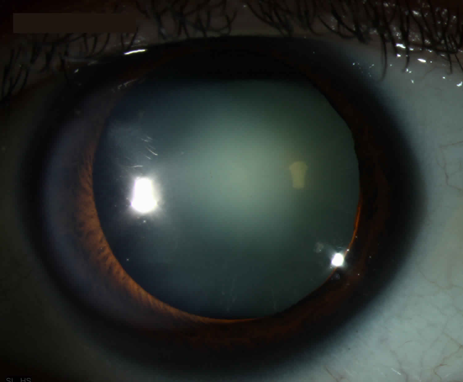
Nuclear sclerotic cataract
Nuclear sclerotic cataracts are secondary to opacification of the lens nucleus which typically occurs as a part of normal aging. The nucleus in this cataract has a “milky” appearance.
Nuclear sclerotic cataract signs and symptoms
You might not have any symptoms at first, when cataracts are mild. But as cataracts grow, they can cause changes in your vision. For example, you may notice that:
- Your vision is cloudy or blurry
- Double vision in a single eye
- Decrease in distance vision
- Colors look faded or loss of color discrimination ability
- You can’t see well at night or difficulty with night driving
- Need for brighter light for reading and other activities
- Sensitivity to light and glare – lamps, sunlight, or headlights seem too bright
- You see a halo around lights
- You see double (this sometimes goes away as the cataract gets bigger)
- You have to change the prescription for your glasses or contact lens often
- Myopic refractive shift in which you notice as an improvement in your near vision
These symptoms can be a sign of other eye problems, too. Be sure to talk to your eye doctor if you have any of these problems.
Over time, cataracts can lead to vision loss.
Nuclear sclerotic cataract signs:
- Gradual opacification of the central portion of the lens nucleus
- Age-related, involves the hardening (sclerosis) and either yellowing or brown to black darkening (brunescense) of the lens nucleus
Risk factor for cataracts
Factors that increase your risk of cataracts include:
- Increasing age
- Diabetes
- Excessive exposure to sunlight
- Smoking
- Obesity
- High blood pressure
- Previous eye injury or inflammation
- Previous eye surgery
- Prolonged use of corticosteroid medications
- Drinking excessive amounts of alcohol
- Have a family history of cataracts
- Have had an eye injury, eye surgery, or radiation treatment on your upper body
- Take steroids (medicines used to treat a variety of health problems, like arthritis and rashes)
If you’re worried you might be at risk for cataracts, talk with your doctor. Ask if there is anything you can do to lower your risk.
What causes cataracts?
Most cataracts are caused by normal changes in your eyes as you get older.
Cataracts form when the proteins in the lens of your eye clump together, making your lens cloudy.
When you’re young, the lens of your eye is normally clear, allowing light to pass through. The lens helps focus the light on your retina (the light-sensitive layer of tissue in the back of your eye) so you can see things clearly, whether they’re up close or far away.
Your lens is mostly made of water and proteins. Normally, the proteins line up in a special way so that your lens is completely clear and light can pass through easily. If the proteins in your lens clump together, the lens gets cloudy.
Around age 40, the proteins in the lens of your eye start to break down and clump together. This clump makes a cloudy area on your lens — or a cataract. Over time, the cataract gets more severe and clouds more of the lens.
Cataracts keep you from seeing clearly because light can’t pass through the clumps of proteins in your lens very easily. Over time, the clumps of proteins get bigger and thicker, making it harder for you to see. The lens may also develop a yellow or brown tint, which can change how you see colors.
Doctors and researchers don’t know exactly what makes some people get cataracts, but they do know that there are things that can make cataracts form faster, including:
- Smoking
- Drinking too much alcohol
- Spending too much time in the sun without sunglasses
Some health problems and treatments can also make cataracts more likely, including:
- Diabetes
- A serious eye injury
- Eye surgery to treat glaucoma or another eye condition
- Taking steroids — medicines used to treat a variety of health problems, like arthritis or allergies — for a long time
- Getting radiation treatment for cancer or other diseases
Cataracts prevention
No studies have proved how to prevent cataracts or slow the progression of cataracts. But doctors think several strategies may be helpful, including:
- Wear sunglasses and a hat with a brim to block the sun. Ultraviolet light from the sun may contribute to the development of cataracts. Wear sunglasses that block ultraviolet B (UVB) rays when you’re outdoors.
- Quit smoking.
- Reduce alcohol use. Excessive alcohol use can increase the risk of cataracts.
- Eat healthy. Eat plenty of fruits and vegetables — especially dark, leafy greens like spinach, kale, and collard greens. Adding a variety of colorful fruits and vegetables to your diet ensures that you’re getting many vitamins and nutrients. Fruits and vegetables have many antioxidants, which help maintain the health of your eyes. Studies haven’t proved that antioxidants in pill form can prevent cataracts. But, a large population study recently showed that a healthy diet rich in vitamins and minerals was associated with a reduced risk of developing cataracts. Fruits and vegetables have many proven health benefits and are a safe way to increase the amount of minerals and vitamins in your diet.
- Have regular eye examinations. Eye examinations can help detect cataracts and other eye problems at their earliest stages. Ask your doctor how often you should have an eye examination.
- Get a dilated eye exam. If you’re age 60 or older, get a dilated eye exam at least once every 2 years.
- Manage other health problems. Follow your treatment plan if you have diabetes or other medical conditions that can increase your risk of cataracts.
Cataracts diagnosis
To determine whether you have a cataract, your doctor will review your medical history and symptoms, and perform an eye examination. Your doctor may conduct several tests, including:
- Visual acuity test. A visual acuity test uses an eye chart to measure how well you can read a series of letters. Your eyes are tested one at a time, while the other eye is covered. Using a chart or a viewing device with progressively smaller letters, your eye doctor determines if you have 20/20 vision or if your vision shows signs of impairment.
- Slit-lamp examination. A slit lamp allows your eye doctor to see the structures at the front of your eye under magnification. The microscope is called a slit lamp because it uses an intense line of light, a slit, to illuminate your cornea, iris, lens, and the space between your iris and cornea. The slit allows your doctor to view these structures in small sections, which makes it easier to detect any tiny abnormalities.
- Retinal exam. To prepare for a retinal exam, your eye doctor puts drops in your eyes to open your pupils wide (dilate). This makes it easier to examine the back of your eyes (retina). Using a slit lamp or a special device called an ophthalmoscope, your eye doctor can examine your lens for signs of a cataract.
Nuclear sclerosis treatment
Surgery is the only way to get rid of a cataract, but you may not need to get surgery right away.
Talk with your eye doctor about whether surgery is right for you. Most eye doctors suggest considering cataract surgery when your cataracts begin to affect your quality of life or interfere with your ability to perform normal daily activities, such as reading or driving at night.
It’s up to you and your doctor to decide when cataract surgery is right for you. For most people, there is no rush to remove cataracts because they usually don’t harm the eye. But cataracts can worsen faster in people with diabetes.
Delaying the procedure generally won’t affect how well your vision recovers if you later decide to have cataract surgery. Take time to consider the benefits and risks of cataract surgery with your doctor.
If you choose not to undergo cataract surgery now, your eye doctor may recommend periodic follow-up exams to see if your cataracts are progressing. How often you’ll see your eye doctor depends on your situation.
Early on, you may be able to make small changes to manage your cataracts. You can do things like:
- Use brighter lights at home or work
- Wear anti-glare sunglasses
- Use magnifying lenses for reading and other activities
New glasses or contacts
A new prescription for eyeglasses or contact lenses can help you see better with cataracts early on.
Surgery
Your eye doctor might suggest surgery if your cataracts start getting in the way of everyday activities like reading, driving, or watching TV. During cataract surgery, the doctor removes the clouded lens and replaces it with a new, artificial lens (also called an intraocular lens, or IOL). This surgery is very safe, and 9 out of 10 people who get it can see better afterwards.
Most people don’t need to rush into surgery. Waiting to have surgery usually won’t harm your eyes or make surgery more difficult later. Remember these tips:
- Tell your doctor if cataracts are getting in the way of your everyday activities
- See your doctor for regular check-ups
- Ask your doctor about the benefits and risks of cataract surgery
- Encourage family members to get checked for cataracts, since they can run in families.
- Chapter 11 – Biology and Diseases of Dogs. Laboratory Animal Medicine (Second Edition) American College of Laboratory Animal Medicine 2002, Pages 395-458 https://doi.org/10.1016/B978-012263951-7/50014-4[↩]
- Beyer, Eric & Ebihara, Lisa & Berthoud, Viviana. (2013). Connexin Mutants and Cataracts. Frontiers in pharmacology. 4. 43. 10.3389/fphar.2013.00043. [↩][↩]
- Nuclear cataract, incipient stage. https://webeye.ophth.uiowa.edu/eyeforum/atlas/pages/nuclear-cataract-incipient-stage.html[↩]


